FNA Cytology of Metastatic Malignancies of Unknown Primary...
142
FNA Cytology of Metastatic Malignancies of Unknown Primary Site Tarik M. Elsheikh Jan F. Silverman
Transcript of FNA Cytology of Metastatic Malignancies of Unknown Primary...

FNA Cytology of Metastatic
Malignancies of Unknown Primary Site
Tarik M. Elsheikh Jan F. Silverman
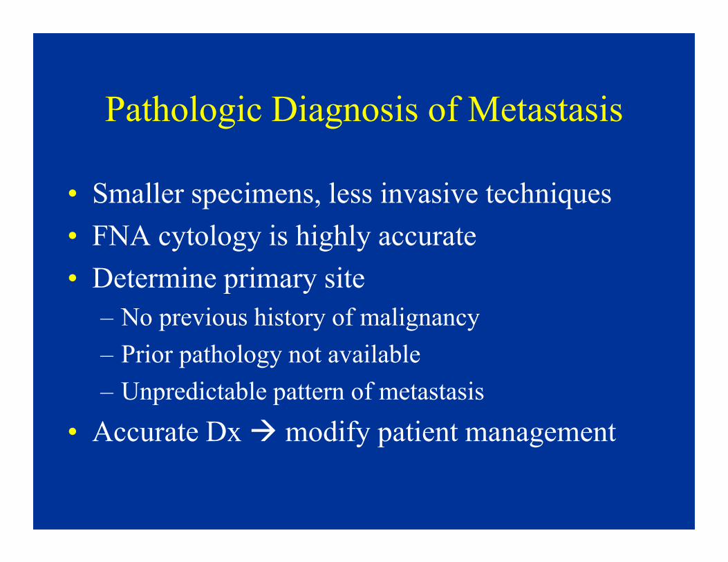
Pathologic Diagnosis of Metastasis
• Smaller specimens, less invasive techniques
• FNA cytology is highly accurate
• Determine primary site• Determine primary site
– No previous history of malignancy
– Prior pathology not available
– Unpredictable pattern of metastasis
• Accurate Dx � modify patient management
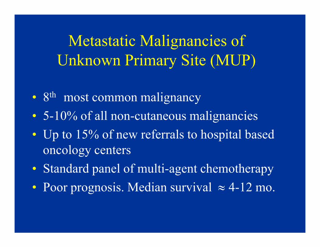
Metastatic Malignancies of
Unknown Primary Site (MUP)
• 8th most common malignancy
• 5-10% of all non-cutaneous malignancies
• Up to 15% of new referrals to hospital based
oncology centers
• Standard panel of multi-agent chemotherapy
• Poor prognosis. Median survival ≈ 4-12 mo.
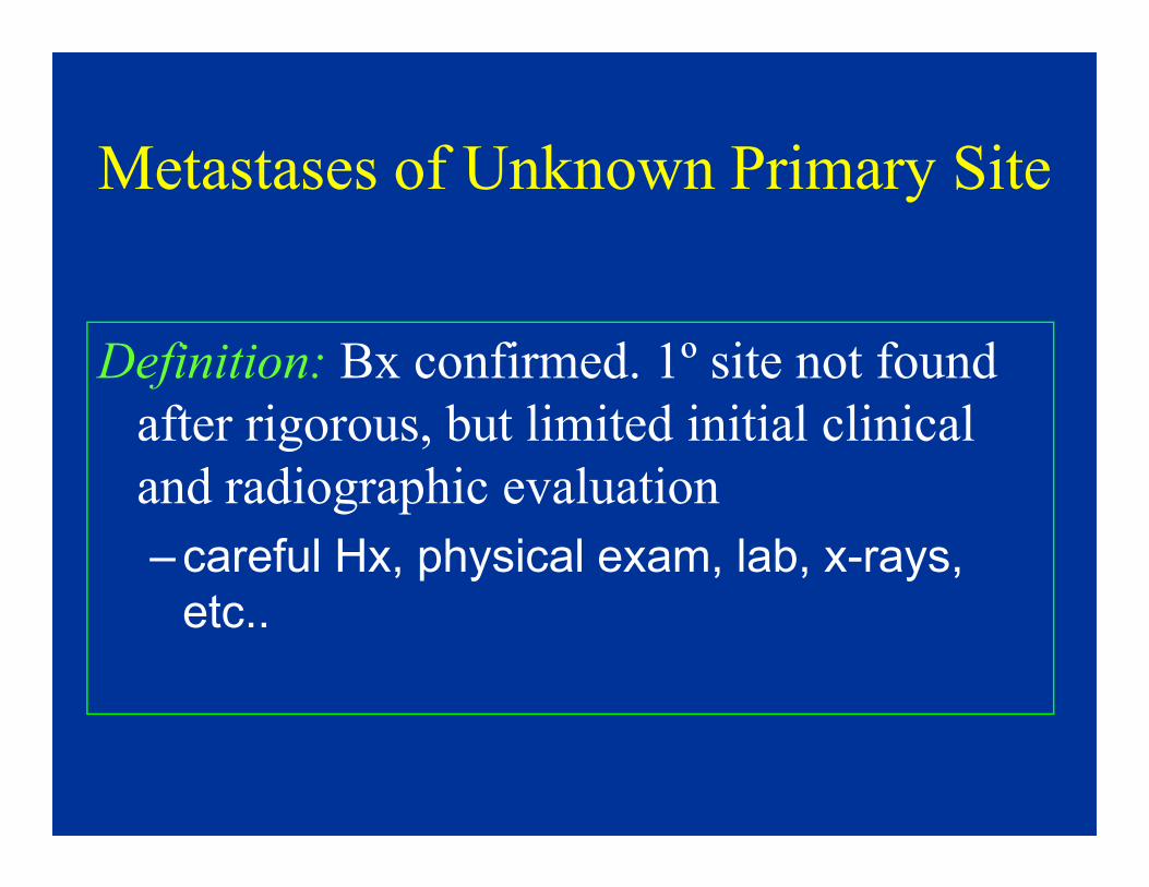
Metastases of Unknown Primary Site
Definition: Bx confirmed. 1º site not found
after rigorous, but limited initial clinical after rigorous, but limited initial clinical
and radiographic evaluation
–careful Hx, physical exam, lab, x-rays,
etc..

Is Workup of MUP Necessary?
• Optimal management may be organ-
specific, and rely on accurate determination
of primary site
• Inability to ID a primary � major clinical • Inability to ID a primary � major clinical
challenge
– Patient anxiety:
• ? Inadequate evaluation by physician
• ? Prognosis improved if primary is found

Cost Effectiveness of Pathologic Workup• Extensive radiological exams & serum tumor markers –
often unsuccessful in finding 1º site
• Pathologic evaluation (including extended IHC panel) is
more cost effective than clinical workup
Cost per Success Theoretical cost-Cost per
patient
Success
rate
Theoretical cost-
effectiveness ratio
Clinical tests
alone
$ 18,000 * 20 % $ 250,000
IHC panel** $ 2,000 70 % $ 2,900
* excluding physician charges
** panel of 6 tests Wick et al 1999

Cost Effectiveness of Pathologic Workup 2
• Overutilization occurs in individual cases or
by individual pathologists
– Too many Ab’s in 30% of cases– Too many Ab’s in 30% of cases
– Unnecessary IHC in 10% of cases
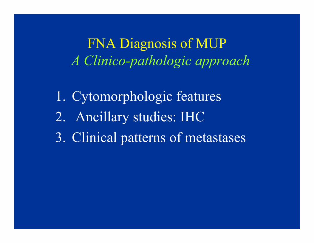
FNA Diagnosis of MUP
A Clinico-pathologic approach
1. Cytomorphologic features
2. Ancillary studies: IHC2. Ancillary studies: IHC
3. Clinical patterns of metastases

FNA Diagnosis of MUP 2
A Clinico-pathologic approach
1. Cytomorphologic features
• Histologic types (specific cell lineage):
adenoca, squamous ca, melanoma, etc.
• Morphologic patterns (non specific cell • Morphologic patterns (non specific cell
lineage): small cell, large cell, oncocytic,
spindle, etc.
2. Ancillary studies: IHC
3. Clinical patterns of metastases

CYTOMORPHOLOGIC PATTERNS OF MUP
Specific Cell Lineage Cell Pattern / Type
Squamous CA
Sarcoma
Adenocarcinoma
Lymphoma
Small Cell
Oncocytic/Granular
Melanoma Clear Cell
Pleomorphic/Giant Cell
Spindle cell
Polygonal, Large Cell

Case 1
• CT guided FNA biopsy of a kidney mass
in a 68 year old woman.
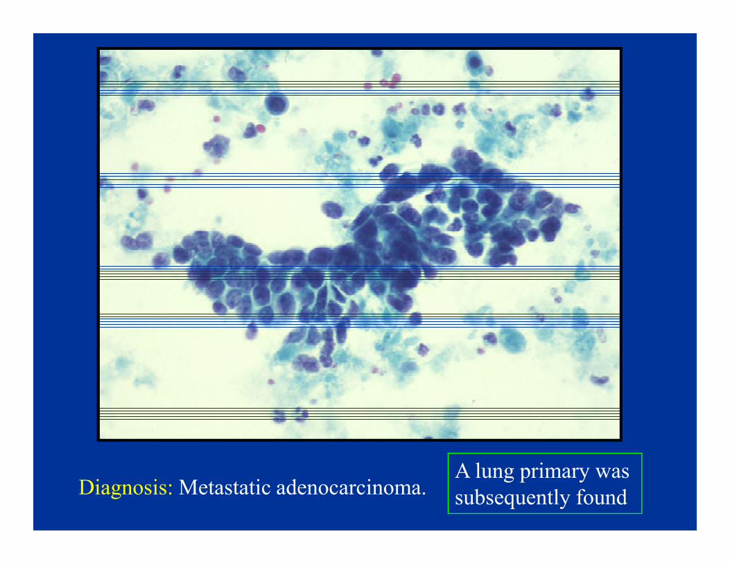
Diagnosis: Metastatic adenocarcinoma. A lung primary was
subsequently found
Adenocarcinoma
• Most common MUP (60%)
• W-M differentiated adenocarcinoma �
median survival ≈ 3-6 months
• Lung & pancreas: most common (40%)• Lung & pancreas: most common (40%)
– GI tract
– Liver
• Nonspecific diagnosis � 1º vs. MET

Columnar/ductal
Prostate
Microacinar
Breast
Mucinous
Thyroid
Papillary
Adenocarcinoma
Morphologic Patterns of
Differentiated Adenocarcinoma (W-M)
Pancreas
Bile duct
Colon
Lung(BAC)
Breast
Carcinoid
Low grade
COLON
Endometrioid
CA
Hyperchromatic
Lung
Pancreas
Prostate
Bile duct
Stomach
Hypochromatic
High gradeProstate
NEC
Thyroid
Granulosa CT
Breast
Ovary
Pancreas
GIT
Chordoma
Thyroid
Ovary
Kidney
Endometrium
Breast
Lung

Adenocarcinoma
Columnar/ductal
Low grade
Hyperchromatic Hypochromatic
High grade

Adenocarcinoma: Low Grade Columnar/ductal
• Cohesive clusters and geographic flat sheets

Low Grade Columnar/Ductal
• Uniform cell population with bland appearance
• Low N/C ratio, finely granular chromatin, small nucleoli
• Round to elongated nuclei, luminal borders

Low Grade Columnar/Ductal
AdenocarcinomaCarcinoid
– Pancreas
– Breast
– Bile duct
– Lung (BAC)
– Colon
– Carcinoid Cholangiocarcinoma

High Grade Columnar/Ductal
Adenocarcinoma
• Cohesive clusters and flat sheets
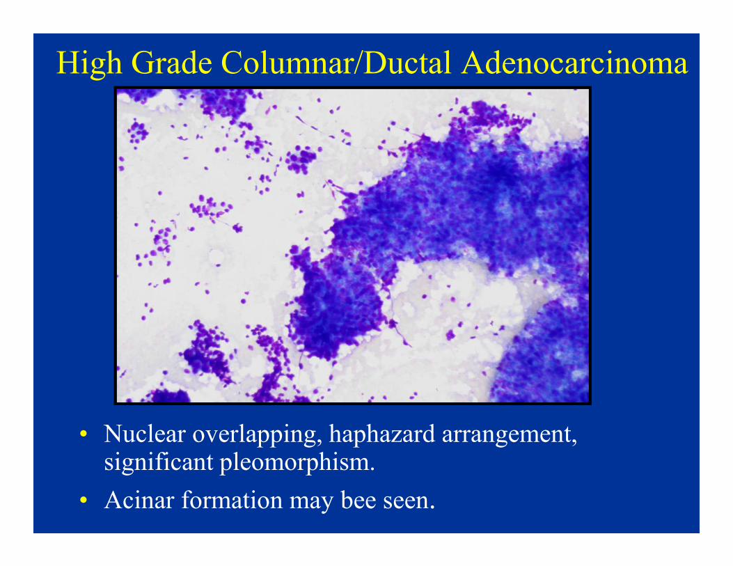
High Grade Columnar/Ductal Adenocarcinoma
• Nuclear overlapping, haphazard arrangement, significant pleomorphism.
• Acinar formation may bee seen.
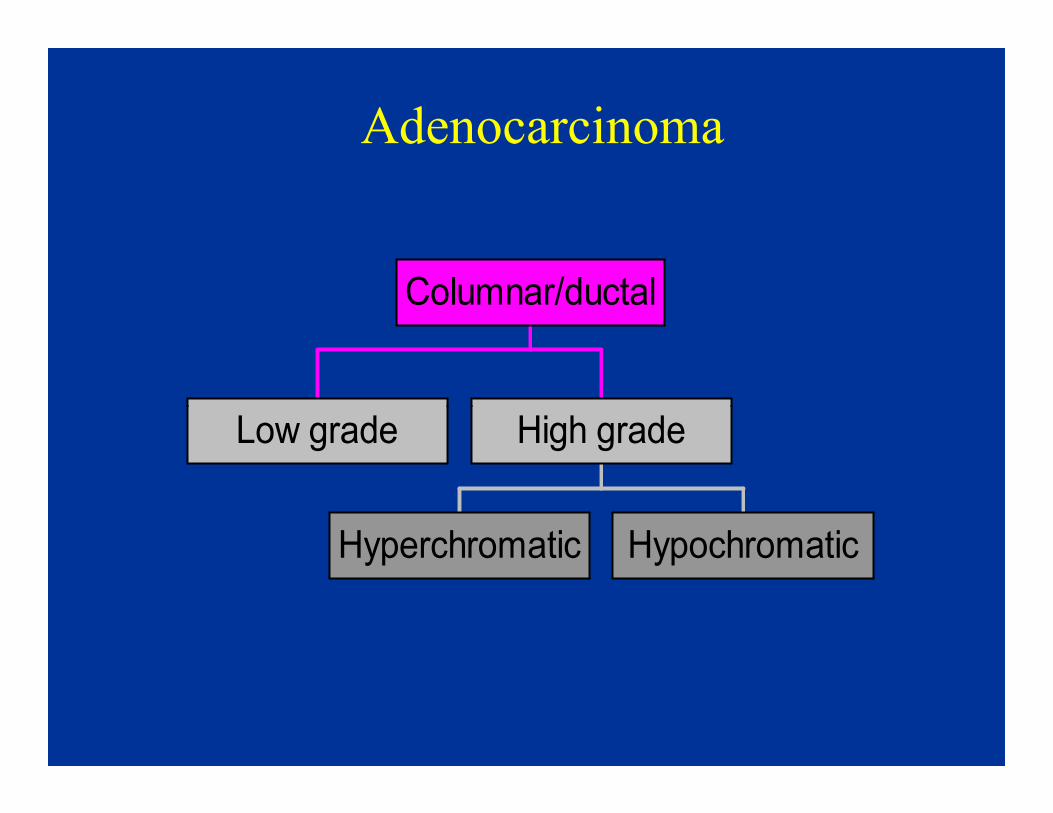
Adenocarcinoma
Columnar/ductal
Low grade
Hyperchromatic Hypochromatic
High grade

High Grade Columnar/Ductal
Adenocarcinoma
• Hypochromatic
• Lung
• Pancreas
• Bile duct
• Prostate
• Stomach

High Grade Columnar/Ductal
Adenocarcinoma
• Hyperchromatic
– COLON– COLON
– Endometrioid
CA (endometrium, ovary, cervix)
– Bile duct
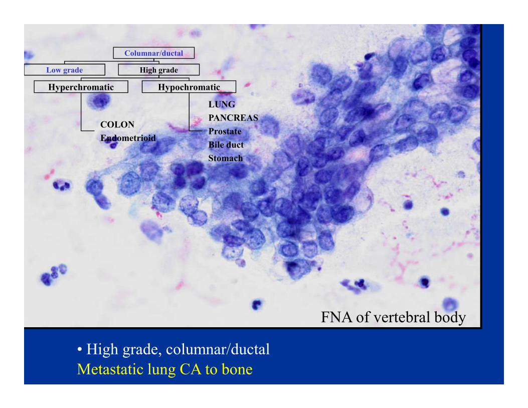
Columnar/ductal
Low grade High grade
Hyperchromatic Hypochromatic
LUNG
PANCREAS
Prostate
Bile duct
Stomach
COLON
Endometrioid
• High grade, columnar/ductal
FNA of vertebral body
Metastatic lung CA to bone

Columnar/ductal
Low grade High grade
Hyperchromatic Hypochromatic
LUNG
PANCREAS
Prostate
Bile duct
Stomach
COLON
Endometrioid
Metastatic pancreatic CA to liver
FNA of liver
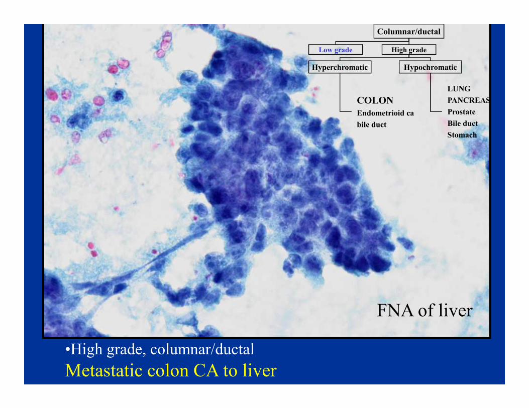
Columnar/ductal
Low grade High grade
Hyperchromatic Hypochromatic
LUNG
PANCREAS
Prostate
Bile duct
Stomach
COLON
Endometrioid ca
bile duct
•High grade, columnar/ductal
Metastatic colon CA to liver
FNA of liver

CYTOMORPHOLOGIC PATTERNS OF
METASTASIS OF UNKNOWN PRIMARY ORGIN
Specific Cell Lineage Cell Pattern / Type
Squamous CA
Sarcoma
Adenocarcinoma
Lymphoma
Small Cell
Oncocytic/Granular
Melanoma Clear Cell
Pleomorphic/Giant Cell
Spindle cell
Polygonal, Large Cell
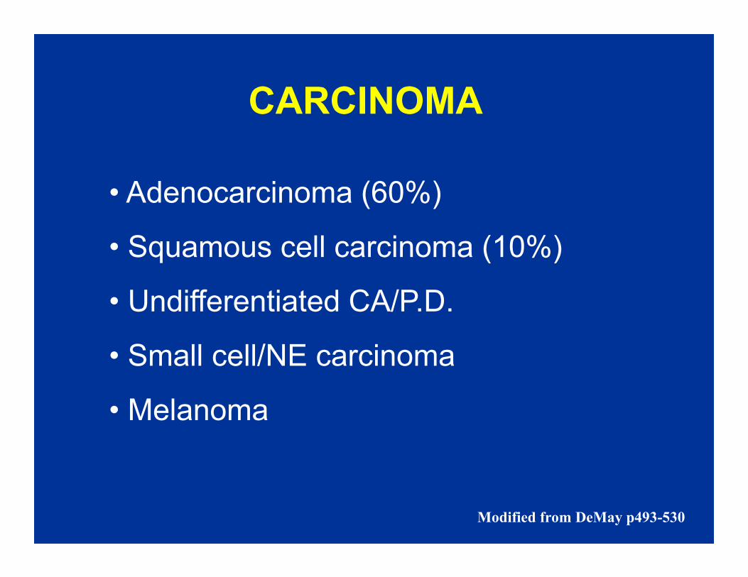
CARCINOMA
• Adenocarcinoma (60%)
• Squamous cell carcinoma (10%)
• Undifferentiated CA/P.D.• Undifferentiated CA/P.D.
• Small cell/NE carcinoma
• Melanoma
Modified from DeMay p493-530
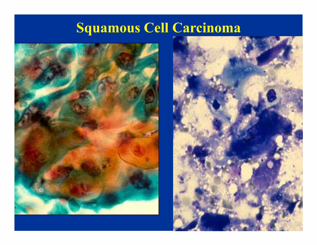
Squamous Cell Carcinoma

MELANOMA
• Metastasis to unusual sites
• Mimics other malignancies
• Primary occult or not apparent by
history


Melan - A

Malignant Melanoma Variants
• Rhabdoid
• Signet-ring
• Spindle• Spindle
• Myxoid
• Desmoplastic
• Ballon Cell
• Small Cell

Signet-Ring Melanoma Ballon Cell
Spindle Cell Small Cell MM

Rhabdoid MM

Pigmented dendritic histiocytes

SARCOMA
• Very unusual unknown primary
• Primary site usually obvious
• Diff Dx: Sarcomatoid carcinoma / • Diff Dx: Sarcomatoid carcinoma /
melanoma
• Spindle, epitheliod, pleomorphic,
small cell, myxoid

An 81 year old woman was identified as
Case 2
An 81 year old woman was identified as
having a right hilar lung mass. FNA
biopsy was performed.


Case 2
DIAGNOSIS
Metastatic Hurthle cell carcinoma
of the thyroid

A CT guided FNA biopsy of a single
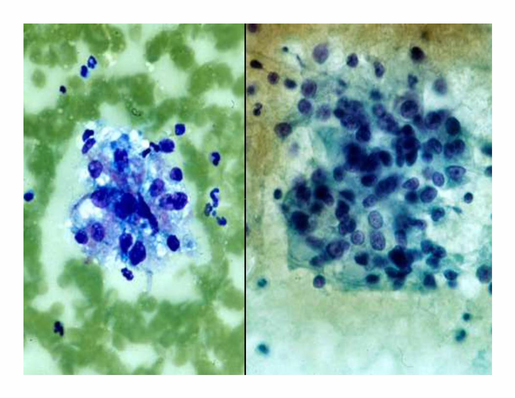
Case 3
A CT guided FNA biopsy of a single
mass involving the anterior right lobe of
liver was performed in a 72 year old
female


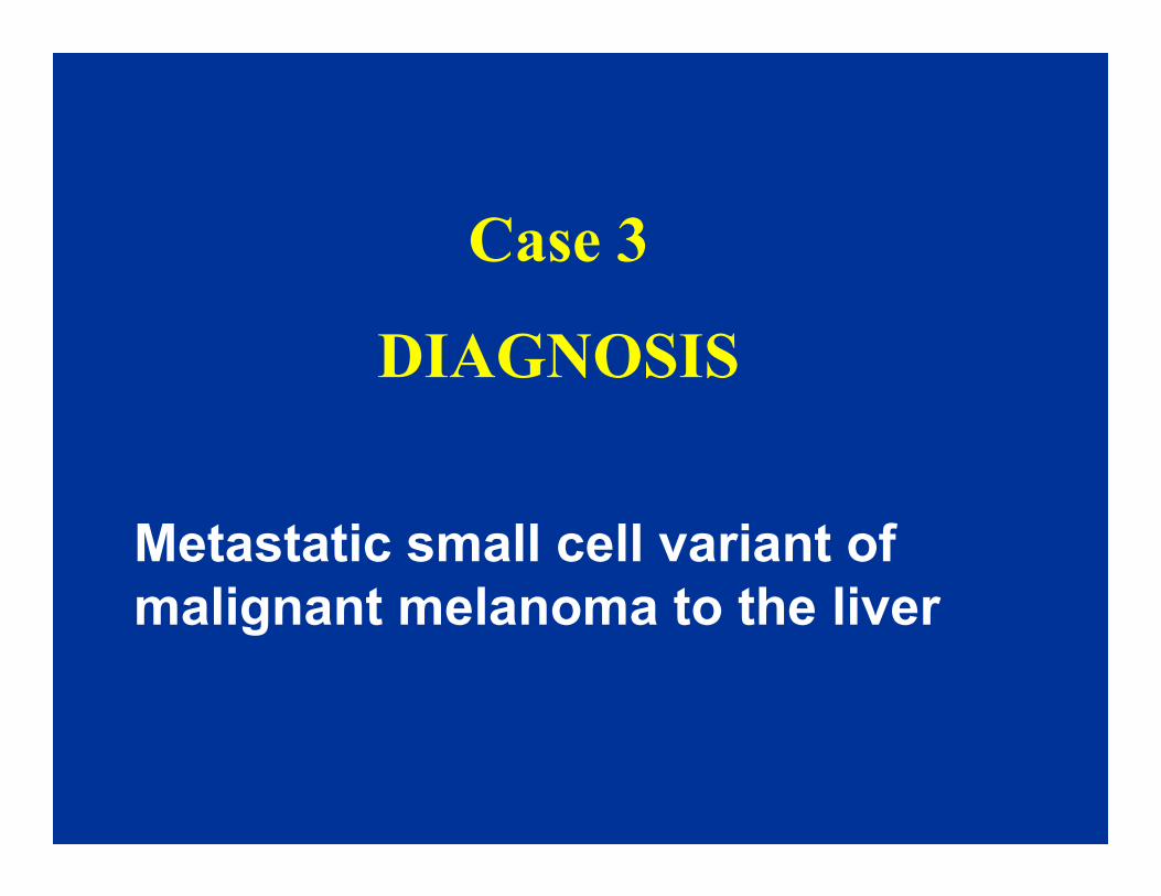
Case 3
DIAGNOSIS
Metastatic small cell variant of
malignant melanoma to the liver

Case 4
53 year old male presented with a 6
cm sacral mass and pain in his legs. A
FNA biopsy was performed
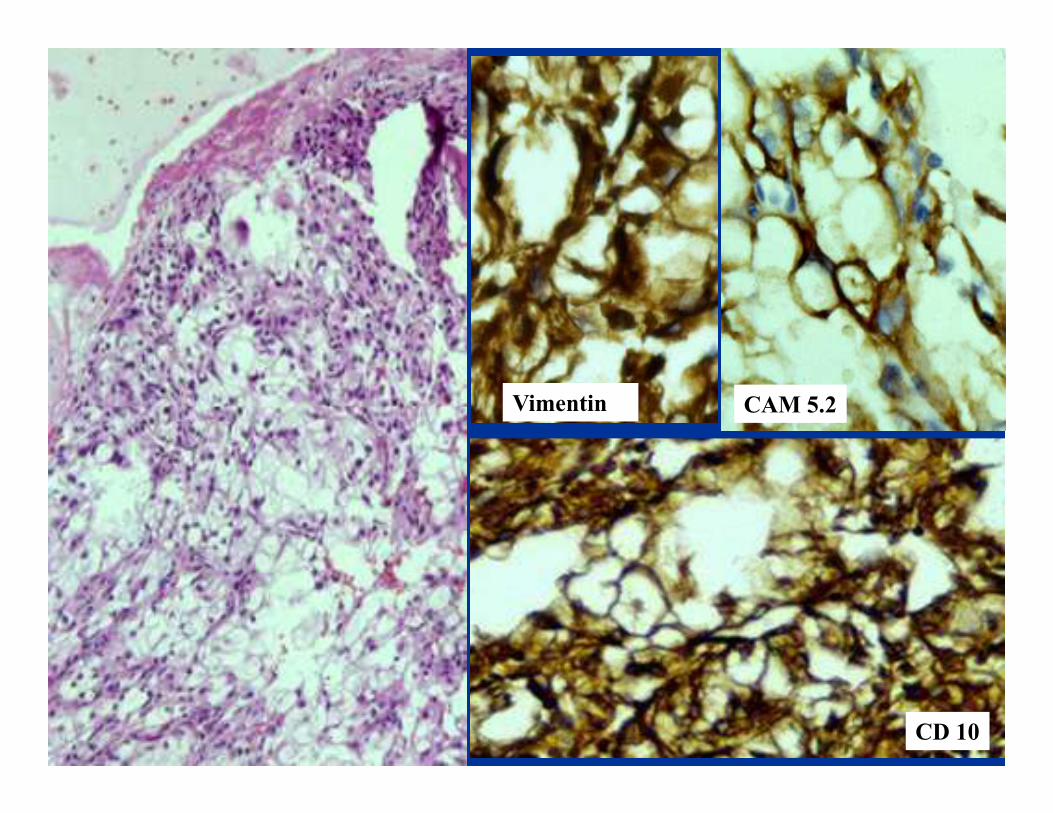
CAM 5.2Vimentin CAM 5.2Vimentin
CD 10

Case 4
DIAGNOSIS
Metastatic conventional clear cell
carcinoma of the kidney

CYTOMORPHOLOGIC PATTERNS OF
METASTASIS OF UNKNOWN PRIMARY ORGIN
Cell Pattern / Type
Small Cell
Oncocytic/Granular
Clear Cell
Pleomorphic/Giant Cell
Spindle cell
Polygonal, Large Cell

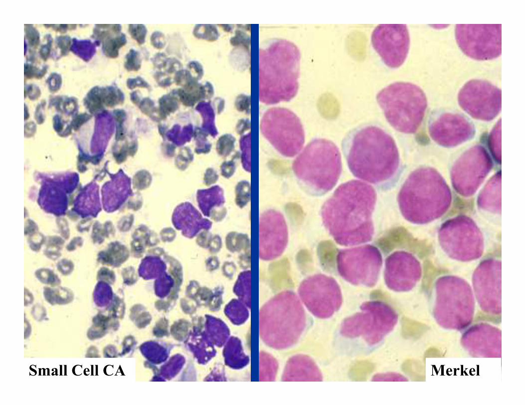
Small Cell Tumors
• Poorly differentiated carcinomas
Squamous Cell Carcinoma
• Neuroendocrine tumors
Carcinoids / Islet cell tumors, ect.
Small cell (neuroendocrine) carcinoma
Squamous Cell Carcinoma
Adenocarcinoma
• Lymphomas
• Small blue cell tumors of childhood
• Some sarcomas (synovial)
• Melanoma variant
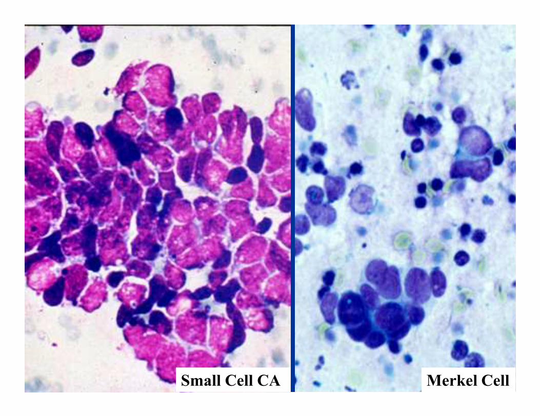
Small Cell CA Merkel Cell

Lymphoma
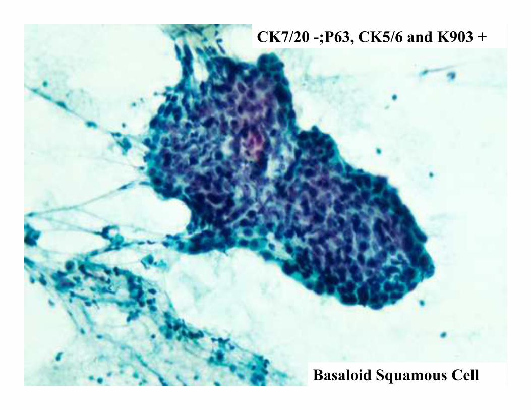
CK7/20 -;P63, CK5/6 and K903 +
Basaloid Squamous Cell

Pleomorphic / Giant Cells
• Sarcomas
i.e., Malignant fibrous histiocytoma, etc.
• Germ cell tumors
• Carcinomas
Lung, Pancreas, Liver, Thyroid, etc.
• Neuroendocrine tumors
Pheochromocytoma
Choriocarcinoma
• Lymphoreticular neoplasms
Anaplastic large cell lymphoma (Ki-1)
• Melanoma
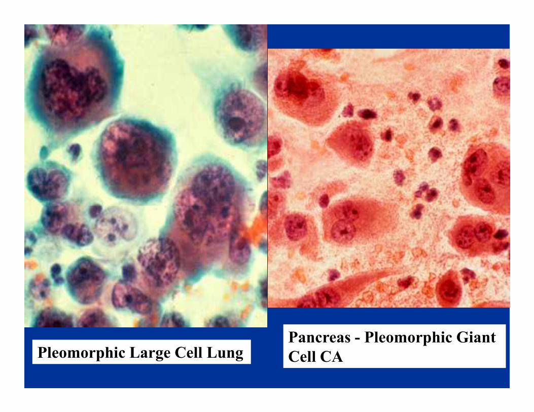
Pleomorphic Large Cell LungPancreas - Pleomorphic Giant
Cell CA
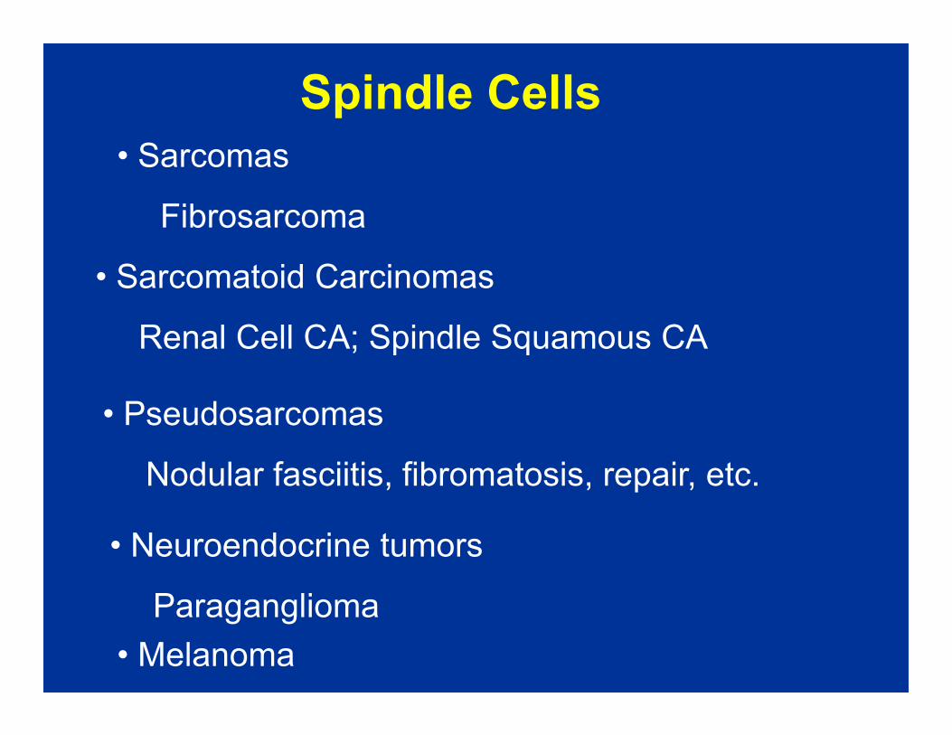
Spindle Cells
• Sarcomas
Fibrosarcoma
• Sarcomatoid Carcinomas
Renal Cell CA; Spindle Squamous CA
• Neuroendocrine tumors
Paraganglioma
• Pseudosarcomas
Nodular fasciitis, fibromatosis, repair, etc.
• Melanoma

Sarcomatoid Squamous Cell CA

Melanoma

Sarcomatoid Renal Cell
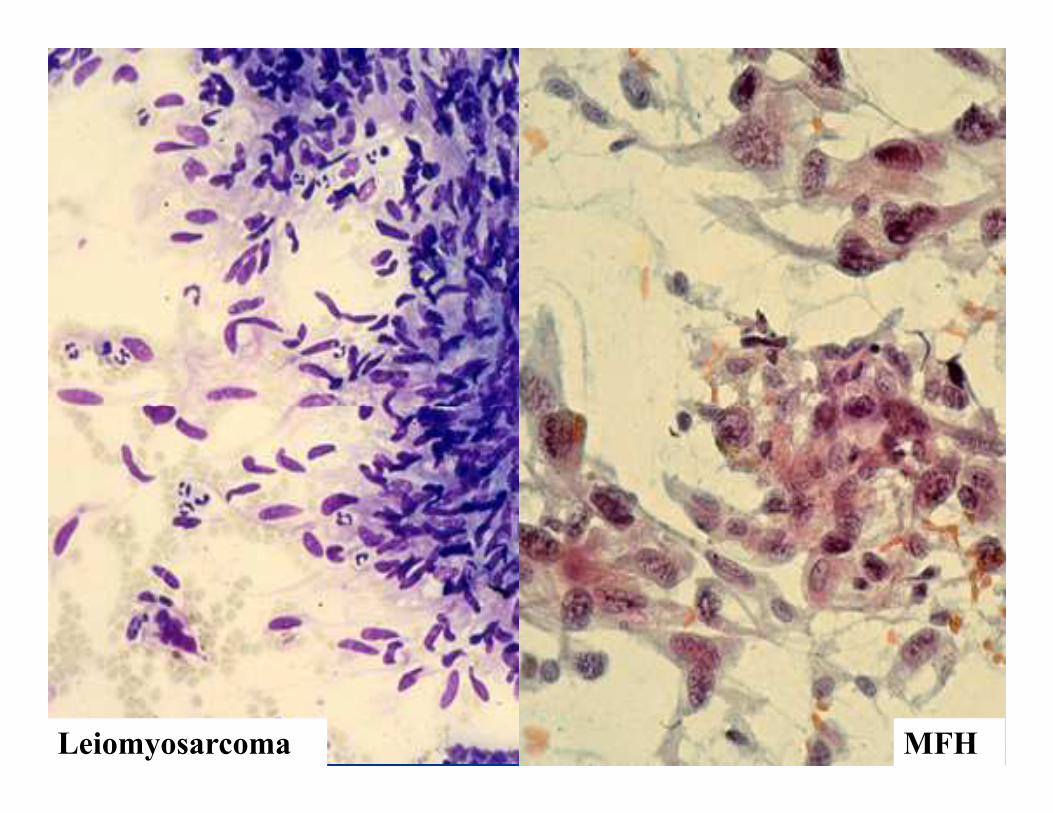
Leiomyosarcoma MFH

Granular Cell Neoplasms
• Carcinomas (Adenomas)
Kidney, Liver, Salivary Gland, Glassy Cell (cervix)
• Oncocytic / Hurthle Neoplasms
Kidney, Thyroid, etc.
• Apocrine - Breast, Sweat Gland
• Soft Tissue Tumors - Granular Cell Tumor
Others: Muscle, Alveolar Soft Parts Sarcoma
• Neuroendocrine Tumors - Carcinoid, Paraganglioma
• Melanoma
• Hilar / Leydig Cell Tumor
DDX: Nonspecific degenerationModified from DeMay

Hurthle Cell CA Renal Cell CA
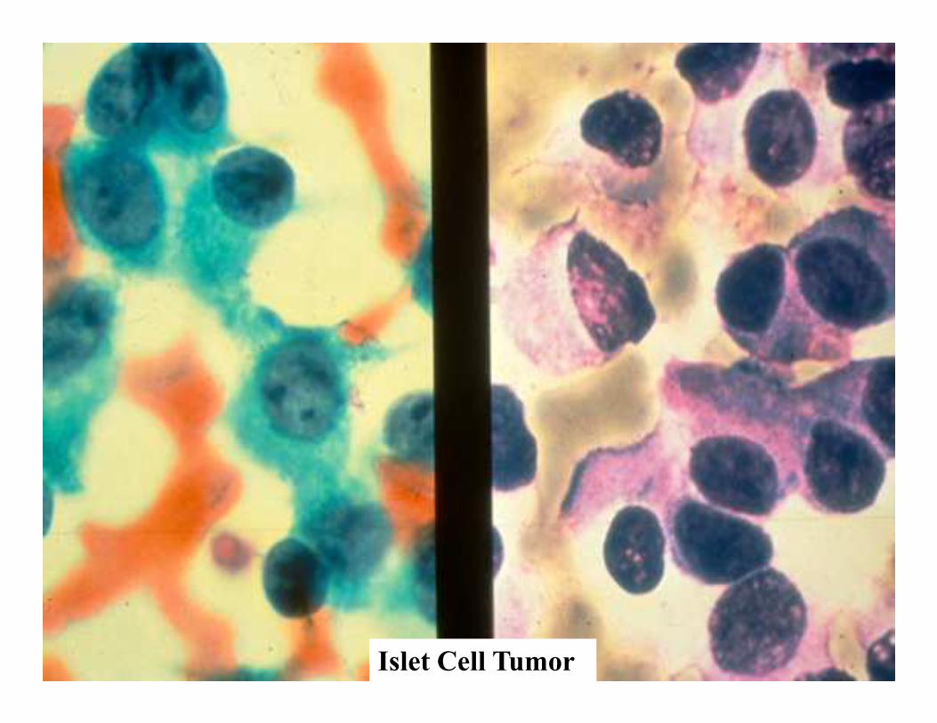
Islet Cell Tumor

Oncocytic Neuroendocrine Warthin’s

Clear cell Tumors
• Oncocytic neoplasms
• Acinic / Acinar Tumors
• Carcinomas
KIDNEY, also Ovary, Liver, Adrenal, Salivary Gland,
lung GYN, Thyroid
• Acinic / Acinar Tumors
• Neuroendocrine Tumors (i.e., paragaglioma)
• Soft Tissue Tumors (i.e., clear cell sarcoma)
• Lymphoma - very rare
• Germ Cell Tumors
• Melanoma (ballon cells)

Clear Cell - Kidney Yolk-Sac CA

Paraganglioma

Intranuclear Cytoplasmic Inclusions
• Thyroid
Papillary CA, others
• Lung
Bronchioloalveolar CABronchioloalveolar CA
• Liver
Favors HCC
• Melanoma
• Many others
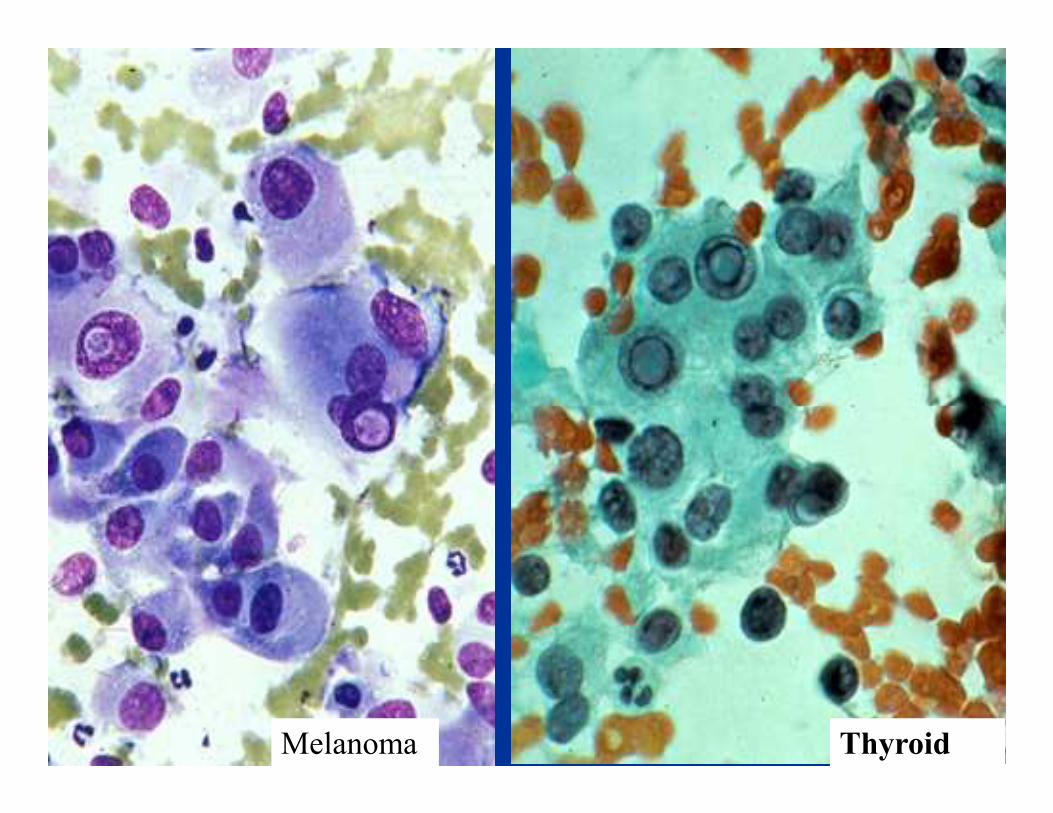
Melanoma Thyroid

Microacinar Complexes
• Prostate
• Thyroid • Thyroid
• Carcinoid / Islet (Rosettes)
• Others - Granulosa cell tumor, other
SRCT of childhood

Thyroid - Follicular CA Carcinoid

Prostrate CA
PSA +

Hyaline Globules
• Carcinoma (Rhabdoid)
Wide variety, often PD malignancies
• Sarcomas• Sarcomas
• Lymphoma
• Melanoma (Rhabdoid)
• Hepatocellular, renal, ovary

Melanoma

Pleomorphic Giant Cell - Pancreas

Single Cell
Adeno CA
BREAST
Pancreas
Other Tumors
Small Cell CA
MesotheliomaPancreas
Stomach
Prostate
Mesothelioma
Carcinoids
Melanoma
Hematopoeitic
Small Cell CA Merkel

Neuroblastoma
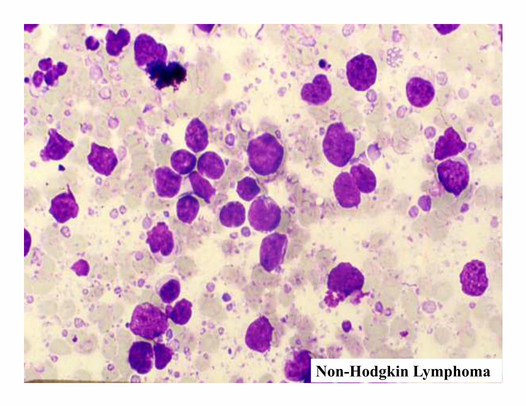
Non-Hodgkin Lymphoma

Gastric CA

Adrenal Cortex
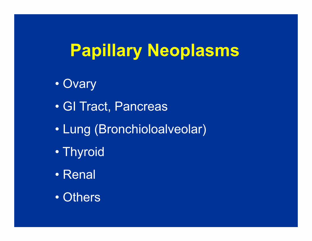
Papillary Neoplasms
• Ovary
• GI Tract, Pancreas
• Lung (Bronchioloalveolar)
• Thyroid
• Renal
• Others
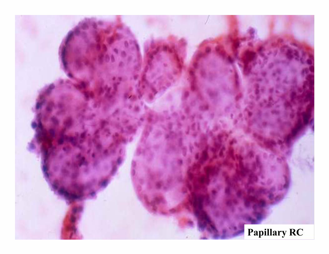
Papillary RC
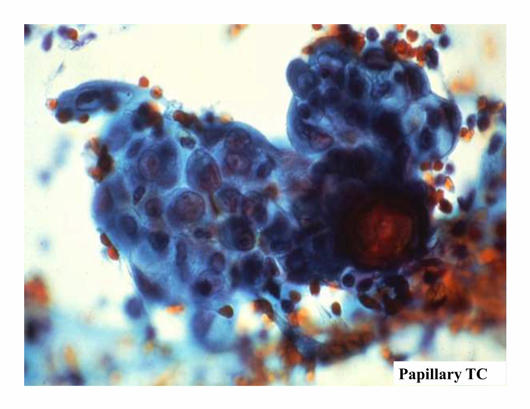
Papillary TC

Plasmacytoid Cells
• Plasma Cells
• Carcinoid / Islet
• Melanoma
• Breast CA
• Pleomorphic adenoma

Multiple Myeloma
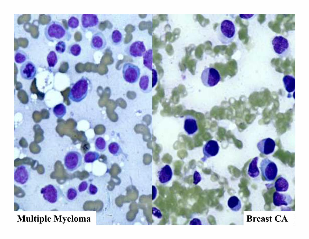
Multiple Myeloma Breast CA


Colloid (Mucinous) Neoplasms
• Colloid Carcinomas
GI tract, Breast, Ovary, Pancreas
• Pseudomyxoma peritonei (appendix)
• Myxoid sarcomas
• Melanoma (Rare)

Colon - Colloid CA

Mucin Positivity excludes:
• LYMPHOMA / LEUKEMIA
• SARCOMA (except chordoma)
• MELANOMA
Modified from DeMay

72 year old male presented with a
Case 5
72 year old male presented with a
single lung mass. FNA biopsy was
performed


CK 20

Case 5
DIAGNOSIS
Metastatic colon cancer to the
lung
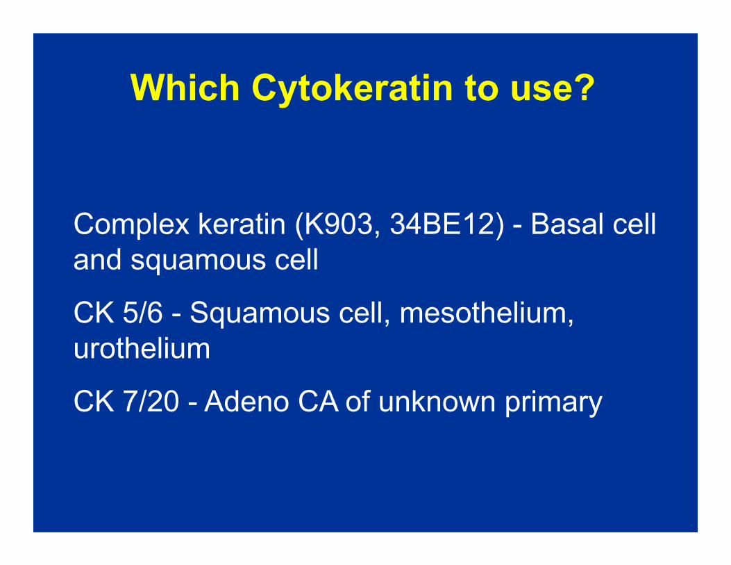
Which Cytokeratin to use?
Complex keratin (K903, 34BE12) - Basal cell
and squamous cell
CK 5/6 - Squamous cell, mesothelium,
urothelium
CK 7/20 - Adeno CA of unknown primary

IHC MARKERS FOR INTESTINAL CA
• CK 7/20
• Villin - Colorectal, pancreas. Occasionally in
non - GI i.e. endometrial, RCC (brush border
staining)staining)
• CDX2 - Intestinal tumors, also bladder adeno,
ovarian mucinous
Strong uniform CDX-2 +/with or without villin
- favors colorectal

Organ-specific and Organ-associated
Markers
Antibodies to: Identifying: Also identifies:
Prostatic specific antigen (PSA)
Prostatic acid phosphatase (PAP)
Gross cystic disease fluid protein -15
Thyroglobulin
Thyroid transcription factor-1 (TTF-1)
Uroplakin
Prostrate Carcinoma
Prostrate Carcinoma
Breast Carcinoma
Thyroid carcinoma
Thyroid and Lung carcinomas
Urothelial carcinomas
-----
Neuroendocrine carcinomas
Salivary gland, sweat gland tumors
-----
Rare other carcinomas
-----Uroplakin
Inhibin
Hep PAR-1
LCA, B&T
Urothelial carcinomas
Adrenal
Liver
Lymphoid
-----
Sex cord / stromal, granular cell
Modified from Pathol case Review 4(6), p254, 1999
Pathol case Review 4(6), p150, 2001

PSA +
Prostrate CA

IMMUNOHISTOCHEMICAL DETECTION OF
TTF-1 IN LUNG TUMORS
Adenocarcinoma 72.5%
Squamous carcinoma 10%
Large cell carcinoma 25.8%
Large cell neuoendocrine carcinoma 75.0%Large cell neuoendocrine carcinoma 75.0%
Typical carcinoid 30.5%
Atypical carcinoid 100%
Small cell carcinoma 94.1%
Alveolar adenoma 100%
Ordonez, N., Adv Anat Path 7:124, 2000

TTF-1 + / Adeno CA TTF-1 + / Small Cell CA
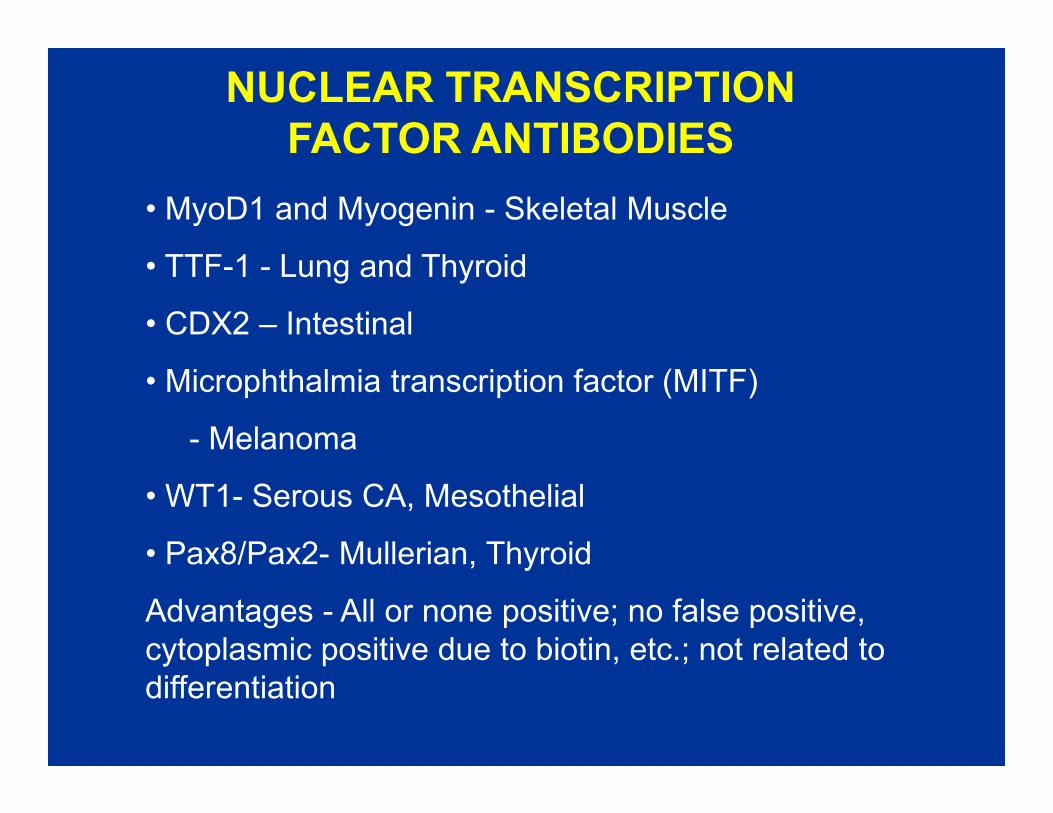
NUCLEAR TRANSCRIPTION
FACTOR ANTIBODIES
• MyoD1 and Myogenin - Skeletal Muscle
• TTF-1 - Lung and Thyroid
• CDX2 – Intestinal
• Microphthalmia transcription factor (MITF)
- Melanoma
• WT1- Serous CA, Mesothelial
• Pax8/Pax2- Mullerian, Thyroid
Advantages - All or none positive; no false positive,
cytoplasmic positive due to biotin, etc.; not related to
differentiation
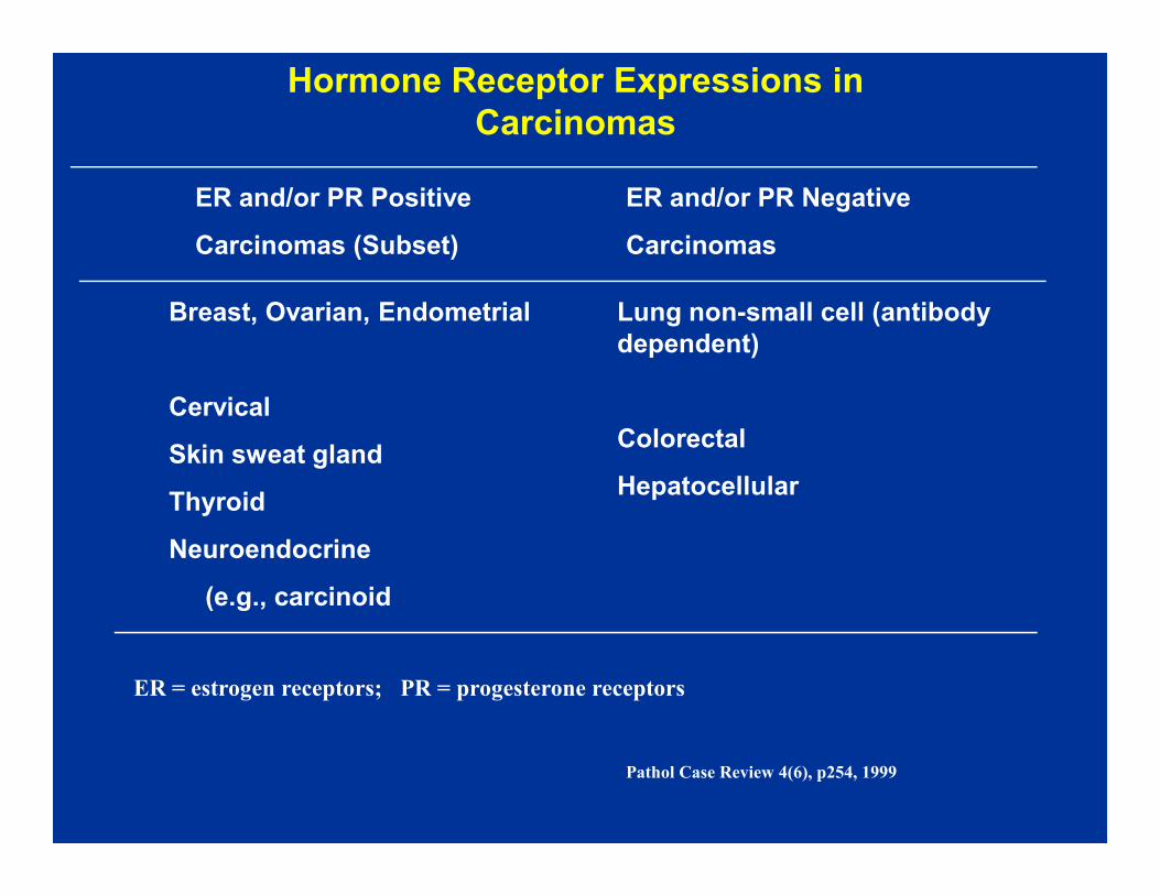
Hormone Receptor Expressions in
Carcinomas
ER and/or PR Positive
Carcinomas (Subset)
Breast, Ovarian, Endometrial
Cervical
Skin sweat gland
Lung non-small cell (antibody
dependent)
Colorectal
ER and/or PR Negative
Carcinomas
Skin sweat gland
Thyroid
Neuroendocrine
(e.g., carcinoid
Colorectal
Hepatocellular
Pathol Case Review 4(6), p254, 1999
ER = estrogen receptors; PR = progesterone receptors

Breast CA / ER +
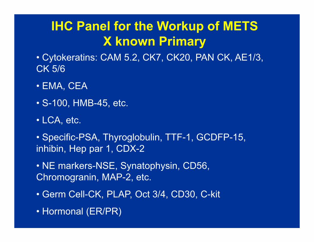
IHC Panel for the Workup of METS
X known Primary
• Cytokeratins: CAM 5.2, CK7, CK20, PAN CK, AE1/3,
CK 5/6
• EMA, CEA
• S-100, HMB-45, etc.
• LCA, etc.• LCA, etc.
• Specific-PSA, Thyroglobulin, TTF-1, GCDFP-15,
inhibin, Hep par 1, CDX-2
• NE markers-NSE, Synatophysin, CD56,
Chromogranin, MAP-2, etc.
• Germ Cell-CK, PLAP, Oct 3/4, CD30, C-kit
• Hormonal (ER/PR)
IHC WORKUP OF UNDIFFERENTIATED/POORLY
DIFFERENTIATED MALIGNANCY
AE-1/3 CD – 45 S-100 PLAP Additional
markers
Carcinoma + - +
-
- Differential
keratins,
EMA
Melanoma - - + - HMB 45, Melanoma - - + - HMB 45,
Melan A
Lymphoma - + - - CD 20, CD
3, CD 30
etc
Germ cell
tumor
-
+
- - + EMA,
OCT-4,
CD-30

Clinical Patterns of Metastasis
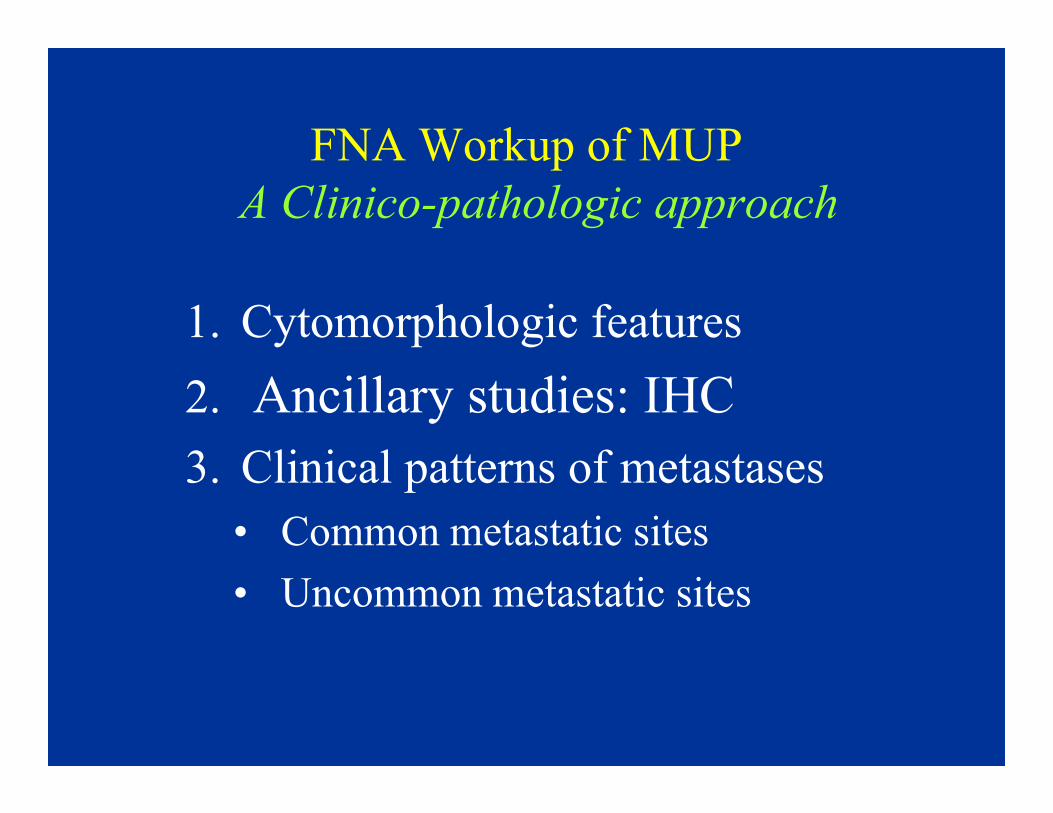
FNA Workup of MUP
A Clinico-pathologic approach
1. Cytomorphologic features
2. Ancillary studies: IHC2. Ancillary studies: IHC
3. Clinical patterns of metastases
• Common metastatic sites
• Uncommon metastatic sites

Metastatic Malignancies
• Determination of primary site is facilitated
by familiarity with cytologic features of the
malignancy and selected use of ICC
• Still, a primary site may not be determined • Still, a primary site may not be determined
because of non-specific cytologic & IHC
features, or an atypical pattern of
dissemination
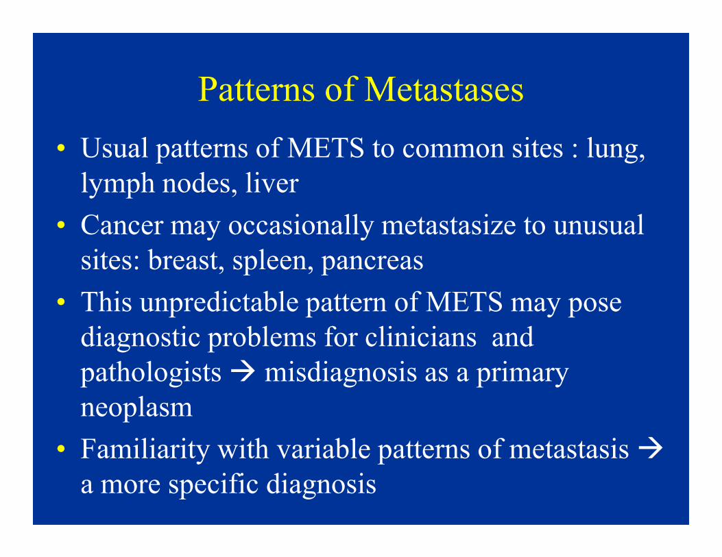
Patterns of Metastases
• Usual patterns of METS to common sites : lung,
lymph nodes, liver
• Cancer may occasionally metastasize to unusual
sites: breast, spleen, pancreas
• This unpredictable pattern of METS may pose
diagnostic problems for clinicians and
pathologists � misdiagnosis as a primary
neoplasm
• Familiarity with variable patterns of metastasis �
a more specific diagnosis

Initial Sites of Metastasis
• Parallel natural drainage pathways of primary malignancy, i.e. related to anatomic location of tumor
• Lymphatic: regional lymph nodes
– head & neck, cervix, melanoma – head & neck, cervix, melanoma
• Vascular: venous pathways
– head & neck, bone, kidney→ lung
– pancreas, stomach, colon → liver
– prostate � axial skeleton via paravertebral veins
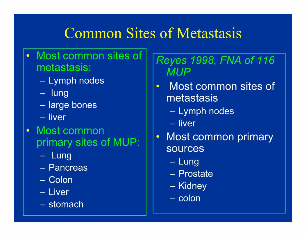
Common Sites of Metastasis
• Most common sites of metastasis:– Lymph nodes
– lung
– large bones
– liver
Reyes 1998, FNA of 116 MUP
• Most common sites of metastasis– Lymph nodes
– liver – liver
• Most common primary sites of MUP:– Lung
– Pancreas
– Colon
– Liver
– stomach
– liver
• Most common primary sources – Lung
– Prostate
– Kidney
– colon
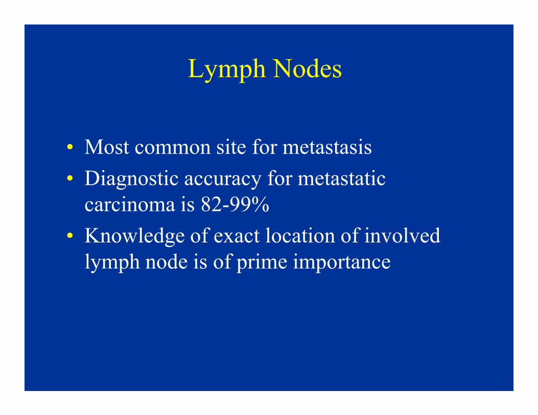
Lymph Nodes
• Most common site for metastasis
• Diagnostic accuracy for metastatic
carcinoma is 82-99%carcinoma is 82-99%
• Knowledge of exact location of involved
lymph node is of prime importance

Lymph Node Metastasis
Lymph nodes Common/Probable primary site or
malignancy
Cervical Head and neck, lung, melanoma, breast
Right supraclavicular Lung, breast, lymphoma
Left supraclavicular Lung, breast, cervix, prostate, lymphoma
Axillary Breast, lung, arm, regional trunk, GI tract
Inguinal Melanoma, trunk, leg, vulva, prostate,
anorectal, bladder
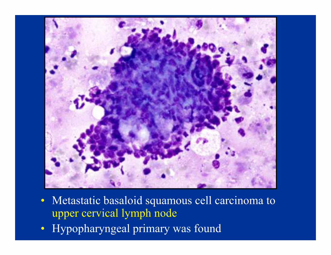
• Metastatic basaloid squamous cell carcinoma to upper cervical lymph node
• Hypopharyngeal primary was found
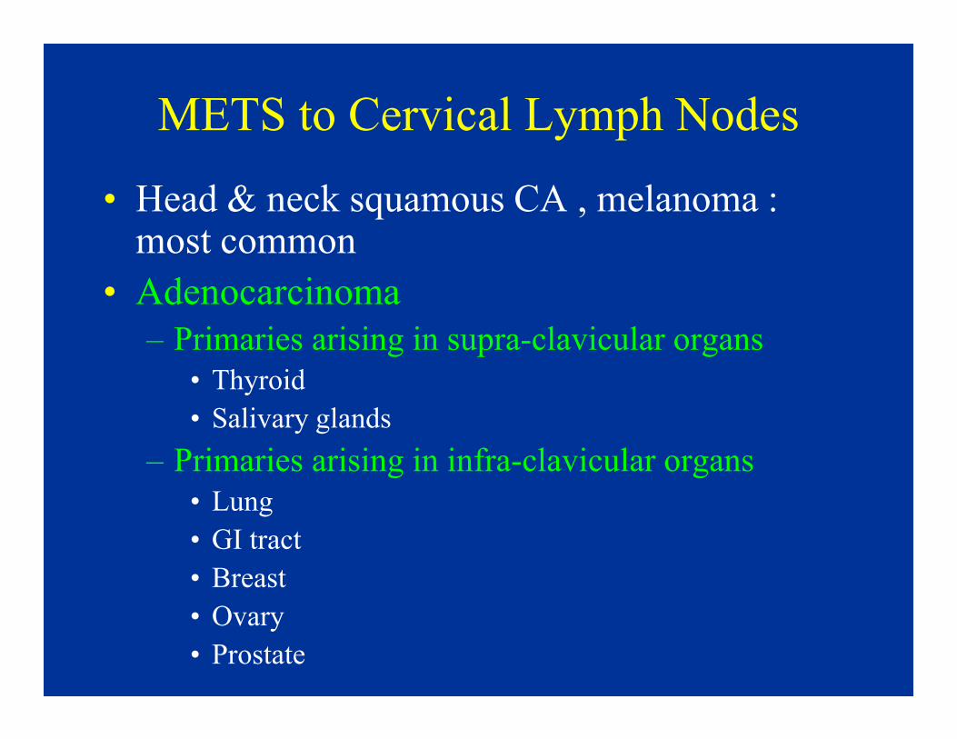
METS to Cervical Lymph Nodes
• Head & neck squamous CA , melanoma : most common
• Adenocarcinoma
– Primaries arising in supra-clavicular organs
• Thyroid• Thyroid
• Salivary glands
– Primaries arising in infra-clavicular organs
• Lung
• GI tract
• Breast
• Ovary
• Prostate

Supraclavicular Lymph Nodes
• Primary sites involving left SCLN (Virchow’s Node) are different from those involving right SCLN
• Cervin et al 1995, FNA of 96 SCLN• Cervin et al 1995, FNA of 96 SCLN
– Pelvic (16/19) & abdominal (6/6) malignancies → LSCLN
– Thorax, breast, head/neck � no difference in metastatic pattern to LSCLN or RSCLN
– Most common primaries: lung/breast > pelvis/testis > abdomen

Case 7. FNA biopsy of left supraclavicular lymph
node. The patient is a 65 year old man with a
remote previous history of malignancy

Diagnosis: Metastatic urothelial carcinoma. The
patient had a previous history of bladder CA

• PD carcinoma may mimic lymphoma
• Diff Dx: large cell lymphoma, neuroendocrine CA, melanoma
Dx: Metastatic large cell CA, lung 1º, involving
cervical lymph node

• Lymphoma may mimic carcinoma
DX: Anaplastic large cell lymphoma (Ki-1),
involving RSCLN

Lung Metastases
• Breast, GIT- common
• Any malignancy �lung
• Multiple nodules, most
commonly
– Miliary:
• Melanoma, kidney,
ovary, thyroid
medullary CA
– Cannon ball:
• Sarcoma, kidney,
melanoma,
colorectal CA

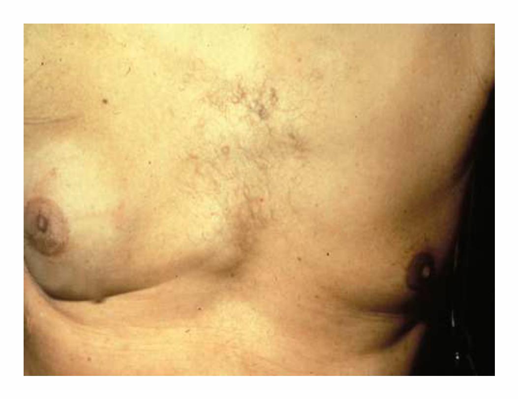
Multiple lung nodules (cannon ball) in 49 yr old woman.
No previous malig.
DX: Metastatic adeno CA c/w colon 1°
CDX2
CK20
•CK7-, CK20+
•CDX2+, TTF1-

Lung Metastases (cont.)
• Diffuse infiltrate or solitary coin lesion
(more problematic)→ rule out primary
lung carcinoma
• Diffuse (6-8 % of pulmonary mets):• Diffuse (6-8 % of pulmonary mets):
– Lung, breast, GI tract, pancreas
• Solitary MET (3-9 % of all solitary
pulmonary nodules):
– Melanoma, breast, colon, kidney, sarcoma,
non-seminomatous GCT
• FNA sensitivity =89%, specificity =96%

• Solitary lung
• IHC: CK 7+, CK 20-, TTF1-, ER+, PR+
Diagnosis: Metastatic breast ca
• Solitary lung mass, 68y F
• Hx breast ca X 1 month, SBR I, 0/18 nodes
breastER/PR

Lung
53 year old male presented with a solitary 3 cm lung
mass. Patient also had an indistinct kindey mass

• FNA of right lower lobe lung masses may
also inadvertently sample benign liver tissue

Lung
PAP
• Multiple lung nodules, 76 y M
• No previous hx of malignancy
5-10% of PD prostate CA either PSA- or PAP-(best to use both)
PSA

Unusual Sites of Metastasis
• Include breast, thyroid, pancreas, kidney,
small bones, eye, spleen
• Uncommonly encountered• Uncommonly encountered
• May pose diagnostic difficulties and lead
to confusion with primary neoplasms
arising in these sites

Mechanisms of Metastasis to
Unusual Sites
• Initial sites of metastasis → lymph nodes or
venous (lung, liver)venous (lung, liver)
• Subsequent (2°) widespread dissemination from initial metastatic site via arterial system
�brain, endocrine glands, small bones, spleen

METS to Thyroid
• Unusual site of involvement in clinical
practice; although autopsy series report 2-
26% of patients with malignancy
• Solitary mass or multiple small nodules• Solitary mass or multiple small nodules
• Direct extension – head & neck squamous
cell CA, adenoid cystic CA
• Kidney > colon, lung, breast > melanoma

METS to Thyroid (2)
• Alien cytology
• Differential diagnosis:
– Renal CC, clear cell type vs. thyroid CA with clear cellsclear cells
– RCC, granular type vs. Hurthle cell neoplasm
• RCA, TTF-1, thyroglobulin
– Plasmacytoma + amyloid vs. Medullary CA
(EMA, kappa/lambda, Calcitonin, CEA)
• Dx of metastasis may prevent inappropriate thyroidectomy

FNA right thyroid nodule,
76 year old female.
Patient had previous Hx
of malignancy X 15 yrs
•Diagnosis: Metastatic
Renal cell CA

SummaryCytopathologic Workup of MUP
• Clinico-pathologic approach
1. Cytomorphologic patterns
• Cell lineage: adenoca, squamous, etc.• Cell lineage: adenoca, squamous, etc.
• Cytomorphologic classification: small cell, large
cell, etc.
2. Ancillary studies – IHC
3. Clinical patterns of metastasis
• Common metastatic sites
• Uncommon metastatic sites

Gene Expression Profiling in MUP
• Confirm existing suspicions or provide new info?
- High agreement with already available CP data
– ? superiority to IHC + clinical info in unresolved
cases: not helpful (Personal experience w AviaraDx)
– Cost: $ 3,350 - 3,750– Cost: $ 3,350 - 3,750
• Prospective studies are needed to assess:
- Effect on patient outcome
- Which profiling methodology /gene panel is best?
• IHC remains crucial component of workup. GEP
may play supportive role in unresolved cases.
Promising future

General Principles Considered in
Analysis of Suspected Metastasis
• Familiar with cytologic features of common malignancies originating in a primary site
• Unusual/alien cytology for a primary site
• Knowledge of common and unusual • Knowledge of common and unusual metastatic patterns of malignancies & possible diagnostic pitfalls
• Produce a potential short list of possible primary sites
• Cytomorphology and IHC can then help arrive at a more specific diagnosis

General Principles Considered in
Analysis of Suspected Metastasis (2)
• Clinical history of previous malignancy
• Review of previous pathology material
• Tissue confirmation in unresolved cases
before definitive treatment