fMRI and ADHD: Understanding the Neurobiological Basis of ...
38
Neuroimaging and ADHD: The Neurobiological Basis of ADHD and Pharmacological Treatment Jeffrey H. Newcorn, MD Icahn School of Medicine at Mount Sinai
Transcript of fMRI and ADHD: Understanding the Neurobiological Basis of ...

Neuroimaging and ADHD: The Neurobiological Basis of ADHD and
Pharmacological Treatment
Jeffrey H. Newcorn, MD
Icahn School of Medicine at Mount Sinai

Locus Coeruleus
VTA
SubstantiaNigra
Posterior Parietal CortexNET, ɑ2A
Sensory input
Prefrontal Cortex D1,
D4, D5NET, ɑ2A
StriatumDAT, D2
NE enhances relevant signal;
regulates DA
DA suppresses irrelevant signal
NE enhances relevant signal
CerebellumD3, D4
Neurobiological Basis of ADHD
Modulators
Nicotinic/ Cholinergic
5-HT1A, 1B
H3
Synthesis/ metabolism
DƁH
COMT
MAO (A)
Interface with:
Glutamate
NMDA/ AMPA
GABA
Newcorn, NCDEU, 2008
Objectives of This Presentation
• Discuss different types of imaging techniques and their application to ADHD
• Examine results of studies using neuroimaging to elucidate the neurobiological basis of ADHD
• Show results of studies examining effects of medications for ADHD on brain function
• Present our own work on mechanisms of action and differential response to methylphenidate and atomoxetine

What Can PET Studies Tell Us?
• Opportunities
– Receptor binding to confirm hypothesized activity
– Glucose utilization (proxy for regional brain activity) while performing a task
• Disadvantages
– Single dose challenge - does not study actual clinical utility; better for stimulants than other drugs; still there are issues
– Desired radioligand often not available (e.g., NET)
– Expensive (multiple scans; cost of radioligand development; may require a cyclotron)

DA D2 Receptors
DA
DA
DA
DA DADA
DA
DA Synapse
DA Transporters
DAT:Increased:70% Dougherty et al, 1999
17% Krause et al, 2000
17% Dresel et al, 2000
30% McGough et al., 2001
39% Cheon et al, 2003*
34% Spencer et al., 2005
15% Spencer et al., 2007
No Change:van Dyck et al, 2002
Jucaite et al., 2005* (striatum)
Volkow et al., 2007 (putamen)
Decreased:16%↓ Jucaite et al., 2005* (midbrain)
13%↓ Volkow et al, 2007 (caudate)
DA D2:Increased: Ilgin et al., 2001*
No Change: Jucaite et al., 2005*
Decreased: Volkow et al., 2007*In children
DA Transporters and Receptors in ADHD

Striatal Dopamine Transporter Alterations in ADHD: Pathophysiology or Adaptation to Psychostimulants?
Fusar-Poli et al., Am J Psychiatry. 2012;169(3):264-272.

D2-Raclopride DAT-Cocaine
Density of DAT and D2/D3 in Adults With ADHD vs. Healthy Controls
[11C]raclopride distribution volume ratio images revealed 1 cluster with lower D2/D3
availability in ADHD participants than controls in the left hemisphere. This cluster included brain regions of the dopamine reward pathway–ventral caudate, accumbens, and midbrain regions, as well as the hypothalamic region
Volkow et al., JAMA, 2009 Sep 9;302(10):1084-91

Relationship Between Trait Motivation and DA Transporter and D2/D3 Receptor Availability
Scattergram showing the regression between the measures of DA D2/D3 receptor and of DAT availability in the NAcc and in the midbrain regions and Trait Motivation (MPQ Achievement scale) in ADHD participants (circles) and in controls (x).
Volkow et al., Mol Psychiatry, 2010 Sep 21

What Can MRI and fMRI Tell Us?
• Potential uses
– Pharm MRI: document site of activity (in animals)
– Structural MRI: regional brain volume and white matter connections; good for longitudinal studies
– fMRI: demonstrate regional activity in relation to task performance (animals; humans)
– fMRI: connections between brain regions/networks
• Limitations
– fMRI highly task dependent; task-based scans are lengthy; hard to obtain data on multiple tasks
– Difficulty scanning children

Developmental Trajectories of Cerebellum & Caudate Nucleus in ADHD
Castellanos et al. JAMA. 2002;288:1740-1748.
• Normalization of reduced
caudate volume in ADHD
by mid-adolescence
• Reduced cerebellar
volume in ADHD persists
through adolescence
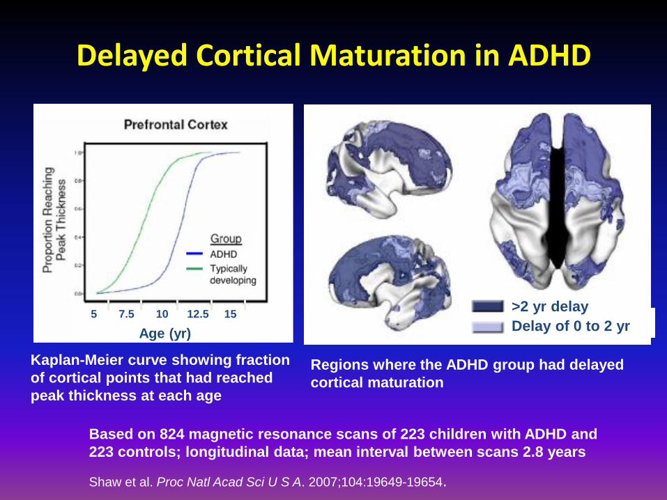
Delayed Cortical Maturation in ADHD
Regions where the ADHD group had delayed
cortical maturation
>2 yr delay
Delay of 0 to 2 yr
Kaplan-Meier curve showing fraction
of cortical points that had reached
peak thickness at each age
5 7.5 10 12.5 15
Age (yr)
Based on 824 magnetic resonance scans of 223 children with ADHD and
223 controls; longitudinal data; mean interval between scans 2.8 years
Shaw et al. Proc Natl Acad Sci U S A. 2007;104:19649-19654.

Normal ADHD Adult vs Normal Controls(fMRI During Perceptual Task)*
ADHD
Adults with ADHD process information less efficiently
*Stroop task utilized.MGH-NIMR Center & Harvard-MIT CITP, Bush G, et al. Biol Psychiatry. 1999;45(12):1542-1552.
Normal Controls
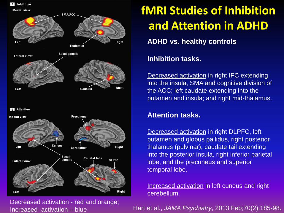
fMRI Studies of Inhibition and Attention in ADHD
Hart et al., JAMA Psychiatry, 2013 Feb;70(2):185-98.
ADHD vs. healthy controls
Inhibition tasks.
Decreased activation in right IFC extending
into the insula, SMA and cognitive division of
the ACC; left caudate extending into the
putamen and insula; and right mid-thalamus.
Attention tasks.
Decreased activation in right DLPFC, left
putamen and globus pallidus, right posterior
thalamus (pulvinar), caudate tail extending
into the posterior insula, right inferior parietal
lobe, and the precuneus and superior
temporal lobe.
Increased activation in left cuneus and right
cerebellum.
Decreased activation - red and orange;
Increased activation – blue

Plichta & Scheres, Neurosci Biobehavioral Rev, 38: 125–134, 2014
Meta-analysis Showing Ventral Striatal Hypo-responsiveness in ADHD During
Reward Anticipation

Resting State MRI: What Can We Learn About Pathophysiology and Treatment?
• Efficient method to study changes in and across networks, rather than individual regions
– Scans are short; larger n studies possible
– Instrumental in developing a new model of what ADHD is
– Possible to test in large numbers of subjects
• Excellent for hypothesis generation
• Excellent for cross-site studies and building larger data based

Default Mode Network: Functional Connectivity of ACC, Precuneus, PCC and VMPFC
ACC Seed: A robust negative or antiphasic relationship was noted between the ACC seed region and default-mode network components (i.e., an increase in ACC activity predicts a decrease in default-mode activity). ADHD–related decreases in functional connectivity were noted between the ACC and precuneus.
Precuneus Seed: ADHD-related decreases in precuneus/ACC connectivity, and among precuneus and other default-mode network components, including VMPFC and anterior portions of PCC.
Castellanos et al., Biol Psychiatry, 2007

Anti-correlated Task-Positive and Task-Negative Networks
Sonuga-Barke and Castellanos, Neurosci Biobehav Rev, 31:977–986, 2007

Summary: Using Imaging and ADHD Pathophysiology
• Studies have both confirmed hypothesizd neurobiological bases of ADHD and also generated new hypotheses about the disorder
• Current conceptualizations include a greater number of inter-digitating brain regions and an expanding array of neural circuits
• Moving away from region of interet analyses toward a broader conceptualization focusing on communication across regions

Imaging ADHD Treatment

At typical therapeutic doses (0.3-0.5 mg/kg), MPH occupies >50% of DATs
0
20
40
60
80
100
0.0 0.2 0.4 0.6 0.8 1.0
Dose (mg/kg)
Typical Dose
(0.5 mg/kg)
Placebo 20 mg 40 mg
Dopamine Transporter Occupancy by Methylphenidate
% D
AT
Occ
up
ancy
Volkow et al; J Neurosci, 2001
Volkow et al., Synapse, 2002

MPH Occupancy of NET at Clinically Relevant Doses in Humans
Hannestad et al., Biol Psychiatry, 2010
Demonstrates activity of an established treatment
in a different neurotransmitter system; examines
differences in activity across multiple brain regions
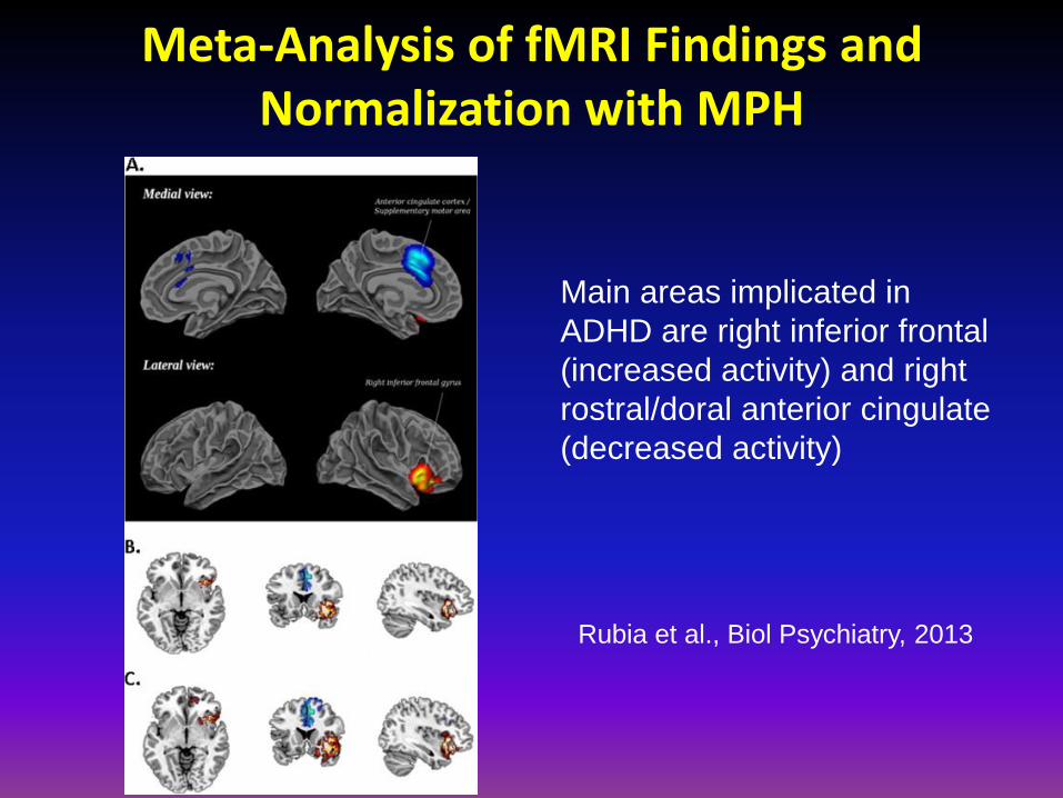
Meta-Analysis of fMRI Findings and Normalization with MPH
Main areas implicated in
ADHD are right inferior frontal
(increased activity) and right
rostral/doral anterior cingulate
(decreased activity)
Rubia et al., Biol Psychiatry, 2013

MPH Increases Suppression of Default-mode Activity in Precuneus and mPFC in Children with ADHD
Peterson et al., Am J Psychiatry 2009

MPH Normalizes OFC Activation for Reward Processing in Medication-naïve
Children with ADHD
Rubia et al., Neuropharmacology, 57:640–652, 2009
Rewarded – non-rewarded target trials on CPT:
MPH (single dose challenge) enhanced activation in mesial frontal cortex
(above), and anterior cingulate and caudate (not shown)

MPH Attentuation of mPFC Activity During an Emotional Stroop Task*
Positively valenced distraction interactions Negatively valenced distraction interactions
Posner et al., Psychiatry Research: Neuroimaging, 2011 *Using a different task to show an alternative
effect of a treatment

Atomoxetine Increases Right IFG Activation: Association with Improved Inhibitory Control
Chamberlain et al., Biol Psychiatry, 2009;65:550–555

Guanfacine Enhances Activation of dl-PFC During a
Cued Alerting Task*
Clerkin et al., Biol Psychiatry, 2009
Single dose challenge with guanfacine and placebo in healthy adults

Summary: Using Brain Imaging to Study ADHD Pathophysiology and Treatment
• Convergent evidence re: change in brain structure or function in association with treatment
• Several methods; each providing different information
– Receptor binding; metabolism (PET)
– Regional volume (MRI) acutely and over time; Regional activation (fMRI); Regional blood flow (pharm MRI)
– Connectivity (fMRI; Resting state); DTI (MRI)
• Methodologic issues in how to use/study medication
– Single dose challenge vs. clinical trial
– Acute discontinuation designs
– Common and unique effects of medications (discussed later)

Methylphenidate – Atomoxetine Crossover (MACRO) Study
Common and Unique Effects of Methylphenidate and Atomoxetine:
Understanding Mechanisms of Action and Predicting Differential Response

ATX MPH
MPH ATX
MACRO Study
Randomize
Methylphenidate
6-8 wks
Atomoxetine
6-8 wksAtomoxetine
6-8 wks
Open-label
treatment
2 wk
washout
2 wk
washout
Methylphenidate
6-8 wks
Baseline
fMRI
N = 56
EOT
fMRI
N = 36

fMRI Scan Procedures
• 3.0 Tesla Siemens Allegra head-
only MRI scanner with Echo
Planar Imaging (EPI) hardware
• Stimuli projected via LCD system
onto rear projection screen at
head of MRI bore
• Fiber optic response pad on right
hand
• Image Acquisition
• Spin-echo T2-weighted structural image
• 2D EPI sequence depicting BOLD signal
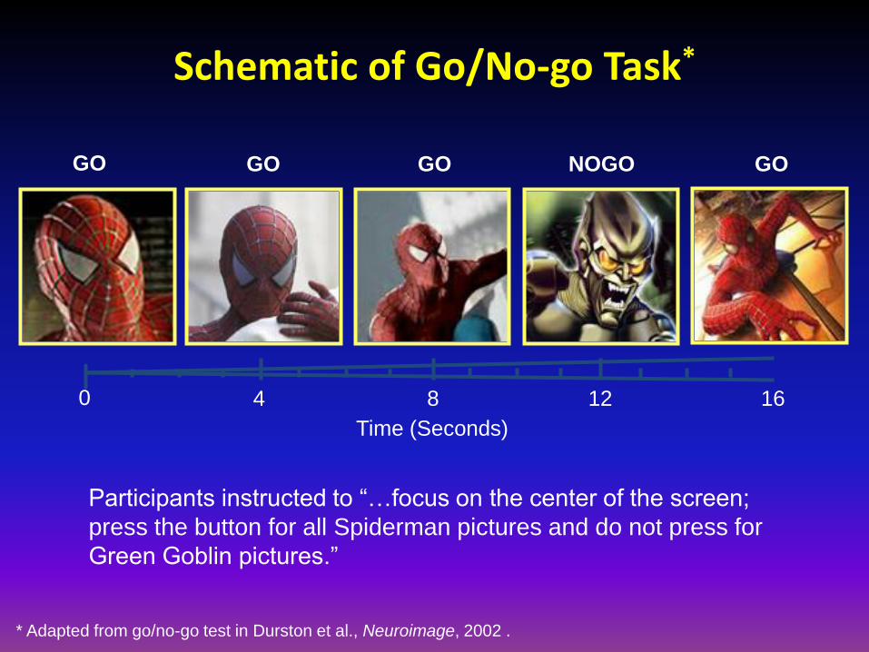
0 4 8 12 16
Time (Seconds)
GO GO GO GONOGO
Participants instructed to “…focus on the center of the screen;
press the button for all Spiderman pictures and do not press for
Green Goblin pictures.”
Schematic of Go/No-go Task*
* Adapted from go/no-go test in Durston et al., Neuroimage, 2002 .
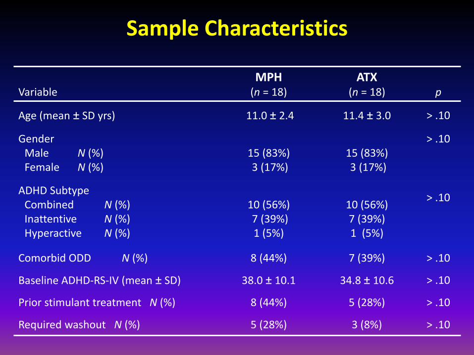
Sample Characteristics
VariableMPH
(n = 18)ATX
(n = 18) p
Age (mean ± SD yrs) 11.0 ± 2.4 11.4 ± 3.0 > .10
GenderMale N (%)Female N (%)
15 (83%)3 (17%)
15 (83%)3 (17%)
> .10
ADHD SubtypeCombined N (%)Inattentive N (%)Hyperactive N (%)
10 (56%)7 (39%)1 (5%)
10 (56%)7 (39%)1 (5%)
> .10
Comorbid ODD N (%) 8 (44%) 7 (39%) > .10
Baseline ADHD-RS-IV (mean ± SD) 38.0 ± 10.1 34.8 ± 10.6 > .10
Prior stimulant treatment N (%) 8 (44%) 5 (28%) > .10
Required washout N (%) 5 (28%) 3 (8%) > .10

Second-Level (Group) Analysis
• General linear model (GLM)
• Multiple regression
• Regressor 1: Medication (MPH vs. ATX)‡
• Regressor 2: ADHD-RS-IV change score‡
• Regressor 3: Interaction term
• Threshold: p < 0.01, kappa = 100 voxels
‡ Variable centered on zero

Differential Activation Profiles in Association with Response to MPH and ATX
Schulz et al., Arch Gen Psychiatry, 2012
- Pre and post-treatment (7 weeks) scans in 18 subjects treated with MPH and
18 subjects treated with ATX in randomized clinical trials
- Regression analysis incorporates change in regional activation and change
in ADHD-RS ratings in the same model

Conclusions
• First evidence of common and distinct frontoparietal
therapeutic mechanisms of action for stimulant and non-
stimulant treatments in youth with ADHD
• Common therapeutic mechanisms for MPH and ATX in motor cortex
• Possible attenuation in prepotency of inhibited “GO” responses
• Cannot rule-out practice and other non-specific factors
• MPH and ATX had divergent therapeutic actions on task-
positive and task-negative brain regions
• Divergent effects on inferior frontal activation indicate therapeutic
actions not solely attributable to promiscuous NET

Conclusions
• Atomoxetine “drives” mental effort
• Enhances inferior frontal inhibitory mechanisms
• Enhances cognitive control mechanisms in anterior cingulate cortex
• Homeostatic gains in posterior cingulate activation
• Amelioration of oft-reported prefrontal hypoactivation
• Methylphenidate increases neural efficiency
• Posterior cingulate deactivation reduces distracting
mental processes
• Reduces need for inferior frontal inhibitory effort
• Reduces need for anterior cingulate control effort
• Amelioration of poster cingulate hyperactivation

Acknowledgements
Collaborators: Mount Sinai Clinical Translational ADHD Program
• Kurt Schulz, PhD
• Jin Fan, PhD
• Suzanne Clerkin, PhD
• Jeffrey Halperin, PhD
• Anne-Claude Bédard, PhD
• Iliyan Ivanov, MD
Special Thanks to:
• Beth Krone, PhD
• Hanna Oltarzewska
• Robyn Palmero, PhD