Fluoride containing-bioactive-glasses-and-bioglasss-45 s5-form-apatite-in-low-ph-cell-culture-medium
5
See discussions, stats, and author profiles for this publication at: https://www.researchgate.net/publication/259574403 Fluoride-containing bioactive glasses and Bioglass (R) 45S5 form apatite in low pH cell culture medium ARTICLE in MATERIALS LETTERS · JANUARY 2014 Impact Factor: 2.49 · DOI: 10.1016/j.matlet.2013.12.102 CITATIONS 7 READS 177 5 AUTHORS, INCLUDING: Furqan Ali Shah University of Gothenburg 11 PUBLICATIONS 28 CITATIONS SEE PROFILE Delia S Brauer Friedrich Schiller University Jena 61 PUBLICATIONS 813 CITATIONS SEE PROFILE Robert G Hill Queen Mary, University of London 273 PUBLICATIONS 4,164 CITATIONS SEE PROFILE Karin A Hing Queen Mary, University of London 67 PUBLICATIONS 2,315 CITATIONS SEE PROFILE All in-text references underlined in blue are linked to publications on ResearchGate, letting you access and read them immediately. Available from: Furqan Ali Shah Retrieved on: 03 March 2016
-
Upload
elsenz-toothpaste -
Category
Healthcare
-
view
36 -
download
0
Transcript of Fluoride containing-bioactive-glasses-and-bioglasss-45 s5-form-apatite-in-low-ph-cell-culture-medium
Seediscussions,stats,andauthorprofilesforthispublicationat:https://www.researchgate.net/publication/259574403
Fluoride-containingbioactiveglassesandBioglass(R)45S5formapatiteinlowpHcellculturemedium
ARTICLEinMATERIALSLETTERS·JANUARY2014
ImpactFactor:2.49·DOI:10.1016/j.matlet.2013.12.102
CITATIONS
7
READS
177
5AUTHORS,INCLUDING:
FurqanAliShah
UniversityofGothenburg
11PUBLICATIONS28CITATIONS
SEEPROFILE
DeliaSBrauer
FriedrichSchillerUniversityJena
61PUBLICATIONS813CITATIONS
SEEPROFILE
RobertGHill
QueenMary,UniversityofLondon
273PUBLICATIONS4,164CITATIONS
SEEPROFILE
KarinAHing
QueenMary,UniversityofLondon
67PUBLICATIONS2,315CITATIONS
SEEPROFILE
Allin-textreferencesunderlinedinbluearelinkedtopublicationsonResearchGate,
lettingyouaccessandreadthemimmediately.
Availablefrom:FurqanAliShah
Retrievedon:03March2016
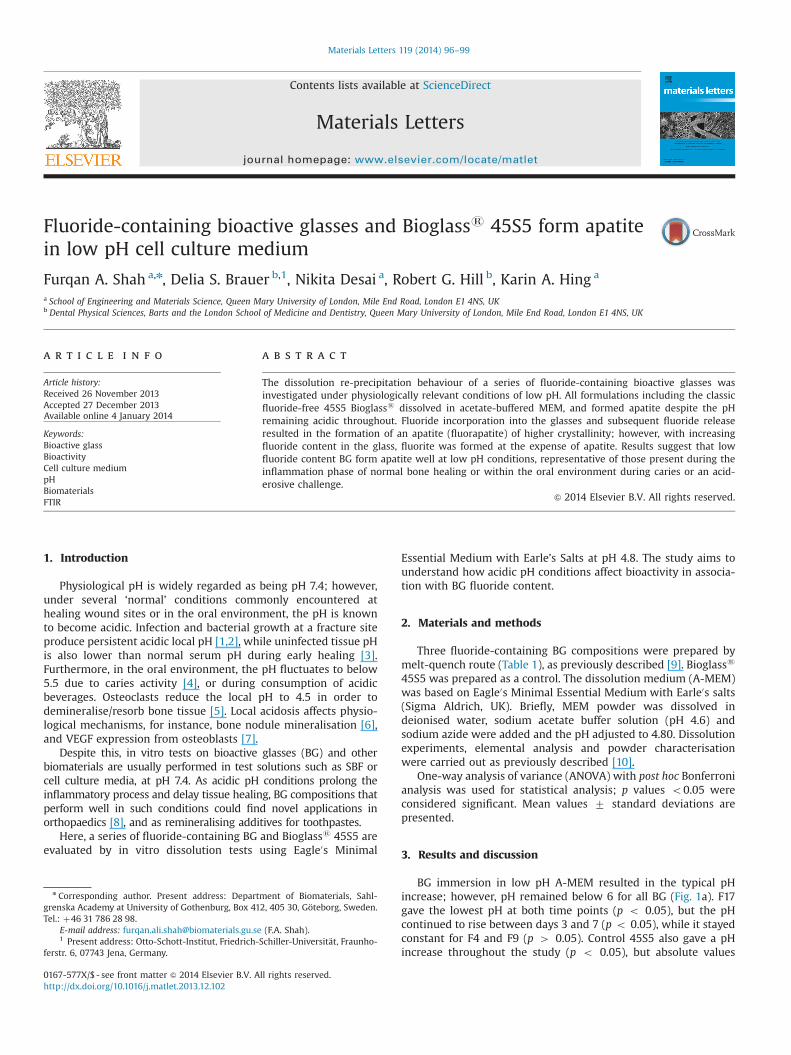
Fluoride-containing bioactive glasses and Bioglasss 45S5 form apatitein low pH cell culture medium
Furqan A. Shah a,n, Delia S. Brauer b,1, Nikita Desai a, Robert G. Hill b, Karin A. Hing a
a School of Engineering and Materials Science, Queen Mary University of London, Mile End Road, London E1 4NS, UKb Dental Physical Sciences, Barts and the London School of Medicine and Dentistry, Queen Mary University of London, Mile End Road, London E1 4NS, UK
a r t i c l e i n f o
Article history:Received 26 November 2013Accepted 27 December 2013Available online 4 January 2014
Keywords:Bioactive glassBioactivityCell culture mediumpHBiomaterialsFTIR
a b s t r a c t
The dissolution re-precipitation behaviour of a series of fluoride-containing bioactive glasses wasinvestigated under physiologically relevant conditions of low pH. All formulations including the classicfluoride-free 45S5 Bioglasss dissolved in acetate-buffered MEM, and formed apatite despite the pHremaining acidic throughout. Fluoride incorporation into the glasses and subsequent fluoride releaseresulted in the formation of an apatite (fluorapatite) of higher crystallinity; however, with increasingfluoride content in the glass, fluorite was formed at the expense of apatite. Results suggest that lowfluoride content BG form apatite well at low pH conditions, representative of those present during theinflammation phase of normal bone healing or within the oral environment during caries or an acid-erosive challenge.
& 2014 Elsevier B.V. All rights reserved.
1. Introduction
Physiological pH is widely regarded as being pH 7.4; however,under several ‘normal’ conditions commonly encountered athealing wound sites or in the oral environment, the pH is knownto become acidic. Infection and bacterial growth at a fracture siteproduce persistent acidic local pH [1,2], while uninfected tissue pHis also lower than normal serum pH during early healing [3].Furthermore, in the oral environment, the pH fluctuates to below5.5 due to caries activity [4], or during consumption of acidicbeverages. Osteoclasts reduce the local pH to 4.5 in order todemineralise/resorb bone tissue [5]. Local acidosis affects physio-logical mechanisms, for instance, bone nodule mineralisation [6],and VEGF expression from osteoblasts [7].
Despite this, in vitro tests on bioactive glasses (BG) and otherbiomaterials are usually performed in test solutions such as SBF orcell culture media, at pH 7.4. As acidic pH conditions prolong theinflammatory process and delay tissue healing, BG compositions thatperform well in such conditions could find novel applications inorthopaedics [8], and as remineralising additives for toothpastes.
Here, a series of fluoride-containing BG and Bioglasss 45S5 areevaluated by in vitro dissolution tests using Eagle0s Minimal
Essential Medium with Earle’s Salts at pH 4.8. The study aims tounderstand how acidic pH conditions affect bioactivity in associa-tion with BG fluoride content.
2. Materials and methods
Three fluoride-containing BG compositions were prepared bymelt-quench route (Table 1), as previously described [9]. Bioglasss
45S5 was prepared as a control. The dissolution medium (A-MEM)was based on Eagle0s Minimal Essential Medium with Earle0s salts(Sigma Aldrich, UK). Briefly, MEM powder was dissolved indeionised water, sodium acetate buffer solution (pH 4.6) andsodium azide were added and the pH adjusted to 4.80. Dissolutionexperiments, elemental analysis and powder characterisationwere carried out as previously described [10].
One-way analysis of variance (ANOVA) with post hoc Bonferronianalysis was used for statistical analysis; p values o0.05 wereconsidered significant. Mean values 7 standard deviations arepresented.
3. Results and discussion
BG immersion in low pH A-MEM resulted in the typical pHincrease; however, pH remained below 6 for all BG (Fig. 1a). F17gave the lowest pH at both time points (p o 0.05), but the pHcontinued to rise between days 3 and 7 (p o 0.05), while it stayedconstant for F4 and F9 (p 4 0.05). Control 45S5 also gave a pHincrease throughout the study (p o 0.05), but absolute values
Contents lists available at ScienceDirect
journal homepage: www.elsevier.com/locate/matlet
Materials Letters
0167-577X/$ - see front matter & 2014 Elsevier B.V. All rights reserved.http://dx.doi.org/10.1016/j.matlet.2013.12.102
n Corresponding author. Present address: Department of Biomaterials, Sahl-grenska Academy at University of Gothenburg, Box 412, 405 30, Göteborg, Sweden.Tel.: þ46 31 786 28 98.
E-mail address: [email protected] (F.A. Shah).1 Present address: Otto-Schott-Institut, Friedrich-Schiller-Universität, Fraunho-
ferstr. 6, 07743 Jena, Germany.
Materials Letters 119 (2014) 96–99
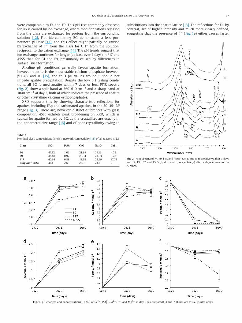
were comparable to F4 and F9. This pH rise commonly observedfor BG is caused by ion exchange, where modifier cations releasedfrom the glass are exchanged for protons from the surroundingsolution [12]. Fluoride-containing BG demonstrate a less pro-nounced pH rise [13], and this effect might partially be causedby exchange of F� from the glass for OH� from the solution,reciprocal to the cation exchange [14]. The pH trends suggest thation exchange continues for longer (at least over 7 days) in F17 and45S5 than for F4 and F9, presumably caused by differences insurface layer formation.
Alkaline pH conditions generally favour apatite formation;however, apatite is the most stable calcium phosphate betweenpH 4.5 and 10 [15], and thus pH values around 5 should notimpede apatite precipitation. Despite the low pH testing condi-tions, all BG formed apatite within 7 days or less. FTIR spectra(Fig. 2) show a split band at 560–610 cm�1 and a sharp band at1040 cm�1 at day 3, both of which indicate the presence of apatiteor other crystalline calcium orthophosphates.
XRD supports this by showing characteristic reflections forapatites, including FAp and carbonated apatites, in the 30–351 2θrange (Fig. 3). There are, however, distinct differences with glasscomposition. 45S5 exhibits peak broadening on XRD, which istypical for apatite formed by BG, as the crystallites are usually inthe nanometre size range [16] and of poor crystallinity owing to
substitutions into the apatite lattice [13]. The reflections for F4, bycontrast, are of higher intensity and much more clearly defined,suggesting that the presence of F� (Fig. 1e) either causes faster
Table 1Nominal glass compositions (mol%); network connectivity [11] of all glasses is 2.1.
Glass SiO2 P2O5 CaO Na2O CaF2
F4 47.12 1.02 21.98 25.13 4.75F9 44.88 0.97 20.94 23.93 9.28F17 40.68 0.88 18.98 21.69 17.76Bioglasss 45S5 46.1 2.6 26.9 24.3 –
Fig. 1. pH changes and concentrations (7SD) of Ca2þ , PO3�4 , Si4þ , F� , and Mg2þ at day 0 (as-prepared), 3 and 7. (Lines are visual guides only).
Fig. 2. FTIR spectra of F4, F9, F17, and 45S5 (a, c, e, and g, respectively) after 3 daysand F4, F9, F17 and 45S5 (b, d, f, and h, respectively) after 7 days immersion inA-MEM.
F.A. Shah et al. / Materials Letters 119 (2014) 96–99 97
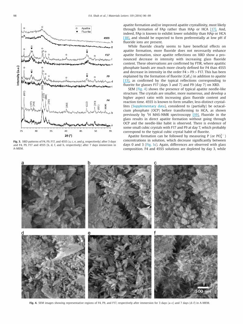
apatite formation and/or improved apatite crystallinity, most likelythrough formation of FAp rather than HAp or HCA [17]. And,indeed, FAp is known to exhibit lower solubility than HAp or HCA[18], and should be expected to form preferentially at low pH iffluoride ions are present.
While fluoride clearly seems to have beneficial effects onapatite formation, more fluoride does not necessarily enhanceapatite formation, since apatite reflections on XRD show a pro-nounced decrease in intensity with increasing glass fluoridecontent. These observations are confirmed by FTIR, where apatiticphosphate bands are much more clearly defined for F4 than 45S5and decrease in intensity in the order F44F94F17. This has beenexplained by the formation of fluorite (CaF2) in addition to apatite[13], as confirmed by the typical reflections corresponding tofluorite for glasses F17 (days 3 and 7) and F9 (day 7) on XRD.
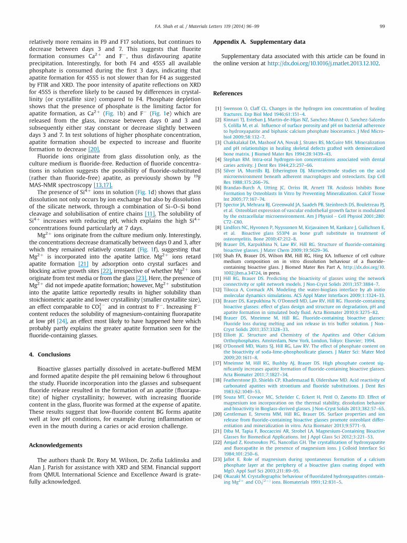
SEM (Fig. 4) shows the presence of typical apatite needle-likestructure. The crystals are smaller, more numerous, and develop ahigher aspect ratio with increasing glass fluoride content andreaction time. 45S5 is known to form smaller, less-distinct crystal-lites (Supplementary data), considered to (partially) be octacal-cium phosphate (OCP) before transforming to HCA, as shownpreviously by 1H MAS-NMR spectroscopy [19]. Fluoride in theglass results in direct apatite formation without going throughOCP and the needle-like habit is observed. There is evidence ofsome small cubic crystals with F17 and F9 at day 7, which probablycorrespond to the typical cubic crystal habit of fluorite.
Apatite formation can be followed by measuring P (or PO3�4 )
concentrations in solution, which decrease significantly betweendays 0 and 3 (Fig. 1c). Again, differences are observed with glasscomposition. F4 and 45S5 solutions are depleted by day 3, while
Fig. 3. XRD patterns of F4, F9, F17, and 45S5 (a, c, e, and g, respectively) after 3 daysand F4, F9, F17 and 45S5 (b, d, f, and h, respectively) after 7 days immersion inA-MEM.
Fig. 4. SEM images showing representative regions of F4, F9, and F17, respectively after immersion for 3 days (a–c) and 7 days (d–f) in A-MEM.
F.A. Shah et al. / Materials Letters 119 (2014) 96–9998
relatively more remains in F9 and F17 solutions, but continues todecrease between days 3 and 7. This suggests that fluoriteformation consumes Ca2þ and F� , thus disfavouring apatiteprecipitation. Interestingly, for both F4 and 45S5 all availablephosphate is consumed during the first 3 days, indicating thatapatite formation for 45S5 is not slower than for F4 as suggestedby FTIR and XRD. The poor intensity of apatite reflections on XRDfor 45S5 is therefore likely to be caused by differences in crystal-linity (or crystallite size) compared to F4. Phosphate depletionshows that the presence of phosphate is the limiting factor forapatite formation, as Ca2þ (Fig. 1b) and F� (Fig. 1e) which arereleased from the glass, increase between days 0 and 3 andsubsequently either stay constant or decrease slightly betweendays 3 and 7. In test solutions of higher phosphate concentration,apatite formation should be expected to increase and fluoriteformation to decrease [20].
Fluoride ions originate from glass dissolution only, as theculture medium is fluoride-free. Reduction of fluoride concentra-tions in solution suggests the possibility of fluoride-substituted(rather than fluoride-free) apatite, as previously shown by 19FMAS-NMR spectroscopy [13,17].
The presence of Si4þ ions in solution (Fig. 1d) shows that glassdissolution not only occurs by ion exchange but also by dissolutionof the silicate network, through a combination of Si–O–Si bondcleavage and solubilisation of entire chains [11]. The solubility ofSi4þ increases with reducing pH, which explains the high Si4þ
concentrations found particularly at 7 days.Mg2þ ions originate from the culture medium only. Interestingly,
the concentrations decrease dramatically between days 0 and 3, afterwhich they remained relatively constant (Fig. 1f), suggesting thatMg2þ is incorporated into the apatite lattice. Mg2þ ions retardapatite formation [21] by adsorption onto crystal surfaces andblocking active growth sites [22], irrespective of whether Mg2þ ionsoriginate from test media or from the glass [23]. Here, the presence ofMg2þ did not impede apatite formation; however, Mg2þ substitutioninto the apatite lattice reportedly results in higher solubility thanstoichiometric apatite and lower crystallinity (smaller crystallite size),an effect comparable to CO2�
3 and in contrast to F� . Increasing F�
content reduces the solubility of magnesium-containing fluorapatiteat low pH [24], an effect most likely to have happened here whichprobably partly explains the greater apatite formation seen for thefluoride-containing glasses.
4. Conclusions
Bioactive glasses partially dissolved in acetate-buffered MEMand formed apatite despite the pH remaining below 6 throughoutthe study. Fluoride incorporation into the glasses and subsequentfluoride release resulted in the formation of an apatite (fluorapa-tite) of higher crystallinity; however, with increasing fluoridecontent in the glass, fluorite was formed at the expense of apatite.These results suggest that low-fluoride content BG forms apatitewell at low pH conditions, for example during inflammation oreven in the mouth during a caries or acid erosion challenge.
Acknowledgements
The authors thank Dr. Rory M. Wilson, Dr. Zofia Luklinska andAlan J. Parish for assistance with XRD and SEM. Financial supportfrom QMUL International Science and Excellence Award is grate-fully acknowledged.
Appendix A. Supplementary data
Supplementary data associated with this article can be found inthe online version at http://dx.doi.org/10.1016/j.matlet.2013.12.102.
References
[1] Swenson O, Claff CL. Changes in the hydrogen ion concentration of healingfractures. Exp Biol Med 1946;61:151–4.
[2] Kinnari TJ, Esteban J, Martin-de-Hijas NZ, Sanchez-Munoz O, Sanchez-SalcedoS, Colilla M, et al. Influence of surface porosity and pH on bacterial adherenceto hydroxyapatite and biphasic calcium phosphate bioceramics. J Med Micro-biol 2009;58:132–7.
[3] Chakkalakal DA, Mashoof AA, Novak J, Strates BS, McGuire MH. Mineralizationand pH relationships in healing skeletal defects grafted with demineralizedbone matrix. J Biomed Mater Res 1994;28:1439–43.
[4] Stephan RM. Intra-oral hydrogen-ion concentrations associated with dentalcaries activity. J Dent Res 1944;23:257–66.
[5] Silver IA, Murrills RJ, Etherington DJ. Microelectrode studies on the acidmicroenvironment beneath adherent macrophages and osteoclasts. Exp CellRes 1988;175:266–76.
[6] Brandao-Burch A, Utting JC, Orriss IR, Arnett TR. Acidosis Inhibits BoneFormation by Osteoblasts In Vitro by Preventing Mineralization. Calcif TissueInt 2005;77:167–74.
[7] Spector JA, Mehrara BJ, Greenwald JA, Saadeh PB, Steinbrech DS, Bouletreau PJ,et al. Osteoblast expression of vascular endothelial growth factor is modulatedby the extracellular microenvironment. Am J Physiol – Cell Physiol 2001;280:C72–C80.
[8] Lindfors NC, Hyvonen P, Nyyssonen M, Kirjavainen M, Kankare J, Gullichsen E,et al. Bioactive glass S53P4 as bone graft substitute in treatment ofosteomyelitis. Bone 2010;47:212–8.
[9] Brauer DS, Karpukhina N, Law RV, Hill RG. Structure of fluoride-containingbioactive glasses. J Mater Chem 2009;19:5629–36.
[10] Shah FA, Brauer DS, Wilson RM, Hill RG, Hing KA. Influence of cell culturemedium composition on in vitro dissolution behaviour of a fluoride-containing bioactive glass. J Biomed Mater Res Part A, http://dx.doi.org/10.1002/jbm.a.34724, in press.
[11] Hill RG, Brauer DS. Predicting the bioactivity of glasses using the networkconnectivity or split network models. J Non-Cryst Solids 2011;357:3884–7.
[12] Tilocca A, Cormack AN. Modeling the water-bioglass interface by ab initiomolecular dynamics simulations. ACS Appl Mater Interfaces 2009;1:1324–33.
[13] Brauer DS, Karpukhina N, O’Donnell MD, Law RV, Hill RG. Fluoride-containingbioactive glasses: effect of glass design and structure on degradation, pH andapatite formation in simulated body fluid. Acta Biomater 2010;6:3275–82.
[14] Brauer DS, Mneimne M, Hill RG. Fluoride-containing bioactive glasses:Fluoride loss during melting and ion release in tris buffer solution. J Non-Cryst Solids 2011;357:3328–33.
[15] Elliott JC. Structure and Chemistry of the Apatites and Other CalciumOrthophosphates. Amsterdam, New York, London, Tokyo: Elsevier; 1994.
[16] O’Donnell MD, Watts SJ, Hill RG, Law RV. The effect of phosphate content onthe bioactivity of soda-lime-phosphosilicate glasses. J Mater Sci: Mater Med2009;20:1611–8.
[17] Mneimne M, Hill RG, Bushby AJ, Brauer DS. High phosphate content sig-nificantly increases apatite formation of fluoride-containing bioactive glasses.Acta Biomater 2011;7:1827–34.
[18] Featherstone JD, Shields CP, Khademazad B, Oldershaw MD. Acid reactivity ofcarbonated apatites with strontium and fluoride substitutions. J Dent Res1983;62:1049–53.
[19] Souza MT, Crovace MC, Schröder C, Eckert H, Peitl O, Zanotto ED. Effect ofmagnesium ion incorporation on the thermal stability, dissolution behaviorand bioactivity in Bioglass-derived glasses. J Non-Cryst Solids 2013;382:57–65.
[20] Gentleman E, Stevens MM, Hill RG, Brauer DS. Surface properties and ionrelease from fluoride-containing bioactive glasses promote osteoblast differ-entiation and mineralization in vitro. Acta Biomater 2013;9:5771–9.
[21] Diba M, Tapia F, Boccaccini AR, Strobel LA. Magnesium-Containing BioactiveGlasses for Biomedical Applications. Int J Appl Glass Sci 2012;3:221–53.
[22] Amjad Z, Koutsoukos PG, Nancollas GH. The crystallization of hydroxyapatiteand fluorapatite in the presence of magnesium ions. J Colloid Interface Sci1984;101:250–6.
[23] Jallot E. Role of magnesium during spontaneous formation of a calciumphosphate layer at the periphery of a bioactive glass coating doped withMgO. Appl Surf Sci 2003;211:89–95.
[24] Okazaki M. Crystallographic behaviour of fluoridated hydroxyapatites contain-ing Mg2þ and CO3
(2�) ions. Biomaterials 1991;12:831–5.
F.A. Shah et al. / Materials Letters 119 (2014) 96–99 99