Children in High Fluoridated Areas Have Lower Iq Nih Study 2012

This is a repository copy of Fluoridated elastomers: Effect on the microbiology of plaque .
White Rose Research Online URL for this paper:http://eprints.whiterose.ac.uk/402/
Article:
Benson, P.E., Douglas, C.W.I. and Martin, M.V. (2004) Fluoridated elastomers: Effect on the microbiology of plaque. American Journal of Orthodontics and Dentofacial Orthopedics, 126 (3). pp. 325-330. ISSN 0889-5406
https://doi.org/10.1016/j.ajodo.2003.07.007
[email protected]://eprints.whiterose.ac.uk/
Reuse
Unless indicated otherwise, fulltext items are protected by copyright with all rights reserved. The copyright exception in section 29 of the Copyright, Designs and Patents Act 1988 allows the making of a single copy solely for the purpose of non-commercial research or private study within the limits of fair dealing. The publisher or other rights-holder may allow further reproduction and re-use of this version - refer to the White Rose Research Online record for this item. Where records identify the publisher as the copyright holder, users can verify any specific terms of use on the publisher’s website.
Takedown
If you consider content in White Rose Research Online to be in breach of UK law, please notify us by emailing [email protected] including the URL of the record and the reason for the withdrawal request.

Fluoridated Elastomers – Effect on the Microbiology of
Plaque
P.E. Benson PhD FDS (Orth)1
C.W.I. Douglas BSc PhD1
M.V. Martin PhD FRCPath2
1 School of Clinical Dentistry, University of Sheffield, UK. 2 Liverpool University Dental Hospital, Liverpool, UK. Address for correspondence: Dr P. E. Benson, Department of Oral Health and Development, School of Clinical Dentistry, Claremont Crescent, Sheffield, S10 2TA. Tel: 0114 271 7885 Fax: 0114 271 7843 E-mail: p.benson@sheffield .ac.uk Keywords: Orthodontic, dental plaque, fluoride, elastomers, randomised clinical trial
1

Abstract
Objective: To investigate the effect of fluoridated elastomeric ligatures on the
microbiology of the local dental plaque in vivo.
Design: A randomised, prospective, longitudinal clinical trial, employing a split
mouth, crossover design.
Sample population and setting: 30 individuals at the beginning of their treatment
with fixed orthodontic appliances in the Orthodontic Departments of Liverpool and
Sheffield Dental Hospitals.
Method: The study consisted of two experimental periods of six weeks with a
washout period between. Fluoridated elastomers were randomly allocated at the first
visit to be placed around brackets on 12, 11, 33; or 22, 21, 43. Non-fluoridated
elastomers were placed on the contra-lateral teeth. A standard non-antibacterial
fluoridated toothpaste and mouthwash were supplied. After 6 weeks (visit 2) the
elastomers were removed, placed in transport media and plated on agar within two
hours. Non-fluoridated elastomers were placed on all brackets for one visit to allow
for a washout period. At visit 3 fluoridated elastomers were placed on the contra-
lateral teeth to visit 1. At visit 4 the procedures at visit 2 were repeated. Therefore the
experiment was performed over a total of four treatment visits, with samples collected
on visit 2 and 4. A logistic regression was performed with the dependent variable
being the presence or absence of streptococcal or anaerobic growth. A mixed effects
ANOVA was carried out with the dependent variable being the percent streptococcal
or anaerobic bacterial count.
Results: The only significant independent variables were the subject variable
(p=<0.001) for the percent streptococcal and anaerobic bacterial count and the visit
variable for the percent streptococcal count (p=<0.001). The use of fluoridated or
non-fluoridated elastomers was not significant for either percent streptococcal count
(p=0.288) or anaerobic count (p=0.230).
Conclusion: Fluoridated elastomers are not effective at reducing local streptococcal
or anaerobic bacterial growth after a clinical relevant time in the mouth.
2

Introduction
Plaque is a major aetiological factor in the development of dental caries. The control
of plaque is fundamental in the control of caries and periodontitis. It has been shown
that the placement of a fixed orthodontic appliance leads to both an increase in the
levels1 and a change in the composition of dental plaque2. Sakamaki and Bahn3
showed an increase in lactobacillus index and salivary lactobacillus counts with the
placement of orthodontic bands. Corbett et al4 demonstrated an increase in the level
of Streptococcus mutans in the plaque surrounding an orthodontic appliance. Scheie
et al5 demonstrated an increase in the levels of Streptococcus mutans in saliva and
suggested that the placement of an orthodontic appliance leads to the creation of
new retentive areas favouring the local growth of this organism.
Elastomeric ligatures that release stannous fluoride are commercially available6.
Maltz and Emilson7 showed that stannous and cupric fluoride had a superior
antimicrobial effect on streptococci and lactobacilli over sodium or ammonium
fluoride and suggested that the metal ions play a large part in their bactericidal effect.
A reduction in the level of Streptococcus mutans in the saliva with these elastomers
has been demonstrated8. If fluoridated elastomers are shown to adversely affect local
cariogenic bacteria, they will be important in the reduction of enamel demineralisation
around orthodontic brackets.
The objective of this study was to investigate the effect of fluoridated elastomeric
ligatures on the bacterial count of dental plaque forming on the ligatures in vivo.
3

Materials and Methods
This was a prospective, randomised clinical trial, employing a split mouth, crossover
design. Volunteers were recruited from patients about to start their orthodontic
treatment with upper and lower fixed appliances in the orthodontic departments of
Liverpool University Dental Hospital and the Charles Clifford Dental Hospital,
Sheffield. Patients who were pregnant; diabetic; using an antimicrobial mouthwash;
using any complicating medicine or patients with a history of antibiotic use in the last
two months were excluded. All patients visited a hygienist who provided standardized
oral hygiene instruction. They had to achieve a score of 1 on the Index of Oral
Cleanliness prior to placement of fixed appliances
Ethical approval was obtained from the Local Research Ethics Committees. Eligible
patients were invited to participate in the study at a visit before the fixed appliances
were placed. Informed, written consent was given by the patients and their parents
agreeing to enter the study on the visit the appliances were placed. This was usually
two weeks after the initial discussion.
The following procedures were carried out:
Visit 1: The fixed appliance brackets and bands were placed. The patients were
randomly allocated to having the fluoridated elastomers (Fluor-I-Ties, Ortho Arch Co.,
Schaumburg, IL USA) either on the upper left lateral incisor, upper left central incisor
and lower right canine; or the upper right lateral incisor, upper right central incisor
and lower left canine. The randomisation was carried out using computer generated
random numbers in a block design. The allocation was concealed in consecutively
4

numbered, sealed, opaque envelopes. Conventional non-fluoridated elastomers were
placed on the remaining teeth. The patients were provided with a standard
fluoridated toothpaste (Aquafresh; monofluorophosphate 0.75% w/w and sodium
fluoride 0.01% w/w; total fluoride 1,055ppm SmithKline Beecham Consumer
Healthcare, Middlesex, UK.), with no antimicrobial ingredients and a daily fluoride
mouthrinse (Fluorigard, 0.05% NaF Colgate-Palmolive (UK) Ltd, Surrey, UK).
Visit 2: Six weeks later at the first adjustment appointment, the elastomers on the
upper incisors and lower canines were aseptically removed, placed in separate
containers with a pre-reduced transport medium and coded. These were taken to the
laboratory within ten minutes. The adjustment to the appliance was carried out and
non-fluoridated elastomers were placed on all the teeth to allow for a washout period
of at least six weeks.
Visit 3: The appliance was adjusted and the fluoridated elastomers placed on the
contra-lateral teeth to the first appointment. Therefore, if at appointment 2 the patient
had the fluoridated module placed on the upper left incisors and lower right canine, at
appointment 3 the fluoridated module was placed on the upper right incisors and
lower left canine. Non-fluoridated elastomers were placed on the remaining teeth.
Visit 4: Six weeks later, the procedures carried out during appointment 2 were
repeated.
Microbiology
On arrival in the laboratory each elastomeric sample that was collected on visit 2 and
4 was vortexed for 30 seconds, serially diluted and 100µl aliquots plated onto blood
5

agar (blood agar base number 2, Lab M, Bury Lancs supplemented with horse blood
5% v/v, Oxoid) and Mitis Salivarius (MS) agar (Difco, USA) supplemented with
sucrose (20% w/v) and bacitracin (0.2 units/ml). Plates were incubated for up to 7
days at 37°C either in CO 2 or anaerobically on pre-reduced plates under an
atmosphere of 80% N2, 10% H, 10%CO2.
Cultures were assessed for total aerobic, total anaerobic and total Mitis Salivarius
colony forming units (cfu). Wherever possible, cultures with colonies in the range 30-
300 cfu were chosen for counting.
Representative colonies recovered on the Mitis Salivarius Bacitracin medium were
Gram-stained to confirm that they were streptococci. Each streptococcal colony type
was then speciated using the Rapid ID32 Strep system (bioMérieux, Basingstoke,
UK). Although Streptococcus sanguis and S.parasanguis were recovered on the Mitis
Salivarius Bacitracin agar plates in small numbers, growth was dominated by
S.mutans. No S.sobrinus or other species of mutans streptococci were found with the
identification system employed.
Statistics
Sample Size Calculation
A sample size calculation was carried out using data from two previous studies4, 9.
This suggested that a sample size of 30 would be sufficient to detect a difference in
the Streptococcus mutans count of 30 percent to a power of 0.85 with a significance
level of 0.05.
6
Hypothesis Testing
The streptococcus count was expressed as a percentage of the total aerobic count.
There were a number of occasions when no streptococci were recovered, therefore a
logistic regression was carried out to assess whether there were any factors that
affected the presence or absence of bacterial growth. The dependent variable was
categorical yes or no indicating the presence or absence of bacterial growth. The
covariates were subject, visit, fluoride or non-fluoride elastomeric, tooth type,
dominant or non-dominant toothbrushing hand side, and number of days in the
mouth. A mixed effects ANOVA was used to investigate the positive bacterial counts.
The dependent variable was either the percentage streptococcal count or the
anaerobic bacterial count, these were transformed to log10 values as they were found
to be positively skewed. The random variable was the subject. The fixed factors
included gender of patient, visit, fluoride or non-fluoride elastomeric, tooth type and
dominant or non-dominant toothbrushing hand side. Covariates included age and the
number of days the elastomer was in place.
Results
Thirty patients were recruited to the study. Three patients dropped out before any
microbiology could be obtained. Several samples were lost due to failure of the
fluoridated elastomers between appointments, debonding of brackets and
irregularities in laboratory procedure. A total of 220 elastomers were collected from
27 patients. There were 18 females and 9 males. The average age was 14.2 years
(sd 2.1, range 11.8 – 20.6). The elastomers were in the mouth for an average of 39.4
days (sd 9.0, range 28 – 67) for the first visit and 41.3 days (sd 9.1, range 28 – 63)
for the second visit.
7

The results of the logistic regression are revealed in Table I. This shows that for the
dependent variable, presence or absence of bacterial count, there were no significant
independent variables for either the streptococcal count or the anaerobic growth.
There were a higher proportion of negative streptococcal growths following the first
visit (27%) than following the second visit (7%). The proportion of the total variation
explained by both models was 94 percent, which suggests that the amount of
variation not explained by the analysis was low and therefore these were good
models to explain the presence or absence of streptococcal growth and anaerobic
bacterial growth.
The results of the mixed effects analysis of variance are shown in Table II. There
were significant results for the subject factor for percentage streptococcal and
anaerobic growths (p<0.001). The only other significant factor for the percent
streptococcal growth was whether the ligature was collected on the first or second
visit.
Figure 1 shows two boxplots of the bacterial counts from the streptococcus growth
media for ligatures collected on the first and second visits. This shows that the
median and interquartile range of the streptococcal counts was greater for the
ligatures collected on the second visit.
There was no significant effect of the fluoridated elastomeric ligatures on the
bacterial growth either in terms of the percent streptococcal count (p=0.288) or the
total anaerobic count (p=0.230). The bacterial counts were positively skewed and
therefore the median and interquartile descriptors for the fluoridated and non-
8

fluoridated elastomers were calculated. These are revealed in Table III. Boxplots of
these data are shown in Figure 2 and Figure 3.
Discussion
This study has shown that after a clinically relevant period of time in the mouth there
were no significant differences in percent streptococcal or anaerobic bacterial counts
in plaque obtained from fluoridated elastomers compared with conventional
elastomers. It must be concluded that fluoridated elastomers are not effective at
reducing streptococcal or anaerobic bacterial growth in plaque on a ligature and so
probably also that surrounding an orthodontic bracket between adjustment visits.
The reason for this is unknown, but is most likely explained by the fact that
fluoridated elastomers release high levels of fluoride initially, but this release rapidly
drops to a point where it will not affect bacterial growth and metabolism6. The
currently accepted view is that levels of fluoride that are bactericidal are far higher
than are likely to be present in the mouth10.
The short-term effects of fluoridated elastomers on the oral flora have been
demonstrated previously. Wilson and Gregory8 compared a group of patients wearing
fluoridated elastomers with a group wearing conventional elastomers. They showed
that the percent Streptococcus mutans count as a proportion of the total
streptococcal count reduced significantly in unstimulated whole saliva samples after
one week in individuals supplied with the fluoridated elastomers. However they found
that the streptococcal count then rose to baseline levels in the second week.
9

Hallgren et al9 looked at the effect on plaque of bonding the brackets with glass
ionomer cement compared with conventional composite. Using a split mouth design
on 12 individuals undergoing orthodontic treatment, they collected pooled plaque
from test and control quadrants and found that the proportion of Streptococcus
mutans was lower around brackets bonded with the glass ionomer cement. They also
found that the concentration of lactic acid in plaque samples taken from glass
ionomer cement was lower11. However, these findings might have been due to
differences in the properties of the two materials other than their fluoride content12.
Another reason for the apparent lack of effect of fluoridated elastomers on the plaque
flora might be due to higher levels of bacteria found on the elastomeric material.
Forsberg et al13 found that most patients had a higher bacterial count on teeth ligated
with conventional elastomers compared with teeth ligated with steel ligatures. In the
present investigation it was noticed that clinically there was a marked deterioration in
the physical properties of fluoridated elastomers in the mouth. They were
considerably swollen compared with the conventional elastomers after six weeks and
several were missing when the patient returned (Figure 4). This has been noted
previously14. Wiltshire15 found that the fluoridated elastomers doubled their weight
after 1 month in the mouth, while the weight of the non-fluoride elastomers remained
virtually unchanged. It could be that the plaque inhibiting effect of the fluoride is
cancelled out by the deterioration in the physical properties of the elastomeric, which
leads to a higher bacterial load in fluoridated elastomers.
In this investigation there was a higher proportion of samples with no streptococcal
growth collected from the first visit compared with the second visit. This confirms the
10

work of Schiei et al5 who found that there was a transient decrease in plaque and
salivary Streptococcus mutans levels in the first few weeks of placing a fixed
orthodontic appliance. They suggested that this was due to reduction in streptococcal
reservoirs following the banding procedure. After three months the proportion of
Streptococcus mutans had risen above pre-treatment levels.
Although fluoridated elastomers have not been shown to reduce the proportion of
streptococci or number of anaerobic bacteria in plaque, they might help to reduce the
prevalence and severity of demineralisation during orthodontics16, 17 by raising the
concentration of fluoride in the plaque adjacent to the bracket. This will help to
restore a positive balance between remineralisation and demineralisation and would
be a valuable area of further research.
Conclusion
Fluoridated elastomers are not effective at reducing streptococcal or anaerobic
bacterial growth in local plaque surrounding an orthodontic bracket after a mean time
of 40 days in the mouth.
Individual oral hygiene instruction for patients is likely to be more effective at
reducing local plaque.
Acknowledgements
This study was supported by a General Research Grant from the Royal College of
Surgeons of Edinburgh.
11

References
1. Alstad S, Zachrisson BU. Longitudinal study of periodontal condition
associated with orthodontic treatment in adolescents. Am J Orthod, 1979; 76:
277-86.
2. Huser MC, Baehni PC, Lang R. Effects of orthodontic bands on microbiologic
and clinical parameters. Am J Orthod Dentofacial Orthop, 1990; 97: 213-8.
3. Sakamaki ST, Bahn AN. Effect of orthodontic banding on localized oral
lactobacilli. J Dent Res, 1968; 47: 275-9.
4. Corbett JA, Brown LR, Keene HJ, Horton IM. Comparison of Streptococcus
mutans concentrations in non-banded and banded orthodontic patients. J Dent
Res, 1981; 60: 1936-42.
5. Scheie AA, Arneberg PAL, Krogstad O. Effect of orthodontic treatment on
prevalence of Streptococcus mutans in plaque and saliva. Scand J Dent Res,
1984; 92: 211-7.
6. Wiltshire WA. Determination of fluoride from fluoride-releasing elastomeric
ligature ties. Am J Orthod Dentofacial Orthop, 1996; 110: 383-7.
7. Maltz M, Emilson CG. Susceptibility of oral bacteria to various fluoride salts. J
Dent Res, 1982; 61: 786-90.
8. Wilson TG, Gregory RL. Clinical effectiveness of fluoride-releasing elastomers.
I: Salivary Streptococcus mutans numbers. Am J Orthod Dentofacial Orthop,
1995; 107: 293-7.
9. Hallgren A, Oliveby A, Twetman S. Caries associated microflora in plaque
from orthodontic appliances retained with glass ionomer cement. Scand J
Dent Res, 1992; 100: 140-3.
12

10. ten Cate JM. Current concepts on the theories of the mechanism of action of
fluoride. Acta Odontol Scand, 1999; 57: 325-9.
11. Hallgren A, Oliveby A, Twetman S. L(+)-lactic acid production in plaque from
orthodontic appliances retained with glass ionomer cement. Br J Orthod, 1994;
21: 23-6.
12. Svanberg M, Ljunglof S, Thilander B. Streptococcus mutans and
Streptococcus sanguis in plaque from orthodontic bands and brackets. Eur J
Orthod, 1984; 6: 132-6.
13. Forsberg CM, Brattstrom V, Malmberg E, Nord CE. Ligature wires and
elastomeric rings: two methods of ligation, and their association with microbial
colonization of Streptococcus mutans and lactobacilli. Eur J Orthod, 1991; 13:
416-20.
14. Miethke RR. Comment on determination of fluoride from fligature ties. Am J
Orthod Dentofacial Orthop, 1997; 111: 33A.
15. Wiltshire WA. In vitro and in vivo fluoride release from orthodontic elastomeric
ligature ties. Am J Orthod Dentofacial Orthop, 1999; 115: 288-92.
16. Banks PA, Chadwick SM, Asher-McDade C, Wright JL. Fluoride-releasing
elastomerics--a prospective controlled clinical trial. Eur J Orthod, 2000; 22:
401-7.
17. Mattick CR, Mitchell L, Chadwick SM, Wright J. Fluoride-releasing elastomeric
modules reduce decalcification: a randomized controlled trial. J Orthod, 2001;
28: 217-9.
13

Legends
Figures
Figure 1 - Boxplots showing median, interquartile and ranges of the percent
streptococcal count for the samples collected at visit 2 and visit 4.
Figure 2 - Boxplots showing median, interquartile and ranges of the percent
streptococcal count for the fluoridated and the non-fluoridated elastomers.
Figure 3 - Boxplots showing median, interquartile and ranges of the bacterial counts
from the anaerobic growth media for the fluoridated and the non-fluoridated
elastomers.
Figure 4 - Fluoridated elastomer on the upper right lateral incisor after six weeks in
the mouth demonstrating the swollen appearance compared with the conventional
elastomers on the other teeth. The fluoridated elastomer on the upper right central
has been lost.
Tables
Table 1 - Results of the logistic regression showing the p values for the independent
variables, where the dependent variable was the presence or absence of bacterial
growth on the anaerobic or Streptococcus growth media.
14

Table II - Results of the mixed effects analysis of variance showing the p values for
the independent variables, where the dependent variable is the percent streptococcal
or anaerobic bacterial counts.
Table III - Descriptive data for the bacterial counts from the percent streptococcal and
anaerobic bacterial counts for the fluoride and non-fluoride elastomers.
15

Figures
Figure 1
Boxplots showing median, interquartile and ranges of the percent
streptococcal count for the samples collected at visit 2 and visit 4.
16

Figure 2
Boxplots showing median, interquartile and ranges of the percent
streptococcal count for the fluoridated and the non-fluoridated
elastomers.
17
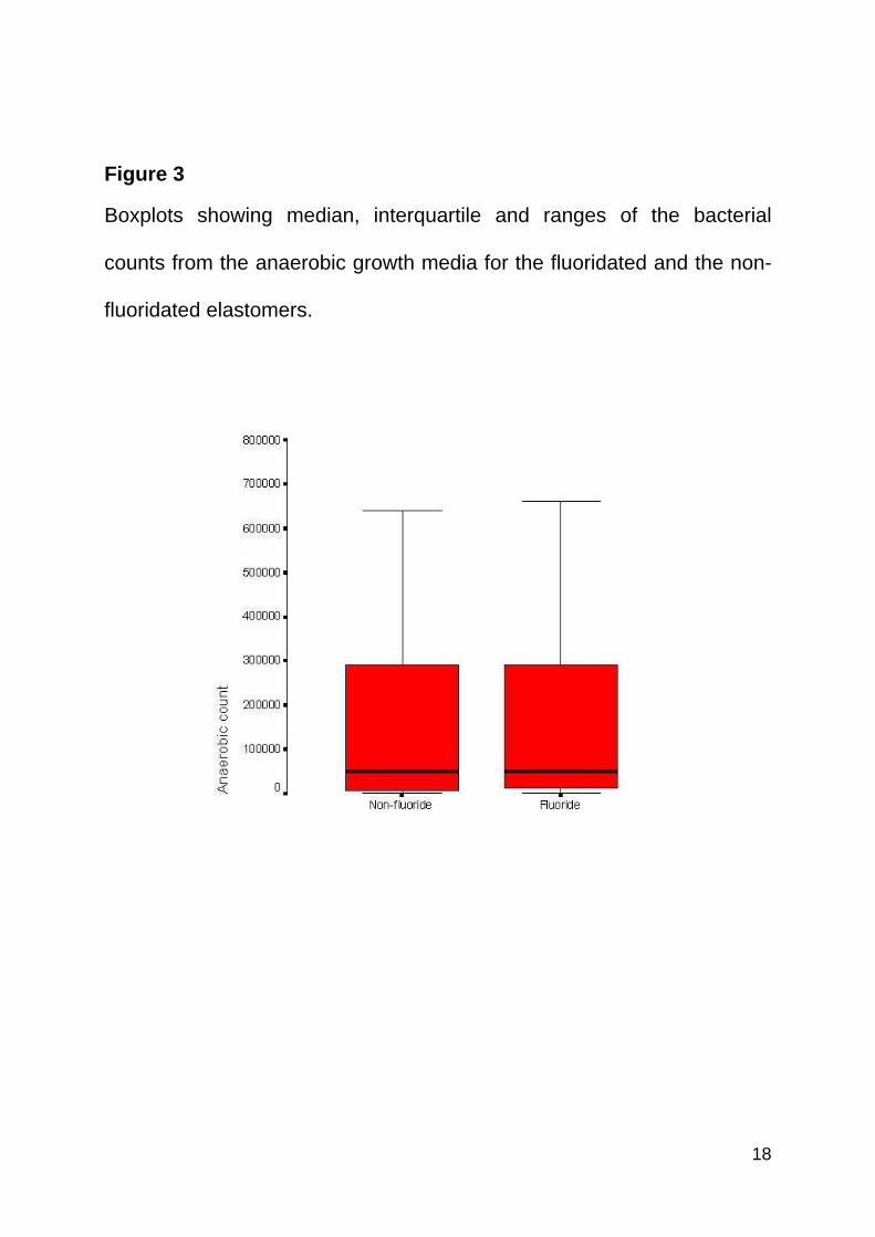
Figure 3
Boxplots showing median, interquartile and ranges of the bacterial
counts from the anaerobic growth media for the fluoridated and the non-
fluoridated elastomers.
18

Figure 4
Fluoridated elastomer on the upper right lateral incisor after six weeks in
the mouth demonstrating the swollen appearance compared with the
conventional elastomers on the other teeth. The fluoridated elastomer on
the upper right central has been lost.
19

Tables
Table I
Results of the logistic regression showing the p values for the
independent variables, where the dependent variable was the presence
or absence of bacterial growth on the anaerobic or Streptococcus growth
media.
Independent Variable Percent Streptococcal
Count Anaerobic Count
Subject 1.000 1.000
Fluoride or Non-fluoride elastomer 0.886 0.130
Tooth 0.961 0.573
Dominant or non-dominant hand 0.943 0.969
Visit 2 or 4 0.787 0.866
Number of days in mouth 0.105 0.880
20

Table II
Results of the mixed effects analysis of variance showing the p values
for the independent variables, where the dependent variable is the
percent streptococcal or anaerobic bacterial counts.
Independent Variable Percent Streptococcal Count Anaerobic Count
Fluoride 0.288 0.230
Tooth 0.394 0.346
Dominant 0.884 0.992
Visit <0.001 0.301
Subject <0.001 <0.001
Days 0.894 0.418
Age 0.283 0.307
Gender 0.530 0.937
21
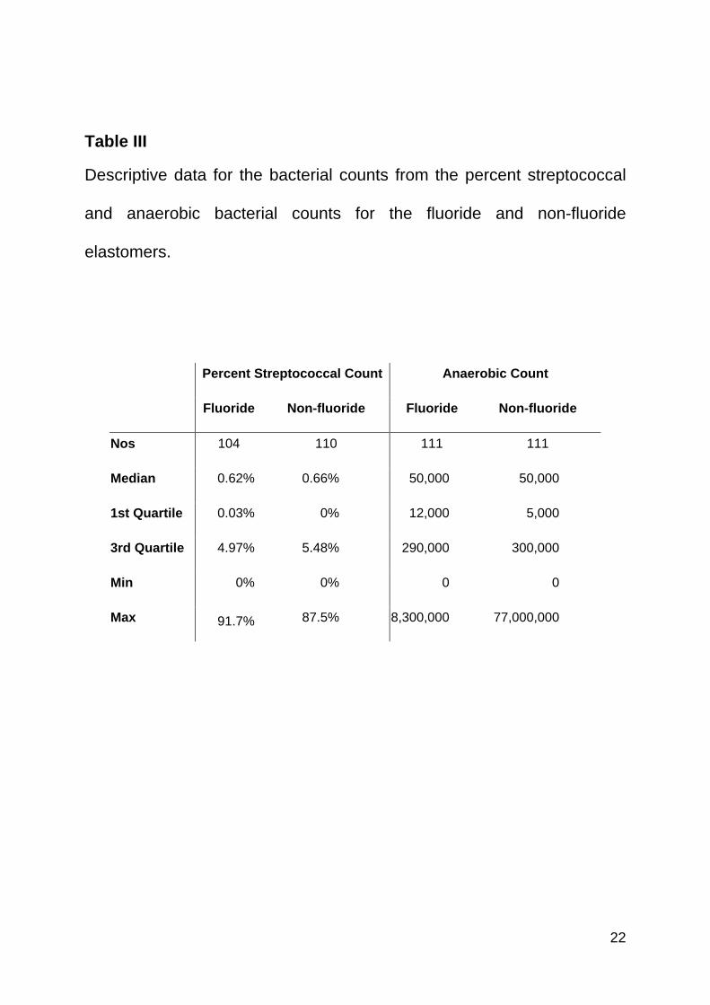
Table III
Descriptive data for the bacterial counts from the percent streptococcal
and anaerobic bacterial counts for the fluoride and non-fluoride
elastomers.
Percent Streptococcal Count Anaerobic Count
Fluoride Non-fluoride Fluoride Non-fluoride
Nos 104 110 111 111
Median 0.62% 0.66% 50,000 50,000
1st Quartile 0.03% 0% 12,000 5,000
3rd Quartile 4.97% 5.48% 290,000 300,000
Min 0% 0% 0 0
Max 91.7% 87.5% 8,300,000 77,000,000
22