Fluorescent Cellular Stains - Biotium... • 3 Lipid Stains and Membrane Potential Dyes Phospholipid...
12
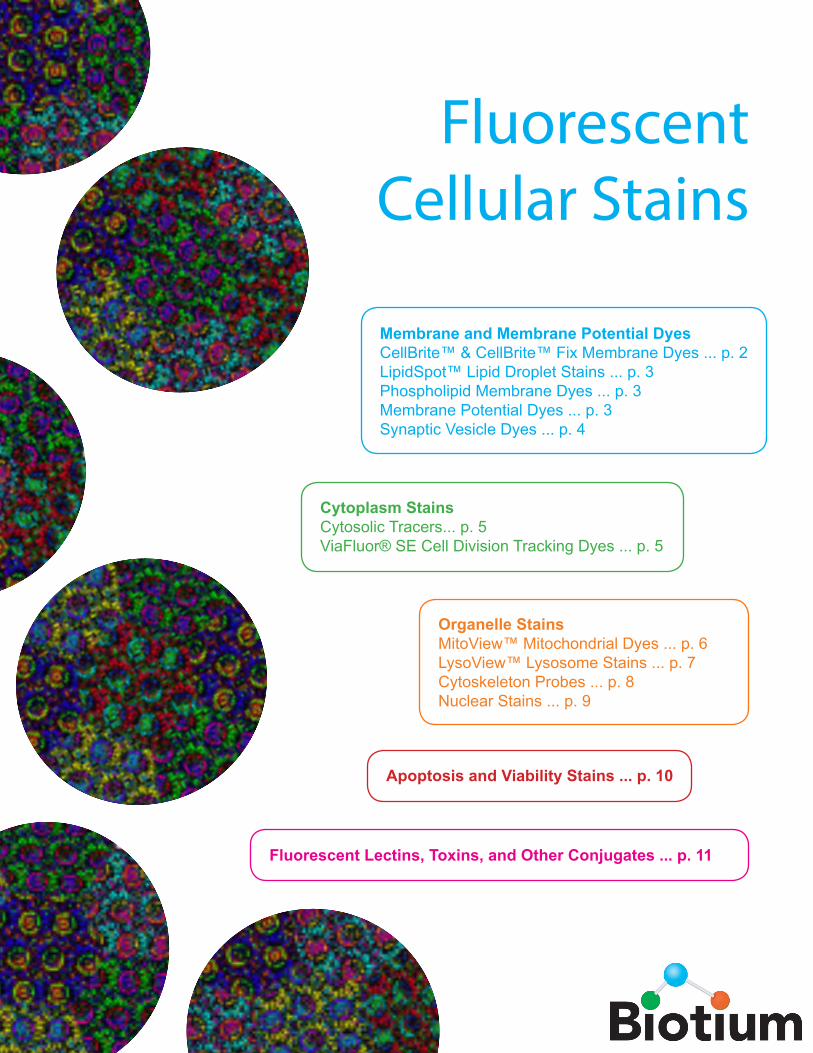
Fluorescent Cellular Stains Membrane and Membrane Potential Dyes CellBrite™ & CellBrite™ Fix Membrane Dyes ... p. 2 LipidSpot™ Lipid Droplet Stains ... p. 3 Phospholipid Membrane Dyes ... p. 3 Membrane Potential Dyes ... p. 3 Synaptic Vesicle Dyes ... p. 4 Cytoplasm Stains Cytosolic Tracers... p. 5 ViaFluor® SE Cell Division Tracking Dyes ... p. 5 Organelle Stains MitoView™ Mitochondrial Dyes ... p. 6 LysoView™ Lysosome Stains ... p. 7 Cytoskeleton Probes ... p. 8 Nuclear Stains ... p. 9 Apoptosis and Viability Stains ... p. 10 Fluorescent Lectins, Toxins, and Other Conjugates ... p. 11
Transcript of Fluorescent Cellular Stains - Biotium... • 3 Lipid Stains and Membrane Potential Dyes Phospholipid...
Fluorescent Cellular Stains
Membrane and Membrane Potential DyesCellBrite™ & CellBrite™ Fix Membrane Dyes ... p. 2LipidSpot™ Lipid Droplet Stains ... p. 3Phospholipid Membrane Dyes ... p. 3Membrane Potential Dyes ... p. 3Synaptic Vesicle Dyes ... p. 4
Cytoplasm Stains Cytosolic Tracers... p. 5 ViaFluor® SE Cell Division Tracking Dyes ... p. 5
Organelle StainsMitoView™ Mitochondrial Dyes ... p. 6LysoView™ Lysosome Stains ... p. 7Cytoskeleton Probes ... p. 8Nuclear Stains ... p. 9
Apoptosis and Viability Stains ... p. 10
Fluorescent Lectins, Toxins, and Other Conjugates ... p. 11
2 • www.biotium.com
Fluorescent Membrane Stains
Catalog No. Product Ex/Em (nm) Unit Size
30024 CellBrite™ Blue Cytoplasmic Membrane Labeling Kit 366/441 50 assays
30021 CellBrite™ Green Cytoplasmic Membrane Labeling Dye 484/501 1 mL
30022 CellBrite™ Orange Cytoplasmic Membrane Labeling Dye 549/565 1 mL
30023 CellBrite™ Red Cytoplasmic Membrane Labeling Dye 644/665 1 mL
30070 CellBrite™ NIR680 Cytoplasmic Membrane Labeling Dye 683/724 100 uL
30077 CellBrite™ NIR750 Cytoplasmic Membrane Labeling Dye 748/780 100 uL
30078 CellBrite™ NIR770 Cytoplasmic Membrane Labeling Dye 767/806 100 uL
30079 CellBrite™ NIR790 Cytoplasmic Membrane Labeling Dye 786/820 100 uL
Catalog No. Product Ex/Em (nm) Unit Size
60011 DiO 484/501 50 mg
60015 Neuro-DiO 484/501 25 mg
60019 Neuro-DiO in vegetable oil 484/501 200 uL
60035 Dilinoleyl DiO (FAST DiO™) 484/499 5 mg
60012 DiOC14(3), hydroxyethanesulfonate 484/501 50 mg
60010 DiI 549/565 50 mg
60018 DiI in vegetable oil 549/565 0.5 mL
60034 Dilinoleyl DiI (FAST DiI™) 549/565 5 mg
60016 Neuro-DiI 549/565 25 mg
60014 DiD 644/655 50 mg
60017 DiR 748/780 25 mg
Other Lipophilic Carbocyanine Dyes
CellBrite™ Cytoplasmic Membrane Dyes
Fast DiI and Fast DiO are trademarks of Thermo Fisher Scientific
CellBrite™ Cytoplasmic Membrane DyesLipophilic carbocyanine dyes label membranes in a wide variety of cell types. The dyes are weakly fluorescent in aqueous phase, but become highly fluorescent in lipid bilayers. Staining is very stable with low toxicity and very little dye transfer in between cells, making the dyes suitable for long-term cell labeling and tracking studies. Cell populations can be labeled with different fluorescent colors for identification after mixing. Double labeling can identify cells that have fused or formed stable clusters. Cells can be fixed with formaldehyde either before or after staining, but plasma membrane staining has poor tolerance for detergent permeabilization.
Unlike PKH dyes, CellBrite™ dyes do not require a complicated hypoosmotic labeling protocol. They are ready-to-use dye delivery solutions that can be added directly to normal culture media to label suspended or adherent cells in culture. We offer a selection of dyes with fluorescence ranging from blue to near-infrared. The CellBrite™ NIR dyes are suitable for near-IR small animal imaging.
Other Lipophilic Carbocyanine Dyes for Membrane LabelingBiotium offers a selection of stand-alone carbocyanine dyes. Neuro-DiO, Neuro-DiI, and dilinoleyl dyes have structural features designed to make the probes diffuse faster in cell membranes. Near-infrared DiR is useful for small animal imaging.
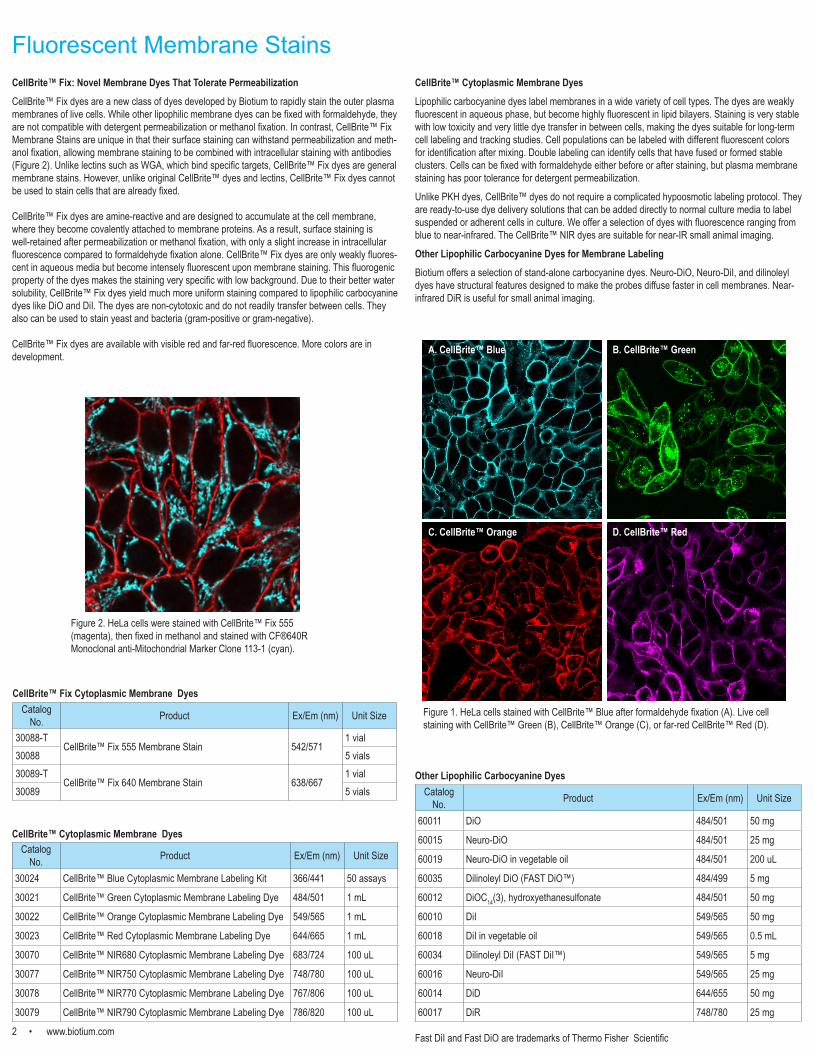
Figure 1. HeLa cells stained with CellBrite™ Blue after formaldehyde fixation (A). Live cell staining with CellBrite™ Green (B), CellBrite™ Orange (C), or far-red CellBrite™ Red (D).
CellBrite™ Fix: Novel Membrane Dyes That Tolerate PermeabilizationCellBrite™ Fix dyes are a new class of dyes developed by Biotium to rapidly stain the outer plasma membranes of live cells. While other lipophilic membrane dyes can be fixed with formaldehyde, they are not compatible with detergent permeabilization or methanol fixation. In contrast, CellBrite™ Fix Membrane Stains are unique in that their surface staining can withstand permeabilization and meth-anol fixation, allowing membrane staining to be combined with intracellular staining with antibodies (Figure 2). Unlike lectins such as WGA, which bind specific targets, CellBrite™ Fix dyes are general membrane stains. However, unlike original CellBrite™ dyes and lectins, CellBrite™ Fix dyes cannot be used to stain cells that are already fixed.
CellBrite™ Fix dyes are amine-reactive and are designed to accumulate at the cell membrane, where they become covalently attached to membrane proteins. As a result, surface staining is well-retained after permeabilization or methanol fixation, with only a slight increase in intracellular fluorescence compared to formaldehyde fixation alone. CellBrite™ Fix dyes are only weakly fluores-cent in aqueous media but become intensely fluorescent upon membrane staining. This fluorogenic property of the dyes makes the staining very specific with low background. Due to their better water solubility, CellBrite™ Fix dyes yield much more uniform staining compared to lipophilic carbocyanine dyes like DiO and DiI. The dyes are non-cytotoxic and do not readily transfer between cells. They also can be used to stain yeast and bacteria (gram-positive or gram-negative).
CellBrite™ Fix dyes are available with visible red and far-red fluorescence. More colors are in development.
Catalog No. Product Ex/Em (nm) Unit Size
30088-TCellBrite™ Fix 555 Membrane Stain 542/571
1 vial30088 5 vials30089-T
CellBrite™ Fix 640 Membrane Stain 638/6671 vial
30089 5 vials
CellBrite™ Fix Cytoplasmic Membrane Dyes
Figure 2. HeLa cells were stained with CellBrite™ Fix 555 (magenta), then fixed in methanol and stained with CF®640R Monoclonal anti-Mitochondrial Marker Clone 113-1 (cyan).
A. CellBrite™ Blue B. CellBrite™ Green
C. CellBrite™ Orange D. CellBrite™ Red
www.biotium.com • 3
Lipid Stains and Membrane Potential Dyes
Phospholipid Membrane ProbesThese membrane probes are derived from natural phospholipids by modifying the head group with a fluorescent dye or biotin. The probes are useful for studies of vesicle trafficking and membrane fusion. Red fluorescent phospholipids like TRITC-, Rhodamine-, and Texas Red®-DHPE have been used as fluorescence acceptors in combination with NBD-DHPE in membrane-fusion assays.
Slow-Responding Membrane Potential DyesTranslational (or slow-responding) membrane potential dyes undergo a change in their membrane distribution as a result of changes in membrane potential.
The fluorescence of DiBAC4(3) is enhanced with membrane depolarization. The rate of fluorescence response of the dye is slower than styryl dyes like the ANEPPS dyes, but the fluorescence change is significantly larger. DiOC2(3) has been used for measuring membrane potential in bacteria. The green fluorescent dye forms red fluorescent aggregates with increasing membrane potential, allowing ratiometric potential measurements. DiOC5(3) and DiOC6(3) are two of the most widely used carbocyanine dyes for membrane potential measurements. Tetramethylrhodamine ethyl ester (TMRE) and Tetramethylrhodamine methyl ester (TMRM) can be used for quantitative measurements of membrane potential and mitochondrial membrane potential.
Fast-Responding Membrane Potential DyesFast-responding membrane potential dyes are styryl dyes that undergo changes in fluorescence intensity in response to changes in membrane potential, on the order of 2-10% change in fluorescence per 100 mV. The dyes also undergo spectral shift with changes in membrane potential, allowing ratiometric measurements. Fast response dyes have been used to measure electrical activity in neural and cardiac cells.
Di-8-ANNEPPS is more hydrophobic and better retained in the outer leaflet of the plasma membrane than Di-4-ANNEPS, and therefore is more suitable for long-term membrane potential studies. It is also more photostable and less phototoxic than Di-4-ANNEPS. Di-4-ANNEPS has been used for studies of human stem cell-derived cardiomyocytes.
Di-2-ANEPEQ (also known as JPW 1114) is a highly water soluble fast-responding dye that is usually introduced into cells by microinjection. Di-8-ANEPQ and Di-12-ANEPQ are successively more hydrophobic, and have been used for potential-sensitive retrograde labeling of neurons.
RH237, RH414, RH421, and RH795 are fast-responding potentiometric probes generally used for functional imaging of neurons. RH421 exhibits >20% fluorescence change per 100 mV on neuroblastoma cells. These dyes can differ in their physiological effects, for example RH414 causes arterial constriction during cortex staining, while the spectrally similar dye RH795 does not.
DiO/DPA Membrane Potential KitThe membrane localization of the fluorescence quencher dipicrylamine (DPA) is a function of the polarity and magnitude of membrane potential. The DiO/DPA system detects cytoplasmic membrane potential changes using the principle of fluorescence resonance energy transfer (FRET). The green fluorescent membrane dye DiO is a “stationary” FRET donor while DPA acts as a mobile FRET acceptor, resulting in a membrane potential-dependent quenching of fluorescence by FRET. The DiO/DPA system has been reported to produce a fluorescence signal change of >56% in HEK-293 cells and >25% in neuronal cultures and brain slices per 100 mV membrane potential change.
Catalog No. Product Ex/Em (nm) Unit Size
60022 Biotin-DHPE N/A 10 mg
60023 Biotin-X-DHPE N/A 5 mg
60024 Fluorescein-DHPE 496/519 5 mg
60025 NBD-PE 463/536 10 mg
60028 TRITC-DHPE 540/566 1 mg
60026 Rhodamine-DHPE 560/581 5 mg
60027 Texas Red®-DHPE 582/601 1 mg
Catalog No. Product Ex/Em (nm) (MeOH)1 Unit Size
61011 DiBAC4(3) 493/516 25 mg
70008 DiOC2(3) 482/497 100 mg
70007 DiOC5(3) 482/497 100 mg
70009 DiOC6(3) 484/501 100 mg
70016 TMRE 549/574 25 mg
70017 TMRM 548/573 25 mg
61010 Di-4-ANEPPS 496/705 5 mg
61012 Di-8-ANEPPS 498/713 5 mg
61013 Di-2-ANEPEQ (JPW 1114) See note 2 5 mg
61014 Di-8-ANEPPQ See note 2 5 mg
61015 Di-12-ANEPPQ See note 2 5 mg
61018 RH237 528/782 5 mg
61016 RH414 532/706 5 mg
61017 RH421 515/704 25 mg
61019 RH795 530/712 5 mg
30037 DiO/DPA Membrane Potential Kit 484/501 (DiO)406/- (DPA) 1 kit
1Spectral properties of styryl dyes are highly dependent on environment. In cell membranes, spectra are typically blue shifted by as much as 20 nm for Abs/Ex and 80 nm for Em.2Spectra expected to be similar to Di-4-ANEPPS and Di-8-ANEPPS, but red-shifted 10-20 nm.
Texas Red is a registered trademark of Thermo Fisher Scientific
Phospholipid Probes
Membrane Potential Dyes
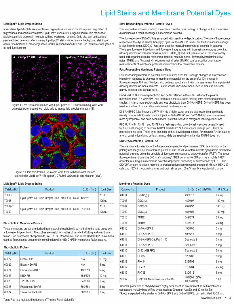
LipidSpot™ Lipid Droplet StainsIntracellular lipid droplets are cytoplasmic organelles involved in the storage and regulation of triglycerides and cholesterol esters. LipidSpot™ dyes are fluorogenic neutral lipid stains that rapidly stain lipid droplets in live cells with no wash step required. Cells also can be fixed and permeabilized before or after staining. LipidSpot™ stains show minimal background staining of cellular membranes or other organelles, unlike traditional dyes like Nile Red. Available with green or far-red fluorescence.
Figure 1. Live HeLa cells stained with LipidSpot™ 610. Prior to staining, cells were untreated (A) or treated with oleic acid to induce lipid droplet formation (B).
Figure 2. Oleic acid-treated HeLa cells were fixed with formaldehyde and stained with LipidSpot™ 488 (green), CF®594 WGA (red), and Hoechst (blue).
A. LipidSpot™ 610, untreated B. LipidSpot™ 610, oleic acid
Catalog No. Product Ex/Em (nm) Unit Size
70065-TLipidSpot™ 488 Lipid Droplet Stain, 1000X in DMSO 420/511
20 uL
70065 125 uL
70069-TLipidSpot™ 610 Lipid Droplet Stain, 1000X in DMSO 610/663
20 uL
70069 125 uL
LipidSpot™ Lipid Droplet Stains
4 • www.biotium.com
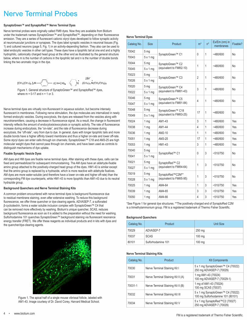
Nerve Terminal ProbesSynaptoGreen™ and SynaptoRed™ Nerve Terminal Dyes Nerve terminal probes were originally called FM® dyes. Now they are available from Biotium under the trademark names SynaptoGreen™ and SynaptoRed™, depending on their fluorescence emission. They are a series of fluorescent cationic styryl dyes developed to follow synaptic activity at neuromuscular junctions or synapses. The dyes label synaptic vesicles in neuronal tissues (Fig. 1) and cultured neurons (page 5, Fig. 1) in an activity-depending fashion. They also can be used to label endocytic vesicles in other cell types. These dyes have a lipophilic tail at one end and a highly hydrophilic, cationically charged head group at the other end as illustrated by the general structure below, where m is the number of carbons in the lipophilic tail and n is the number of double bonds linking the two aromatic rings in the dye.
Nerve terminal dyes are virtually non-fluorescent in aqueous solution, but become intensely fluorescent in membranes. Following nerve stimulation, the dye molecules are internalized in newly formed endocytic vesicles. During exocytosis, the dyes are released from the vesicles along with neurotransmitters, causing a decrease in fluorescence signal. As a result, the change in fluorescent intensity reflects the amount of endocytosis/exocytosis or synaptic activity. The rate of fluorescence increase during endocytosis, the “on-rate”, and the rate of fluorescence decrease during exocytosis, the “off-rate”, vary from dye to dye. In general, dyes with longer lipophilic tails and more double bonds have a higher affinity toward membrane and thus a higher on-rate and lower off-rate. Some styryl dyes can enter cells through ion channels; SynaptoGreen™ C18 and AM3-25 are high molecular weight dyes that cannot pass through ion channels, and have been used as controls to distinguish mechanisms of dye uptake.
Fixable Synaptic Vesicle DyesAM dyes and HM dyes are fixable nerve terminal dyes. After staining with these dyes, cells can be fixed and permeabilized for subsequent immunostaining. The AM dyes have an aldehyde-fixable amino group attached to the positively-charged head group of the dyes. HM1-43 is similar except that the amino group is replaced by a hydrazide, which is more reactive with aldehyde fixatives. AM dyes are more water-soluble (and therefore have a lower on-rate and higher off-rate) than the corresponding FM dye counterparts, while HM1-43 is more lipophilic than AM1-43 due to its neutral hydrazide group.
Background Quenchers and Nerve Terminal Staining KitsA common problem encountered with nerve terminal dyes is background fluorescence due to residual membrane staining, even after extensive washing. To reduce this background fluorescence, we offer three quencher or dye-clearing agents. ADVASEP-7, a sulfonated β-cyclodextrin, forms a water soluble inclusion complex with SynaptoGreen™ C4 that can be removed more effectively by washing. Biotium’s unique quencher, SCAS, reduces background fluorescence as soon as it is added to the preparation without the need for washing. Sulforhodamine 101 quenches SynaptoGreen™ background staining via fluorescent resonance energy transfer (FRET). We offer these reagents as individual products and in kits with dyes and the quencher/dye-clearing agents.
Figure 1. General structure of SynaptoGreen™ and SynaptoRed™ dyes, where m = 0-17 and n = 1 or 3.
Catalog No. Product Kit Components
70030 Nerve Terminal Staining Kit I 5 x 1 mg SynaptoGreen™ C4 (70022)250 mg ADVASEP-7 (70029)
70031 Nerve Terminal Staining Kit II (A) 1 mg AM1-43 (70024)100 mg ADVASEP-7 (70029-1)
70031-1 Nerve Terminal Staining Kit II (B) 1 mg of AM1-43 (70024) 100 mg SCAS (70037)
70032 Nerve Terminal Staining Kit III 5 x 1 mg SynaptoGreen™ C4 (70022)100 mg Sulforhodamine 101 (80101)
70034 Nerve Terminal Staining Kit V 5 x 1 mg SynaptoRed™C2 (70027)250 mg ADVASEP-7 (70029)
Catalog No. Size Product m* n* Ex/Em (nm) in membranes Fixable?
70042 5 mgSynaptoGreen™ C1 0 1 ~480/600 No
70043 5 x 1 mg
70044 5 mg SynaptoGreen™ C2 (equivalent to FM®2-10) 1 1 ~480/600 No
70045 5 x 1 mg
70023 5 mgSynaptoGreen™ C3 2 1 ~480/600 No
70026 5 x 1 mg
70020 5 mg SynaptoGreen™ C4 (equivalent to FM®1-43) 3 1 ~480/600 No
70022 5 x 1 mg
70046 5 mg SynaptoGreen™ C5 (equivalent to FM®1-84) 4 1 ~480/600 No
70047 5 x 1 mg
70048 5 mg SynaptoGreen™ C18(equivalent to FM®3-25) 17 1 ~480/600 No
70049 5 x 1 mg
70024 1 mg AM1-43 3 1 ~480/600 Yes
70038 1 mg AM1-44 4 1 ~480/600 Yes
70036 1 mg AM2-10 1 1 ~480/600 Yes
70051 1 mg AM3-25 17 1 ~480/600 Yes
70053 1 mg HM1-43 3 1 ~480/600 Yes
70040 5 mgSynaptoRed™ C1 0 3 ~510/750 No
70041 5 x 1 mg
70021 5 mg SynaptoRed™ C2(equivalent to FM®4-64) 1 3 ~510/750 No
70027 5 x 1 mg
70019 5 mg SynaptoRed™C2M**(equivalent to FM®5-95) 1 3 ~510/750 No
70028 5 x 1 mg
70025 1 mg AM4-64 1 3 ~510/750 Yes
70039 1 mg AM4-65 3 3 ~510/750 Yes
70050 1 mg AM4-66 4 3 ~510/750 Yes
*See Figure 1 for general dye structures. **The positively-charged end of SynaptoRed C2M is a trimethylammonium group. FM is a registered trademark of Thermo Fisher Scientific.
Figure 1. The apical half of a single mouse vibrissal follicle, labeled with AM1-43. Image courtesy of Dr. David Corey, Harvard Medical School.
Nerve Terminal Staining Kits
Nerve Terminal Dyes
Catalog No. Product Unit Size
70029 ADVASEP-7 250 mg
70037 SCAS 100 mg
80101 Sulforhodamine 101 100 mg
Background Quenchers
FM is a registered trademark of Thermo Fisher Scientific.
www.biotium.com • 5
Catalog No. Product Ex/Em (nm) Unit Size
80104 DBA/Terbium for membrane fusion assay 276/490, 545* 1 set
80105 SDIP/Europium for membrane fusion assay 250-320/~610* 1 set
80012 DPX N/A 500 mg
90010 ANTS 353/520 500 mg
Cytosolic Tracers and Cell Division TrackingCF® Dye HydrazidesWe offer a wide selection of our bright and photostable CF® dyes as water-soluble, aldehyde-fixable hydrazides for microinjection as neuronal tracers (Figure 1). See page 11.
Calcein and Calcein AMCalcein is a water soluble fluorescein derivative widely used for the study of cell membrane integrity that can be introduced by microinjection. Calcein AM is a membrane-permeant AM ester of calcein that can be loaded into cells by incubation, where it is cleaved by esterases to release the dye. Calcein AM also can be used to quantitate viable cells (see page 10).
Other Fluorescent TracersLucifer Yellow CH and Lucifer Yellow cadaverine are aldehyde-fixable green fluorescent cytosolic tracers. Hydroxystilbamidine (equivalent to Fluoro-Gold™) has been used extensively as a retrograde tracer for neurons and as a histochemical stain. Sulforhodamines are highly water soluble red fluorescent dyes that can be used as polar tracers for neuronal morphology and intercellular junctions. Sulforhodamine B also has been used as a fixed cell protein stain for colorimetric quantitation of cell number.
Biotin DerivativesBiocytin is a cellular tracer that can be introduced by microinjection; biocytin hydrazide is an aldehyde-fixable analog. Biotin ethylenediamine (equivalent to Neurobiotin™) is used as an anterograde and transneuronal tracer. Fluorescent biotin derivatives can be used for cell tracing or detection of biotin binding sites. We also offer conjugates of biotin and the biotin-binding protein streptavidin with our bright and photostable CF® dyes. See page 11.
Catalog No. Product Ex/Em (nm) Unit Size
80013 Calcein (high purity) 494/517 100 mg
30026 Calcein AM Cell Viability Assay Kit
494/517 (hydrolyzed product)
1000 assays
80011 Calcein AM 1 mg
80011-1 Calcein AM, 4 mM in anhydrous DMSO 100 uL
80011-2 Calcein AM, 1 mg/mL in anhydrous DMSO 1 mL
80011-3 Calcein AM 20 x 50 ug
80015 Lucifer Yellow CH, lithium salt
428/536
25 mg
80016 Lucifer Yellow CH, potassium salt 25 mg
80018 Lucifer Yellow Cadaverine 25 mg
80017 Lucifer Yellow Cadaverine Biotin-X, dipotassium salt 428/532 10 mg
80014 Hydroxystilbamidine (Fluoro-Gold™) 361/536 10 mg
80023 Hydroxystilbamidine (Fluoro-Gold™), 4% in H2O 361/536 200 uL
80100 Sulforhodamine B 565/586 5 g
80101 Sulforhodamine 101 586/605 500 mg
80102 Sulforhodamine G 529/548 5 g
90055 Biocytin N/A 100 mg
90060 Biocytin hydrazide N/A 25 mg
90057 Biotin ethylenediamine, hydrobromide (Neurobiotin™) N/A 25 mg
90075 Biotin ethylenediamine, hydrochloride N/A 25 mg
80019 Fluorescein-biotin 494/518 5 mg
90062 Biotin-4-fluorescein 494/523 10 mg
80022 Biotin-rhodamine 110 502/524 5 mg
80017 Lucifer Yellow Cadaverine Biotin-X, dipotassium salt 428/532 10 mg
Neurobiotin is a trademark of Vector Laboratories. Fluoro-Gold is a trademark of Fluorochrome.*For complex.
Reagents for membrane fusion assaysThe principle of DPA/Terbium vesicle fusion assays is based on the fact that contact of the chelator dipicolinic acid (DPA) with terbium (III) instantly forms a complex that is ~10,000 times more fluorescent than free Tb3+. In the assay, separate vesicle populations are loaded with DPA or terbium. Fusion of the two types of vesicles results in fluorescence at 490 and 545. Biotium developed SDIP/Europium as an alternative to DPA/Tb3+ for vesicle fusion assays. The combination of SDIP/Eu3+ generates intense red fluorescence at 610 , with brighter intensity compared to DPA/Tb3+. Each set includes 10 mg SDIP and 25 mg EuCl3.
DPX is a positively charged quencher that has been used with the fluorescent dye ANTS for studies of vesicle fusion or membrane permeability. The complex of ANTS-DPX has minimal fluorescence, but when it is diluted due to membrane fusion or leakage, the dye becomes increasingly fluorescent.
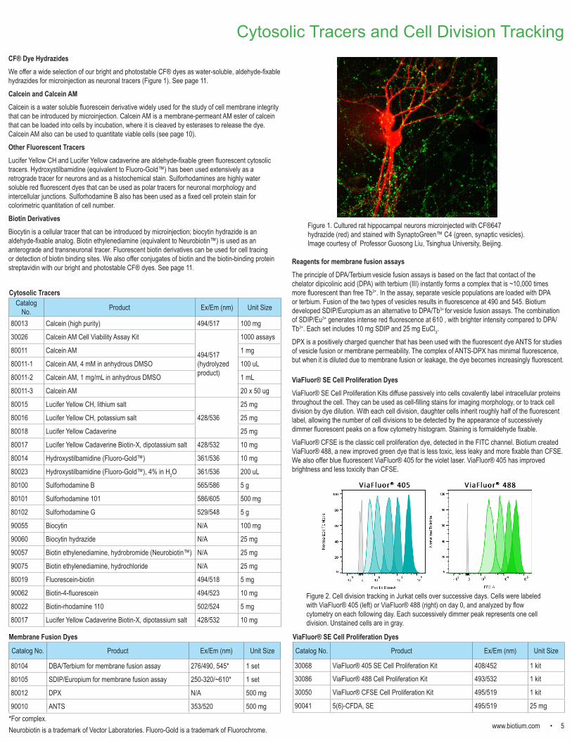
Figure 1. Cultured rat hippocampal neurons microinjected with CF®647 hydrazide (red) and stained with SynaptoGreen™ C4 (green, synaptic vesicles). Image courtesy of Professor Guosong Liu, Tsinghua University, Beijing.
Cytosolic Tracers
Membrane Fusion Dyes
ViaFluor® SE Cell Proliferation Dyes ViaFluor® SE Cell Proliferation Kits diffuse passively into cells covalently label intracellular proteins throughout the cell. They can be used as cell-filling stains for imaging morphology, or to track cell division by dye dilution. With each cell division, daughter cells inherit roughly half of the fluorescent label, allowing the number of cell divisions to be detected by the appearance of successively dimmer fluorescent peaks on a flow cytometry histogram. Staining is formaldehyde fixable.
ViaFluor® CFSE is the classic cell proliferation dye, detected in the FITC channel. Biotium created ViaFluor® 488, a new improved green dye that is less toxic, less leaky and more fixable than CFSE. We also offer blue fluorescent ViaFluor® 405 for the violet laser. ViaFluor® 405 has improved brightness and less toxicity than CFSE.
Catalog No. Product Ex/Em (nm) Unit Size
30068 ViaFluor® 405 SE Cell Proliferation Kit 408/452 1 kit
30086 ViaFluor® 488 Cell Proliferation Kit 493/532 1 kit
30050 ViaFluor® CFSE Cell Proliferation Kit 495/519 1 kit
90041 5(6)-CFDA, SE 495/519 25 mg
ViaFluor® SE Cell Proliferation Dyes
Figure 2. Cell division tracking in Jurkat cells over successive days. Cells were labeled with ViaFluor® 405 (left) or ViaFluor® 488 (right) on day 0, and analyzed by flow cytometry on each following day. Each successively dimmer peak represents one cell division. Unstained cells are in gray.
6 • www.biotium.com
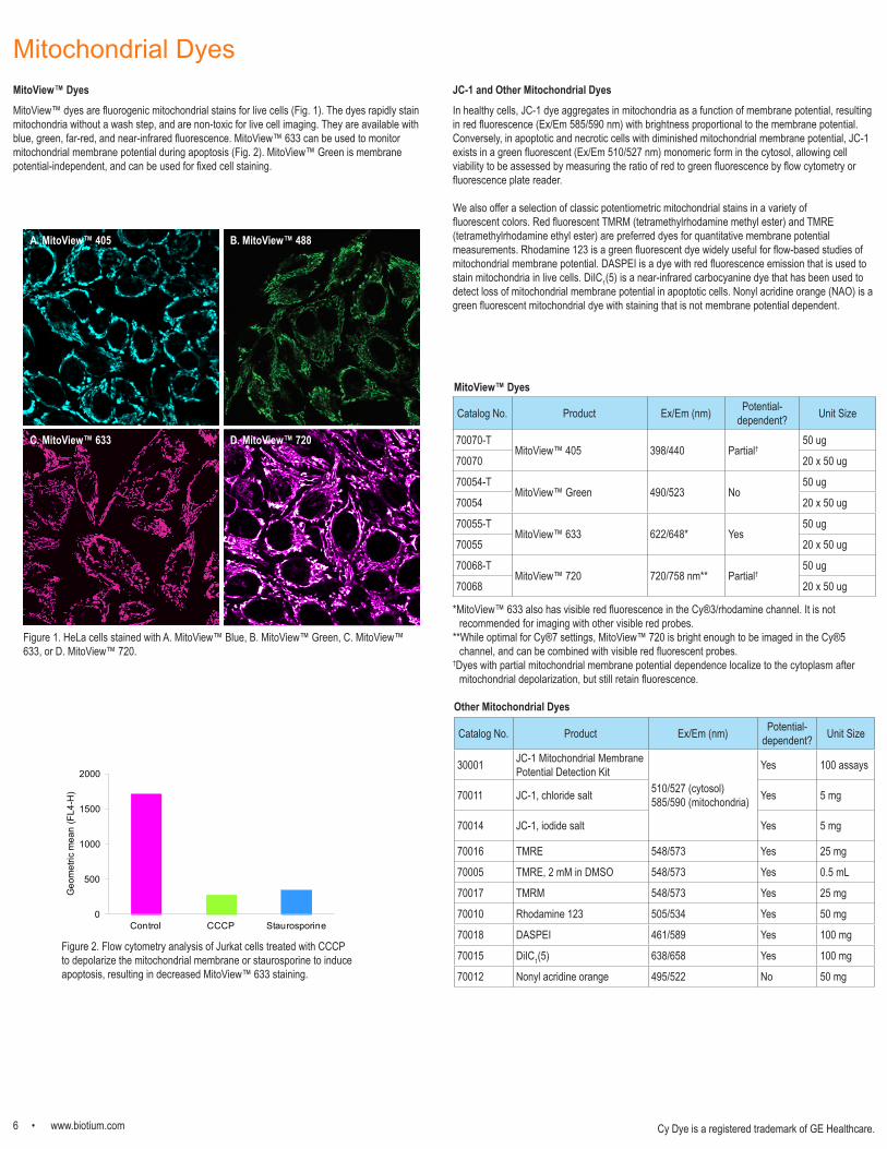
Mitochondrial DyesMitoView™ DyesMitoView™ dyes are fluorogenic mitochondrial stains for live cells (Fig. 1). The dyes rapidly stain mitochondria without a wash step, and are non-toxic for live cell imaging. They are available with blue, green, far-red, and near-infrared fluorescence. MitoView™ 633 can be used to monitor mitochondrial membrane potential during apoptosis (Fig. 2). MitoView™ Green is membrane potential-independent, and can be used for fixed cell staining.
0
500
1000
1500
2000
Control CCCP Staurosporine
Geo
met
ric m
ean
(FL4
-H)
Figure 2. Flow cytometry analysis of Jurkat cells treated with CCCP to depolarize the mitochondrial membrane or staurosporine to induce apoptosis, resulting in decreased MitoView™ 633 staining.
Figure 1. HeLa cells stained with A. MitoView™ Blue, B. MitoView™ Green, C. MitoView™ 633, or D. MitoView™ 720.
Catalog No. Product Ex/Em (nm) Potential-dependent? Unit Size
70070-TMitoView™ 405 398/440 Partial†
50 ug
70070 20 x 50 ug
70054-TMitoView™ Green 490/523 No
50 ug
70054 20 x 50 ug
70055-TMitoView™ 633 622/648* Yes
50 ug
70055 20 x 50 ug
70068-TMitoView™ 720 720/758 nm** Partial†
50 ug
70068 20 x 50 ug
*MitoView™ 633 also has visible red fluorescence in the Cy®3/rhodamine channel. It is not recommended for imaging with other visible red probes.
**While optimal for Cy®7 settings, MitoView™ 720 is bright enough to be imaged in the Cy®5 channel, and can be combined with visible red fluorescent probes.
†Dyes with partial mitochondrial membrane potential dependence localize to the cytoplasm after mitochondrial depolarization, but still retain fluorescence.
A. MitoView™ 405 B. MitoView™ 488
C. MitoView™ 633 D. MitoView™ 720
Catalog No. Product Ex/Em (nm) Potential-dependent? Unit Size
30001 JC-1 Mitochondrial Membrane Potential Detection Kit
510/527 (cytosol)585/590 (mitochondria)
Yes 100 assays
70011 JC-1, chloride salt Yes 5 mg
70014 JC-1, iodide salt Yes 5 mg
70016 TMRE 548/573 Yes 25 mg
70005 TMRE, 2 mM in DMSO 548/573 Yes 0.5 mL
70017 TMRM 548/573 Yes 25 mg
70010 Rhodamine 123 505/534 Yes 50 mg
70018 DASPEI 461/589 Yes 100 mg
70015 DiIC1(5) 638/658 Yes 100 mg
70012 Nonyl acridine orange 495/522 No 50 mg
MitoView™ Dyes
Other Mitochondrial Dyes
JC-1 and Other Mitochondrial DyesIn healthy cells, JC-1 dye aggregates in mitochondria as a function of membrane potential, resulting in red fluorescence (Ex/Em 585/590 nm) with brightness proportional to the membrane potential. Conversely, in apoptotic and necrotic cells with diminished mitochondrial membrane potential, JC-1 exists in a green fluorescent (Ex/Em 510/527 nm) monomeric form in the cytosol, allowing cell viability to be assessed by measuring the ratio of red to green fluorescence by flow cytometry or fluorescence plate reader.
We also offer a selection of classic potentiometric mitochondrial stains in a variety of fluorescent colors. Red fluorescent TMRM (tetramethylrhodamine methyl ester) and TMRE (tetramethylrhodamine ethyl ester) are preferred dyes for quantitative membrane potential measurements. Rhodamine 123 is a green fluorescent dye widely useful for flow-based studies of mitochondrial membrane potential. DASPEI is a dye with red fluorescence emission that is used to stain mitochondria in live cells. DiIC1(5) is a near-infrared carbocyanine dye that has been used to detect loss of mitochondrial membrane potential in apoptotic cells. Nonyl acridine orange (NAO) is a green fluorescent mitochondrial dye with staining that is not membrane potential dependent.
Cy Dye is a registered trademark of GE Healthcare.

www.biotium.com • 7
Lysosome StainsLysoView™ DyesLysoView™ dyes are fluorescent stains for imaging lysosome localization and morphology in live cells. LysoView™ dyes belong to a family of lysosomotropic dyes that contain weakly basic amines that accumulate in acidic organelles. LysoView™ dyes are available with blue, green, visible red, and far-red fluorescence. Red-fluorescent LysoView™ 540 and far-red fluorescent LysoView™ 633 dye fluorescence is pH-sensitive (Figure 2), resulting in specific lysosomal staining without a wash step. We also offer LysoView™ 650, a far-red lysosome dye that is compatible with super-resolution imaging by SIM and STED.
Figure 3. LysoView™ 633 compared to LysoTracker® Deep Red. Live HeLa cells were stained for 10 minutes at 37oC with 1X LysoView 633 or 50 nM LysoTracker® Deep Red (Thermo Fisher Scientific). LysoView™ 633 (A) showed more specific lysosomal staining with less cytoplasmic staining compared to LysoTracker® Deep Red (B).
A. LysoView™ 633
LysoTracker is a registered trademark of Thermo Fisher ScientificCy Dye is a registered trademark of GE Healthcare.
Figure 1. Live HeLa cells stained with (A) LysoView™ 405, (B) LysoView™ 488, (C) LysoView™ 540 (red) and Hoechst 33342 (blue), or (D) LysoView™ 650 (magenta) and MitoView™ Green.
A. Before UV exposure B. After UV exposure
Figure 4. UV-activated lysosomal fluorescence with "Light-On" LysoView™ 555. HeLa cells were stained with 1 uM Light-on LysoView™ 555 for 15 minutes at 37oC, then imaged using a Zeiss LSM 700 confocal microscope using a 40X objective and imaging settings for Cy®3. A. Before UV exposure, fluorescence was not detectable. B. After five seconds of exposure to UV light from a short arc lamp, bright red fluorescence localized to lysosomes was observed.
Catalog No. Product Ex/Em (nm) Unit Size
70061-TLysoView™ 405, 1000X in DMSO 318, 400/464
10 uL
70061 50 uL
70061-TLysoView™ 488, 1000X in DMSO 506/532
10 uL
70061 50 uL
70061-TLysoView™ 540, 1000X in DMSO 541/634 *
10 uL
70061 50 uL
70058-TLysoView™ 633 (1000X after reconstitution) 634/659*
100 uL**
70058 10 x 100 uL**
70059-TLysoView™ 650, 1000X in DMSO 650/675
10 uL
70059 50 uL
70060-T"Light-On" LysoView™ 555, 1 mM in DMSO 554/583 *
10 uL
70060 50 uL
B LysoTracker® Deep Red
*pH ≤ 5**After reconstitution
Figure 2. pH dependence of LysoView™ 540 (A) and LysoView™ 633 (B) fluorescence emission.
550 600 650 700 750 800
Em
issi
on
Wavelength (nm)
pH 2pH 3pH 4.6pH 6pH 7pH 8pH 9pH 9.6pH 11.6
pH 6
pH 7
pH ≥ 8
pH ≤ 4.6
625 650 675 700 725
Em
issi
on
Wavelength (nm)
pH 2pH 3pH 4pH 5pH 6pH 7pH 8pH 9
pH ≤ 5
pH 6
pH 7
pH 8
pH 9
A
B
“Light-On” LysoView™ 555: a unique photo-activated dyeWe also offer “Light-On" LysoView™ 555, a UV-activatable lysosome stain. In cells, the dye initially shows low fluorescence, but brief exposure to UV excitation from a mercury arc lamp activates bright red fluorescence localizing to lysosomes (Figure 4). Lysosomal fluorescence fades over the course of several minutes after UV exposure, but can be re-activated in the same cells multiple times by exposure to UV light. Therefore the dye provides a novel tool for UV-activated, reversible fluorescence imaging of lysosomes.
C. LysoView™ 540 D. LysoView™ 650
A. LysoView™ 405 B. LysoView™ 488
LysoView™ Dyes
8 • www.biotium.com
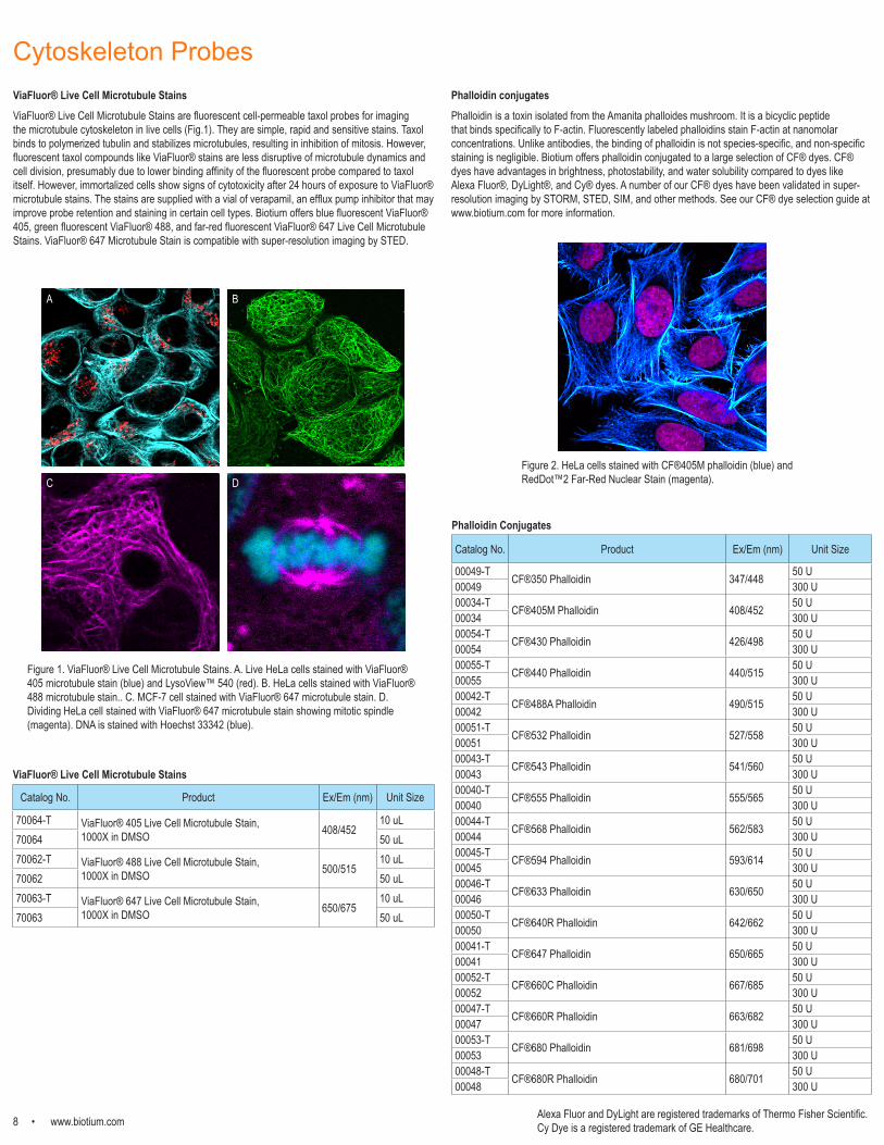
Cytoskeleton ProbesViaFluor® Live Cell Microtubule StainsViaFluor® Live Cell Microtubule Stains are fluorescent cell-permeable taxol probes for imaging the microtubule cytoskeleton in live cells (Fig.1). They are simple, rapid and sensitive stains. Taxol binds to polymerized tubulin and stabilizes microtubules, resulting in inhibition of mitosis. However, fluorescent taxol compounds like ViaFluor® stains are less disruptive of microtubule dynamics and cell division, presumably due to lower binding affinity of the fluorescent probe compared to taxol itself. However, immortalized cells show signs of cytotoxicity after 24 hours of exposure to ViaFluor® microtubule stains. The stains are supplied with a vial of verapamil, an efflux pump inhibitor that may improve probe retention and staining in certain cell types. Biotium offers blue fluorescent ViaFluor® 405, green fluorescent ViaFluor® 488, and far-red fluorescent ViaFluor® 647 Live Cell Microtubule Stains. ViaFluor® 647 Microtubule Stain is compatible with super-resolution imaging by STED.
A B
C D
Figure 1. ViaFluor® Live Cell Microtubule Stains. A. Live HeLa cells stained with ViaFluor® 405 microtubule stain (blue) and LysoView™ 540 (red). B. HeLa cells stained with ViaFluor® 488 microtubule stain.. C. MCF-7 cell stained with ViaFluor® 647 microtubule stain. D. Dividing HeLa cell stained with ViaFluor® 647 microtubule stain showing mitotic spindle (magenta). DNA is stained with Hoechst 33342 (blue).
Catalog No. Product Ex/Em (nm) Unit Size
70064-T ViaFluor® 405 Live Cell Microtubule Stain, 1000X in DMSO 408/452
10 uL70064 50 uL70062-T ViaFluor® 488 Live Cell Microtubule Stain,
1000X in DMSO 500/51510 uL
70062 50 uL70063-T ViaFluor® 647 Live Cell Microtubule Stain,
1000X in DMSO 650/67510 uL
70063 50 uL
Phalloidin conjugatesPhalloidin is a toxin isolated from the Amanita phalloides mushroom. It is a bicyclic peptide that binds specifically to F-actin. Fluorescently labeled phalloidins stain F-actin at nanomolar concentrations. Unlike antibodies, the binding of phalloidin is not species-specific, and non-specific staining is negligible. Biotium offers phalloidin conjugated to a large selection of CF® dyes. CF® dyes have advantages in brightness, photostability, and water solubility compared to dyes like Alexa Fluor®, DyLight®, and Cy® dyes. A number of our CF® dyes have been validated in super-resolution imaging by STORM, STED, SIM, and other methods. See our CF® dye selection guide at www.biotium.com for more information.
Catalog No. Product Ex/Em (nm) Unit Size
00049-T CF®350 Phalloidin 347/448 50 U00049 300 U00034-T CF®405M Phalloidin 408/452 50 U00034 300 U00054-T CF®430 Phalloidin 426/498 50 U00054 300 U00055-T CF®440 Phalloidin 440/515 50 U00055 300 U00042-T CF®488A Phalloidin 490/515 50 U00042 300 U00051-T CF®532 Phalloidin 527/558 50 U00051 300 U00043-T CF®543 Phalloidin 541/560 50 U00043 300 U00040-T CF®555 Phalloidin 555/565 50 U00040 300 U00044-T CF®568 Phalloidin 562/583 50 U00044 300 U00045-T CF®594 Phalloidin 593/614 50 U00045 300 U00046-T CF®633 Phalloidin 630/650 50 U00046 300 U00050-T CF®640R Phalloidin 642/662 50 U00050 300 U00041-T CF®647 Phalloidin 650/665 50 U00041 300 U00052-T CF®660C Phalloidin 667/685 50 U00052 300 U00047-T CF®660R Phalloidin 663/682 50 U00047 300 U00053-T CF®680 Phalloidin 681/698 50 U00053 300 U00048-T CF®680R Phalloidin 680/701 50 U00048 300 U
Figure 2. HeLa cells stained with CF®405M phalloidin (blue) and RedDot™2 Far-Red Nuclear Stain (magenta).
ViaFluor® Live Cell Microtubule Stains
Phalloidin Conjugates
Alexa Fluor and DyLight are registered trademarks of Thermo Fisher Scientific. Cy Dye is a registered trademark of GE Healthcare.
www.biotium.com • 9
Nuclear Stains
Catalog No. Product Ex/Em (nm) Unit Size
40081-T NucSpot® 470 Nuclear Stain,1000X in DMSO 500/515
10 uL40081 50 uL40081-T NucSpot® Live 488 Nuclear Stain,
1000X in DMSO 500/515 10 uL
40081 50 uL40082-T NucSpot® Live 650 Nuclear Stain,
1000X in DMSO 650/67510 uL
40082 50 uL40060-T
RedDot™1 Far-Red Nuclear Stain, 200X in water 662/694*
25 uL40060 250 uL40060-1 1 mL40061-T
RedDot™2 Far-Red Nuclear Stain, 200X in DMSO 662/694*
25 uL40061 250 uL40061-1 1 mL40011 DAPI
358/461*
10 mg40009 DAPI, dilactate 10 mg40043 DAPI, 10 mg/mL in H2O 1 mL23002 EverBrite™ Mounting Medium with DAPI 10 mL23004 EverBrite™ Hardset Mounting Medium with DAPI 10 mL40044 Hoechst 33258, 10 mg/mL in H2O 10 mL40045 Hoechst 33258, pentahydrate 100 mg40046 Hoechst 33342, 10 mg/mL in H2O 10 mL40047 Hoechst 33342, trihydrochloride trihydrate 100 mg40012 DMAO, 2 mM in DMSO 503/530* 1 mL40016 Propidium iodide (PI)
535/617 **100 mg
40017 Propidium iodide, 1 mg/mL in H2O 10 mL40048 Propidium iodide buffer, 50 ug/mL in PBS 2 mL
40039 Acridine Orange, 10 mg/mL in H2O 500/525*460/650*** 10 mL
40037 7-AAD 546/647 * 1 mg40010 Ethidium Homodimer I
528/617**1 mg
40014 Ethidium Homodimer I, 2 mM in DMSO 0.5 mL40050 Ethidium Homodimer III
530/620**1 mg
40051 Ethidium Homodimer III, 1 mM in DMSO 200 uL
RedDot™1 and RedDot™2 Far-Red Nuclear StainsRedDot™1 and RedDot™2 are far-red nuclear counterstains for the Cy®5 channel. RedDot™1 rapidly and specifically stains nuclei in live cells (see page 11, Fig. 3), and can be used for cell cycle analysis by flow cytometry or for cell normalization for In Cell Western™. It also has been used to stain nuclei in live flatworms. Note: Similar to Draq5™, RedDot™1 shows cytotoxicity within four hours of staining. For long-term live cell imaging, we recommend using NucSpot® Live dyes.
RedDot™2 is membrane impermeant and can be used to selectively stain dead cells, or as a nuclear counterstain for fixed cells. RedDot™2 shows better nuclear specificity in fixed cells than Draq7™, which requires a blocking step for nuclear-specific staining (Fig. 2).
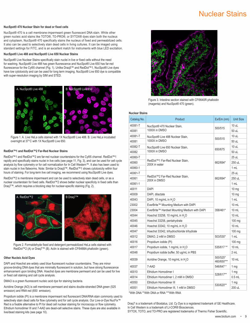
Figure 3. Intestine section stained with CF®640R phalloidin (magenta) and NucSpot® 470 (green).
Other Nucleic Acid DyesDAPI and Hoechst are widely used blue fluorescent nuclear counterstains. They are minor groove-binding DNA dyes that are minimally fluorescent in solution, but have strong fluorescence enhancement upon binding DNA. Hoechst dyes are membrane permeant and can be used for live or fixed cell staining and cell cycle analysis.
DMAO is a green fluorescent nucleic acid dye for staining bacteria. Acridine Orange (AO) is cell membrane permeant and stains double-stranded DNA green (525 emission) and RNA red (650 emission).
Propidium iodide (PI) is a membrane impermeant red fluorescent DNA/RNA stain commonly used to selectively stain dead cells for flow cytometry and for cell cycle analysis. Our Live-or-Dye NucFix™ Red is a fixable alternative to PI for dead cell nuclear staining for microscopy or flow cytometry. Ethidium homodimer III and 7-AAD are dead-cell selective stains. These dyes are also available in live/dead staining kits (see page 10).
*With DNA **With DNA or RNA ***With RNA
Figure 2. Formaldehyde fixed and detergent permeabilized HeLa cells stained with RedDot™2 (A) or Draq7™ (B). Actin is stained with CF®488A phalloidin (green).
Draq7 is a trademark of Biostatus, Ltd. Cy Dye is a registered trademark of GE Healthcare. In Cell Western is a trademark of LI-COR® Biosciences.SYTOX, TOTO, and TO-PRO are registered trademarks of Thermo Fisher Scientific.
A. RedDot™2 B Draq7™
NucSpot® 470 Nuclear Stain for dead or fixed cellsNucSpot® 470 is a cell membrane-impermeant green fluorescent DNA stain. While other green nucleic acid stains like TOTO®, TO-PRO®, or SYTOX® dyes stain both the nucleus and cytoplasm, NucSpot® 470 specifically stains the nucleus of fixed and permeabilized cells. It also can be used to selectively stain dead cells in living cultures. It can be imaged using standard settings for FITC, and is an excellent match for instruments with blue LED excitation.
NucSpot® Live 488 and NucSpot® Live 650 Nuclear StainsNucSpot® Live Nuclear Stains specifically stain nuclei in live or fixed cells without the need for washing. NucSpot® Live 488 has green fluorescence and NucSpot® Live 650 has far-red fluorescence for the Cy®5 channel (Fig. 1). Unlike Draq5™ and RedDot™1, NucSpot® Live dyes have low cytotoxicity and can be used for long term imaging. NucSpot® Live 650 dye is compatible with super-resolution imaging by SIM and STED.
A. NucSpot® Live 488 B. NucSpot® Live 650
Figure 1. A. Live HeLa cells stained with 1X NucSpot® Live 488. B. Live HeLa incubated overnight at 37°C with 1X NucSpot® Live 650.
Nuclear Stains
10 • www.biotium.com
CF® Dye Annexin V Conjugates and Cell Viability KitsFluorescent conjugates of Annexin V can be used to stain phosphatidylserine on the surface of apoptotic cells. We offer Annexin V conjugates of our exceptionally bright and photostable CF® dyes for flow cytometry or fluorescence microscopy. See p. 11 for a complete list. Our CF®488A and CF®594 Annexin V conjugates have been validated in real-time kinetic imaging studies using the IncuCyte® Live Cell Analysis System (Essen Bioscience). Near infrared CF® dye Annexin V conjugates (CF®680 through CF®790) are supplied as preservative-free lyophilized solids, suitable for in vivo imaging.
We also offer kits combining CF®488A Annexin V with red dead cell nucleic acid stains PI or 7-AAD. The Apoptosis and Necrosis Kit Plus includes CF®488A and EthD-III, while the Apoptotic, Necrotic and Healthy Cells Kit Plus also includes Hoechst to stain the total cell population. See page 9 for more information about nucleic acid stains.
Apoptosis and Viability StainsNucView® Caspase-3 SubstratesNucView® Caspase-3 Substrates are cleaved by caspases to stain the nuclei of apoptotic cells with fluorescence. Unlike FLICA substrates, NucView® substrates do not inhibit caspase activity, allowing caspase-3/7 activity to be monitored in individual intact cells in real time.
Green fluorogenic NucView® 488 Caspase-3 Substrate has been validated in more than a hundred published studies and cell types, and hs been validated in real-time kinetic studies using the IncuCyte® Live Cell Analysis System (Essen Bioscience). We also offer blue fluorogenic NucView® 405 and orange fluorogenic NucView® 530 for multi-color flexibility.
Catalog No. Product Ex/Em (nm)
10402 NucView® 488 Caspase-3 Substrate, 1 mM in DMSO 504/534 10403 NucView® 488 Caspase-3 Substrate, 1 mM in PBS 504/534 10405 NucView® 405 Caspase-3 Substrate, 1 mM in DMSO 429/469 10407 NucView® 405 Caspase-3 Substrate, 1 mM in PBS 429/469 10406 NucView® 530 Caspase-3 Substrate, 1 mM in DMSO 528/563 10408 NucView® 530 Caspase-3 Substrate, 1 mM in PBS 528/563
30067 Dual Apoptosis Assay with NucView® 488 Caspase-3 Substrate and CF®594 Annexin V
504/534 (NucView®)593/614 (Annexin)
30073 Dual Apoptosis Assay with NucView® 488 Caspase-3 Substrate and CF®640R Annexin V
504/534 (NucView®)642/662 (Annexin)
30062 NucView® 488 and MitoView™ 633 Apoptosis Kit 504/534 (NucView®)622/648 (MitoView™)
30072 NucView® 488 and RedDot™2 Apoptosis & Necrosis Kit 504/534 (NucView®)662/694 (RedDot™)
30060 CF®488A Annexin V and PI Apoptosis Assay Kit 490/515 (Annexin)535/617 (PI)
30061 CF®488A Annexin V and 7-AAD Apoptosis Assay Kit 490/515 (Annexin)546/647 (7-AAD)
30065 Apoptosis and Necrosis Quantitation Kit Plus 490/515 (Annexin)530/620 (EthD-III)
30066 Apoptotic, Necrotic and Healthy Cells Quantitation Kit Plus358/461 (Hoechst)490/515 (Annexin)530/620 (EthD-III)
30026 Calcein AM Cell Viability Assay Kit 494/517
30002 Viability/Cytotoxity Assay Kit for Animal Live & Dead Cells 494/517 (Calcein)530/620 (EthD-III)
32002 Live-or-Dye™ 350/448 347/448 32003 Live-or-Dye™ 405/452 408/452 32009 Live-or-Dye™ 405/545 395/545 32004 Live-or-Dye™ 488/515 490/515 32005 Live-or-Dye™ 568/583 562/583 32006 Live-or-Dye™ 594/614 561/624 32007 Live-or-Dye™ 640/662 642/662 32008 Live-or-Dye™ 750/777 755/777 32010 Live-or-Dye NucFix™ Red 520/610
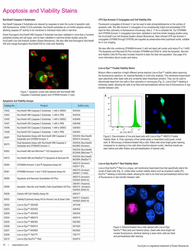
Live-or-Dye™ Fixable Viability StainsBiotium offers a selection of eight different amine-reactive Live-or-Dye™ viability stains spanning the fluorescence spectrum, for maximal flexibility in multi-color analysis. The membrane-impermeant dyes selectively enter dead cells and covalently label intracellular proteins. They can be used to discriminate dead from live cells in flow cytometry or microscopy (Fig. 2). Live-or-Dye™ labeling is extremely stable, allowing the cells to be fixed and permeabilized without loss of fluorescence or dye transfer between cells.
Figure 1. Apoptotic Jurkat cells stained with NucView® 488 Caspase-3 Substrate (green) and CF®594 Annexin V (red).
Figure 3. Ethanol-treated HeLa cells stained with Live-or-Dye NucFix™ Red (red) and Hoechst (blue). Dead cells show bright red nuclear fluorescence. Identical staining is seen when cells are fixed and permeabilized after staining.
Live-or-Dye NucFix™ Red Viability StainLive-or-Dye NucFix™ Red is a unique, cell membrane-impermeant dye that specifically stains the nuclei of dead cells (Fig. 3). Unlike other nuclear viability stains such as propidium iodide (PI), NucFix™ labeling is extremely stable, allowing the cells to be fixed and permeabilized without loss of fluorescence or dye transfer between cells.
Figure 2. Discrimination of live and dead cells with Live-or-Dye™ 488/515 Fixable Viability Stain. A. Flow cytometry of live (white peak) or heat-killed (solid peak) Jurkat cells. B. Microscopy of ethanol-treated HeLa cells. Killed cells show bright green staining, compared to no staining in live cells (blue Hoechst-stained nuclei). Identical results are seen before and after fixation and permeabilization of stained cells..
B
IncuCyte is a registered trademark of Essen Bioscience.
A
Apoptosis and Viability Assays
www.biotium.com • 11
CF® Dye Lectins, Toxins, and Other ConjugatesCF® Dye ConjugatesBiotium offers a wide selection of probes for cell staining conjugated to CF® dyes, our line of next-generation fluorescent dyes. CF® dyes have advantages in brightness, photostability, and water solubility compared to dyes like Alexa Fluor®, DyLight®, and Cy® dyes. A number of our CF® dyes have been validated in super-resolution imaging by STORM, STED, SIM, and other methods. See our CF® dye selection guide at www.biotium.com for more information.
Conjugate Application
Annexin V Binds phosphatidylserine, apoptotic cell surface marker
Biotin Cytoplasmic tracer (see p. 5); biotin binding site detection
a-Bungarotoxin (a-BTX) Acetylcholine receptor/neuromuscular junction probe
Bovine serum albumin (BSA) Fluid-phase endocytosis tracer; in vivo blood flow tracer
Cholera Toxin Subunit B Lipid raft, endocytic vesicle, and neuronal tracing
Concanavalin A (Con A) Binds α-D-mannosyl and α-D-glucosyl groups, stains yeast cell wall
Dextran, anionic and fixable Fluid-phase endocytosis tracer available in a variety of molecular weights
Hydrazide Fixable, water-soluble cytoplasmic tracer (see p. 5)
Phalloidin Filamentous actin probe (see p. 8 for product list)
Peanut agglutinin (PNA) Lectin specific for terminal b-galactose
Streptavidin Detection of biotinylated probes
Transferrin (human) Recycling endosome tracer
Wheat germ agglutinin (WGA) Binds N-acetyl-D-glucosamine and sialic acid; bacterial Gram stain, stains yeast bud scars
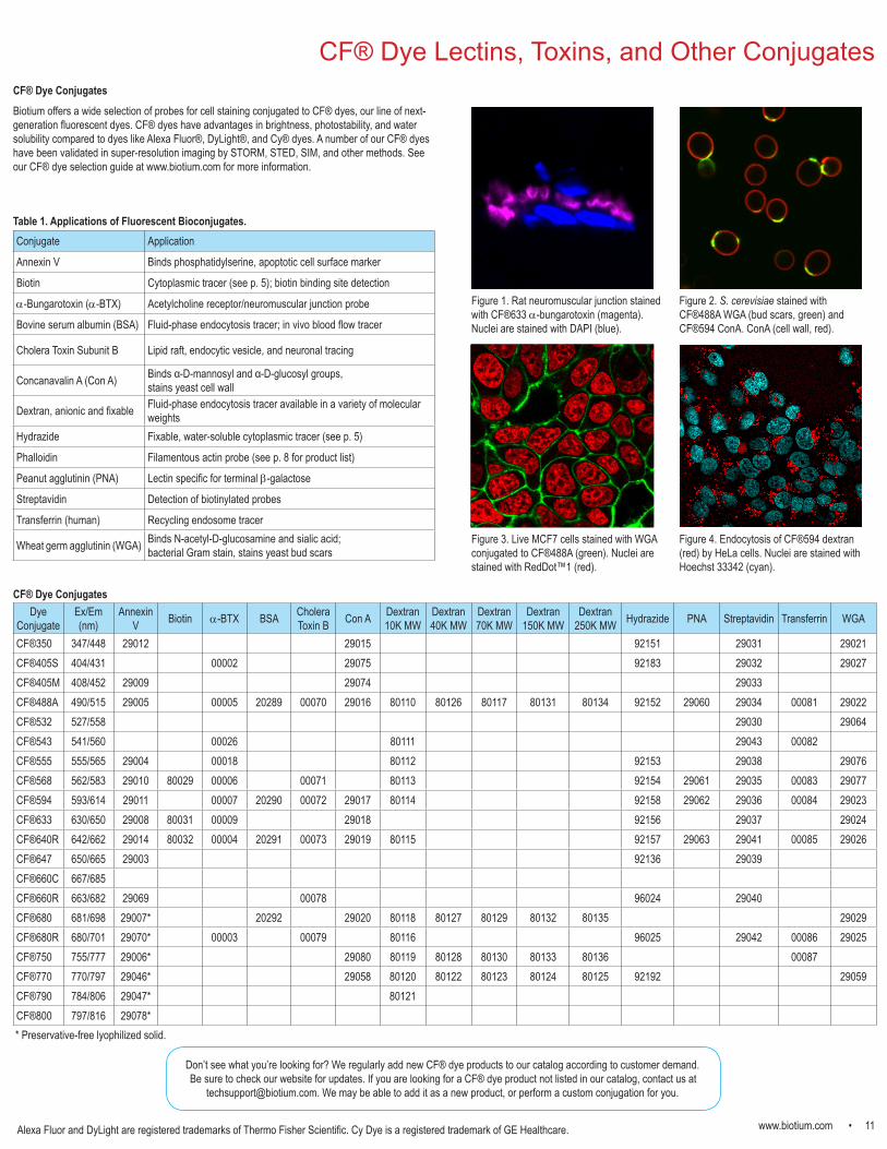
Figure 1. Rat neuromuscular junction stained with CF®633 a-bungarotoxin (magenta). Nuclei are stained with DAPI (blue).
Dye Conjugate
Ex/Em (nm)
Annexin V Biotin a-BTX BSA Cholera
Toxin B Con A Dextran10K MW
Dextran40K MW
Dextran70K MW
Dextran150K MW
Dextran250K MW Hydrazide PNA Streptavidin Transferrin WGA
CF®350 347/448 29012 29015 92151 29031 29021CF®405S 404/431 00002 29075 92183 29032 29027CF®405M 408/452 29009 29074 29033CF®488A 490/515 29005 00005 20289 00070 29016 80110 80126 80117 80131 80134 92152 29060 29034 00081 29022CF®532 527/558 29030 29064CF®543 541/560 00026 80111 29043 00082CF®555 555/565 29004 00018 80112 92153 29038 29076CF®568 562/583 29010 80029 00006 00071 80113 92154 29061 29035 00083 29077CF®594 593/614 29011 00007 20290 00072 29017 80114 92158 29062 29036 00084 29023CF®633 630/650 29008 80031 00009 29018 92156 29037 29024CF®640R 642/662 29014 80032 00004 20291 00073 29019 80115 92157 29063 29041 00085 29026CF®647 650/665 29003 92136 29039CF®660C 667/685 CF®660R 663/682 29069 00078 96024 29040CF®680 681/698 29007* 20292 29020 80118 80127 80129 80132 80135 29029CF®680R 680/701 29070* 00003 00079 80116 96025 29042 00086 29025CF®750 755/777 29006* 29080 80119 80128 80130 80133 80136 00087CF®770 770/797 29046* 29058 80120 80122 80123 80124 80125 92192 29059CF®790 784/806 29047* 80121CF®800 797/816 29078*
Figure 2. S. cerevisiae stained with CF®488A WGA (bud scars, green) and CF®594 ConA. ConA (cell wall, red).
Don’t see what you’re looking for? We regularly add new CF® dye products to our catalog according to customer demand. Be sure to check our website for updates. If you are looking for a CF® dye product not listed in our catalog, contact us at
[email protected]. We may be able to add it as a new product, or perform a custom conjugation for you.
Figure 3. Live MCF7 cells stained with WGA conjugated to CF®488A (green). Nuclei are stained with RedDot™1 (red).
Alexa Fluor and DyLight are registered trademarks of Thermo Fisher Scientific. Cy Dye is a registered trademark of GE Healthcare.
Figure 4. Endocytosis of CF®594 dextran (red) by HeLa cells. Nuclei are stained with Hoechst 33342 (cyan).
* Preservative-free lyophilized solid.
Table 1. Applications of Fluorescent Bioconjugates.
CF® Dye Conjugates
Biotium, Inc.
Toll Free: 800-304-5357Phone: 510-265-1027
Fax: 510-265-1352
General [email protected]
Quotes and [email protected]
Technical [email protected]