Flp-In T-REx Core Kit - assets.thermofisher.com · • ™Once the Flp-In T-REx™ host cell line...
52
USER GUIDE For Research Use Only. Not for human or animal therapeutic or diagnostic use. Flp-In ™ T-REx ™ Core Kit For Generating Stable, Inducible Mammalian Expression Cell Lines by Flp Recombinase-Mediated Integration Catalog number K6500-01 Revision date 1 August 2012 Publication Part number 25-0366 MAN0000184
Transcript of Flp-In T-REx Core Kit - assets.thermofisher.com · • ™Once the Flp-In T-REx™ host cell line...

user guide
For Research Use Only. Not for human or animal therapeutic or diagnostic use.
Flp-In™ T-REx™ Core Kit For Generating Stable, Inducible Mammalian Expression Cell Lines by Flp Recombinase-Mediated Integration
Catalog number K6500-01
Revision date 1 August 2012 Publication Part number 25-0366
MAN0000184


Contents
Contents and Storage ................................................................................................................................. iv
Introduction ................................................................................................................... 1 The Flp-In™ T-REx™ System ........................................................................................................................ 1
Methods ....................................................................................................................... 10 Propagation and Maintenance of Plasmids ............................................................................................ 10
Generating Flp-In™ T-REx™ Host Cell Lines ........................................................................................... 12
Generating Flp-In™ T-REx™ Expression Cell Lines ................................................................................ 22
Appendix ...................................................................................................................... 29 Recipes ......................................................................................................................................................... 29
Zeocin™ Selection Reagent ........................................................................................................................ 31
Blasticidin .................................................................................................................................................... 33
pFRT/lacZeo Vector ................................................................................................................................... 34
pcDNA™6/TR ............................................................................................................................................. 36
pOG44 Vector .............................................................................................................................................. 38
Accessory Products .................................................................................................................................... 40
Technical Support ....................................................................................................................................... 42
Purchaser Notification ............................................................................................................................... 43
References .................................................................................................................................................... 44
iii

Contents and Storage
Shipping and storage
The Flp-In™ T-REx™ Core Kit is shipped at room temperature. Upon receipt, remove the vectors and primers and store at −30°C to −10°C. Store the tetracycline at 2°C to 8°C protected from exposure to light. For long-term storage (> 6 months), store the tetracycline at −30°C to −10°C protected from exposure to light.
Kit contents The Flp-In™ T-REx™ Core Kit contains the following reagents. Store at −30°C to −10°C.
Note: For more information about pcDNA™/FRT/TO and pcDNA™5/FRT/TO/CAT, refer to the pcDNA™5/FRT/TO manual.
Reagent Concentration Amount Comments
pFRT/lacZeo 40 µL at 0.5 µg/µL in TE, pH 8.0
20 µg Flp-In™ target site vector for creation of stable mammalian cell lines containing an integrated Flp Recombination Target (FRT) site
pcDNA™6/TR 40 µL at 0.5 µg/µL in TE, pH 8.0
20 µg Vector for constitutive expression of the Tet repressor
pOG44 40 µL at 0.5 µg/µL in TE, pH 8.0
20 µg Vector for transient expression of the Flp recombinase
pcDNA™5/FRT/TO 40 µL at 0.5 µg/µL in TE, pH 8.0
20 µg Inducible expression vector for cloning your gene of interest
pcDNA™5/FRT/TO/CAT 40 µL at 0.5 µg/µL in TE, pH 8.0
20 µg Positive control vector expressing the CAT gene
CMV Forward Primer (21-mer)
— 2 µg (306 pmoles), lyophilized in TE, pH 8.0
5´-CGCAAATGGGCGGTAGGCGTG-3´
BGH Reverse Primer (18-mer)
— 2 µg (358 pmoles), lyophilized in TE, pH 8.0
5´-TAGAAGGCACAGTCGAGG-3´
Tetracycline — 5 g Induction agent
iv

Introduction
The Flp-In™ T-REx™ System
Overview The Flp-In™ T-REx™ System allows the generation of stable mammalian cell lines exhibiting tetracycline-inducible expression of a gene of interest from a specific genomic location. To generate these cell lines, the Flp-In™ T-REx™ System involves the following major steps:
1. Independent integration of the following two plasmids into the genome of the mammalian cell line of choice to generate a Flp-In™ T-REx™ host cell line:
• A plasmid containing a Flp Recombination Target (FRT) site
• A plasmid expressing the Tet repressor
2. Integration of an expression vector containing your gene of interest under the control of a tetracycline-inducible promoter into the genome via Flp recombinase-mediated DNA recombination at the FRT site (O'Gorman et al., 1991).
3. Induction of the gene of interest by the addition of tetracycline.
Major Components of the system
The major components of the Flp-In™ T-REX™ System include:
• A Flp-In™ target site vector, pFRT/lacZeo, for generation of a Flp-In™ host cell line containing an integrated FRT site.
• A Tet repressor expression plasmid, pcDNA™6/TR, for expression of the Tet repressor under the control of the human CMV promoter.
• An expression plasmid containing a FRT site linked to the hygromycin resistance gene for Flp recombinase-mediated integration and selection of a stable cell line expressing your gene of interest under the control of a tetracycline-regulated CMV/TetO2 promoter.
• A Flp recombinase expression plasmid, pOG44, for expression of the Flp recombinase under the control of the human CMV promoter.
• A control expression plasmid containing the chloramphenicol acetyl transferase (CAT) gene, which when cotransfected with pOG44 into your Flp-In™ T-REx™ host cell line, expresses CAT upon induction with tetracycline.
Advantages of the Flp-In™ T-REx™ system
Use of the Flp-In™ T-REx™ System to generate stable expression cell lines provides a number of advantages: • Once the Flp-In™ T-REx™ host cell line containing an integrated FRT site has
been created, subsequent generation of Flp-In™ T-REx™ cell lines expressing the gene(s) of interest is rapid and efficient.
• The Flp-In™ T-REx™ System allows the generation of isogenic, inducible stable cell lines.
• The Flp-In™ T-REx™ System permits polyclonal selection of stable expression cell lines.
continued on next page
1

The Flp-In™ T-REx™ System, Continued
Generating Flp-In™ T-REx™ host cell lines
The Flp-In™ T-REx™ System streamlines the generation of stable, inducible mammalian expression cell lines by taking advantage of a Saccharomyces cerevisiae-derived DNA recombination system. This DNA recombination system uses a recombinase (Flp) and site-specific recombination (Craig, 1988; Sauer, 1994) to facilitate integration of the gene of interest into a specific site in the genome of mammalian cells.
To generate a Flp-In™ T-REx™ host cell line, you will sequentially transfect two plasmids into the mammalian cell line of choice:
1. pFRT/lacZeo target site vector: The pFRT/lacZeo vector contains a lacZ-Zeocin™ fusion gene whose expression is controlled by the SV40 early promoter (see the Appendix, pages 34–35 for more information). A FRT site has been inserted just downstream of the ATG initiation codon of the lacZ-Zeocin™ fusion gene. The FRT site (see page 5 for more information) serves as the binding and cleavage site for the Flp recombinase. After transfection with pFRT/lacZeo, cells are selected with Zeocin™ Selection Reagent. Zeocin™-resistant clones are screened to identify those containing a single integrated FRT site. The resulting Flp-In™ host cell line contains a single integrated FRT site and expresses the lacZ-Zeocin™ fusion gene (see the following figure).
2. pcDNA™6/TR: The pcDNA™6/TR plasmid constitutively expresses the Tet repressor under the control of the human CMV promoter (see the following figure).
Note: Integration of the pFRT/lacZeo and pcDNA™6/TR plasmids into the genome is random and occurs independently.
�������������� ���������� �������������� �������������� ������������� ���������������������������� ������ �������������������� ����! ��
"��������#$%&����'������� �� ��������������������� ���������������������� ����! ����������������������� ������� �����(������ � � ������� �����������������)*�������� ����! ��
������������� ����! ��
����������
��� � ����������� ��� �������
)*����� ����������������� ����+� �������
��� ���
��� � ����������� ��� �������
)*����� ����������������� ����+� �������
��� ���
���� ���
)*����� ��������������
����������� �������
!"#$���%�������
�����������)*������� ����! ��
continued on next page
2

The Flp-In™ T-REx™ System, Continued
Generating inducible expression cell lines
Once you have generated a Flp-In™ T-REx™ host cell line, you will integrate the pcDNA™5/FRT/TO expression vector containing your gene of interest into the cells via Flp recombinase-mediated DNA recombination at the FRT site (O'Gorman et al., 1991). This step requires the following plasmids:
1. pcDNA™5/FRT/TO expression vector: The pcDNA™5/FRT/TO plasmid contains your gene of interest under the control of a tetracycline-regulated, hybrid human cytomegalovirus (CMV)/TetO2 promoter. The vector also contains the hygromycin resistance gene with a FRT site embedded in the 5′ coding region. The hygromycin resistance gene lacks a promoter and the ATG initiation codon.
2. pOG44: The pOG44 plasmid constitutively expresses the Flp recombinase (Broach et al., 1982; Broach and Hicks, 1980; Buchholz et al., 1996) under the control of the human CMV promoter.
The pOG44 plasmid and the pcDNA™5/FRT/TO vector containing your gene of interest are cotransfected into the Flp-In™ T-REx™ host cell line. Upon cotransfection, the Flp recombinase expressed from pOG44 mediates a homologous recombination event between the FRT sites (integrated into the genome and on pcDNA™5/FRT/TO) such that the pcDNA™5/FRT/TO construct is inserted into the genome at the integrated FRT site (see the figure on page 4). Insertion of pcDNA™5/FRT/TO into the genome at the FRT site brings the SV40 promoter and the ATG initiation codon (from pFRT/lacZeo) into proximity and frame with the hygromycin resistance gene, and inactivates the lacZ-Zeocin™ fusion gene (see the figure on page 4). Thus, stable Flp-In™ T-REx™ expression cell lines can be selected for the following phenotypes:
• Blasticidin resistance
• Hygromycin resistance
• Zeocin™ Selection Reagent sensitivity
• Lack of β-galactosidase activity
Once the pcDNA™5/FRT/TO vector has been stably integrated into the genome at the FRT site, expression of the recombinant protein of interest can be induced by the addition of tetracycline. For more information about the mechanism of tetracycline-induced expression of the gene of interest, refer to pages 6–7.
continued on next page
3

The Flp-In™ T-REx™ System, Continued
Diagram of the Flp-In™ T-REx™ system
The following figure illustrates the major features of the Flp-In™ T-REx™ System as described on the previous page.
��������#$%,����-��*����� ���'���������� � �����+��������� ��������./-�0� ���������������1 ���-/22� ������������������)*������� ����! ���
"��������������� �����*��������������-/22������3�������������+������� ��� ����'���
������1������������� ������ �����������������������������#$%,����-�*����� ���'������
4��������� �������������*����� ���������+��������������������� ������� �������������� ��������� � ' ����������������)*����� ��������������
������ �������� �����������������������������������.����0�
�����&����'�����'���
��� � �������������� ��� �������
()*���)��� �( ��
�'+
������
���
����,-��$',
��������� �
����������������
�����������)*������� ����! �����
���
�������� ()*���)���
��� �������������������� � ������
����,-���$', �'+
�����������)*��)*����� ��� ����! ��
)*����� �������������� ���� �����������
��� ��� �(���
�������� ()*���)���
��� �������������������� � ������
����,-���$', �'+
�����������)*��)*����� ��� ����! ��
)*����� �������������� ���� �����������
)*����� ��������+��������� �������
��� ��� �(���
��$�$�"�)�!���
�
2�)*����� ����������������� �������� �� ��+������������� � ��������������� ���
continued on next page
4

The Flp-In™ T-REx™ System, Continued
Flp recombinase-mediated DNA recombination
In the Flp-In™ T-REx™ System, integration of your pcDNA™5/FRT/TO expression construct into the genome occurs via Flp recombinase-mediated intermolecular DNA recombination. The hallmarks of Flp-mediated recombination are :
• Recombination occurs between specific FRT sites (see the following section) on the interacting DNA molecules.
• Recombination is conservative and requires no DNA synthesis; the FRT sites are preserved following recombination and there is minimal opportunity for introduction of mutations at the recombination site.
• Strand exchange requires only the small 34 bp minimal FRT site (see the following section).
For more information about the Flp recombinase and conservative site-specific recombination, refer to published reviews (Craig, 1988; Sauer, 1994).
Note: If your cell line contains multiple integrated FRT sites, Flp-mediated intramolecular recombination may also occur. Intramolecular recombination may result in:
• Excision of the intervening DNA if the FRT sites are directly repeated (i.e. integration of multiple FRT sites on the same DNA strand)
• DNA inversion if the sites are in opposing orientations
• Deletion of genomic sequences
FRT sites As described above, Flp recombinase-mediated recombination occurs between specific FRT sites. The FRT site, originally isolated from Saccharomyces cerevisiae, serves as a binding site for Flp recombinase and has been well-characterized (Gronostajski and Sadowski, 1985; Jayaram, 1985; Sauer, 1994; Senecoff et al., 1985). The minimal FRT site consists of a 34 bp sequence containing two 13 bp imperfect inverted repeats separated by an 8 bp spacer that includes an Xba I restriction site (see the following figure). An additional 13 bp repeat is found in most FRT sites, but is not required for cleavage (Andrews et al., 1985). While Flp recombinase binds to all three of the 13 bp repeats, strand cleavage actually occurs at the boundaries of the 8 bp spacer region (see the following figure for cleavage sites (CS)) (Andrews et al., 1985; Senecoff et al., 1985).
�����������������������������������������������������
�����"!�����#�$�
��
��
��������� ����
continued on next page
5

The Flp-In™ T-REx™ System, Continued
Tetracycline regulation in the Flp-In™ T-REx™ system
The Flp-In™ T-REx™ System uses regulatory elements from the E. coli Tn10-encoded tetracycline (Tet) resistance operon (Hillen and Berens, 1994; Hillen et al., 1983) to allow tetracycline-regulated expression of your gene of interest from pcDNA™5/FRT/TO. The mechanism of tetracycline regulation in the system is based on the binding of tetracycline to the Tet repressor and derepression of the promoter controlling expression of the gene of interest (Yao et al., 1998). In the system, expression of your gene of interest is repressed in the absence of tetracycline and induced in the presence of tetracycline (Yao et al., 1998).
Expression of your gene of interest from the pcDNA™5/FRT/TO vector is controlled by the human cytomegalovius (CMV) promoter into which 2 tandem copies of the tet operator 2 (TetO2) sequence have been inserted. The TetO2 sequences consist of 2 copies of the 19 nucleotide sequence:
5´-TCCCTATCAGTGATAGAGA-3´
separated by a 2 base pair spacer (Hillen and Berens, 1994; Hillen et al., 1983). Each 19 nucleotide TetO2 sequence serves as the binding site for 2 molecules of the Tet repressor.
Mechanism of repression/ derepression
In the absence of tetracycline, the Tet repressor (expressed from the pcDNA™6/TR plasmid) forms a homodimer that binds with extremely high affinity to each TetO2 sequence in the promoter of the pcDNA™5/FRT/TO vector (Hillen and Berens, 1994). The 2 TetO2 sites in the promoter of pcDNA™5/FRT/TO serve as binding sites for 4 molecules (or 2 homodimers) of the Tet repressor (see the figure on page 7). The affinity of the Tet repressor for the tet operator is KB = 2 × 10¹¹ M-1 (as measured under physiological conditions), where KB is the binding constant (Hillen and Berens, 1994). Binding of the Tet repressor homodimers to the TetO2 sequences represses transcription of your gene of interest. Upon addition, tetracycline binds with high affinity to each Tet repressor homodimer in a 1:1 stoichiometry and causes a conformational change in the repressor that renders it unable to bind to the Tet operator. The association constant, KA, of tetracycline for the Tet repressor is 3 × 10⁹ M-1 (Hillen and Berens, 1994). The Tet repressor:tetracycline complex then dissociates from the Tet operator and allows induction of transcription from the gene of interest (see the figure on page 7).
For more information about the TetR gene, see the Appendix, page 36.
continued on next page
6

The Flp-In™ T-REx™ System, Continued
Diagram of tetracycline regulation
The following figure illustrates the mechanism of tetracycline-regulated repression and derepression of the gene of interest in the Flp-In™ T-REx™ System.
����$�$� $�$�
��$',
$�$� $�$�
��$', *�����.���$���#$
����$�$� $�$�
��$',
$�$� $�$�
��$', *�����.���$���#$
���� ��$', ��$', *�����.���$���#$
� $�$ .���0
�� ��������������.����0������ � ���*���������������#$%&��� �����������������)*��������
"� ���������� ������ ������������������"�.���-"0��5+������ �������#$%,����-��'�����(������� ���������� �� ����������������� ��������
4� 6������� � ��(��������� ���.���0�� ���������������� �����
2� 7 �� ���������������������� �������+�������������� ����������� ������(��������������������������������5+�����(���� ��+�� ������������� �� ������������������� ��������
/0���##�����������##�%
/0���##���������##�%
$�$�$�$�$�$�$�$�
$�$� $�$�$�$� $�$�
$�$�$�$�
$�$�
$�$�
��$�����
continued on next page
7

The Flp-In™ T-REx™ System, Continued
Experimental outline
To create a stable Flp-In™ T-REx™ cell line expressing your gene of interest at a site-specific genomic locus, you will perform the steps listed below (refer to page 9 for a diagram).
1. Transfect the Flp-In™ target site vector, pFRT/lacZeo into the mammalian cell line of choice and screen for integrants containing a single FRT site.
2. Transfect the Tet repressor expression plasmid, pcDNA™6/TR, into a single site integrant (from Step 1of this protocol) to generate the Flp-In™ T-REx™ host cell line.
3. Clone your gene of interest into the pcDNA™5/FRT/TO expression vector.
4. Co-transfect your pcDNA™5/FRT/TO construct and the Flp recombinase expression vector, pOG44, into your Flp-In™ T-REx™ host cell line to generate your Flp-In™ T-REx™ expression cell line.
5. Induce expression of the gene of interest with tetracycline.
6. Assay for expression of your recombinant protein of interest.
Note: The positive control vector containing the CAT gene can be cotransfected into your Flp-In™ T-REx™ host cell line with pOG44 to demonstrate that the system is working properly.
continued on next page
8

The Flp-In™ T-REx™ System, Continued
Diagram of the experimental outline
The following figure illustrates the major steps necessary to generate a Flp-In™ T-REx™ expression cell line.
����������������������� ����������������� ��������� ��������� �������������������������������� ����! ����8��������������������
�����*� � ����� ������ �����������9�� ������������� �������� ' ���
2��! ����������������� �������� ����������#$%,����-���*����� ���'������
,�� ����������������*����� ���'�����������-/22� �������������������)*������� ����! ����8������������������ ������ ������������
:��%����������*������������� ��
��� ��� � �������������� ��� ���
��� ������������������������������������������������������������������� !�"#
������.�+�$���#$
�����
��
�
����������������
��� � ����� �
���
������!!��
������
������
+�$���������
!!��
������
�1�
����
� !!�"��#�
������
����������� ()*���)��� ��� � ���������
����,-���$', �(���
������.�+�$���#$
������.�+�$���#$
���������
���$���
��� ��� ���� �� !"#$
���%��
�������
/��2
.3 ���
������!!��
��$�
� ���
���� ��
*!�4
�� +��
��������� �
�����������������
����
��� ���
������!!��
�( ��
()*���
)������� �
�
�,-��$'
,
������
&��%������������ ������ ��+����*����� ��������+���������� ��������
4��������������#$%&���� ������������������� ����! ���������������������������������)*������� ����! �������8������������������*� � ����� ��������������� � � ����� �����������9�� ������������� �������� ' ���
"��8����������������8�+�������������� ������ ���� �������������1 ������ ����� ��������������� ���
9

Methods
Propagation and Maintenance of Plasmids
Introduction The following section contains guidelines for maintaining and propagating the pFRT/lacZeo, pcDNA™6/TR, and pOG44 vectors. For information about maintaining and propagating the pcDNA™5/FRT/TO expression vector, refer to the vector manual.
General molecular biology techniques
For assistance with E. coli transformations, restriction enzyme analysis, DNA biochemistry, and plasmid preparation, refer to Molecular Cloning: A Laboratory Manual (Sambrook et al., 1989) or Current Protocols in Molecular Biology (Ausubel et al., 1994).
E. coli strain Many E. coli strains are suitable for the propagation of the pFRT/lacZeo, pcDNA™6/TR, and pOG44 vectors. We recommend that you propagate the vectors in E. coli strains that are recombination deficient (recA) and endonuclease A deficient (endA).
For your convenience, TOP10 and DH5α™-T1R E. coli are available as chemically competent or electrocompetent (TOP10 only) cells from Life Technologies (see page 40 for ordering information).
Transformation method
You may use any method of choice for transformation. Chemical transformation is the most convenient for many researchers. Electroporation is the most efficient and the method of choice for large plasmids.
Maintenance of plasmids
The pFRT/lacZeo, pcDNA™6/TR, and pOG44 vectors contain the ampicillin gene to allow selection of the plasmid using ampicillin. Note: The pcDNA™6/TR plasmid also contains the blasticidin resistance gene to allow selection using blasticidin (see page 11).
To propagate and maintain the pFRT/lacZeo, pcDNA™6/TR, and pOG44 plasmids, we recommend using the following procedure:
1. Transform a recA, endA E. coli strain like TOP10, DH5α™-T1R, JM109, or equivalent.
2. Select transformants on LB agar plates containing 50 to 100 μg/mL ampicillin. For fast and easy microwaveable preparation of Low Salt LB agar containing ampicillin, imMedia™ Amp Agar (Catalog no. Q601-20) is available from Life Technologies. For more information, call Technical Support (see page 42).
3. Prepare a glycerol stock from each transformant containing plasmid for long-term storage (see page 11).
continued on next page
10

Propagation and Maintenance of Plasmids, Continued
Selection of pcDNA™6/TR in E. coli using Blasticidin
To propagate and maintain the pcDNA™6/TR plasmid in E. coli using blasticidin selection, follow Steps 1 and 2 of the protocol on page 10. Select transformants on Low Salt LB agar plates containing 100 μg/mL blasticidin.
Note: To facilitate selection of blasticidin-resistant E. coli, the salt concentration of the LB medium must remain low (< 90 mM) and the pH must be 7.0. Failure to lower the salt content of your LB medium will result in non-selection due to inhibition of the drug unless a higher concentration of blasticidin is used. A recipe to prepare Low Salt LB is provided in the Appendix, page 29.
Preparing a glycerol stock
After identifying the correct clone, be sure to purify the colony and make a glycerol stock for long-term storage. It is also a good idea to keep a DNA stock of your plasmid at –20°C.
1. Streak the original colony out on an LB plate containing 50 μg/mL ampicillin. Incubate the plate at 37°C overnight.
2. Isolate a single colony and inoculate into 1–2 mL of LB containing 50 μg/mL ampicillin.
3. Grow the culture to mid-log phase (OD600 = 0.5–0.7).
4. Mix 0.85 mL of culture with 0.15 mL of sterile glycerol and transfer to a cryovial.
5. Store at −80°C.
11

Generating Flp-In™ T-REx™ Host Cell Lines
Introduction Before you can create a stable Flp-In™ T-REx™ cell line(s) which inducibly expresses your gene of interest, you will first need to generate a stable Flp-In™ T-REx™ host cell line. You will generate the Flp-In™ T-REx™ host cell line by independently transfecting the pFRT/lacZeo and pcDNA™6/TR plasmids into your mammalian cell line of interest. Once generated, the Flp-In™ T-REx™ host cell line will exhibit the following features:
• Contains a single integrated FRT site (introduced by transfection of the pFRT/lacZeo plasmid).
• Stably expresses the Tet repressor (introduced by transfection of the pcDNA™6/TR plasmid).
Guidelines to generate a Flp-In™ T-REx™ host cell line are provided in this section.
Experimental Outline
The following table outlines the steps necessary to generate a Flp-In™ T-REx™ host cell line. While it is possible to cotransfect pFRT/lacZeo and pcDNA™6/TR, we recommend that you first generate a stable cell line containing a single integrated FRT site (from pFRT/lacZeo), and then use this cell line as the host for the pcDNA™6/TR plasmid.
Step Action Page
1 Transfect the pFRT/lacZeo target site vector into the mammalian cell line of choice and select for Zeocin™-resistant transfectants.
13–18
2 Pick 20 Zeocin™-resistant foci and expand each clone. 18
3 Isolate genomic DNA and use Southern blot analysis to test for the number of integrated FRT sites.
18–19
4 Select the single integrants and screen for �β-galactosidase activity.
19
5 Select the clone which exhibits the highest �β-galactosidase activity and use this clone as the host for the pcDNA™6/TR plasmid.
19
6 Transfect the pcDNA™6/TR plasmid into the host cell line containing the single integrated FRT site (see Step 5). Select for blasticidin-resistant transfectants.
20–21
7 Pick 20 blasticidin-resistant foci and screen for the clone which expresses the highest level of Tet repressor.
21
Note Reminder: Once you have generated your Flp-In™ T-REx™ host cell line, the cells should exhibit the following phenotypes:
• Zeocin™ resistance
• β-galactosidase activity
• Blasticidin resistance
• Expression of the Tet repressor
continued on next page
12

Generating Flp-In™ T-REx™ Host Cell Lines, Continued
The Flp-In™ T-REx™-293 host cell line which contains a single integrated FRT site and stably expresses the Tet repressor is available from Life Technologies (see page 40 for ordering information). If you wish to inducibly express your gene of interest in 293 cells, you may want to use this cell line as the host and proceed directly to generate your stable expression cell line.
Alternatively, several Flp-In™ host cell lines which contain a single integrated FRT site are also available from Life Technologies (see page 40 for ordering information). You may transfect the pcDNA™6/TR plasmid into these cell lines to generate Flp-In™ T-REx™ host cell lines.
For more information about the Flp-In™ T-REx™-293 cell line or the Flp-In™ cell lines, refer to our Web site (www.lifetechnologies.com) or call Technical Support (see page 42).
�) -
;;)$#%�
�-$
IMPORTANT! We have observed down-regulation of the viral CMV promoter and subsequent loss of gene expression when pcDNA™5/FRT-based expression constructs are introduced into Flp-In™-3T3 or Flp-In™-BHK cells. We recommend that you do not use 3T3 or BHK cells to generate your Flp-In™ T-REx™ host cell line.
Plasmid preparation
Plasmid DNA for transfection into eukaryotic cells must be very clean and free from phenol and sodium chloride. Contaminants will kill the cells and salt will interfere with lipids decreasing transfection efficiency. We recommend isolating plasmid DNA using the PureLink® HiPure Miniprep Kit or the PureLink® HiPure Midiprep Kit (see page 40 for ordering information).
Methods of transfection
For established cell lines (e.g. HeLa, COS-1), consult original references or the supplier of your cell line for the optimal method of transfection. We recommend that you follow exactly the protocol for your cell line. Pay particular attention to medium requirements, when to pass the cells, and at what dilution to split the cells. Further information is provided in Current Protocols in Molecular Biology (Ausubel et al., 1994).
Methods for transfection include calcium phosphate (Chen and Okayama, 1987; Wigler et al., 1977), lipid-mediated (Felgner et al., 1989; Felgner and Ringold, 1989) and electroporation (Chu et al., 1987; Shigekawa and Dower, 1988). If you wish to use a cationic lipid-based reagent for transfection, we recommend using Lipofectamine™ 2000 Reagent (see page 40 for ordering information). For more information, refer to www.lifetechnologies.com or contact Technical Support (page 42).
Zeocin™ Selection Reagent
The pFRT/lacZeo plasmid contains a lacZ-Zeocin™ fusion gene under the control of the SV40 early promoter. Expression of the lacZ-Zeocin™ fusion gene allows selection of stable integrants using Zeocin™ antibiotic. The resulting stable integrants can then be screened by assaying for expression of β-galactosidase. For more information about preparing and handling Zeocin™ Selection Reagent, refer to the Appendix, pages 31–32.
continued on next page
13

Generating Flp-In™ T-REx™ Host Cell Lines, Continued
Determination of Zeocin™ sensitivity
To successfully generate a stable cell line containing an integrated FRT site and expressing the β-galactosidase-Zeocin™ fusion protein, you need to determine the minimum concentration of Zeocin™ Selection Reagent required to kill your untransfected mammalian cell line. Typically, concentrations ranging from 50–1000 μg/mL Zeocin™ Selection Reagent are sufficient to kill most untransfected mammalian cell lines, with the average being 100–400 μg/mL. We recommend that you test a range of concentrations (see the following protocol) to ensure that you determine the minimum concentration necessary for your cell line. Refer to the Appendix, pages 31–32 for instructions on how to prepare and store Zeocin™ Selection Reagent.
1. Plate or split a confluent plate so the cells will be approximately 25% confluent. Prepare a set of 7 plates. Allow cells to adhere overnight.
2. The next day, substitute culture medium with medium containing varying concentrations of Zeocin™ Selection Reagent (0, 50, 100, 250, 500, 750, and 1000 μg/mL Zeocin™ Selection Reagent).
3. Replenish the selective media every 3–4 days, and observe the percentage of surviving cells.
4. Note the percentage of surviving cells at regular intervals to determine the appropriate concentration of Zeocin™ Selection Reagent that kills the cells within 1–2 weeks after addition of Zeocin™ Selection Reagent.
Effect of Zeocin™ Selection Reagent on sensitive and resistant cells
Zeocin™ Selection Reagent‘s method of killing is quite different from other antibiotics including hygromycin, G418, and blasticidin. Cells do not round up and detach from the plate. Sensitive cells may exhibit the following morphological changes upon exposure to Zeocin™ Selection Reagent:
• Vast increase in size (similar to the effects of cytomegalovirus infecting permissive cells).
• Abnormal cell shape.
• Presence of large empty vesicles in the cytoplasm (breakdown of the endoplasmic reticulum and Golgi apparatus, or other scaffolding proteins).
• Breakdown of plasma and nuclear membrane (appearance of many holes in these membranes).
Eventually, these "cells" will completely break down and only "strings" of protein remain.
Zeocin™-resistant cells should continue to divide at regular intervals to form distinct colonies. There should not be any distinct morphological changes in Zeocin™-resistant cells when compared to cells not under selection with Zeocin™ Selection Reagent. For more information about Zeocin™ Selection Reagent and its mechanism of action, refer to the Appendix, pages 31–32.
continued on next page
14

Generating Flp-In™ T-REx™ Host Cell Lines, Continued
Transfection considerations
Once the appropriate Zeocin™ concentration to use for selection is determined, you are ready to transfect the pFRT/lacZeo plasmid into your mammalian cell line of choice. Before beginning, you will need to consider the following factors:
• Insertion of the FRT site into the genome: Integration of the pFRT/lacZeo plasmid containing the FRT site into the genome will occur randomly. Subsequent integration of the pcDNA™5/FRT/TO expression plasmid containing your gene of interest will occur through Flp recombinase-mediated recombination at the genomic FRT site.
• Transfection efficiency of your cell line: The aim of most users will be to create stable cell lines containing a single integrated FRT site (“single integrants”; see the following Note). The probability of obtaining stable integrants containing a single FRT site or multiple FRT sites will depend upon the transfection efficiency of your cell line and the amount of DNA transfected. To increase the likelihood of obtaining single integrants, you will need to lower the transfection efficiency by limiting the amount of plasmid DNA that you transfect (see Recommendation on the next page).
• Selection of foci: You will select for stable transfectants by plating cells in medium containing Zeocin™ Selection Reagent. Zeocin™-resistant foci can then be screened by Southern blot analysis to identify single integrants. To increase the chances of obtaining single integrants, we recommend that you pick foci from plates that have been transfected with the least amount of plasmid DNA.
• Chromosomal position effects: Because integration of the pFRT/lacZeo plasmid into the genome occurs randomly, expression levels of the lacZ-Zeocin™ fusion gene will be dependent on the transcriptional activity of the surrounding sequences at the integration site (i.e. chromosomal position effect). Once you have obtained single integrants, you may want to screen the Zeocin™-resistant clones for those expressing the highest β-galactosidase levels. Those clones expressing the highest levels of β-galactosidase should contain single FRT sites which have integrated into the most transcriptionally active regions.
• Antibiotic concentration: Single integrants will express only a single copy of the lacZ-Zeocin™ fusion gene and therefore, may be more sensitive to Zeocin™ selection than multiple integrants. If you have previously used your mammalian cell line for transfection and Zeocin™ selection, note that you may need to use lower concentrations of Zeocin™ Selection Reagent to obtain single integrants.
continued on next page
15

Generating Flp-In™ T-REx™ Host Cell Lines, Continued
Note Because transfection efficiency is dependent upon the nature of your cell line and the amount of DNA transfected, it is possible to generate a cell line containing multiple integrated FRT sites. In theory, cotransfection of your pcDNA™5/FRT/TO construct and pOG44 into these cells will allow integration of your gene of interest into multiple genomic loci. We do not recommend generating a Flp-In™ T-REx™ host cell line which contains multiple integrated FRT sites for the following reasons:
• Because expression of your gene of interest is based upon a repression/derepression mechanism, the amount of Tet repressor produced in your Flp-In™ T-REx™ host cell line may not be sufficient to repress basal transcription of your gene if the expression plasmid can integrate into multiple genomic loci.
• Proper tetracycline regulated expression of your gene of interest may be more difficult to achieve.
• The presence of multiple integrated FRT sites in the genome may increase the occurrence of chromosomal rearrangements or unexpected recombination events in your host cell line.
As mentioned previously on page 15, we recommend that you transfect your mammalian cell line with a limiting amount of pFRT/lacZeo plasmid. We generally use 250 ng–2 μg of plasmid DNA per 4 × 10⁶ cells for transfection, but the amount of plasmid DNA may vary due to the nature of the cell line, the transfection efficiency of your cells, and the method of transfection used. When transfecting your mammalian cell line of choice, we suggest that you try a range of plasmid DNA concentrations (e.g. 0.25, 0.5, 1, 2, 5 μg/mL DNA) to optimize transfection conditions for your cell line.
We generally use electroporation to transfect cells, but other methods of transfection are suitable. For a protocol to electroporate cells, refer to Current Protocols in Molecular Biology, Unit 9.3 (Ausubel et al., 1994). Note that if you use calcium phosphate or lipid-mediated transfection methods, the amount of total DNA required for transfection is typically higher than for electroporation (usually between 10 and 20 μg DNA). Depending on the amount of pFRT/lacZeo plasmid that you use for transfection, you may need to supplement your plasmid DNA with carrier DNA (e.g. salmon sperm DNA).
�) -
;;)$#%�
�-$
continued on next page
16

Generating Flp-In™ T-REx™ Host Cell Lines, Continued
Possible sites for linearization of pFRT/lacZeo
To obtain stable transfectants, we recommend that you linearize the pFRT/lacZeo plasmid before transfection. While linearizing the vector may not improve the efficiency of transfection, it increases the chances that the vector does not integrate in a way that disrupts the ATG-FRT-lacZ-Zeocin™ cassette or other elements necessary for expression in mammalian cells. The following table lists unique sites that may be used to linearize your construct prior to transfection. Other restriction sites are possible.
Note: We generally use Sca I to linearize pFRT/lacZeo.
Enzyme Restriction Site (bp) Location Supplier
Tth111 I 125 Backbone Many
Apa I 5617 Backbone Life Technologies (Catalog no. 15440-019)
Swa I 6075 Backbone New England Biolabs, Sigma, Takara
Xmn I 6487 Ampicillin gene Many
ScaI I 6606 Ampicillin gene Life Technologies (Catalog no. 15436-017)
Bsa I 7021 Ampicillin gene New England Biolabs
Eam1105 I
7087 Ampicillin gene AGS*, Fermentas, Takara
Sap I 8092 Backbone New England Biolabs *Angewandte Gentechnologie Systeme
continued on next page
17

Generating Flp-In™ T-REx™ Host Cell Lines, Continued
Selection of stable pFRT/lacZeo integrants
Once you have determined the appropriate Zeocin™ concentration to use for selection, you can generate a stable cell line with pFRT/lacZeo.
1. Transfect mammalian cells with the appropriate amount of pFRT/lacZeo (see page 17) using the desired protocol. Remember to include a plate of untransfected cells as a negative control.
2. 24 hours after transfection, wash the cells and add fresh medium to the cells.
3. 48 hours after transfection, split the cells into fresh medium. Split the cells such that they are no more than 25% confluent. If the cells are too dense, the antibiotic will not kill the cells. Antibiotics work best on actively dividing cells.
4. Incubate the cells at 37°C for 2–3 hours until they have attached to the culture dish.
5. Remove the medium and add fresh medium containing Zeocin™ Selection Reagent at the pre-determined concentration required for your cell line.
6. Feed the cells with selective medium every 3–4 days until foci can be identified.
7. Pick at least 20 Zeocin™-resistant foci and expand each clone to test for the number of integrated FRT sites. Isolate genomic DNA and use Southern blot analysis to distinguish between single and multiple integrants (see the following section and page 19). Select the single integrants and proceed to Step 8.
8. Screen the single integrants for β-galactosidase activity (see page 19). Select those clones which exhibit the highest levels of β-galactosidase expression to use as your host for the pcDNA™6/TR plasmid.
9. Once you have obtained a stable Flp-In™ cell line, you can use this cell line to isolate a stable cell line expressing the Tet repressor from the pcDNA™6/TR plasmid (see pages 36–37). Note: Remember to maintain the Flp-In™ host cell line in medium containing the appropriate amount of Zeocin™ Selection Reagent.
Isolation of genomic DNA
Once you have obtained Zeocin™-resistant foci, you will need to expand the cells and isolate genomic DNA. You may use any standard protocol to isolate genomic DNA from your cells. Protocols may be found in Current Protocols in Molecular Biology (Ausubel et al., 1994) or Molecular Cloning: A Laboratory Manual (Sambrook et al., 1989). For easy isolation of genomic DNA, the Easy-DNA™ Kit (Catalog no. K1800-01) is available separately. Call Technical Support for more information (see page 42).
continued on next page
18

Generating Flp-In™ T-REx™ Host Cell Lines, Continued
Screening clones by Southern blot analysis
You can use Southern blot analysis to determine the number of integrated FRT sites present in each of your Zeocin™-resistant clones. When performing Southern blot analysis, you should consider the following factors:
• Probe: We recommend that you use a fragment of the lacZ gene (100–500 bp) as the probe to screen your samples. Mammalian cells do not contain an endogenous lacZ gene, therefore, a lacZ probe should allow you to identify those clones which contain pFRT/lacZeo DNA. To label the probe, we generally use a standard random priming kit (e.g. Ambion, DECAprime II™ Kit, Catalog no. 1455). Other random priming kits are suitable.
• Restriction digest: When choosing a restriction enzyme to digest the genomic DNA, we recommend choosing an enzyme that cuts at a single known site outside of the lacZ gene in the pFRT/lacZeo vector. Hybridization of the lacZ probe to digested DNA should then allow you to detect a single band containing the lacZ gene from pFRT/lacZeo. We generally use Hind III to digest genomic DNA from the Zeocin™-resistant clones. pFRT/lacZeo contains a single Hind III site within the FRT site.
• Protocol: You may use any Southern blotting protocol of your choice. Refer to Current Protocols in Molecular Biology (Ausubel et al., 1994) or Molecular Cloning: A Laboratory Manual (Sambrook et al., 1989) for detailed protocols.
What you Should see
If you digest genomic DNA from your transfectants with Hind III and use a lacZfragment as a probe in your Southern analysis, you should be able to easily distinguish between single and multiple FRT integrants:
• DNA from single integrants should contain only one hybridizing band corresponding to a single copy of the integrated pFRT/lacZeo plasmid.
• DNA from multiple integrants should contain more than one hybridizing band. If the pFRT/lacZeo plasmid integrates into multiple chromosomal locations, the bands may be of varying sizes.
Assay for β-Galactosidase activity
After identifying single integrants, proceed to screen the clones for β-galactosidase expression. You may assay for β-galactosidase expression by activity assay using cell-free lysates (Miller, 1972) or by staining the cells for activity. Life Technologies offers the β-Gal Assay Kit (Catalog no. K1455-01) and the β-Gal Staining Kit (Catalog no. K1465-01) for fast and easy detection of β-galactosidase expression. Select those clones expressing the highest levels of β-galactosidase (if desired) to use as the host cell lines for the pcDNA™6/TR plasmid.
continued on next page
19

Generating Flp-In™ T-REx™ Host Cell Lines, Continued
Determination of Blasticidin sensitivity
Once you have generated a Flp-In™ cell line containing a single integrated FRT site, you will transfect the pcDNA™6/TR plasmid into these cells to generate a Flp-In™ T-REx™ host cell line. The pcDNA™6/TR plasmid contains the blasticidin resistance gene (bsd) to allow selection of stable transfectants. Before proceeding, we recommend that you determine the minimum concentration of blasticidin required to kill your untransfected Flp-In™ host cell line. Use the protocol on page 14 with the following exceptions:
• Typically, concentrations ranging from 2.5–10 μg/mL blasticidin are sufficient to kill most untransfected mammalian cell lines.
• Selection should be complete within 10 days after addition of the antibiotic.
Refer to the Appendix, page 33 for instructions on how to prepare and store blasticidin.
Possible sites for linearization of pcDNA™6/TR
As with transfection of pFRT/lacZeo, we also recommend that you linearize the pcDNA™6/TR plasmid before transfection. The following table lists unique sites that may be used to linearize your construct prior to transfection. Other restriction sites are possible.
Enzyme Restriction Site (bp) Location Supplier
Bst1107 I 4470 Backbone AGS*, Fermentas, Takara
Sap I 4733 Backbone New England Biolabs
BspLU11 I 4849 Backbone Boehringer-Mannheim
Eam1105 I 5739 Ampicillin gene AGS*, Fermentas, Takara
Fsp I 5961 Ampicillin gene Many *Angewandte Gentechnologie Systeme.
continued on next page
20

Generating Flp-In™ T-REx™ Host Cell Lines, Continued
Selection of stable pcDNA™6/TR
integrants
Once you have determined the appropriate blasticidin concentration to use for selection, you can generate a stable cell line expressing the Tet repressor from pcDNA™6/TR. Reminder: Since you are using transfecting pcDNA™6/TR into the Flp-In™ host cell line, remember to maintain cells in medium containing Zeocin™ Selection Reagent.
1. Transfect your Flp-In™ host cell line with the pcDNA™6/TR plasmid using the desired protocol. Remember to include a plate of untransfected cells as a negative control.
2. 24 hours after transfection, wash the cells and add fresh medium to the cells.
3. 48 hours after transfection, split the cells into fresh medium. Split the cells such that they are no more than 25% confluent.
4. Incubate the cells at 37°C for 2–3 hours until they have attached to the culture dish.
5. Remove the medium and add fresh medium containing blasticidin at the pre-determined concentration required for your cell line.
6. Feed the cells with selective medium every 3–4 days until foci can be identified.
7. Pick at least 20 blasticidin-resistant foci and expand them to test for tetracycline-inducible gene expression by transiently transfecting with the pcDNA™5/FRT/TO/CAT positive control plasmid expressing CAT. Screen for those clones which exhibit the lowest levels of basal transcription and the highest levels of CAT expression after addition of tetracycline (see the following section). For guidelines to prepare and use tetracycline, see pages 25 and 28.
8. Once you have obtained a stable Flp-In™ T-REx™ cell line, you can use this cell line to isolate a stable cell line expressing your gene of interest from the pcDNA™5/FRT/TO plasmid. Note: Remember to maintain the Flp-In™ T-REx™ host cell line in medium containing the appropriate amount of Zeocin™ Selection Reagent and blasticidin.
IMPORTANT! Because tetracycline-regulated expression in the Flp-In™ T-REx™ System is based on a repression/derepression mechanism, the amount of Tet repressor expressed in the Flp-In™ T-REx™ host cell line from pcDNA™6/TR will determine the level of transcriptional repression of the Tet operator sequences in your pcDNA™5/FRT/TO expression construct. Tet repressor levels should be sufficiently high to suitably repress basal level transcription. Therefore, when screening your blasticidin-resistant clones, select clones which exhibit the lowest basal levels of CAT expression (see above) to use as the host(s) for your pcDNA™5/FRT/TO construct. These clones should be exhibiting the highest levels of Tet repressor expression.
Assay for CAT protein
The CAT protein expressed from the pcDNA™5/FRT/TO/CAT control plasmid is approximately 32 kDa in size. You may assay for CAT expression by ELISA assay, Western blot analysis, fluorometric assay, or radioactive assay (Ausubel et al., 1994; Neumann et al., 1987). The FAST CAT® Chloramphenicol Acetyltransferase Assay Kit (Catalog no. F-2900) is available from Life Technologies to assay for CAT protein.
21

Generating Flp-In™ T-REx™ Expression Cell Lines
Introduction Once you have established your Flp-In™ T-REx™ host cell line, you may cotransfect your pcDNA™5/FRT/TO construct and the pOG44 expression plasmid into the host cell line to generate a stable Flp-In™ T-REx™ expression cell line which inducibly expresses your gene of interest. Integration of the pcDNA™5/FRT/TO construct into the genome will occur at the FRT site in the Flp-In™ T-REx™ host cells. The pcDNA™5/FRT/TO plasmid contains the hygromycin resistance gene to allow selection of stable cell lines (see the following section). For more information about the pcDNA™5/FRT/TO plasmid and cloning your gene of interest into pcDNA™5/FRT/TO, refer to the vector manual. For more information about the pOG44 plasmid, see the following section.
IMPORTANT! The hygromycin resistance gene in the pcDNA™5/FRT/TO vector lacks an ATG initiation codon and a promoter to drive expression of the gene. Transfection of pcDNA™5/FRT/TO plasmid alone into a Flp-In™ T-REx™ host cell line will not confer hygromycin resistance to the cells containing the plasmid. The ATG initiation codon and the SV40 promoter required for expression of the hygromycin resistance gene are brought into proximity and frame with the gene only through Flp recombinase-mediated recombination between the FRT sites in the pcDNA™5/FRT/TO plasmid and the Flp-In™ T-REx™ host cell line.
If you wish to express your gene of interest in 293 cells, you may want to use the Flp-In™ T-REx™-293 host cell line available from Life Technologies to establish your expression cell line (see page 40 for ordering information). For more information, refer to our World Wide Web site (www.lifetechnologies.com) or call Technical Support (see page 42).
�) -
;;)$#%�
�-$
Note It is also possible to cotransfect pcDNA™5/FRT/TO and pOG44 into a Flp-In™ host cell line to generate an expression cell line. Flp-In™ host cell lines contain a single integrated FRT site, but do not express the Tet repressor. Cotransfection of pcDNA™5/FRT/TO and pOG44 into a Flp-In™ host cell line would allow integration of the pcDNA™5/FRT/TO construct into the genome via the FRT sites. However, in this case, the TetO2 sequences in the hybrid CMV/TetO2 promoter of pcDNA™5/FRT/TO are inert and the CMV/TetO2 promoter functions to allow constitutive expression of your gene of interest at levels similar to the native CMV promoter.
pOG44 plasmid You will cotransfect the pOG44 plasmid and your pcDNA™5/FRT/TO construct into your Flp-In™ T-REx™ host cell line to generate stable cell lines that inducibly express your protein of interest. Cotransfection of pOG44 and pcDNA™5/FRT/TO allows expression of Flp recombinase and integration of the pcDNA™5/FRT/TO plasmid into the genome via the FRT sites. Once the pcDNA™5/FRT/TO construct has integrated into the genome, the Flp recombinase is no longer required. The continued presence of Flp recombinase would actually be detrimental to the cells because it could mediate excision of your pcDNA™5/FRT/TO construct.
The pOG44 plasmid lacks an antibiotic resistance marker for selection in mammalian cells. Thus, the plasmid and therefore, Flp recombinase expression, will gradually be lost from transfected cells as they are cultured and selected in hygromycin.
continued on next page
22
Generating Flp-In™ T-REx™ Expression Cell Lines, Continued
Flp recombinase The FLP gene was originally isolated from the Saccharomyces cerevisiae 2μ plasmid (Broach et al., 1982; Broach and Hicks, 1980) (see the Appendix, page 38 for more information). When tested in mammalian cells, the Flp recombinase has been shown to possess optimum recombination activity near 30°C and relatively low activity at 37°C, a result consistent with its physiological role in yeast (Buchholz et al., 1996).
The FLP gene in pOG44 is further limited in its activity because it contains a point mutation that encodes a Flp recombinase with a phenylalanine to leucine amino acid substitution at position 70 (Buchholz et al., 1996). The resulting Flp recombinase (flp-F70L) exhibits increased thermolability at 37°C in mammalian cells when compared to the native Flp recombinase (Buchholz et al., 1996). Studies have shown that the Flp recombinase expressed from pOG44 possesses only 10% of the activity at 37°C of the native Flp recombinase (Buchholz et al., 1996).
IMPORTANT! When generating Flp-In™ T-REx™ expression cell lines, it is important to remember that you are selecting for a relatively rare recombination event since you want recombination and integration of your pcDNA™5/FRT/TO construct to occur only through the FRT site and for a limited time. In this case, using a highly inefficient Flp recombinase is beneficial and may decrease the occurrence of other undesirable recombination events.
Note Reminder: Integration of the pcDNA™5/FRT/TO construct into the genome via the FRT sites will result in the following events (see page 4 for a diagram):
• Insertion of the hygromycin resistance gene downstream of the SV40 early promoter and the ATG initiation codon (provided by pFRT/lacZeo).
• Insertion of the plasmid containing the CMV/TetO2 promoter, your gene of interest, and the BGH polyadenylation signal upstream of the lacZ-Zeocin™ fusion gene.
• Disruption of the functional lacZ-Zeocin™ transcriptional unit caused by loss of the SV40 early promoter and the ATG initiation codon and insertion of the cassette containing the CMV/TetO2 promoter, gene of interest, and the BGH polyadenylation signal.
As a result, your Flp-In™ T-REx™ expression cell lines should exhibit the following phenotype:
• Hygromycin resistance
• Zeocin™ Selection Reagent sensitivity
• Lack of β-galactosidase activity
• Blasticidin resistance
• Tetracycline-regulated expression of the gene of interest
continued on next page
23

Generating Flp-In™ T-REx™ Expression Cell Lines, Continued
Positive control The pcDNA™5/FRT/TO/CAT plasmid is provided as a positive control vector for mammalian cell transfection and expression and may be used to assay for expression levels in your Flp-In™ T-REx™ expression cell line. If you have several different Flp-In™ T-REx™ host cell lines (cell lines containing FRT sites integrated at different genomic loci), you may want to use the pcDNA™5/FRT/TO/CAT control vector to compare protein expression levels from the various genomic loci. For more information about pcDNA™5/FRT/TO/CATrefer to the pcDNA™5/FRT/TO vector manual.
Hygromycin B The pcDNA™5/FRT/TO vector contains the E. coli hygromycin resistance gene (HPH) (Gritz and Davies, 1983) for selection of transfectants with the antibiotic, hygromycin B (Palmer et al., 1987). When added to cultured mammalian cells, hygromycin B acts as an aminocyclitol to inhibit protein synthesis by disrupting translocation and promoting mistranslation. Hygromycin B liquid is available separately (see page 41 for ordering information).
• Hygromycin B is light sensitive. Store the liquid stock solution at 4°C protected from exposure to light.
• Hygromycin B is toxic. Do not ingest solutions containing the drug.
• Wear gloves, a laboratory coat, and safety glasses or goggles when handling hygromycin B and hygromycin B-containing solutions.
Preparing and storing hygromycin B
The hygromycin B available from Life Technologies is supplied as a 50 mg/mLstock solution in autoclaved, deionized water and is filter-sterilized. The solution is brown in color. The stability of hygromycin B is guaranteed for 6 months, if stored at 4°C. Medium containing hygromycin is stable for up to 6 weeks.
Determination of hygromycin sensitivity
To successfully generate a stable cell line expressing your gene of interest from pcDNA™5/FRT/TO, you need to determine the minimum concentration of hygromycin B required to kill your untransfected Flp-In™ T-REx™ host cell line. Typically, concentrations ranging from 10–400 μg/mL hygromycin B are sufficient to kill most untransfected mammalian cell lines. We recommend that you test a range of concentrations (0, 10, 50, 100, 200, 400, 600 μg/mL hygromycin B) to ensure that you determine the minimum concentration necessary for your Flp-In™ T-REx™ host cell line.
continued on next page
24

Generating Flp-In™ T-REx™ Expression Cell Lines, Continued
Tetracycline Tetracycline is commonly used as a broad spectrum antibiotic and acts to inhibit translation by blocking polypeptide chain elongation in bacteria. In the Flp-In™ T-REx™ System, tetracycline hydrochloride (MW = 480.90) is used as an inducing agent to induce transcription of the gene of interest from the pcDNA™5/FRT/TO expression vector. Tetracycline induces transcription by binding to the Tet repressor homodimer and causing the repressor to undergo a conformational change that renders it unable to bind to the Tet operator. The association constant of tetracycline to the Tet repressor is 3 × 10⁹ M-1 (Takahashi et al., 1991). Tetracycline is supplied with the Flp-In™ T-REx™ Core Kit, but may also be obtained separately (see page 41 for ordering information). Note: The concentrations of tetracycline used to induce gene expression in the Flp-In™ T-REx™ System are generally not high enough to be toxic to mammalian cells.
Tetracycline-reduced serum
When culturing cells in medium containing fetal bovine serum (FBS), note that many lots of FBS contain tetracycline as FBS is generally isolated from cows that have been fed a diet containing tetracycline. If you culture your cells in medium containing FBS that is not reduced in tetracycline, you may observe low basal expression of your gene of interest in the absence of tetracycline. We have cultured our mammalian cells in medium containing FBS that may not be reduced in tetracycline, and have observed undetectable to very low basal expression of CAT from the positive control vector in the absence of additional tetracycline. If your gene of interest produces a toxic protein, you may wish to culture your cells in tetracycline-reduced FBS. For more information, consult the supplier of your serum.
Tetracycline is light sensitive. Store the powdered drug at 4°C in the dark. Prepare medium containing tetracycline immediately before use. Tetracycline is toxic. Do not ingest or inhale the powder or solutions containing the drug.
Wear gloves, a laboratory coat, and safety glasses or goggles when handling tetracycline and tetracycline-containing solutions.
Preparation of Tetracycline
To prepare tetracycline:
1. Weigh out 10 mg of tetracycline (provided in the Flp-In™ T-REx™ Core Kit) and transfer to a sterile 15 mL conical polypropylene tube.
2. Resuspend 10 mg of tetracycline in 10 mL of sterile water. This provides a 1 mg/mL stock solution of tetracycline that is yellow in color.
3. Store the stock solution at –20°C protected from exposure to light.
continued on next page
25

Generating Flp-In™ T-REx™ Expression Cell Lines, Continued
Because correct integration of your pcDNA™5/FRT/TO construct into the genome is dependent on Flp recombinase, the expression levels of Flp recombinase in the cell will determine the efficiency of the recombination reaction. Flp recombinase levels must be sufficiently high to mediate recombination at the FRT sites (single recombination event) and overcome the low intrinsic activity of the enzyme (see page 23). We have varied the ratio of pOG44 and pcDNA™5/FRT/TO expression plasmid that we cotransfect into mammalian Flp-In™ T-REx™ host cells to optimize the recombination efficiency. We recommend that you cotransfect your Flp-In™ T-REx™ host cell line with a ratio of at least 9:1 (w/w) pOG44:pcDNA™5/FRT/TO expression plasmid. Note that this ratio may vary depending on the nature of the cell line. You may want to determine this ratio empirically for your cell line.
�) -
;;)$#%�
�-$
IMPORTANT! When transfecting your Flp-In™ T-REx™ host cell line, be sure to use supercoiled pOG44 and pcDNA™5/FRT/TO plasmid DNA. Flp-mediated recombination between the FRT site on pcDNA™5/FRT/TO and the integrated FRT site in the Flp-In™ T-REx™ host cell line will only occur if the pcDNA™5/FRT/TO plasmid is circularized. The pOG44 plasmid should be circularized to minimize the possibility of the plasmid integrating into the genome.
continued on next page
26

Generating Flp-In™ T-REx™ Expression Cell Lines, Continued
Selection of stable Flp-In™ T-REx™ expression cell lines
Once you have determined the appropriate hygromycin concentration to use for selection in your Flp-In™ T-REx™ host cell line, you can generate a stable cell line expressing your pcDNA™5/FRT/TO construct.
Reminder: Following cotransfection, your Flp-In™ T-REx™ expression clones should become sensitive to Zeocin™ Selection Reagent (see Note on page 18); therefore, your selection medium should not contain Zeocin™ Selection Reagent, however the selection medium should contain blasticidin to select for the pcDNA™6/TR plasmid.
1. Cotransfect your mammalian Flp-In™ T-REx™ host cells with a 9:1 ratio of pOG44:pcDNA™5/FRT/TO plasmid DNA (see above) using the desired protocol. Include a plate with no pOG44 as a Flp recombination control, a plate of untransfected cells as a negative control, and a plate of cells transfected with the pcDNA™5/FRT/TO/CAT plasmid as a positive control.
2. 24 hours after transfection, wash the cells and add fresh medium to the cells.
3. 48 hours after transfection, split the cells into fresh medium. Split the cells such that they are no more than 25% confluent. If the cells are too dense, the antibiotic will not kill the cells. Antibiotics work best on actively dividing cells.
4. Plate the trypsinized cells at 37°C in the presence of hygromycin (at the predetermined concentration for your cell line) immediately. Do not wait for the cells to attach before adding antibiotic. This ensures that only true transfectants survive, and that untransfected cells die off quickly.
5. Feed the cells with selective medium every 3–4 days until foci can be identified.
6. Pick 5–20 hygromycin-resistant foci and expand the cells. Verify that the pcDNA™5/FRT/TO construct has integrated into the FRT site by testing each clone for Zeocin™ sensitivity and lack of β-galactosidase activity.
Note: To screen a polyclonal population of cells, see page 28.
7. Select those clones that are hygromycin-resistant, Zeocin™-sensitive, and LacZ– , and assay for expression of your gene of interest. Note: We have observed in-house that in cells where the FRT site has integrated into a very transcriptionally active locus in the host cell genome (seen more commonly in Flp-in CHO and Flp-in 293 cells but can also happen in Flp-in 3T3 cells and any other Flp-in host cell line), there is some “read-through” transcription and translation of the lacZ-Zeocin ORF post Flp-in, even though the lacZ-Zeocin ORF does not have a bonafide promoter and ATG. In such cases, the hygromycin-resistant clones would also be lacZ positive and Zeocin-resistant. To make sure that the integration is FRT site-specific and not random, we recommend doing a parallel control transfection with no pOG44 present. This should yield no surviving clones upon hygromycin selection, indicating that all the hygromycin-resistant clones obtained in the presence of pOG44 are indeed Flp recombinase-dependent and hence have the gene of interest integrated at the FRT site. Also, a Southern blot analysis of these clones will help verify that they do indeed have proper FRT integration of the gene of interest despite the expression of lacZ (although this is typically not necessary). Post Flp-in, as long as you see hygromycin-resistant clones, we recommend that you select them and assay them for expression of your gene of interest.
continued on next page
27
Generating Flp-In™ T-REx™ Expression Cell Lines, Continued
Polyclonal selection
If you use a single integrant as your Flp-In™ T-REx™ host cell line, all of the hygromycin-resistant foci that you obtain after cotransfection of pcDNA™5/FRT/TO and pOG44 and selection with hygromycin should be isogenic (i.e. pcDNA™5/FRT/TO should integrate into the same genomic locus in every clone, therefore, all clones should be identical). Having isogenic clones should allow you to perform “polyclonal” selection and screening of your hygromycin-resistant cells. If you wish, you do not need to pick and screen separate foci for expression of your protein of interest. After hygromycin selection, simply pool the foci and screen the entire population of cells for tetracycline-regulated expression of your protein of interest.
Induction of gene expression
Guidelines are provided below to induce expression of your protein of interest with tetracycline. Expression conditions may vary depending on the nature of your protein of interest and on the cell line; therefore, some empirical experimentation may be needed to determine the optimal conditions for inducible expression.
• Plate your Flp-In™ T-REx™ expression cell line in medium containing blasticidin and hygromycin.
• To induce expression of the gene of interest, remove medium and add fresh medium containing the appropriate concentration of tetracycline to the cells. In general, we recommend that you add tetracycline to a final concentration of 1 μg/mL (5 μL of a 1 mg/mL stock per 5 mL of medium) to the cells and incubate the cells for 24 hours at 37ºC.
• Harvest the cells and assay for expression of your gene.
Optimization of expression
You may want to vary the concentration of tetracycline (0.1–1 μg/mL) and time of exposure to tetracycline (8 to 48 hours) to optimize or modulate expression for your cell line.
Other inducers You may use doxycycline as an alternative inducing agent in the Flp-In™ T-REx™
System. Doxycycline is similar to tetracycline in its mechanism of action, and exhibits similar dose response and induction characteristics as tetracycline in the Flp-In™ T-REx™ System. Doxycycline has been shown to have a longer half-life than tetracycline (48 hours vs. 24 hours, respectively). Doxycycline may be obtained from Sigma (Catalog no. D9891).
28

Appendix
Recipes
LB (Luria-Bertani) medium and plates
Composition:
10 g Tryptone 10 g NaCl 5 g Yeast Extract pH 7.0
1. Combine the dry reagents above and add deionized, distilled water to 950 mL.
2. Adjust the pH of the solution to 7.0 with NaOH and bring the volume up to 1 liter.
3. Autoclave on liquid cycle for 20 minutes at 15 psi. Allow solution to cool to 55°C and add antibiotic if needed.
4. Store at room temperature or at 4°C.
LB agar plates
1. Prepare LB medium as above, but add 15 g/L agar before autoclaving.
2. Autoclave on liquid cycle for 20 minutes at 15 psi.
3. After autoclaving, cool to ~55°C, add antibiotic (i.e. 50–100 μg/mL of ampicillin), and pour into 10 cm plates.
4. Let harden, then invert and store at 4°C, in the dark.
Low Salt LB medium with Blasticidin
Low Salt LB Medium:
10 g Tryptone 5 g NaCl 5 g Yeast Extract
1. Combine the dry reagents above and add deionized, distilled water to 950 mL. Adjust pH to 7.0 with 1 N NaOH. Bring the volume up to 1 liter. For plates, add 15 g/L agar before autoclaving.
2. Autoclave on liquid cycle at 15 psi and 121°C for 20 minutes.
3. Allow the medium to cool to at least 55°C before adding the blasticidin to 50 μg/mL final concentration.
4. Store plates at 4°C in the dark. Plates containing blasticidin are stable for up to 2 weeks.
continued on next page
29

Recipes, Continued
Phosphate-Buffered Saline (PBS)
137 mM NaCl 2.7 mM KCl 10 mM Na2HPO4 1.8 mM KH2PO4
1. Dissolve the following in 800 mL of deionized water:
8 g NaCl 0.2 g KCl 1.44 g Na2HPO4
0.24 g KH2PO4
2. Adjust pH to 7.4 with concentrated HCl.
3. Bring the volume to 1 liter and autoclave for 20 minutes on liquid cycle.
4. Store at room temperature or at 4°C.
30

Zeocin™ Selection Reagent
Zeocin™ Selection Reagent
Zeocin™ Selection Reagent is a member of the bleomycin/phleomycin family of antibiotics isolated from Streptomyces. Antibiotics in this family are broad spectrum antibiotics that act as strong anti-bacterial and anti-tumor drugs. They show strong toxicity against bacteria, fungi (including yeast), plants, and mammalian cells (Baron et al., 1992; Drocourt et al., 1990; Mulsant et al., 1988; Perez et al., 1989).
The Zeocin™ resistance protein has been isolated and characterized (Calmels et al., 1991; Drocourt et al., 1990). This protein, the product of the Sh ble gene (Streptoalloteichus hindustanus bleomycin gene), is a 13.7 kDa protein that binds Zeocin™ Selection Reagent and inhibits its DNA strand cleavage activity. Expression of this protein in eukaryotic and prokaryotic hosts confers resistance to Zeocin™ Selection Reagent.
Molecular weight, formula, and structure
The formula for Zeocin™ Selection Reagent is C55H86O21N20S2Cu-HCl and the molecular weight is 1527.5. The following diagram shows the structure of Zeocin™ Selection Reagent.
���
��
�
��� ��
� �
�
���
�
������
��
�
�
���
������
�
���� ��
��
��
��
�
��
����
��
����
���
� �
�
�
�
���
�
�
�
�
�
�� �
�
��
��
�� �
Applications of Zeocin™ Selection Reagent
Zeocin™ Selection Reagent is used for selection in mammalian cells (Mulsant et al., 1988); plants (Perez et al., 1989); yeast (Baron et al., 1992); and prokaryotes (Drocourt et al., 1990). Typically, Zeocin™ Selection Reagent concentrations ranging from 50 to 1000 μg/mL are used for selection in mammalian cells. Before transfection, we recommend that you first test the sensitivity of your mammalian host cell to Zeocin™ Selection Reagent as natural resistance varies among cell lines.
continued on next page
31

Zeocin™ Selection Reagent, Continued
Handling Zeocin™ Selection Reagent
• Store Zeocin™ Selection Reagent at –20°C and thaw on ice before use.
• Zeocin™ Selection Reagent is light sensitive. Store drug, plates, and medium containing drug in the dark.
• Wear gloves, a laboratory coat, and safety glasses or goggles when handling solutions containing Zeocin™ Selection Reagent.
• Zeocin™ Selection Reagent is toxic. Do not ingest or inhale solutions containing the drug.
Ordering information
You may purchase Zeocin™ Selection Reagent from Life Technologies. For your convenience, the drug is prepared in autoclaved, deionized water and available in 1.25 mL aliquots at a concentration of 100 mg/mL. The stability of Zeocin™ Selection Reagent is guaranteed for six months, if stored at –20°C.
Amount Catalog no.
1 gram R250-01
5 grams R250-05
32
Blasticidin
Blasticidin Blasticidin S HCl is a nucleoside antibiotic isolated from Streptomyces griseochromogenes which inhibits protein synthesis in both prokaryotic and eukaryotic cells (Takeuchi et al., 1958; Yamaguchi et al., 1965). Resistance is conferred by expression of either one of two blasticidin S deaminase genes: bsd from Aspergillus terreus (Kimura et al., 1994) or bsr from Bacillus cereus (Izumi et al., 1991). These deaminases convert blasticidin S to a non-toxic deaminohydroxy derivative (Izumi et al., 1991).
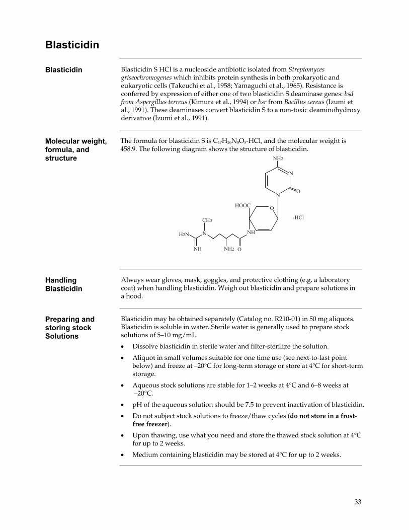
Molecular weight, formula, and structure
The formula for blasticidin S is C17H26N8O5-HCl, and the molecular weight is 458.9. The following diagram shows the structure of blasticidin.
$�"
-
�--
$�
�4
�"$
$�"
$
-$�
$
$
-
�� �
Handling Blasticidin
Always wear gloves, mask, goggles, and protective clothing (e.g. a laboratory coat) when handling blasticidin. Weigh out blasticidin and prepare solutions in a hood.
Preparing and storing stock Solutions
Blasticidin may be obtained separately (Catalog no. R210-01) in 50 mg aliquots. Blasticidin is soluble in water. Sterile water is generally used to prepare stock solutions of 5–10 mg/mL.
• Dissolve blasticidin in sterile water and filter-sterilize the solution.
• Aliquot in small volumes suitable for one time use (see next-to-last point below) and freeze at –20°C for long-term storage or store at 4°C for short-term storage.
• Aqueous stock solutions are stable for 1–2 weeks at 4°C and 6–8 weeks at –20°C.
• pH of the aqueous solution should be 7.5 to prevent inactivation of blasticidin.
• Do not subject stock solutions to freeze/thaw cycles (do not store in a frost-free freezer).
• Upon thawing, use what you need and store the thawed stock solution at 4°C for up to 2 weeks.
• Medium containing blasticidin may be stored at 4°C for up to 2 weeks.
33

pFRT/lacZeo Vector
Map of pFRT/lacZeo
pFRT/lacZeo is a 8106 bp vector that expresses a fusion protein containing β-galactosidase and the Zeocin™ resistance marker under the control of SV40 early promoter. Note that neither the lacZ gene nor the Zeocin™ resistance gene contains its native ATG initiation codon. The ATG initiation codon is placed directly upstream of a FRT site and allows expression of the lacZ-Zeocin™ fusion gene in cells. The figure below summarizes the features of the pFRT/lacZeo. The complete sequence for pFRT/lacZeo is available for downloading from our World Wide Web site (www.lifetechnologies.com) or by contacting Technical Support (see page 42).
������$#�.���������� ��53���6�!��$�%�#
������������������������ ������������ !��"#$������������������������!�% !&&'(#�����������!&� !!&��������� �)*����� �
�����+('�,���"#$-�������!�. /���0����1���������� ��,���"#$-�������/�&� �&�&
������������������������ ���������.&�� .��.������������������!��& !�%%"��������,���-���������� ��������!/�� �&!��2����� ����������/�. �%��
�����
����������������
��� � ����� �
������!!��
������
������
���
���
continued on next page
34

pFRT/lacZeo Vector, Continued
Features of pFRT/lacZeo
The table below describes the relevant features of pFRT/lacZeo. All features have been functionally tested.
Feature Benefit
SV40 early promoter and origin Permits efficient, high-level expression of the lacZ-Zeocin™ fusion gene in mammalian cells and episomal replication in cells expressing the SV40 large T antigen.
ATG initiation codon Allows translation imitation of the LacZ-Zeocin™ fusion protein.
Flp Recombination Target (FRT) site
Encodes a 34 bp (+14 bp non-essential) sequence that serves as the binding and cleavage site for Flp recombinase (Gronostajski and Sadowski, 1985; Jayaram, 1985; Senecoff et al., 1985).
lacZ-Zeocin™ fusion gene Encodes a fusion protein containing �-galactosidase and the Zeocin™ resistance marker to permit selection of stable mammalian cell lines with Zeocin™ Selection Reagent and screening by �β-galactosidase activity assay.
SV40 early polyadenylation signal
Allows efficient transcription termination and polyadenylation of mRNA.
bla promoter Allows expression of the ampicillin (bla) resistance gene.
Ampicillin (bla) resistance gene (β-lactamase)
Allows selection of transformants in E. coli.
pUC origin Permits high-copy number replication and growth in E. coli.
35

pcDNA™6/TR
Map of pcDNA™6/TR
pcDNA™6/TR is a 6662 bp vector that expresses the Tet repressor under the control of the human CMV promoter. The following figure summarizes the features of the pcDNA™6/TR vector. The complete sequence for pcDNA™6/TR is available for downloading from our World Wide Web site (www.lifetechnologies.com) or by contacting Technical Support (see page 42).
™
TetR Gene The TetR gene used in pcDNA™6/TR was originally isolated from the Tn10transposon which confers resistance to tetracycline in E. coli and other enteric bacteria (Postle et al., 1984). The TetR gene from Tn10 encodes a class B Tet repressor and is often referred to as TetR(B) in the literature (Hillen and Berens, 1994).
The TetR gene encodes a repressor protein of 207 amino acids with a calculated molecular weight of 23 kDa. For more information about the Tet repressor and its interaction with the Tet operator, refer to the review by Hillen and Berens (1994).
continued on next page
36

pcDNA™6/TR Vector, Continued
Features of pcDNA™6/TR
The following table describes the relevant features of pcDNA™6/TR. The vector includes the rabbit β-globin intron II to enhance expression of the TetR gene. For a more detailed description of the TetR gene and the Tet repressor, see page 36. All features have been functionally tested.
Feature Benefit
Human cytomegalovirus (CMV) immediate early promoter
Permits high-level expression of the TetR gene (Andersson et al., 1989; Boshart et al., 1985; Nelson et al., 1987).
Rabbit �β-globin intron II (IVS) Enhances expression of the TetR gene (van Ooyen et al., 1979) in cultured cells.
TetR gene Encodes the Tet repressor that binds to tet operator sequences to repress transcription of the gene of interest in the absence of tetracycline (Postle et al., 1984; Yao et al., 1998).
SV40 early polyadenylation signal Permits efficient transcription termination and polyadenylation of mRNA.
f1 origin Allows rescue of single-stranded DNA.
SV40 early promoter and origin Allows efficient, high-level expression of the blasticidin resistance gene in mammalian cells and episomal replication in cells expressing SV40 large T antigen.
EM-7 promoter Synthetic prokaryotic promoter for expression of the blasticidin resistance gene in E. coli.
Blasticidin (bsd) resistance gene Allows selection of stable transfectants in mammalian cells (Kimura et al., 1994).
SV40 early polyadenylation signal Allows efficient transcription termination and polyadenylation of mRNA
pUC origin Permits high-copy number replication and growth in E. coli.
bla promoter Allows expression of the ampicillin (bla) resistance gene.
Ampicillin (bla) resistance gene (�β-lactamase)
Allows selection of transformants in E. coli.
37

pOG44 Vector
Map of pOG44 pOG44 is a 5785 bp vector that expresses the Flp recombinase under the control of the human CMV promoter as previously described (O'Gorman et al., 1991). The vector contains a synthetic intron to enhance expression of the FLP gene. Note that the vector does not contain an antibiotic resistance marker to allow stable selection in mammalian cells. The following figure below summarizes the features of the pOG44 vector. The complete sequence for pOG44 is available for downloading from our World Wide Web site (www.lifetechnologies.com) or by contacting Technical Support (see page 42).
������$#�.����'���&25&��6�!��$�%�#
�3�����������������/� ��&����4�������������������& &&�.'56�+('�������&��� ���/����������������������� ����������.%� ��/��2����� ���������//�� /%%/�,�����������������-�������������������%%% .�%��,�����������������-"��������,���-���������� ���������&/� �%%��,�����������������-
+�$���������!
!��
������
�1�
����
� !!�%�����
������
FLP gene The FLP gene was originally isolated from the Saccharomyces cerevisiae 2μ plasmid (Broach et al., 1982; Broach and Hicks, 1980), and encodes a site-specific recombinase that is a member of the integrase family of recombinases (Argos et al., 1986). The Flp recombinase mediates a site-specific recombination reaction between interacting DNA molecules via the pairing of interacting FRT sites. For more information about site-specific recombination, refer to page 5 and published reviews (Craig, 1988; Sauer, 1994).
The native FLP gene encodes a protein of 423 amino acids with a calculated molecular weight of 49 kDa. The FLP gene expressed from pOG44 encodes a temperature-sensitive Flp recombinase which carries a point mutation (flp-F70L) that results in a change in amino acid 70 from phenylalanine to leucine (Buchholz et al., 1996). For more information about the properties of the flp-F70L protein, refer to page 23 and Buchholz et al., 1996.
continued on next page
38

pOG44 Vector, Continued
Features of pOG44 The table below describes the relevant features of pOG44. All features have been functionally tested.
Feature Benefit
Human cytomegalovirus (CMV) immediate early promoter
Permits high-level expression of the FLP gene (Andersson et al., 1989; Boshart et al., 1985; Nelson et al., 1987).
Synthetic intron Hybrid fragment which contains sequences derived from the adenovirus major late region and an IgG variable region (Huang and Gorman, 1990; O'Gorman et al., 1991) and functions to enhance expression of the FLP gene.
FLP ORF Encodes a temperature-sensitive Flp recombinase (Buchholz et al., 1996) that mediates conservative recombination via FRT sites (O'Gorman et al., 1991).
SV40 late polyadenylation signal Permits efficient transcription termination and polyadenylation of mRNA.
pUC origin Permits high-copy number replication and growth in E. coli.
bla promoter Allows expression of the ampicillin (bla) resistance gene.
Ampicillin (bla) resistance gene (�β-lactamase)
Allows selection of transformants in E. coli.
39

Accessory Products
Introduction The products listed in this section are intended for use with the Flp-In™ T-REx™
System. For more information, refer to our World Wide Web site (www.lifetechnologies.com) or call Technical Support (see page 42).
Additional reagents
Many of the products included in the Flp-In™ T-REx™ Core Kit as well as other products that may be used in conjunction with the Flp-In™ T-REx™ System are available separately from Life Technologies. Ordering information is provided in the following table.
Item Amount Catalog no.
pcDNA™5/FRT/TO 20 µg V6520-20
pFRT/lacZeo 20 µg V6015-20
pFRT/lacZeo2 20 µg V6022-20
pOG44 20 µg V6005-20
pcDNA™6/TR 20 µg V1025-20
BGH Reverse Primer 2 µg, lyophilized N575-02
Calcium Phosphate Transfection Kit
75 reactions K2780-01
PureLink® HiPure Plasmid Miniprep Kit
100 preps K2100-03
PureLink® HiPure Plasmid Midiprep Kit
25 preps K2100-04
Competent cells Ordering information on a variety of electrophoresis reagents and apparatus available from Life Technologies is provided in the following table.. For more information, visit our website at www.lifetechnologies.com or call Technical Support (see page 42).
Product Quantity Catalog no.
One Shot® TOP10 Chemically Competent E. coli 21 × 50 µl C4040-03
One Shot® TOP10 Electrocomp™ E. coli 21 × 50 µL C4040-52
One Shot® DH5α™-T1R Max Efficiency® Competent Cells 21 × 50 µL 12297-016
continued on next page
40

Accessory Products, Continued
Selection agents The selection agents required for use in the Flp-In™ T-REx™ System may be obtained separately. Ordering information is provided in the following table.
Item Amount Catalog no.
Zeocin™ Selection Reagent
1 g R250-01
5 g R250-05
Blasticidin 50 mg R210-01
Hygromycin B 1 g 10687010
Other Flp-In™ T-REx™ products
A number of other Flp-In™ T-REx™ products are available to facilitate expression of your gene of interest in the Flp-In™ T-REx™ System. The pcDNA™5/FRT/TO TOPO® TA Expression Kit allows rapid and efficient TOPO® Cloning of Taq-amplified PCR products into the pcDNA™5/FRT/TO-TOPO® vector. The Flp-In™ T-REx™-293 Cell Line contains a single integrated FRT site and stably expresses the Tet repressor, and allows the user to proceed directly to generation of the Flp-In™ T-REx™ expression cell line. For more information about these products, visit our World Wide Web site (www.lifetechnologies.com) or call Technical Support (see page 42).
Item Amount Catalog no.
pcDNA™5/FRT/TO TOPO® TA Expression Kit
20 reactions
K6510-20
Flp-In™ T-REx™-293 Cell Line 3 × 10⁶ cells, frozen R780-07
Flp-In™ host cell lines
For your convenience, Life Technologies has available several mammalian Flp-In™
host cell lines that stably express the lacZ-Zeocin™ fusion gene from pFRT/lacZeo or pFRT/lacZeo2 (Flp-In™-CHO). Each cell line contains a single integrated FRT site as confirmed by Southern blot analysis. By transfecting the pcDNA™6/TR plasmid into these cell lines, you can easily generate Flp-In™ T-REx™ host cell lines. For more information, see our World Wide Web site (www.lifetechnologies.com) or call Technical Support (see page 42).
Cell Line Amount Catalog no.
Flp-In™-293 3 × 10⁶ cells, frozen R750-07
Flp-In™-CV-1 3 × 10⁶ cells, frozen R752-07
Flp-In™-CHO 3 × 10⁶ cells, frozen R758-07
41

Technical Support
Obtaining support
For the latest services and support information for all locations, go to www.lifetechnologies.com/support. At the website, you can: • Access worldwide telephone and fax numbers to contact Technical Support
and Sales facilities • Search through frequently asked questions (FAQs) • Submit a question directly to Technical Support ([email protected]) • Search for user documents, SDSs, vector maps and sequences, application
notes, formulations, handbooks, certificates of analysis, citations, and other product support documents
• Obtain information about customer training • Download software updates and patches
Safety Data Sheets (SDS)
Safety Data Sheets (SDSs) are available at www.lifetechnologies.com/support.
Certificate of Analysis
The Certificate of Analysis provides detailed quality control and product qualification information for each product. Certificates of Analysis are available on our website. Go to www.lifetechnologies.com/support and search for the Certificate of Analysis by product lot number, which is printed on the box.
Limited product warranty
Life Technologies Corporation and/or its affiliate(s) warrant their products as set forth in the Life Technologies’ General Terms and Conditions of Sale found on Life Technologies’ website at www.lifetechnologies.com/termsandconditions. If you have any questions, please contact Life Technologies at www.lifetechnologies.com/support.
42

Purchaser Notification
Limited Use Label License No. 64: Flp-In™ System
Life Technologies Corporation (“Life Technologies”) has a license to sell the Flp-In™ System and its components (“System”) to scientists for research purposes only, under the terms described below. Use of the System for any Commercial Purpose (as defined below) requires the user to obtain commercial licenses as detailed below. Before using the System, please read the terms and conditions set forth below. Your use of the System shall constitute acknowledgment and acceptance of these terms and conditions. If you do not wish to use the System pursuant to these terms and conditions, please contact Life Technologies’ Technical Supports within 10 days to return the unused and unopened System for a full refund. Life Technologies grants you a non-exclusive license to use the enclosed System for research purposes only. The System is being transferred to you in furtherance of, and reliance on, such license. You may not use the System, or the materials contained therein, for any Commercial Purpose without licenses for such purpose. Commercial Purpose includes: any use of the System or Expression Products in a Commercial Product; any use of the System or Expression Products in the manufacture of a Commercial Product; any sale of the System or Expression Products; any use of the System or Expression Products to facilitate or advance research or development of a Commercial Product; and any use of the System or Expression Products to facilitate or advance any research or development program the results of which will be applied to the development of a Commercial Product. "Expression Products” means products expressed with the System, or with the use of any vectors or host strains in the System. “Commercial Product” means any product intended for sale or commercial use. Access to the System must be limited solely to those officers, employees and students of your entity who need access to perform the aforementioned research. Each such officer, employee and student must be informed of these terms and conditions and agree, in writing, to be bound by same. You may not distribute the System or the vectors or host strains contained in it to others. You may not transfer modified, altered, or original material from the System to a third party without written notification to, and written approval from Life Technologies. You may not assign, sub-license, rent, lease or otherwise transfer any of the rights or obligations set forth herein, except as expressly permitted by Life Technologies. Inquiries about licensing for commercial or other uses should be directed to: The Salk Institute for Biological Studies, 10010 North Torrey Pines Road, La Jolla, CA 92037, Attn.: Department of Intellectual Property and Technology Transfer. Phone: 858-453-4100 ext 1703; Fax: 858-450-0509; Email: [email protected] .
Limited Use Label License: Research Use Only
The purchase of this product conveys to the purchaser the limited, non-transferable right to use this product only to perform internal research for the sole benefit of the purchaser. No right to resell this product or any of its components is conveyed expressly, by implication, or by estoppel. This product is for internal research purposes only and is not for use in commercial applications of any kind, including, without limitation, quality control and commercial services such as reporting the results of purchaser’s activities for a fee or other form of consideration. For information on obtaining additional rights, please contact [email protected] or Out Licensing, Life Technologies, 5791 Van Allen Way, Carlsbad, California 92008.
43

References Andersson, S., Davis, D. L., Dahlbäck, H., Jörnvall, H., and Russell, D. W. (1989). Cloning, Structure, and
Expression of the Mitochondrial Cytochrome P-450 Sterol 26-Hydroxylase, a Bile Acid Biosynthetic Enzyme. J. Biol. Chem. 264, 8222-8229.
Andrews, B. J., Proteau, G. A., Beatty, L. G., and Sadowski, P. D. (1985). The FLP Recombinase of the 2
Micron Circle DNA of Yeast: Interaction with its Target Sequences. Cell 40, 795-803. Argos, P., Landy, A., Abremski, K., Egan, J. B., Ljungquist, E. H., Hoess, R. H., Kahn, M. L., Kalionis, B.,
Narayana, S. V. L., and Pierson, L. S. (1986). The Integrase Family of Site-Specific Recombinases: Regional Similarities and Global Diversity. EMBO J. 5, 433-440.
Ausubel, F. M., Brent, R., Kingston, R. E., Moore, D. D., Seidman, J. G., Smith, J. A., and Struhl, K. (1994).
Current Protocols in Molecular Biology (New York: Greene Publishing Associates and Wiley-Interscience).
Baron, M., Reynes, J. P., Stassi, D., and Tiraby, G. (1992). A Selectable Bifunctional b-Galactosidase:
Phleomycin-resistance Fusion Protein as a Potential Marker for Eukaryotic Cells. Gene 114, 239-243.
Boshart, M., Weber, F., Jahn, G., Dorsch-Häsler, K., Fleckenstein, B., and Schaffner, W. (1985). A Very
Strong Enhancer is Located Upstream of an Immediate Early Gene of Human Cytomegalovirus. Cell 41, 521-530.
Broach, J. R., Guarascio, V. R., and Jayaram, M. (1982). Recombination Within the Yeast Plasmid 2mu
Circle is Site-specific. Cell 29, 227-234. Broach, J. R., and Hicks, J. B. (1980). Replication and Recombination Functions Associated with the Yeast
Plasmid, 2 mu Circle. Cell 21, 501-508. Buchholz, F., Ringrose, L., Angrand, P. O., Rossi, F., and Stewart, A. F. (1996). Different Thermostabilities
of FLP and Cre Recombinases: Implications for Applied Site-specific Recombination. Nuc. Acids Res. 24, 4256-4262.
Calmels, T., Parriche, M., Burand, H., and Tiraby, G. (1991). High Efficiency Transformation of
Tolypocladium geodes Conidiospores to Phleomycin Resistance. Curr. Genet. 20, 309-314. Chen, C., and Okayama, H. (1987). High-Efficiency Transformation of Mammalian Cells by Plasmid DNA.
Molec. Cell. Biol. 7, 2745-2752. Chu, G., Hayakawa, H., and Berg, P. (1987). Electroporation for the Efficient Transfection of Mammalian
Cells with DNA. Nucleic Acids Res. 15, 1311-1326. Craig, N. L. (1988). The Mechanism of Conservative Site-Specific Recombination. Ann. Rev. Genet. 22, 77-
105. Drocourt, D., Calmels, T. P. G., Reynes, J. P., Baron, M., and Tiraby, G. (1990). Cassettes of the
Streptoalloteichus hindustanus ble Gene for Transformation of Lower and Higher Eukaryotes to Phleomycin Resistance. Nucleic Acids Res. 18, 4009.
continued on next page
44

References, Continued
Felgner, P. L., Holm, M., and Chan, H. (1989). Cationic Liposome Mediated Transfection. Proc. West.
Pharmacol. Soc. 32, 115-121. Felgner, P. L. a., and Ringold, G. M. (1989). Cationic Liposome-Mediated Transfection. Nature 337, 387-
388. Gritz, L., and Davies, J. (1983). Plasmid-Encoded Hygromycin-B Resistance: The Sequence of Hygromycin-
B-Phosphotransferase Gene and its Expression in E. coli and S. Cerevisiae. Gene 25, 179-188. Gronostajski, R. M., and Sadowski, P. D. (1985). Determination of DNA Sequences Essential for FLP-
mediated Recombination by a Novel Method. J. Biol. Chem. 260, 12320-12327. Hillen, W., and Berens, C. (1994). Mechanisms Underlying Expression of Tn10 Encoded Tetracycline
Resistance. Annu. Rev. Microbiol. 48, 345-369. Hillen, W., Gatz, C., Altschmied, L., Schollmeier, K., and Meier, I. (1983). Control of Expression of the
Tn10-encoded Tetracycline Resistance Genes: Equilibrium and Kinetic Investigations of the Regulatory Reactions. J. Mol. Biol. 169, 707-721.
Huang, M. T. F., and Gorman, C. M. (1990). Intervening Sequences Increase Efficiency of RNA 3´
Processing and Accumulation of Cytoplasmic RNA. Nuc. Acids Res. 18, 937-947. Jayaram, M. (1985). Two-micrometer Circle Site-specific Recombination: The Minimal Substrate and the
Possible Role of Flanking Sequences. Proc. Natl. Acad. Sci. USA 82, 5875-5879. Miller, J. H. (1972). Experiments in Molecular Genetics (Cold Spring Harbor, New York: Cold Spring
Harbor Laboratory). Mulsant, P., Tiraby, G., Kallerhoff, J., and Perret, J. (1988). Phleomycin Resistance as a Dominant Selectable
Marker in CHO Cells. Somat. Cell Mol. Genet. 14, 243-252. Nelson, J. A., Reynolds-Kohler, C., and Smith, B. A. (1987). Negative and Positive Regulation by a Short
Segment in the 5´-Flanking Region of the Human Cytomegalovirus Major Immediate-Early Gene. Molec. Cell. Biol. 7, 4125-4129.
Neumann, J. R., Morency, C. A., and Russian, K. O. (1987). A Novel Rapid Assay for Chloramphenicol
Acetyltransferase Gene Expression. BioTechniques 5, 444-447. O'Gorman, S., Fox, D. T., and Wahl, G. M. (1991). Recombinase-Mediated Gene Activation and Site-
Specific Integration in Mammalian Cells. Science 251, 1351-1355. Palmer, T. D., Hock, R. A., Osborne, W. R. A., and Miller, A. D. (1987). Efficient Retrovirus-Mediated
Transfer and Expression of a Human Adenosine Deaminase Gene in Diploid Skin Fibroblasts from an Adenosine-Deficient Human. Proc. Natl. Acad. Sci. U.S.A. 84, 1055-1059.
Perez, P., Tiraby, G., Kallerhoff, J., and Perret, J. (1989). Phleomycin Resistance as a Dominant Selectable
Marker for Plant Cell Transformation. Plant Mol. Biol. 13, 365-373.
continued on next page
45

46
References, Continued
Sambrook, J., Fritsch, E. F., and Maniatis, T. (1989). Molecular Cloning: A Laboratory Manual, Second
Edition (Plainview, New York: Cold Spring Harbor Laboratory Press). Sauer, B. (1994). Site-Specific Recombination: Developments and Applications. Curr. Opin. Biotechnol. 5,
521-527. Senecoff, J. F., Bruckner, R. C., and Cox, M. M. (1985). The FLP Recombinase of the Yeast 2-micron
Plasmid: Characterization of its Recombination Site. Proc. Natl. Acad. Sci. USA 82, 7270-7274. Shigekawa, K., and Dower, W. J. (1988). Electroporation of Eukaryotes and Prokaryotes: A General
Approach to the Introduction of Macromolecules into Cells. BioTechniques 6, 742-751. Wigler, M., Silverstein, S., Lee, L.-S., Pellicer, A., Cheng, Y.-C., and Axel, R. (1977). Transfer of Purified
Herpes Virus Thymidine Kinase Gene to Cultured Mouse Cells. Cell 11, 223-232. Yao, F., Svensjö, T., Winkler, T., Lu, M., Eriksson, C., and Eriksson, E. (1998). Tetracycline Repressor, tetR,
Rather than the tetR-Mammalian Cell Transcription Factor Fusion Derivatives, Regulates Inducible Gene Expression in Mammalian Cells. Hum. Gene Ther. 9, 1939-1950.
©2012 Life Technologies Corporation. All rights reserved. The trademarks mentioned herein are the property of Life Technologies Corporation or their respective owners.
LIFE TECHNOLOGIES CORPORATION AND/OR ITS AFFILIATE(S) DISCLAIM ALL WARRANTIES WITH RESPECT TO THIS DOCUMENT, EXPRESSED OR IMPLIED, INCLUDING BUT NOT LIMITED TO THOSE OF MERCHANTABILITY, FITNESS FOR A PARTICULAR PURPOSE, OR NON-INFRINGEMENT. TO THE EXTENT ALLOWED BY LAW, IN NO EVENT SHALL LIFE TECHNOLOGIES AND/OR ITS AFFILIATE(S) BE LIABLE, WHETHER IN CONTRACT, TORT, WARRANTY, OR UNDER ANY STATUTE OR ON ANY OTHER BASIS FOR SPECIAL, INCIDENTAL, INDIRECT, PUNITIVE, MULTIPLE OR CONSEQUENTIAL DAMAGES IN CONNECTION WITH OR ARISING FROM THIS DOCUMENT, INCLUDING BUT NOT LIMITED TO THE USE THEREOF.


Headquarters5791 Van Allen Way | Carlsbad, CA 92008 USA | Phone +1 760 603 7200 | Toll Free in USA 800 955 6288For support visit www.lifetechnologies.com/support or email [email protected]
www.lifetechnologies.com