Flexydos3D: A new deformable anthropomorphic 3D …
4
Flexydos3D: A new deformable anthropomorphic 3D dosimeter readout with optical CT scanning Yves De Deene 1,2 , Robin Hill 3 , Peter S Skyt 2,4 and Jeremy Booth 5 1 Dept. of Engineering, Faculty of Science, Macquarie University, Sydney, Australia 2 Institute of Medical Physics, University of Sydney, Sydney, Australia 3 Chris O’Brien Lifehouse, Sydney, Australia 4 Department of Medical Physics, Aarhus University Hospital, Aarhus, Denmark 5 Department of Radiotherapy, Royal North Shore Hospital, Sydney, Australia E-mail: [email protected] Abstract. A new deformable polydimethylsiloxane (PDMS) based dosimeter is proposed that can be cast in an anthropomorphic shape and that can be used for 3D radiation dosimetry of deformable targets. The new material has additional favorable characteristics as it is tissue equivalent for high-energy photons, easy to make and is non-toxic. In combination with dual wavelength optical scanning, it is a powerful dosimeter for dose verification of image gated or organ tracked radiotherapy with moving and deforming targets. 1. Introduction Three-dimensional radiation dosimetry has received growing interest with the implementation of highly conformal radiotherapy treatments [1]. Much research has been conducted towards both the development of different radiation sensitive three-dimensional dosimeters and adequate scanning techniques. Although an acceptable precision and accuracy has been achieved with polymer gel dosimeters and MR scanning [2-4], the procedure is relatively labor intensive and some expertise is needed to acquire reliable quantitative MR images. As an alternative to MR scanning, optical CT scanning has been proposed as an alternative [5]. Different staining dosimeters have been proposed as a better alternative to polymer gel dosimeters including polyurethane based dosimeters, PRESAGE™ [6] and micelle gel dosimeters [7, 8]. We here propose a new 3D dosimeter with favorable properties. In contrast to polyurethane dosimeters such as PRESAGE™, a non-toxic matrix is used and it doesn’t require a specialized chemistry laboratory to construct phantoms. Moreover, as a result of the slow curing time and therefore reduced heat dissipation, it does not display non-uniform densities in the material that in PRESAGE™ dosimeters may give rise to Schlieren artifacts. As a proof-of-principle, we have created some anthropomorphic and highly flexible dosimeter phantoms. 2. Methods and Materials 2.1. Dosimeter fabrication The dosimeter consists of an optically transparent silicone elastomer, a curing agent, chloroform as a source of radiation induced radicals and the leucodye, leucomalachite green (LMG). 8th International Conference on 3D Radiation Dosimetry (IC3DDose) IOP Publishing Journal of Physics: Conference Series 573 (2015) 012025 doi:10.1088/1742-6596/573/1/012025 Content from this work may be used under the terms of the Creative Commons Attribution 3.0 licence. Any further distribution of this work must maintain attribution to the author(s) and the title of the work, journal citation and DOI. Published under licence by IOP Publishing Ltd 1
Transcript of Flexydos3D: A new deformable anthropomorphic 3D …
Flexydos3D: A new deformable anthropomorphic 3D dosimeter readout with optical CT scanning
Yves De Deene1,2, Robin Hill3, Peter S Skyt2,4 and Jeremy Booth5 1Dept. of Engineering, Faculty of Science, Macquarie University, Sydney, Australia 2Institute of Medical Physics, University of Sydney, Sydney, Australia 3Chris O’Brien Lifehouse, Sydney, Australia 4Department of Medical Physics, Aarhus University Hospital, Aarhus, Denmark 5Department of Radiotherapy, Royal North Shore Hospital, Sydney, Australia E-mail: [email protected]
Abstract. A new deformable polydimethylsiloxane (PDMS) based dosimeter is proposed that can be cast in an anthropomorphic shape and that can be used for 3D radiation dosimetry of deformable targets. The new material has additional favorable characteristics as it is tissue equivalent for high-energy photons, easy to make and is non-toxic. In combination with dual wavelength optical scanning, it is a powerful dosimeter for dose verification of image gated or organ tracked radiotherapy with moving and deforming targets.
1. Introduction Three-dimensional radiation dosimetry has received growing interest with the implementation of highly conformal radiotherapy treatments [1]. Much research has been conducted towards both the development of different radiation sensitive three-dimensional dosimeters and adequate scanning techniques. Although an acceptable precision and accuracy has been achieved with polymer gel dosimeters and MR scanning [2-4], the procedure is relatively labor intensive and some expertise is needed to acquire reliable quantitative MR images. As an alternative to MR scanning, optical CT scanning has been proposed as an alternative [5]. Different staining dosimeters have been proposed as a better alternative to polymer gel dosimeters including polyurethane based dosimeters, PRESAGE™ [6] and micelle gel dosimeters [7, 8]. We here propose a new 3D dosimeter with favorable properties. In contrast to polyurethane dosimeters such as PRESAGE™, a non-toxic matrix is used and it doesn’t require a specialized chemistry laboratory to construct phantoms. Moreover, as a result of the slow curing time and therefore reduced heat dissipation, it does not display non-uniform densities in the material that in PRESAGE™ dosimeters may give rise to Schlieren artifacts. As a proof-of-principle, we have created some anthropomorphic and highly flexible dosimeter phantoms.
2. Methods and Materials
2.1. Dosimeter fabrication The dosimeter consists of an optically transparent silicone elastomer, a curing agent, chloroform as a source of radiation induced radicals and the leucodye, leucomalachite green (LMG).
8th International Conference on 3D Radiation Dosimetry (IC3DDose) IOP PublishingJournal of Physics: Conference Series 573 (2015) 012025 doi:10.1088/1742-6596/573/1/012025
Content from this work may be used under the terms of the Creative Commons Attribution 3.0 licence. Any further distributionof this work must maintain attribution to the author(s) and the title of the work, journal citation and DOI.
Published under licence by IOP Publishing Ltd 1
2.2. Dosimeter properties The concentration of chloroform and LMG was varied in order to optimize the sensitivity. The dosimeter was also found to be sensitive to UV light. To facilitate the optimization of the chemical composition of the dosimeter, a UV light source was used to expose the dosimeter to a source of ionizing radiation for various time periods. Once a preliminary idea on sensitivity was obtained, the dosimeter was further analyzed by irradiation with X-ray photon beams from a clinical linear accelerator. We assumed that the dose response upon UVC exposure correlates well with the dose response of high energetic X-rays, which has been confirmed through several additional experiments.
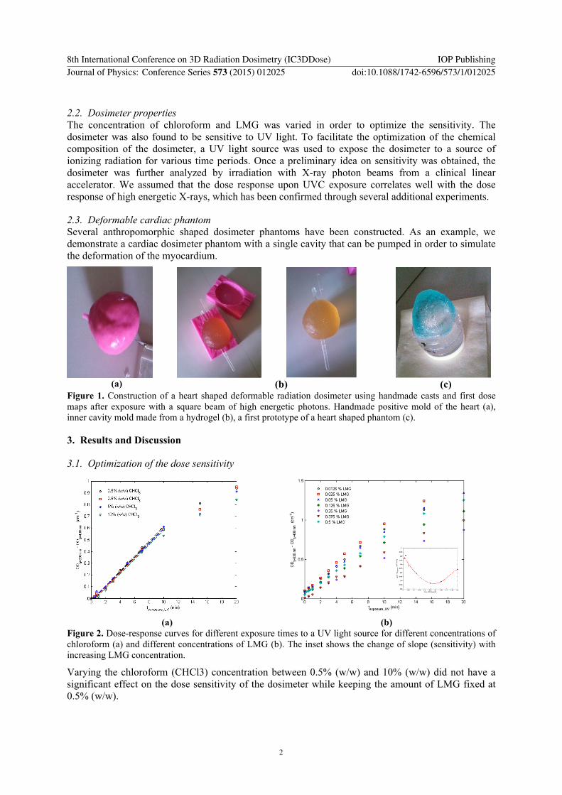
2.3. Deformable cardiac phantom Several anthropomorphic shaped dosimeter phantoms have been constructed. As an example, we demonstrate a cardiac dosimeter phantom with a single cavity that can be pumped in order to simulate the deformation of the myocardium.
(a) (b) (c)
Figure 1. Construction of a heart shaped deformable radiation dosimeter using handmade casts and first dose maps after exposure with a square beam of high energetic photons. Handmade positive mold of the heart (a), inner cavity mold made from a hydrogel (b), a first prototype of a heart shaped phantom (c).
3. Results and Discussion
3.1. Optimization of the dose sensitivity
Varying the chloroform (CHCl3) concentration between 0.5% (w/w) and 10% (w/w) did not have a significant effect on the dose sensitivity of the dosimeter while keeping the amount of LMG fixed at 0.5% (w/w).
(a) (b)
Figure 2. Dose-response curves for different exposure times to a UV light source for different concentrations of chloroform (a) and different concentrations of LMG (b). The inset shows the change of slope (sensitivity) with increasing LMG concentration.
8th International Conference on 3D Radiation Dosimetry (IC3DDose) IOP PublishingJournal of Physics: Conference Series 573 (2015) 012025 doi:10.1088/1742-6596/573/1/012025
2
The optimum concentration of LMG is found to be 0.025% (w/w). For LMG concentrations higher than 0.5% (w/w) the dosimeter became turbid. For the optimum composition (0.025% (w/w) LMG), the dose sensitivity for high energetic photon beams (6 MV) was found to be 0.01 cm-1.Gy-1.
3.2. Tissue equivalence The basic composition of the dosimeter is polydimethylsiloxane. Within the range of 100 kV to 20 MV, the dosimeter is highly tissue equivalent. The relative electron density of the dosimeter as compared to the electron density of water is in the order of 1.0059 (for a chain length of 10). For higher chain lengths the electron density converges to that of water.
n = 10 n = 20 H 8.7 % 8.4 % C 33.6 % 33.6 % O 20.4 % 20.4 % Si 37.3 % 37.6 %
(e/ewater) 1.0059 1.0036 (a) (b)
Figure 3. Mass attenuation coefficient as a function of photon energies for FlexyDos3D (black) and water (blue) calculated using the XCOM NIST online database [9]. (b) Polydimethylsiloxone (PDMS) is the main component in FlexyDos3D. The weight fraction of atoms is listed for different chain lengths.
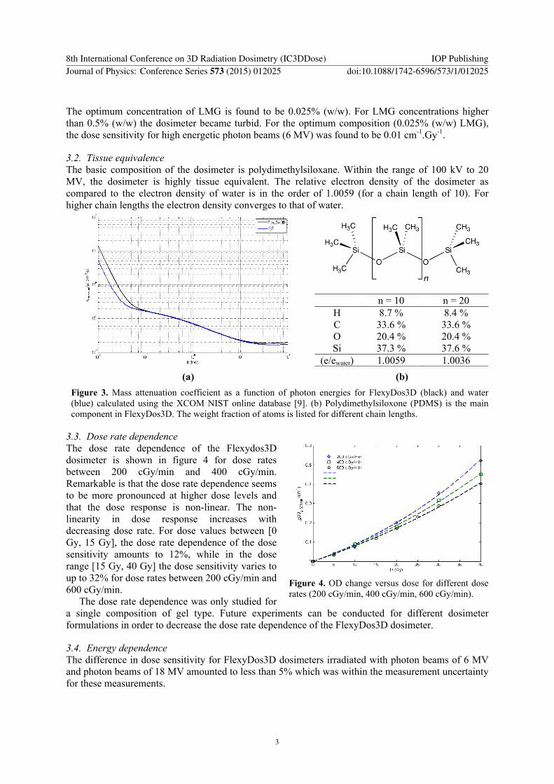
3.3. Dose rate dependence The dose rate dependence of the Flexydos3D dosimeter is shown in figure 4 for dose rates between 200 cGy/min and 400 cGy/min. Remarkable is that the dose rate dependence seems to be more pronounced at higher dose levels and that the dose response is non-linear. The non-linearity in dose response increases with decreasing dose rate. For dose values between [0 Gy, 15 Gy], the dose rate dependence of the dose sensitivity amounts to 12%, while in the dose range [15 Gy, 40 Gy] the dose sensitivity varies to up to 32% for dose rates between 200 cGy/min and 600 cGy/min.
The dose rate dependence was only studied for a single composition of gel type. Future experiments can be conducted for different dosimeter formulations in order to decrease the dose rate dependence of the FlexyDos3D dosimeter.
3.4. Energy dependence The difference in dose sensitivity for FlexyDos3D dosimeters irradiated with photon beams of 6 MV and photon beams of 18 MV amounted to less than 5% which was within the measurement uncertainty for these measurements.
Figure 4. OD change versus dose for different dose rates (200 cGy/min, 400 cGy/min, 600 cGy/min).
8th International Conference on 3D Radiation Dosimetry (IC3DDose) IOP PublishingJournal of Physics: Conference Series 573 (2015) 012025 doi:10.1088/1742-6596/573/1/012025
3
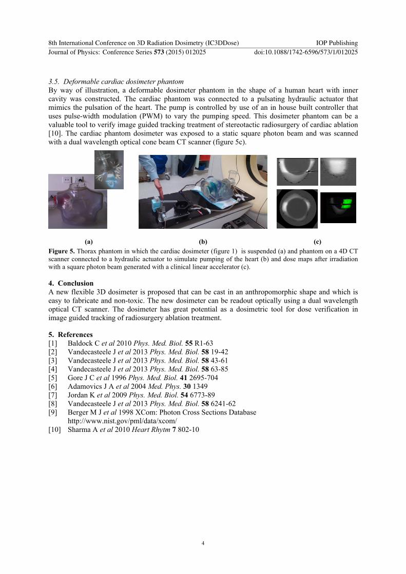
3.5. Deformable cardiac dosimeter phantom By way of illustration, a deformable dosimeter phantom in the shape of a human heart with inner cavity was constructed. The cardiac phantom was connected to a pulsating hydraulic actuator that mimics the pulsation of the heart. The pump is controlled by use of an in house built controller that uses pulse-width modulation (PWM) to vary the pumping speed. This dosimeter phantom can be a valuable tool to verify image guided tracking treatment of stereotactic radiosurgery of cardiac ablation [10]. The cardiac phantom dosimeter was exposed to a static square photon beam and was scanned with a dual wavelength optical cone beam CT scanner (figure 5c).
(a) (b) (c) Figure 5. Thorax phantom in which the cardiac dosimeter (figure 1) is suspended (a) and phantom on a 4D CT scanner connected to a hydraulic actuator to simulate pumping of the heart (b) and dose maps after irradiation with a square photon beam generated with a clinical linear accelerator (c).
4. Conclusion A new flexible 3D dosimeter is proposed that can be cast in an anthropomorphic shape and which is easy to fabricate and non-toxic. The new dosimeter can be readout optically using a dual wavelength optical CT scanner. The dosimeter has great potential as a dosimetric tool for dose verification in image guided tracking of radiosurgery ablation treatment.
5. References [1] Baldock C et al 2010 Phys. Med. Biol. 55 R1-63 [2] Vandecasteele J et al 2013 Phys. Med. Biol. 58 19-42 [3] Vandecasteele J et al 2013 Phys. Med. Biol. 58 43-61 [4] Vandecasteele J et al 2013 Phys. Med. Biol. 58 63-85 [5] Gore J C et al 1996 Phys. Med. Biol. 41 2695-704 [6] Adamovics J A et al 2004 Med. Phys. 30 1349 [7] Jordan K et al 2009 Phys. Med. Biol. 54 6773-89 [8] Vandecasteele J et al 2013 Phys. Med. Biol. 58 6241-62 [9] Berger M J et al 1998 XCom: Photon Cross Sections Database http://www.nist.gov/pml/data/xcom/ [10] Sharma A et al 2010 Heart Rhytm 7 802-10
8th International Conference on 3D Radiation Dosimetry (IC3DDose) IOP PublishingJournal of Physics: Conference Series 573 (2015) 012025 doi:10.1088/1742-6596/573/1/012025
4