PNS Structure of Nerves Cranial Nerves Spinal Nerves Sensory receptors Motor control.

Human Fingerprints –a combination of nerves and skin
Les Bush December 2011 Page 1
Contents Pages 1. Introduction 1-2
2. The skin of hands and feet - a unique special tissue 2-4
3. The primary ridges as the basis of how fingerprints appear. 4-13
4. Development of the Primary Ridge Surface Groove 14-16
5. How does the sweat appear on the surface of the friction skin? 16-20
6. Fingerprints and the identification of a human source 20-21
7. Conclusion 22-23
8. Bibliography 23-24
1. Introduction “What a piece of work is man! how noble in reason! how infinite in faculty! In form and moving, how express and admirable! in action, how like an angel! in apprehension, how like a god! the beauty of the world! the paragon of animals!” (William Shakespeare) To be human is a true state of giftedness passed to each individual by the contributions our parents made when exchanging their wealth of ancestral genetic development. At birth the new organism commences to engage the world and learn through an enhanced brain coupled to special senses. The human brain can analyse a wide variety of sensory information resulting in either a primitive or advanced response. Being part of the animal kingdom means we have the general behavioural traits of survival and reproduction of our species. The advanced and specialised responses come from the variety and sophistication of special senses. These special senses give us abilities to separate distinct flavours, to accurately observe things stationary and moving, to smell the differences and uniqueness of fauna and flora, to hear the variety of noises both close and afar and to discern the texture of surfaces held either lightly or powerfully. Naturally each special sense has in human terms a long developmental and evolutionary pathway that has now been mapped by technology analysing the DNA of our species. Aristotle has been quoted as saying “they who see things from the beginning will have the clearest view of them”. The scope of this paper is to focus on the sensory aspects of touch in association with the biological development of fingerprints. Two areas of the human form exhibit a specialised surface skin morphology they are; the palmar surface of the hands and the plantar surface of the feet. Closer observations

Human Fingerprints –a combination of nerves and skin
Les Bush December 2011 Page 2
reveal a uniformly spaced and regularly constructed grid of raised ridges. Housed within these raised sections of skin lie a pattern of sensory cells distributed in three dimensional planes over the surface area. Because of this sophisticated design of nerve arrangements we are able to detect, pressure, and pain, temperature and texture. In particular the tips of our fingers are capable of detecting the separation of two points of pressure such that both unsighted and sighted humans are able to read Braille language.
Figure 1: Image of actual skin showing the anatomy of a fingerprint 2. The skin of hands and feet - a unique special tissue To understand how special the palmar and plantar skin is requires a journey through the evolutionary development from simpler animal forms with primitive volar pads to the modern human body with advanced hand movements. The outer skin or epidermis covering the body is classified as stratified squamous epithelium. On human hands and feet there are five layers of epithelium tissues on friction skin areas while all other parts of the body have four layers of epidermis, the missing tissue layer being the stratum lucidium or clear layer. The epithelium cell makes up the majority of lining materials for both internal and external parts of the body. Inside each cell are specialised microscopic organelles and the cell nucleus contains our unique DNA. Outside each cell a community of similar cells that become joined together by cell junctions such as desmosomes and for a specialised purpose are referred to as a tissue. Tissues of different types that collectively perform a function are known as an organ. When we have a number of skin organs working together such as the epidermis, dermis, hairs and nails the result is an organ system, the Integumentary system. The integument combines with other systems such as nervous and vascular as well as

Human Fingerprints –a combination of nerves and skin
Les Bush December 2011 Page 3
many others to complete the organism, the human body. The key for an organism’s survival is having each system perform its specific task while not interfering with the function of the other systems. If the human brain never had to make a decision that affected the performance of the organism the whole system will operate highly effectively. By making choices we often place stress on the separate systems and sometimes extend their design. At certain times in evolutionary terms this extension has resulted in specific modifications to the organism a process conforming to Darwin’s principle of natural selection. Also in the performance of criminal acts humans extend the nervous stress on their body through higher than normal production of sweat. Alternatively brick laying as an occupation will, through abrasive actions, reduce the amount of skin present on the hands. Both these examples of human choice have a temporary effect on the body and are reversible by the removal of the cause. Once a normal balance is restored the organism is in harmony, internally and externally, a state referred to as homeostasis. When the body is in sleeping mode there is harmony between all the systems and we wake from this resting state feeling refreshed. The theory of friction ridge unit development had its genesis in our evolutionary specialisation derived from the primitive epidermal wart. Inez Whipple postulated that the wart consisted of a section of raised skin tissue with a hair follicle and a sweat gland. Whipple published her paper in 1904 and relied upon anthropologic evidence and studies of Primatology. Evolutionary specialisation over many thousands of years has improved the skin design pattern on the palmar and plantar surfaces by merging the separate warts into ridges and maintaining an even distance between sweat pores. As oils from the hair follicle would have reduced rather than enhanced grip the hands and feet of modern humans are now covered by thick glabrous (no hair) skin with the surface contour arranged in patterns of friction ridge formations, fingerprints. In collaboration with these and other changes a requirement for a larger brain with a sophisticated nervous system were needed to facilitate the special senses of touch, taste, sight, hearing and smelling. The process known as friction ridge unit fusion assumes badly that during biological development individual units join (fuse) together into an initial continuous ridge (primary ridge) which subsequently forms the surface friction skin. A mature friction ridge unit as defined by Ashbaugh consists of an approximate cuboidal area of stratified skin (epidermis) supported by the underlying dermis. The structure of a fully developed ridge unit has two side edges formed by secondary ridge actions, two free ends available to adjoin a similar ridge unit, a centrally located sweat pore opening inside an ostium, approximately four dermal papillae, a stratified squamous epithelium of five layers, an epidermal sweat duct, associated nerve tissues and a primary ridge that runs between the two free ends at the level of the basement membrane. Due to the purpose of cell junctions such as desmosomes is to hold in place the individual cells of stratum basal and stratum intermedium this postulated theory of ridge unit fusion requires an explanation as to how the “free ends” of two adjoining ridge units combine their tissue. Additionally the stratum basal is composed of one type of keratinocyte cells so the possibility of defining where the ‘free ends’ of any unit can be observed appears difficult. The other tissue structures occurring during this phase of development are the sweat gland anlagen. The position

Human Fingerprints –a combination of nerves and skin
Les Bush December 2011 Page 4
of each anlage would be approximately in the centre of any friction ridge unit and cannot assist in defining the “free end” boundaries of a ridge unit. An alternate and more plausible theory for primary ridge development is known as the neuro-ectoderm theory. The influence of embryonic nerve cells (Merkel from Schwann cells) invading the epidermis from the dermis and establishing an approximate hexagonal grid of spaced nerves above the area of the volar pad is an important precursor to the development of the foetal primary ridges. The collaboration of the skin and nervous tissues ensures both gripping and sensory functions of the surface skin are met. Once again the biological timings of when certain tissues develop are important for understanding how the organism brings together its various specialised organs. Humans develop in three phases of gestation known as trimesters and following birth the organism progressively grows to maturation after which ageing occurs. The human body comprises numerous organ systems that independently and interdependently carry out bodily functions. There are three primary germ layers from which all organ systems are developed, the ectoderm, endoderm and mesoderm. The ectoderm forms two organ systems the Integument and the Nervous. It is not surprising that many special senses are supported by these two systems and exclusively in the sense of touch. 3. The primary ridges as the basis of how fingerprints appear. Primary ridge development occurs along the basement membrane and becomes visible in histological foetal preparations between 12 – 16 week gestation the early part of the second trimester. The purpose of the basement membrane is to separate the functions of the epidermis from the dermis. The epidermis eventually differentiates into stratified squamous epithelium becoming an effective barrier to the environment as well as other important functions. The dermis is mostly mesenchyme tissues and houses the organs of the Nervous and Cardio-vascular systems as well as eccrine sweat glands and hair roots belonging to the epidermis. It is possible with post mortem samples of friction skin that by long immersion in water the epidermis will separate from the dermis along the plane of the basement membrane a process known generally as delaminating or ‘de-gloving’.

Human Fingerprints –a combination of nerves and skin
Les Bush December 2011 Page 5
Figure 2: Stained section of foetal skin at 10 weeks as prepared by Babler showing the epidermis with some periderm bleb cells attached. The epidermis is comprised of the stratum basal (columnar cells attached to the basement membrane) and stratum intermediate. The dermis is the larger area of the section below the basement membrane and with an accumulation of cells and nerves positioned close to the basement membrane. Studies by Moore from an 8 week embryo were stained to highlight the pathways of nerve tissue. It is quite apparent that the distribution of nerve tissue to the area below the basement membrane precedes any primary ridges and supports the theory of the interaction of nerves in the roles of primary ridge development and subsequent anlagen for sweat gland dermal ducts.

Human Fingerprints –a combination of nerves and skin
Les Bush December 2011 Page 6
Figure 3: This image by Babler shows a stained section of the dermis from a 14 week foetus. The primary ridges are impressed into the dermis and appear in dark stain as do sweat gland anlagen. This is a very good example of the process of how anastomosis between anlagen establishes the patterns of the primary ridges. The variables of anlagen formation and rapid growth of the epidermis are both occurring over the stress associated with the individually shaped volar pad and strongly support the principle of unique formation of the primary ridge pattern. The even spacing of the anlagen highlights the sophistication of the associated neural network. A single anlage that does not form into a primary ridge would appear on the surface skin as a mature single ridge unit (dot). In the above stained section the anlagen have developed distinct circular (conical) depressions into the dermis and accumulated a denser stain deposit due to being deeper than the level of the primary ridges.

Human Fingerprints –a combination of nerves and skin
Les Bush December 2011 Page 7
Figure 4: Stained section from a 14 week foetus prepared by Babler showing the primary ridge pattern with the locations of the sweat gland anlagen. It is hypothesised that anastomoses (joining) between these localised sites (anlagen) results in the development of the primary ridges and ultimately the fingerprint pattern.

Human Fingerprints –a combination of nerves and skin
Les Bush December 2011 Page 8
Figure 5: Bush plan view drawings of the surface of the basement membrane (BM) during early primary ridge (PR) development. Merkel cells (MC) present in the basal layer initiate the site of anlagen development and coincides with the linking of these sites as they mature to form the primary ridge. The progression of development is influenced by a combination of the shape of the volar pad (VP) and biological development in a radio-ulnar direction. These two drawings hypothesise the changes generally occurring at the basement membrane (BM) between gestational weeks 8-11 weeks on the left and then 10-13 weeks on the right. On the distal phalange Merkel cells (MC) arrive from the dermis establish a hexagonal grid and stimulate surrounding stratum basal cells to proliferate and thus localise at these sites. The volar pad (VP) is a raised three dimensional mound of collagen tissue whose curved surfaces influence variations in the pattern of primary ridges (PR). Between 10-13 weeks gestation the primary ridges develop by anastomoses in a radio-ulnar direction and link the epithelial localisation sites now associated with sweat gland anlagen. The linear continuity of any primary ridge will depend on arriving at the Merkel cell site while maintaining an even spacing between other ridges. The volar pad influence generally decreases after the 13th week and primary ridges continue to develop and cover the entire palmar or palmar surfaces.
Human Fingerprints –a combination of nerves and skin
Les Bush December 2011 Page 9
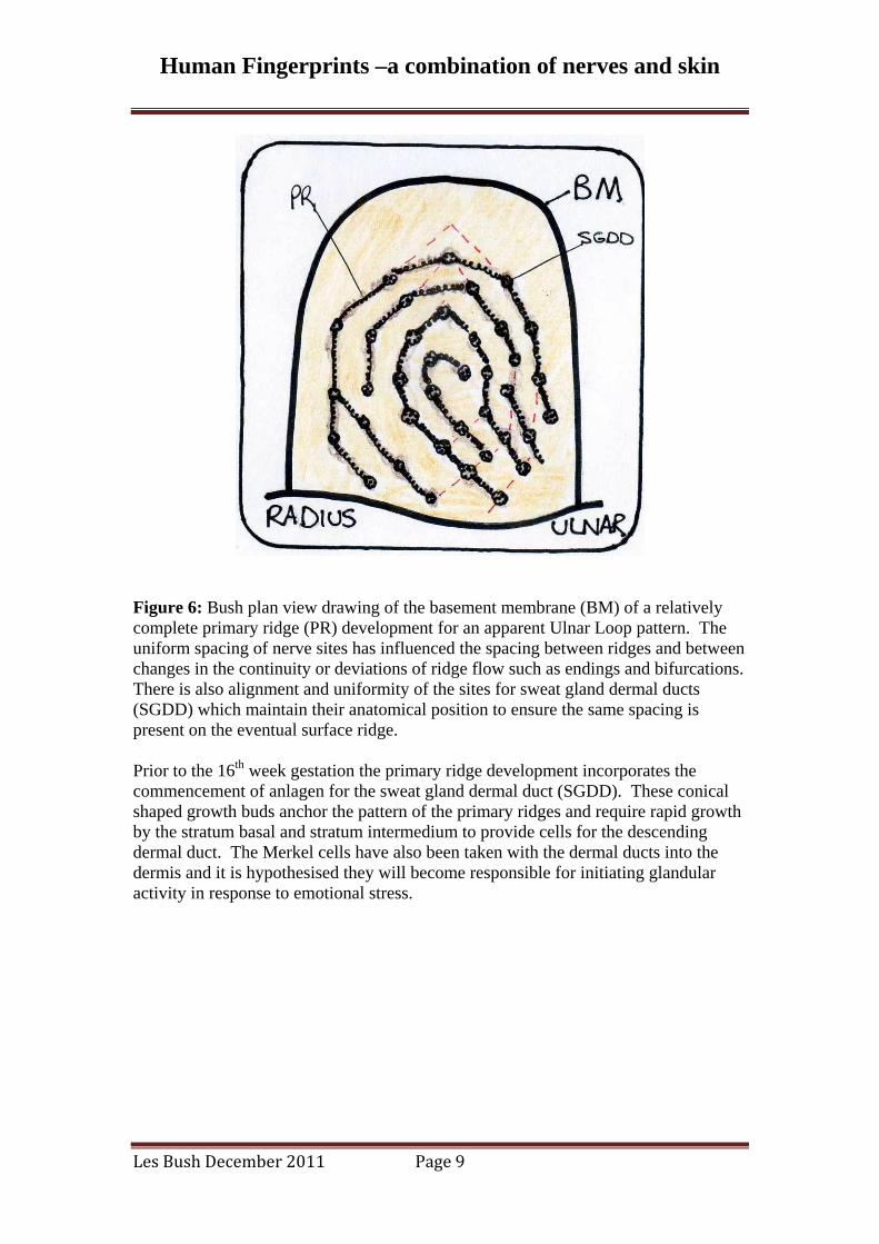
Figure 6: Bush plan view drawing of the basement membrane (BM) of a relatively complete primary ridge (PR) development for an apparent Ulnar Loop pattern. The uniform spacing of nerve sites has influenced the spacing between ridges and between changes in the continuity or deviations of ridge flow such as endings and bifurcations. There is also alignment and uniformity of the sites for sweat gland dermal ducts (SGDD) which maintain their anatomical position to ensure the same spacing is present on the eventual surface ridge. Prior to the 16th week gestation the primary ridge development incorporates the commencement of anlagen for the sweat gland dermal duct (SGDD). These conical shaped growth buds anchor the pattern of the primary ridges and require rapid growth by the stratum basal and stratum intermedium to provide cells for the descending dermal duct. The Merkel cells have also been taken with the dermal ducts into the dermis and it is hypothesised they will become responsible for initiating glandular activity in response to emotional stress.

Human Fingerprints –a combination of nerves and skin
Les Bush December 2011 Page 10
Figure 7: Bush drawings covering the period of gestation from 8 – 13 weeks showing a cross section of the epidermis (E) and dermis (D). The periderm (P) covers the epidermis which is composed of the stratum basal (B) and stratum intermediate (I). Merkel cells (MC) appear as extensions of the Schwann cell (SC) and penetrate through the basement membrane (BM) into the stratum basal (B). This penetration is a highly specialised event since the purpose of the basement membrane is to separate the epidermis from the dermis. Within the dermis additional Merkel cells will align evenly once again along the apex of the primary ridge to stimulate the growth of the ridge into the dermis and between anlagen (anastomoses).
Figure 8: Bush drawing representing the period of primary ridge (PR) development between 13 – 16 weeks gestation with new ridges occurring out of sequence with

Human Fingerprints –a combination of nerves and skin
Les Bush December 2011 Page 11
other ridges and adding to the uniqueness of the overall pattern. Sequential timing depends on the relative maturity of each Merkel cell (MC), the shape of the volar pad and the radio-ulnar biological development. The sweat gland anlagen deform the basement membrane (BM), stratum basal (B) and stratum intermediate (I) into a localised conical shape that remains a permanent feature in subsequent differentiated epidermal layers. It is postulated the intra-epidermal duct will use this deformation as the guide to progress the spiral epidermal duct (acrosyringium) to the outer surface. Prior to the end of primary ridge development the stratum intermediate (I) will generally have differentiated into four layers. Also the eccrine sweat gland intra-dermal duct (SGDD) will have reached the hypodermis and formed a gland with an open duct (lumen -internal space) extending from the gland to the basement membrane.
Figure 9: Bush drawing of plan view representations of the stratum basal (SB) between 10 – 14 weeks showing the initial formation of the primary ridge (PR) with the Merkel cells (MC) embedded within the anlagen of the soon to be developed sweat gland dermal duct (SGDD). The drawing on the right represents the proliferation of stratum basal cells along the primary ridge and around the site of the anlagen. Merkel cells within the dermis are also arranged evenly between each localised site to stimulate both the progression of the anastomosis and depth of the primary ridge.

Human Fingerprints –a combination of nerves and skin
Les Bush December 2011 Page 12
Figure 10: Bush plan view drawing illustrating the stratum basal (SB) development of the primary ridge and the sweat gland dermal duct around 16 weeks gestation. The Merkel cell has now descended with the developing duct (SGDD) into the region of the hypodermis where the gland will be differentiated from duct tissue. The connection of localised basal cells between each anlage site is consolidated and the primary ridge (PR) is complete.
Figure 11: Bush drawings of plan view representations of the basement membrane (BM) during gestational period between 10 – 16 weeks highlighting the even spacing

Human Fingerprints –a combination of nerves and skin
Les Bush December 2011 Page 13
between Merkel cells (MC) and between rows of Merkel cells. The left side drawing depicts the period leading up to the formation of primary ridge development. The right side drawing shows the changes to the basement membrane (BM) toward the end of primary ridge (PR) development. The anlagen of the sweat gland dermal ducts (SGDD) are shown as conical depressions inside of which are lumens of the dermal ducts leading down to their glands. The primary ridges are shown as grooves within the basement membrane connecting each of the anlagen. This representation of the basement membrane mirrors the final morphology of the surface friction ridges where ostia are conical depressions housing the pores of intra-epidermal ducts.
Figure 12: Bush drawing depicting the final weeks of basement membrane (BM) development (20 – 24) with a secondary ridge (SR) positioned midway between two primary ridges (PR). Following secondary ridge development the sweat gland epidermal duct (SGED) grows as a solid duct from the basement membrane (BM) spiralling upwards to the surface of the epidermis. Similarly during this phase other Merkel cells already positioned evenly along the primary ridge (dermal side) stimulate the stratum basal to grow conical shaped buds upwards from the basement membrane into the epidermis to form the dermal papillae (DP). Anastomoses also develop across the area of the primary ridges and further stabilises the pattern of ridges and dermal papillae. Located within most dermal papillae are Meissner corpuscle nerves (MCO) responsible for detecting two-point discrimination. The secured position and distribution of these nerves ensures the sophistication of the sensory capability for touch. Without the uniformity of the neural distribution the sensory signal to the brain would be unclear or confused. The requirement for an even distribution of nerves in support of sensory capability reinforces the priority of the neural map upon which the friction skin forms.

Human Fingerprints –a combination of nerves and skin
Les Bush December 2011 Page 14
4. Development of the Primary Ridge Surface Groove Literature research conducted for the purpose of this paper and other papers revealed that an observation of the shallow groove connecting each surface ostia has not been officially defined or explained. The following passage describes the postulated ontogenesis of this feature and attributes it the title of the primary ridge surface groove. Merkel cells embedded within the basal layer are responsible for the development of anlagen and consequently the progression of a primary ridge pattern. Other Merkel cells remaining within the dermis become distributed either side of the developing primary ridge for the purpose of stimulating growth of the primary ridge down into the dermis and consolidating its position. Importantly the sweat gland anlagen deformed both the basement membrane and superior layers of the epidermis into a localised conical shape. A similar process of epidermal layer deformation occurs with the downward growth of primary ridges.
Figure 13: Bush drawing of a cross section of the epidermis (E) around 16 weeks on the left showing a section of primary ridge (PR) development between sites of sweat gland anlagen and prior to dermal papillae development. Secondary ridge development is causing the superior layers of the epidermis to become compressed with the effect of creating the sides of the surface friction ridge (FR). This same process of epidermal layer deformation caused by the primary ridge (PR) growth at the basement membrane (BM) results in a shallow groove (PRSG) on the friction ridge apex. The maximum depth of the primary ridge and consequent deformation of the epidermal layers are variables influencing the size of the primary ridge surface groove (PRSG). The right diagram includes the establishment of Meissner corpuscles (MCO) within the sites of dermal papillae (DP) supporting the theory of the interdependence of the skin and nervous tissue.

Human Fingerprints –a combination of nerves and skin
Les Bush December 2011 Page 15
Figure 14: Bush drawing of a fully developed and differentiated epidermis and basement membrane (BM) between sites of sweat gland development. The resultant effect of the primary ridge development (PR) is the establishment of a surface groove (PRSG) connecting the surface ridge ostia (not shown in this drawing). The surface ridges are not visible until the Periderm (P) layer has reduced, detached and converted itself into vernix caseova.
Figure 15: Bush drawings of plan view sections representing on the left the outer surface of the epidermis (E) between weeks 8 – 16 and on the right 16 - 20 weeks. Initially the epidermis is covered with the periderm (P) during most of the second

Human Fingerprints –a combination of nerves and skin
Les Bush December 2011 Page 16
trimester and its presence precludes observations of the friction ridges. The friction ridges only become observable following both secondary ridge development and separation of the periderm (P) layer. The secondary ridges cause sulci (S) separating each friction ridge (FR). The ostia (SGO) have formed as a result of the effect from the shape of anlagen at the basement membrane. Similarly but less pronounced the primary ridge surface groove (PRSG) is also present and connects each ostia. During the phase depicted in the right drawing the intra-epidermal sweat duct (acrosyringium) has commenced growing upward from the basement membrane as a solid bud toward the outer surface and will generally emerge inside the ostium. Once at the surface the intra-epidermal duct will open its lumen and create the sweat gland pore (SGEDP).
Figure 16: Bush drawing representing friction ridge (FR) development between 20 – 24 weeks showing two fully developed friction ridges with observable surface structures. The epidermis (E) is now able to provide an effective barrier to the environment of the amniotic fluid that is soon to contain pollution from defecation by the rapidly growing foetus. The eccrine sweat gland is functioning and exudate fills the duct from its gland to the outer surface. The primary ridge surface groove (PRSG) connects each ostium (SGO) and performs the function of spreading exudate along the ridge surface. 5. How does the sweat appear on the surface of the friction skin? Eccrine sweat glands have their ontogenesis along the base of the primary ridge pattern with glandular dermal ducts providing stability to the primary ridge structure.

Human Fingerprints –a combination of nerves and skin
Les Bush December 2011 Page 17
The ontogeny of the sweat gland occurs between weeks 12 to 24 of foetal life and comprises two distinct periods of growth and differentiation. Initially the dermal component is established with a coiled gland located in the region of the hypodermis. A coiled glandular duct connecting to a relatively straight dermal duct completes the intra- dermal construction and initiates the second phase of growth. Once again starting from the base of the primary ridge the intra-epidermal duct now traverses upward through the stratified epidermal layers to reach the free surface of the stratum corneum. The distinctive spiral pathway of the intra-epidermal duct (acrosyringium) follows a relief deformation of superior epidermal tissues and enters a conical depression on the surface ridge known as an ostium. Within the ostium the intra-epidermal duct will generally locate the sweat gland pore. The entire tissue structure of the eccrine sweat gland organ is independently maintained to the adjoining epidermis and dermis. Innervations of the sweat glands on palmar and plantar surfaces are subject to specialised conditions associated with emotional stress rather than usual environmental factors. This is a very important physiological factor brought on by the psychology of the criminal when committing a crime.
Figure 17: Bush drawing of the surface morphology of friction ridges showing the relationship between ostia, pores and the primary ridge surface grooves. (E= epidermis, FR = friction ridge, S = sulci, SGO = sweat gland ostium, SGEDP = sweat gland epidermal duct pore, PRSG = primary ridge surface groove and P = stratum periderm)

Human Fingerprints –a combination of nerves and skin
Les Bush December 2011 Page 18
Figure 18: Bush drawings of skin cross sections depicting the process of intra-epidermal sweat gland duct (SGED) development between 16 to 20 weeks. The growth of the epidermal duct is initiated following the development of the sweat gland dermal duct (SGDD) complete with its lumen. The path of the ascending duct is spiral and follows the deformation of epidermal layers caused by the location of the original anlage at the basement membrane (BM). The epidermis (E) has been fully differentiated and resists the progress of the duct which is overcome by using the corkscrew type path (acrosryingium). The final position of the duct on the outer surface follows the conical deformation of the stratum corneum into the conical depression known as an ostium (SGO).

Human Fingerprints –a combination of nerves and skin
Les Bush December 2011 Page 19
Figure 19: Bush drawing of the period between 20 to 24 weeks where the solid mass of duct cells (SGED) opens out to form the lumen and creates the pore (SGP) for the gland. Once the lumen is formed the gland is ready for innervations and begins production of exudate to fill the lumen and maintain its shape. The ostia (SGO) perform the functions of both a reservoir of exudate to supply moisture for assisting surface contact and also to seal off the pore entrance to limit infection.

Human Fingerprints –a combination of nerves and skin
Les Bush December 2011 Page 20
Figure 20: Macro photograph of a friction skin surface showing sweat rising out of the pores along the ridge surface with some spilling into sulci. (source not known) 6. Fingerprints and the identification of a human source The term fingerprints is generally understood to mean that the skin on our fingers containing patterns act like ‘rubber stamps’ resulting in a pattern transfer onto a surface when touched. A more comprehensive understanding reveals that fingerprints are a specialised combination of nerve and skin organs developed harmoniously during the foetal stages of human growth and containing distinctive patterns of uniformly spaced friction ridges supporting the neural network. The permanence of friction ridge skin is maintained by the anatomy and physiology of the Integumentary system. Fingerprint patterns relating to the investigation of a criminal case require experts to develop capabilities in interpreting friction ridge detail for the purpose of matching a transferred fingerprint to a known human source. The reliability of performing fingerprint comparative analysis resulting in individuality of the human source skin can confidently be presented as a reasonably accurate claim. This type of analysis is an established forensic science with over one hundred years of collective empirical data underpinning the principle of the physical uniqueness present in fingerprint patterns. This same biological uniqueness is observable in other phenotype traits such as eye constructions, facial biometrics and voice patterns as a few examples.

Human Fingerprints –a combination of nerves and skin
Les Bush December 2011 Page 21
Figure 21: Photograph of human skin fingerprint pattern on the left (source) and resulting transfer pattern on the right (exemplar). The marked analysis using coloured markers demonstrates the comparison between the source skin (reversed) and inked impression (exemplar). Positive identification (individualisation) establishes that one source of friction skin was responsible for both the exemplar (known sample) and latent (unknown sample) fingerprints. The consequence of this conclusion is that if similar conditions of transfer were repeated for both the exemplar and latent fingerprints using the nominated source skin then another set of similar impressions would be reproduced. The claim of exclusive source of any individualised fingerprint has received criticism due to conditions such as; not all persons have been fingerprinted and neither has all areas of recorded fingerprints been examined and there are limits to how much friction ridge detail an expert can remember. However fingerprint science is supported by the known biological development of friction ridge detail and its patterns. These same biological processes apply to every human across both palmar and plantar areas (with the exception of abnormalities). The extensive scrutiny of the friction ridge detail on the distal phalanges of the hands has substantially proven that duplication of patterns of friction ridge details sufficient for individualisation has not occurred on two humans. The principles of the biological uniqueness and permanence of friction ridge detail meet the requirements for scientific falsifiability and as yet they have not been proven to be untrue. Therefore the likelihood of an individualised impression of an area of friction skin being made by another source is so remote that it is considered a ‘practical impossibility’ (SWGFAST 2010).

Human Fingerprints –a combination of nerves and skin
Les Bush December 2011 Page 22
7. Conclusion The contemporary theory relying upon an understanding that friction ridge units develop at the epidermal-dermal barrier and then somehow ‘fuse’ with adjoining units to form a primary ridge requires further evidence to explain how the ‘free ends’ of ridge units are observed along the stratum basal and secondly how the biological process of ‘fusing’ two separate tissues occurs. Until further evidence is provided this theory should only be classified as a possible hypothesis of friction skin development. Whereas other research involving histological preparations of foetal specimens material have provided extensive knowledge about the factors involved with the development of primary ridges. The work of Bryce Munger and associates clearly shows an interdependent relationship between the neurological map and the pattern of primary ridges. Skin performs a primary role of covering the whole body with the skin of hands and feet being specialised in both structure and functions. The special sense of touch represents a highly sophisticated network of nerve tissues. The arrangement of these nerves combines well with the two primary objectives of the epidermis; to form evenly spaced friction skin permeated on their ridges with evenly spaced sweat gland pores. The sensory grid of nerve tissues has been mapped out very early in gestation as an organisational priority in order to capture all the special sense functions. Localisations of stratum basal cells are made in response to invading Merkel cells that are arranged in an approximate hexagonal grid around the apical pad but also across palmar and plantar areas. The primary ridges develop due to a process of anastomoses between localisations of stratum basal epithelium. Other Merkel cells in the dermis arrange themselves along the apex (dermal side) of each primary ridge to stimulate the increase in depth of the primary ridge groove. Merkel cells with Meissner corpuscles later stimulate the basement membrane and the stratum basal to form dermal papillae. The gestational period over which these actions occur is between weeks 12 – 24 following which the skin has become an effective and specialised covering in support of the sensory organ. The observation and naming of the primary ridge surface groove is exclusive to this paper. Underpinning this observation is the consistency of two separate biological processes, the secondary ridges creating the surface sulci and the sweat gland ostia being similarly formed as a result of the downward conical deformation of the basement membrane by anlage of the sweat gland dermal duct. When the primary ridges initially develop they also cause a deformation of both the basement membrane and the superior layers of the epidermis. This action results in the formation of a shallow surface groove in the centre of the surface friction ridge connecting the ostia locations. By achieving this structure exudate from sweat glands fill the ostia and then overflows into the primary ridge surface groove along the surface of the friction ridge. When friction ridges make contact with a suitable surface the transfer of the friction ridge pattern results in a latent fingerprint. Positive identification (individualisation) of a latent fingerprint establishes that one source of friction skin was responsible for both the exemplar (known sample) and latent (unknown sample) fingerprints. This whole process of creating both the latent and exemplar fingerprints is repeatable and each examination conforms to the

Human Fingerprints –a combination of nerves and skin
Les Bush December 2011 Page 23
scientific expectations of falsifiability. A high degree of confidence can be placed in both the underpinning biological principles of fingerprint uniqueness and permanence as well as the trained ability of a fingerprint expert to forensically undertake a comparative analysis of two or more impressions. 8. Bibliography ASHBAUGH David, “Quantitative - qualitative friction ridge analysis”, CRC Press 1999. BABLER William J, “Embryologic development of epidermal ridges and their configurations”, Birth Defects original article series, Vol 27, n) 2, pp 95 - 112 BUSH Les. AFP 2007 “Golden Hands and Silver Feet”, unpublished BUSH Les. AFP 2008 “The Primary Ridge Surface Groove”, unpublished CUMMINS Harold, “The topographic history of the volar pads (walking pads; tastballen) in the human embryo”, Contributions to Embryology, N0 113 pp103 - 126 plus plates DELL David A, Munger Bryce L., “The early embryogenesis of papillary (sweat duct) ridges in primate glabrous skin: the dermatotopic map of cutaneous mechanoreceptors and dermatoglyphics”, The journal of comparative neurology, 1986, 244 pp 511 - 532. GORDON G., ACTIVE TOUCH The Mechanism of Recognition of Objects by Manipulation: a multi-disciplinary approach. Proceedings of a symposium held at Beaune France July 1977. HALE Alfred R., “Morphogenesis of volar skin in the human fetus”, 1951 thesis pp 147 - 173 plus plates. HASHIMOTO K, Gross B G, Lever W F, “ 1. The intraepidermal eccrine sweat duct”, The journal of investigative dermatology, 1965, Vol 45, No 3 pp 139 - 151 HASHIMOTO K, Gross B G, Lever W F, “ 2. The formation of intradermal portion of the eccrine sweat duct and of the secretory segment during the first half of embryonic life”, The journal of investigative dermatology, 1966, Vol 46, No 6 pp 513 - 529 HASHIMOTO K, “10, Merkel tactile cells in the finger and nail”, J. Anat. (1972), 111, 1, pp 99 - 120. JOHANSSON R.S., Vallbo A B, “Tactile sensibility in the human hand: relative and absolute densities of four types of mechanoreceptive units in glabrous skin”, J. Physiol (1979), 286, pp 283-300.
Human Fingerprints –a combination of nerves and skin
Les Bush December 2011 Page 24
KANDEL E. R., Schwartz J H, Jessell T M, Principles of Neural Science, Third Edition, Appleton and Lange, Connecticut, USA MOORE Susan J., MUNGER Bryce L., “The early ontogeny of the afferent nerves and papillary ridges in human digital glabrous skin”, Developmental Brain Research, 48 (1989) pp 119 - 141. MOROHUNFOLA K., Jones T., Munger B., “The differentiation of the skin and its appendages”, The Anatomical Record 232: 587-611 (1992). MUNGER Bryce L., “Neuro-cutaneous interactions in embryonic development: implications for medical dermatoglyphics”, The morphogenesis of down syndrome, 1991, pp 295 - 304. OKAJIMA Michio, “Dermal and epidermal structures of the volar skin”, Birth, Defects Original Article Series, Volume XV Number 6, pp 179 - 198. SCHEIBERT J., Leurent S, Prevost A, Debregeas, “The role of fingerprints in the coding of tactile information probed with a biometric sensor” Science Vol 323 13 March 2009 WERTHEIM Kasey, Maceo Alice, “Friction ridge skin and pattern formation during the critical stage: fact and theory”, handout material presented at the Centennial conference, London June 2001. WHIPPLE, Inez L. The ventral surface of the mammalian chiridium, Z. Morphol. Anthropol., 7, 261 1904.