Fine-needle aspiration cytology in soft tissue tumors: How ...3)_04_OA_20150502_V2.pdf ·...
5
Journal of Advanced Clinical & Research Insights (2015), 2, 1–5 Journal of Advanced Clinical & Research Insights ● Vol. 2:3 ● May-Jun 2015 1 ORIGINAL ARTICLE Fine-needle aspiration cytology in soft tissue tumors: How far did we go?? K. R. Chatura, Akshi Katyal, S. S. Hiremath Department of Pathology, Jagadguru Jayadeva Murugarajendra Medical College, Davangere, Karnataka, India Abstract Objective: Assignment of the tumors into various cytological categories and to determine specific sub-typing in individual cases. To correlate the cytological features observed with histological parameters and analyse the reasons for discordant diagnosis. Materials and Methods: 27 cases, with both cytological and histopathological details, diagnosed as soft tissue tumors (STT) were included. The lesions were classified into 6 cytological categories. Results: On fine-needle aspiration cytology (FNAC), 14 cases were reported positive for malignant cells and 13 were reported as benign. On histological follow up discordant lesions were further characterized. Discordance was attributable largely to sampling in lipomatous lesions and interpretation errors due to inattention to clinical history, low volume of these lesions in practice, and overlapping cytomorphologic features with other entities. Sensitivity and specificity was 84.61% and 85.71% respectively and overall diagnostic accuracy was 85.18%. Conclusion: Soft tissue tumors are rare neoplasms that pose a significant challenge as a result of their morphologic overlap and biological heterogeneity. ‘Time-honored’ histopathology is recognized as the ‘gold standard’ for evaluation of STT. FNAC can be used as a diagnostic modality in soft tissue lesions due to its lesser cost, ease of performance, safety, along with reasonable specificity and sensitivity. In the current era, where ‘needle is preceding the scalpel’ and the biopsy material is getting limited, our study highlights the role and scope, diagnostic difficulties and pitfalls to be aware of when interpreting these challenging FNACs. Keywords Cytohisto-correlation, errors, soft tissue tumors Correspondence Dr. K. R. Chatura, Department of Pathology, Jagadguru Jayadeva Murugarajendra Medical College, Davangere, Karnataka, India. Email: [email protected] Received 02 February 2015; Accepted 10 April 2015 doi: 10.15713/ins.jcri.56 Introduction Fine-needle aspiration cytology (FNAC) as a diagnostic modality for soft tissue tumors (STTs) is increasingly being used. Their morphologic overlap and biological heterogeneity pose a significant diagnostic challenge. FNAC is popular as a result of its cheap cost, easy performance, safety, along with fair specificity and specificity, both in recurrent cases as well as for the initial diagnosis of STTs especially in terms of sorting out malignant cases. This study aimed at the assignment of the tumors into various cytological categories and to determine specific sub-typing in individual cases. In this study, we attempted to correlate the cytological features observed with histological parameters. The reasons for discordant diagnosis on histological follow-up were analyzed. Materials and Methods We retrospectively retrieved all FNAs of histopathologically diagnosed soft tissue lesions, at the Department of Pathology, during a 2-year period from June 2011 to June 2013. The available clinical information was reviewed along with the histopathological follow-up. Aspirates inadequate for opinion and when not followed by excision were excluded from the present study. FNAs were performed using a 22-24 gauge needle. To obtain adequate material, 3-5 passes were used. Smears stained using Papanicolaou, Hematoxylin and Eosin (H and E), and Giemsa were evaluated and interpreted in conjunction with clinical details to render a final diagnosis. Based on the cytomorphological features, lesions were classified into 6 categories namely spindle cell, lipomatous,
Transcript of Fine-needle aspiration cytology in soft tissue tumors: How ...3)_04_OA_20150502_V2.pdf ·...
Journal of Advanced Clinical & Research Insights (2015), 2, 1–5
Journal of Advanced Clinical & Research Insights ● Vol. 2:3 ● May-Jun 2015 1
O R I G I N A L A R T I C L E
Fine-needle aspiration cytology in soft tissue tumors: How far did we go??K. R. Chatura, Akshi Katyal, S. S. Hiremath
Department of Pathology, Jagadguru Jayadeva Murugarajendra Medical College, Davangere, Karnataka, India
AbstractObjective: Assignment of the tumors into various cytological categories and to determine specifi c sub-typing in individual cases. To correlate the cytological features observed with histological parameters and analyse the reasons for discordant diagnosis.Materials and Methods: 27 cases, with both cytological and histopathological details, diagnosed as soft tissue tumors (STT) were included. The lesions were classifi ed into 6 cytological categories.Results: On fi ne-needle aspiration cytology (FNAC), 14 cases were reported positive for malignant cells and 13 were reported as benign. On histological follow up discordant lesions were further characterized. Discordance was attributable largely to sampling in lipomatous lesions and interpretation errors due to inattention to clinical history, low volume of these lesions in practice, and overlapping cytomorphologic features with other entities. Sensitivity and specifi city was 84.61% and 85.71% respectively and overall diagnostic accuracy was 85.18%.Conclusion: Soft tissue tumors are rare neoplasms that pose a signifi cant challenge as a result of their morphologic overlap and biological heterogeneity. ‘Time-honored’ histopathology is recognized as the ‘gold standard’ for evaluation of STT. FNAC can be used as a diagnostic modality in soft tissue lesions due to its lesser cost, ease of performance, safety, along with reasonable specifi city and sensitivity. In the current era, where ‘needle is preceding the scalpel’ and the biopsy material is getting limited, our study highlights the role and scope, diagnostic diffi culties and pitfalls to be aware of when interpreting these challenging FNACs.
KeywordsCytohisto-correlation, errors, soft tissue tumors
CorrespondenceDr. K. R. Chatura, Department of Pathology, Jagadguru Jayadeva Murugarajendra Medical College, Davangere, Karnataka, India. Email: [email protected]
Received 02 February 2015;Accepted 10 April 2015
doi: 10.15713/ins.jcri.56
Introduction
Fine-needle aspiration cytology (FNAC) as a diagnostic modality for soft tissue tumors (STTs) is increasingly being used. Their morphologic overlap and biological heterogeneity pose a signifi cant diagnostic challenge. FNAC is popular as a result of its cheap cost, easy performance, safety, along with fair specifi city and specifi city, both in recurrent cases as well as for the initial diagnosis of STTs especially in terms of sorting out malignant cases.
This study aimed at the assignment of the tumors into various cytological categories and to determine specifi c sub-typing in individual cases. In this study, we attempted to correlate the cytological features observed with histological parameters. The reasons for discordant diagnosis on histological follow-up were analyzed.
Materials and Methods
We retrospectively retrieved all FNAs of histopathologically diagnosed soft tissue lesions, at the Department of Pathology, during a 2-year period from June 2011 to June 2013. The available clinical information was reviewed along with the histopathological follow-up.
Aspirates inadequate for opinion and when not followed by excision were excluded from the present study.
FNAs were performed using a 22-24 gauge needle. To obtain adequate material, 3-5 passes were used. Smears stained using Papanicolaou, Hematoxylin and Eosin (H and E), and Giemsa were evaluated and interpreted in conjunction with clinical details to render a fi nal diagnosis.
Based on the cytomorphological features, lesions were classifi ed into 6 categories namely spindle cell, lipomatous,

FNAC of soft tissue tumors Chatura, et al.
2 Journal of Advanced Clinical & Research Insights ● Vol. 2:3 ● May-Jun 2015
myxoid, infl ammatory, round cell, and pleomorphic type. In cases with mixed components, the specifi c subtype was assigned to the predominant morphological pattern.
The excised tissue specimens of all the cases were processed routinely, stained with H and E and studied.
The cases with a discrepancy between the cytologic and histologic diagnosis were reviewed to determine the reason. The sensitivity, specifi city, positive predictive value, and negative predictive value were calculated considering the false positive and false negative cases for malignant lesions diagnosed on cytology.
Results
Of the 27 cases, a male predominance was noted. Most cases were in age group of 30-50 years, with an average age of 42.11, there were 15 males and 12 females and the extremities were the most common site of involvement. Most of the cases were referred for a primary diagnosis, and only 2 of the evaluated cases were recurrent lesions. On FNAC, 14 cases were interpreted positive for malignant cells whereas 13 were reported as benign conditions or negative for malignant cells [Tables 1 and 2].
The specifi city and sensitivity were calculated for malignancy which was 84.61% and 85.71% respectively. The positive predictive value was found to be 84.61% and negative predictive value 84.61%. The overall diagnostic accuracy was 85.18%.
Among the 13 spindle cell tumors, on cytology, 6 were diagnosed as benign and 7 malignant. Specifi c diagnoses rendered were nerve sheath tumors (4 cases), synovial sarcoma (2 cases), fi brous histiocytoma (3 cases), fi brosarcoma (2 cases),
high grade spindle cell sarcoma (2 cases). Two cases of false negative and one case of false positive were in this category.
Histology was cordant in only one of the 4 lesions in the adipocytic category. The other three, were reported as elastofi broma [Figure 1a], fi brous hamartoma of infancy (FHI) [Figure 1b] and pleomorphic lipoma on histological follow-up, but all had signifi cant adipocyte component.
On cytology, in the myxoid category, 1 benign case was reported as myxoma and 2 cases as myxoid liposarcoma [Figure 1c] and one case as myxofi brosarcoma [Figure 1d]. One myxoid liposarcoma was diagnosed as myoepithelioma on histopathology.
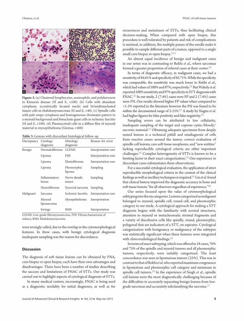
In the infl ammatory category, one case reported as the granulomatous lesion was a nerve sheath tumor on histopathology. Kimura’s [Figure 2a] and infl ammatory pseudotumor (IPT) were the two cases correctly diagnosed.
In the pleomorphic group, both the cases were labeled as sarcoma. The only case in round cell category incorrectly sub-typed as Ewing’s on cytology was a rhabdomyosarcoma (RMS) [Figure 2b] on histology.
In 5 cases, the behavior was accurately categorized as benign or malignant but were wrongly subtyped. Upon review of these, errors were due to sampling in two benign cases and due to interpretation in two benign and one malignant case.
The discrepancy between the cytological and histopathological diagnosis was noted in 9 cases [Table 3]. Upon review, there were 2 false-negative cases, dermatofi broma reported as low grade fi bromyxosarcoma (LGFMS) and neurofi broma as synovial sarcoma and 2 false positive cases, high grade spindle cell sarcoma reported as ischemic fasciitis [Figure 2c] and myxoid liposarcoma as myoepithelioma [Figure 2d].
In 4 cases, error in interpretation was due to their rarity and lack of familiarity with cytological features whereas in 2 cases, they
Table 1: Benign tumors: Correlation of cyto and histopathological diagnosisCytological category
Total cases
Accurately diagnosed
Benign but not
categorized
Benign on FNAC but malignant on
histopathologySpindle 6 4 0 2
Myxoid 1 1 0 0
Infl ammatory 2 1 1 0
Lipomatous 4 1 3 0FNAC: Fine-needle aspiration cytology
Table 2: Malignant tumors: Correlation of cyto and histopathological diagnosisCytological category
Total cases
Accurately diagnosed
Malignant but not
categorized
Malignant on FNAC but benign on histopathology
Spindle 7 6 0 1
Myxoid 3 2 0 1
Infl ammatory 1 1 0 0
Pleomorphic 2 2 0 0
Round cell 1 0 1 0FNAC: Fine-needle aspiration cytology
Figure 1: (a) Braid-like elastic fi bers in elastofi broma (Giemsa, ×400). (b) Mixture of adipose tissue and cluster of bland spindle cells in a myxoid background in fi brous hamartoma of infancy (Papanicolaou [PAP], ×100). (c) Discrete myxoid stromal fragments with univacuolated lipoblasts and curvilinear vessels in myxoid liposarcoma (Giemsa, ×100). (d) Cellular smears with marked pleomorphism in myxofi brosarcoma (PAP, ×400)
dc
ba
Chatura, et al. FNAC of soft tissue tumors
Journal of Advanced Clinical & Research Insights ● Vol. 2:3 ● May-Jun 2015 3
were wrongly called, due to the overlap in the cytomorphological features. In three cases, with benign cytological diagnosis inadequate sampling was the reason for discordance.
Discussion
The diagnosis of soft tissue lesions can be obtained by FNA, core biopsy or open biopsy, each have their own advantages and disadvantages. There have been a number of studies describing the success and limitations of FNAC of STTs. Our study was carried out to highlight aspects of cytological diagnosis of STTs.
In many medical centers, increasingly, FNAC is being used as a diagnostic modality for initial diagnoses, as well as for
recurrences and metastases of STTs, thus facilitating clinical decision-making. When compared with open biopsy, this procedure is well-tolerated by patients and risk of complications is minimal, in addition, the multiple passes of the needle make it possible to sample diff erent parts of a tumor, opposed to a single small core biopsy or open biopsy.[1,2]
An almost equal incidence of benign and malignant cases in our series was in contrasting to Rekhi et al., where sarcomas formed a greater proportion of referral cases at their center.[2]
In terms of diagnostic effi cacy, in malignant cases, we had a sensitivity of 84.61% and specifi city of 85.71%. While the specifi city was comparable, the sensitivity was much lower to Rekhi et al., which had values of 100% and 87%, respectively.[2] But Wakely et al. reported 100% sensitivity and 97% specifi city in STT diagnosis with FNAC.[3] In our study, 2 (7.4%) cases were FP and 2 (7.4%) cases were FN. Our results showed higher FP values when compared to <1-5% reported in the literature however the FN was found to be within the documented range of 2-15%.[2] A study by Nagira et al. had higher fi gures for false positivity and false negativity.[4]
Sampling errors can be attributed to low cellularity, inadequate sampling of the target and copious cystic/bloody/necrotic material.[1] Obtaining adequate specimens from deeply seated lesions is a technical pitfall and misdiagnosis of cells from reactive zones around the tumor, correct evaluation of spindle cell lesions, rare soft tissue neoplasms, and “new entities” lacking reproducible cytological criteria are other important challenges.[5] Complex heterogeneity of STTs is known to be a limiting factor in their exact categorization.[2] Our experience in discordant cases substantiates these observations.
For a successful cytological evaluation, the application of strict reproducible morphological criteria in the context of the clinical fi ndings as well as ancillary techniques is required.[5] Liu et al. found that clinical history improved the diagnostic accuracy in bone and soft tissue lesions “for all observers regardless of experience.”[6]
Our series focused upon the value of cytomorphological subtyping into the six categories. Lesions categorized as malignant belonged to myxoid, spindle cell, round cell, and pleomorphic category in our study. A cytological approach for making a STT diagnosis begins with the familiarity with normal structures, attention to myxoid or metachromatic stromal fragments and a variety of discohesive cells like spindly, round, pleomorphic, polygonal that are indicators of a STT, on aspirates. Cytological categorization with benignancy or malignancy of the subtypes was statistically signifi cant when these features were integrated with clinicoradiological fi ndings.[2]
In terms of exact subtyping, which was off ered in 18 cases, 76% and 75% of the spindle and myxoid tumors and all pleomorphic tumors, respectively, were suitably categorized. The least concordance was seen in lipomatous tumors (25%). This was in contrast to that of Rekhi et al. who reported maximum congruence in lipomatous and pleomorphic cell category and minimum in spindle cell tumors.[2] In the experience of Singh et al., spindle cell lesions were the most diagnostically challenging because of the diffi culties in accurately separating benign lesions from low-grade sarcomas and accurately subclassifying the sarcoma.[7]
Table 3: Lesions with discordant histological follow-upDiscrepancy Cytology
diagnosisHistology diagnosis
Reason for error
Benign Dermatofi broma LGFMS Interpretation rare
Lipoma FHI Interpretation rare
Lipoma Elastofi broma Interpretation rare
Lipoma Pleomorphic lipoma
Sampling
Infl ammatory lesion
Nerve sheath tumor
Sampling
Neurofi broma Synovial sarcoma Sampling
Malignant Sarcoma Ischemic fasciitis Interpretation rare
Myxoid liposarcoma
Myoepithelioma Interpretation
Ewings RMS InterpretationLGFMS: Low-grade fibromyxosarcoma, FHI: Fibrous hamartoma of infancy, RMS: Rhabdomyosarcoma
Figure 2: (a) Clustered lymphocytes, eosinophils, and polykaryocte in Kimura’s disease (H and E, ×100). (b) Cells with abundant cytoplasm, eccentrically located nuclei and bi/multinucleated tumor cells in rhabdomyosarcoma (H and E, ×40). (c) Spindle cells with pale wispy cytoplasm and homogeneous chromatin pattern in a myxoid background and binucleate giant cells in ischemic fasciitis (H and E, ×100). (d) Plasmacytoid cells in a diff use fi lm of myxoid material in myoepithelioma (Giemsa, ×400)
dc
ba

FNAC of soft tissue tumors Chatura, et al.
4 Journal of Advanced Clinical & Research Insights ● Vol. 2:3 ● May-Jun 2015
In the lipomatous category, all 4 cases were categorized as benign, but the exact diagnosis was off ered in only one case. Pleomorphic lipoma was diagnosed only as lipoma, because of inadequate sampling of characteristic “fl oret-like” giant cells. A superfi cial location i.e. neck, in an elderly male, was a helpful clue in excluding a liposarcoma.
In the false positive case of ischemic fasciitis, the reactive stromal cells with nuclear atypia were misinterpreted as malignant cells. Ischemic fasciitis is described as a distinctive fi broblastic proliferation occurring predominantly in elderly, bed-ridden individuals, which can be mistaken clinically, cytologically, and histologically for sarcoma.[8] The cytologic fi ndings in this case, when combined with the history of immobilization following ulnar fracture, should have avoided the misdiagnosis of malignancy in a benign, proliferative lesion.
Two of six false-positive cases were proven to be fi bromatosis though cytodiagnosis was low-grade spindle cell sarcoma. High cellular yield, pleomorphism, and mitosis in the aspirates from fi bromatosis lead to this mistake. Myxoid background, presence of infl ammatory cells and binucleate or trinucleate ganglion cells, pale wispy cytoplasm of spindle cells with homogeneous chromatin pattern should be appreciated for a correct diagnosis. Clinical features are also helpful in diagnosis. But immunocytochemistry has got little role.[9]
In the other case of false positive diagnosis, the presence of plasmacytoid round cells in a diff use myxoid background was mistaken for myxoid liposarcoma. Superfi cial location and absence of characteristic vasculature were ignored. The myxoid stroma in liposarcomas is typically distributed as discrete stromal fragments rather than as a diff use fi lm of myxoid material.[7] Cytologic fi ndings of soft tissue myoepithelioma have been not reported in the literature, except for a case report of imprint cytology.[10]
False negative cytology diagnoses could be attributed to interpretation errors, one due to the rarity and other due to overlap in cytological features. LGFMS reported as dermatofi broma is a rare tumor recently being recognized and seldom diagnosed on FNAC. The cytologic features are not specifi c enough for a defi nitive diagnosis based on FNAC alone; however, correlating the cytologic and clinical fi ndings can narrow the range of diagnosis.[11] One case of synovial sarcoma was reported as neurofi broma due to its para-spinal location and cyotological overlap with limited sampling on USG was the cause for interpretation error.
Sampling error was noted in three of our cases. One case was accurately subtyped as adipocytic but not accurately as pleomorphic lipoma, and the other case was of a nerve sheath tumor which was misinterpreted as a granulomatous lesion, because of superfi cial ulceration. In spindle cell category, a paraspinal neurofi broma was suggested but was a synovial sarcoma on histopathology. From a practical standpoint, it may not be possible to accurately separate low-grade spindle cell and myxoid sarcomas from benign neoplasms based on cytomorphology alone. FNAC in this instance, still allows the rapid separation of patients who will require tissue biopsy for a defi nitive diagnosis,
thereby obviating multiple clinic visits, eliminating unnecessary procedures, and reducing the overall costs.[7]
The problem of inadequate sampling can be avoided by the use of more stringent adequacy criteria. At present with no established adequacy criteria for soft tissue cytology, the number of inadequate cases varies by a pathologist, institution and study. Finding a threshold whereby sensitivity is maximized, and inadequacy rates are minimized is hard to defi ne.[1]
It has been seen that benign lesions are more diffi cult to subclassify than malignant lesions. 10 (71%) were subclassifi ed as sarcoma while in contrast, only 7 (53%) could be subclassifi ed as a specifi c benign lesion. These fi ndings were similar to that of Khalbuss et al. who reported subclassifi cation in 83% and 37% of malignant and benign cases.[1]
Most of the smears, in a study of 5 cases of elastofi broma, were paucicellular, and the cellular component consisted of individual cells or clusters of spindled fi broblasts with bland nuclear features and rare mature adipocytes. The acellular matrix composed of elongated rod-like structures with serrated borders or braid-like or fern-like fi bers and petaloid globules were the diagnostic component. These characteristic cytomorphologic features are not to be mistaken with any other lesions.[12] Our case of elastofi broma was reported as lipoma. A careful review of occupational history i.e., patient was an elderly manual laborer, bilateral inter-scapular location would have helped in the diagnosis. The paucicellular nature of the lesion was inadvertently mistaken, and the acellular matrix was overlooked in our case.
Cytology literature has few reports of FHI, a rare, benign lesion. The mixture of adipose tissue, a cluster of bland spindle cells in a myxoid and collagenous background is helpful to diff erentiate this from other infantile soft tissue lesions on cytology. This diagnosis can be made in the proper clinical setting i.e., clinical history, radiological fi ndings, and location of the lesion with awareness of the cytologic features.[13,14] Interpretation error in our case could be a lack of experience and overemphasis on the adipose tissue fragments as the lesion was situated in the breast.
In the infl ammatory category, FNAC of a retroauricular mass in a female patient, showed dissociated and clustered lymphocytes, eosinophils, and polykaryoctes. A diagnosis of Kimura’s disease was suggested and was confi rmed on histopathology. History, location, and lesional hyperpigmentation with cytological features were recognized to render the diagnosis even in a female patient. It is often diagnosed in male patients.[15]
Although, IPT is placed within the neoplastic category, it is best-regarded as a morphologic entity showing mainly infl ammatory cells with a pleomorphic spindle cell component mimicking a tumor. The term, “pseudotumor” is appropriate, and as the atypical spindle cells are myofi broblastic, it can be termed as an infl ammatory myofi broblastic tumor. Aspirates usually have an infl ammatory background with a predominance of acute or chronic infl ammatory cells in diff erent cases. These aspirates are usually suspected to be malignant due to the presence of pleomorphic spindle cells.[16] The case of IPT in

Chatura, et al. FNAC of soft tissue tumors
Journal of Advanced Clinical & Research Insights ● Vol. 2:3 ● May-Jun 2015 5
our study was a long standing, discharging lesion in the parotid region. The cytological features were reported, and emphasis for excision was communicated.
In the round cell category, presence of tigroid background and multinucleate and binucleate cells could have prompted a diagnosis of RMS. Periodic Acid Schiff positivity of cells favored a diagnosis of Ewing’s sarcoma. Although cytoplasmic glycogen is characteristic for Ewings, it is neither a specifi c or constant fi nding; it can be seen in RMS, lymphoma, and neuroblastoma.[17] In alveolar RMSs, most tumor cells are small and lymphocyte-like, having fi nely granular chromatin. The fi nding of cells with more abundant cytoplasm, eccentrically located nuclei, and bi/multinucleated tumor cells in a background of mucosubstance helps in the diff erential diagnosis.[18]
The relative infrequency of STTs produces unfamiliarity with their histological and cytomorphological features, an increased risk of sampling error due to their tissue heterogeneity and reluctance in soft tissue pathology experts to endorse this technique, which is understandable, if a pathologist has limited exposure and experience to this type of cytologic sample.[19]
In the current study, we have presented clinical, cytological, and histopathological fi ndings of benign and malignant STTs. The specifi c typing was diffi cult in the benign category and was fairly accurate in malignant lesions. The reasons for cytohistological discordance were analyzed. This study helped to document cytological fi ndings in rare cases, which would not have been possible if they were excised.
References
1. Khalbuss WE, Teot LA, Monaco SE. Diagnostic accuracy and limitations of fi ne-needle aspiration cytology of bone and soft tissue lesions: A review of 1114 cases with cytological-histological correlation. Cancer Cytopathol 2010;118:24-32.
2. Rekhi B, Gorad BD, Kakade AC, Chinoy R. Scope of FNAC in the diagnosis of soft tissue tumors – A study from a tertiary cancer referral center in India. Cytojournal 2007;4:20.
3. Wakely PE Jr, Kneisl JS. Soft tissue aspiration cytopathology. Cancer 2000;90:292-8.
4. Nagira K, Yamamoto T, Akisue T, Marui T, Hitora T, Nakatani T, et al. Reliability of fi ne-needle aspiration biopsy in the initial diagnosis of soft -tissue lesions. Diagn Cytopathol 2002;27:354-61.
5. Domanski HA. Fine-needle aspiration cytology of soft tissue lesions: Diagnostic challenges. Diagn Cytopathol 2007;35:768-73.
6. Liu K, Layfi eld LJ, Coogan AC, Ballo MS, Bentz JS, Dodge RK. Diagnostic accuracy in fi ne-needle aspiration of soft tissue and bone lesions. Infl uence of clinical history and experience. Am J Clin Pathol 1999;111:632-40.
7. Singh HK, Kilpatrick SE, Silverman JF. Fine needle aspiration biopsy of soft tissue sarcomas: Utility and diagnostic challenges. Adv Anat Pathol 2004;11:24-37.
8. Kendall BS, Liang CY, Lancaster KJ, McCabe KM, Solanki PH. Ischemic fasciitis. Report of a case with fi ne needle aspiration fi ndings. Acta Cytol 1997;41:598-602.
9. Sengupta S, Mondal SK, Mallick MG, Biswas P, Basu N. Role of cytology in diagnosis of soft tissue sarcomas with special reference to false positive cases. J Cytol 2009;26:15-9.
10. Park SJ, Kim AR, Gu MJ, Choi JH, Shin DS. Imprint cytology of soft tissue myoepithelioma: A case study. Korean J Pathol 2013;47:299-303.
11. Lindberg GM, Maitra A, Gokaslan ST, Saboorian MH, Albores-Saavedra J. Low grade fi bromyxoid sarcoma: Fine-needle aspiration cytology with histologic, cytogenetic, immunohistochemical, and ultrastructural correlation. Cancer 1999;87:75-82.
12. Vincent J, Maleki Z. Elastofi broma: Cytomorphologic, histologic, and radiologic fi ndings in fi ve cases. Diagn Cytopathol 2012;40 Suppl 2:E99-103.
13. Mohanty SK, Dey P. Cytologic diagnosis of fi brous hamartoma of infancy: A case report. Diagn Cytopathol 2003;28:272-3.
14. Gupta R, Singh S. Cytologic diagnosis of fi brous hamartoma of infancy: A case report of a rare soft tissue lesion. Acta Cytol 2008;52:201-3.
15. Chow LT, Yuen RW, Tsui WM, Ma TK, Chow WH, Chan SK. Cytologic features of Kimura’s disease in fi ne-needle aspirates. A study of eight cases. Am J Clin Pathol 1994;102:316-21.
16. Iyer VK. Cytology of soft tissue tumors: Benign soft tissue tumors including reactive, nonneoplastic lesions. J Cytol 2008;25:81-6.
17. González-Cámpora R. Cytoarchitectural fi ndings in the diagnosis of primary soft tissue tumors. Acta Cytol 2001;45:115-46.
18. Atahan S, Aksu O, Ekinci C. Cytologic diagnosis and subtyping of rhabdomyosarcoma. Cytopathology 1998;9:389-97.
19. Tailor HJ, Bhagat VM, Kaptan KB, Italiya SL, Balar HR, Agarwal MP. Diagnostic accuracy of fi ne needle aspiration cytology in soft tissue tumors: Our institutional experience. Int J Res Med Sci 2013;1:443-7.
How to cite this article: Chatura KR, Katyal A, Hiremath SS. Fine-needle aspiration cytology in soft tissue tumors: How far did we go?? J Adv Clin Res Insights 2015;2:1-5.