
Figel 9 4 14 - Roswell Park Comprehensive Cancer Center · ... (–western(blotting (• Cell ......
40
Transcript of Figel 9 4 14 - Roswell Park Comprehensive Cancer Center · ... (–western(blotting (• Cell ......
Overview
• Introduction • Experimental principles
• Molecular biology techniques • DNA – PCR • RNA • Protein – western blotting
• Cell culture
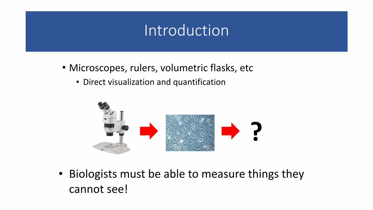
Introduction
• Microscopes, rulers, volumetric flasks, etc • Direct visualization and quantification
• Biologists must be able to measure things they cannot see!
?
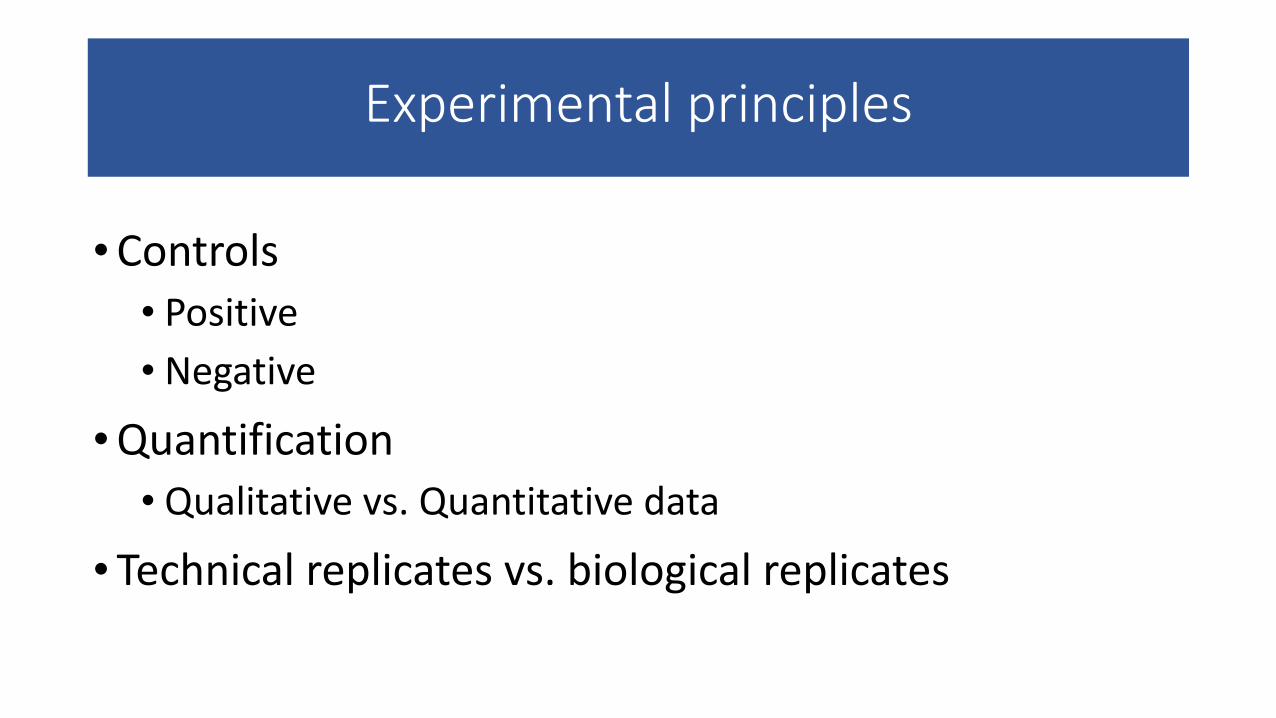
Experimental principles
•Controls • Positive • Negative
•Quantification • Qualitative vs. Quantitative data
• Technical replicates vs. biological replicates
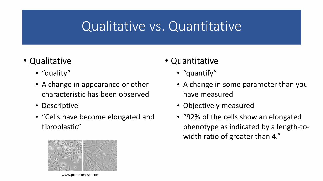
Qualitative vs. Quantitative
• Qualitative • “quality” • A change in appearance or other characteristic has been observed • Descriptive • “Cells have become elongated and fibroblastic”
• Quantitative • “quantify” • A change in some parameter than you have measured • Objectively measured • “92% of the cells show an elongated phenotype as indicated by a length-‐to-‐width ratio of greater than 4.”
www.proteomesci.com
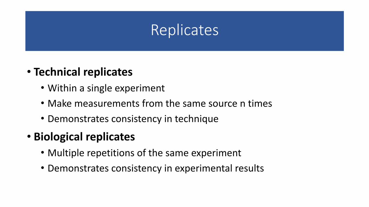
Replicates
• Technical replicates • Within a single experiment • Make measurements from the same source n times • Demonstrates consistency in technique
• Biological replicates • Multiple repetitions of the same experiment • Demonstrates consistency in experimental results
Replicates
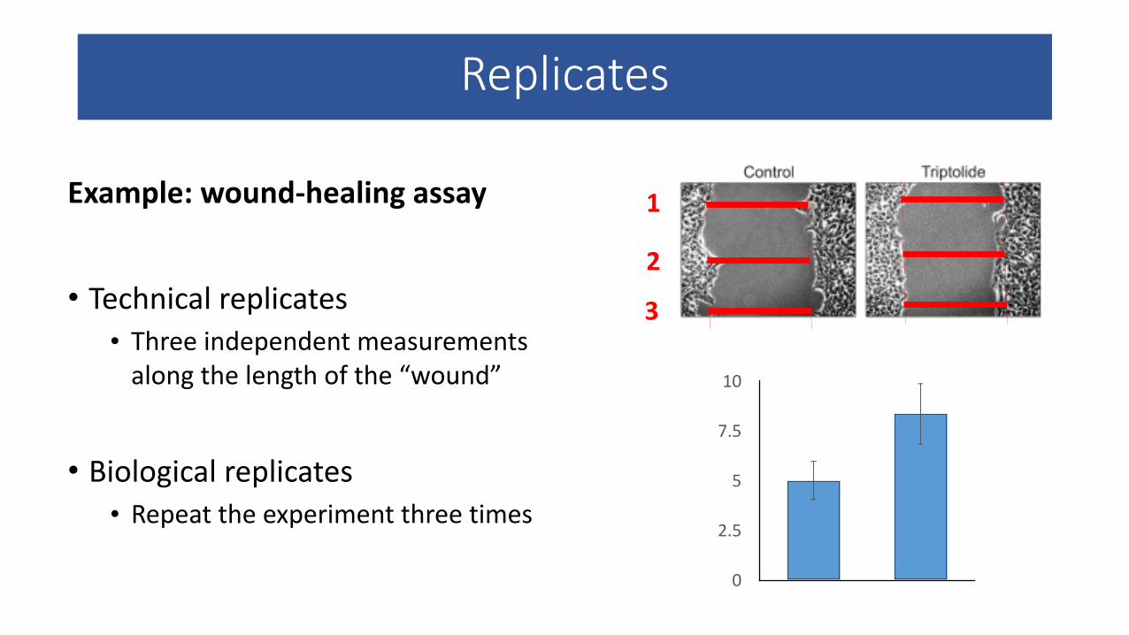
Example: wound-‐healing assay
!• Technical replicates
• Three independent measurements along the length of the “wound”
!• Biological replicates
• Repeat the experiment three times
1
2
3
0
2.5
5
7.5
10
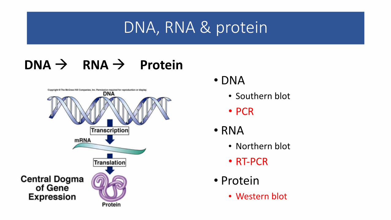
DNA, RNA & protein
• DNA • Southern blot • PCR
• RNA • Northern blot • RT-‐PCR
• Protein • Western blot
DNA à RNA à Protein
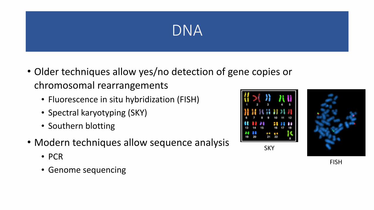
DNA
• Older techniques allow yes/no detection of gene copies or chromosomal rearrangements • Fluorescence in situ hybridization (FISH) • Spectral karyotyping (SKY) • Southern blotting
• Modern techniques allow sequence analysis • PCR • Genome sequencing
SKY
FISH
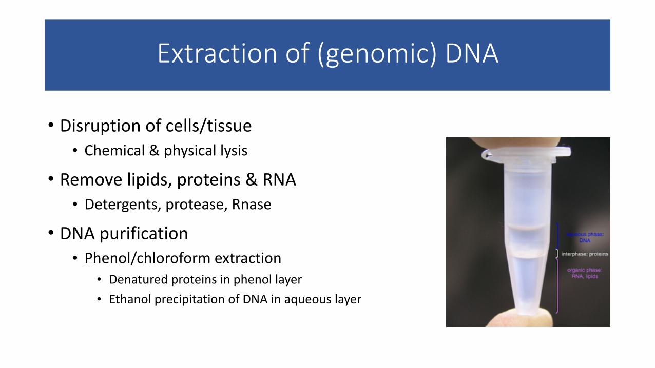
Extraction of (genomic) DNA
• Disruption of cells/tissue • Chemical & physical lysis
• Remove lipids, proteins & RNA • Detergents, protease, Rnase
• DNA purification • Phenol/chloroform extraction
• Denatured proteins in phenol layer • Ethanol precipitation of DNA in aqueous layer
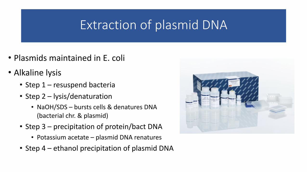
Extraction of plasmid DNA
• Plasmids maintained in E. coli • Alkaline lysis • Step 1 – resuspend bacteria • Step 2 – lysis/denaturation
• NaOH/SDS – bursts cells & denatures DNA (bacterial chr. & plasmid)
• Step 3 – precipitation of protein/bact DNA • Potassium acetate – plasmid DNA renatures
• Step 4 – ethanol precipitation of plasmid DNA
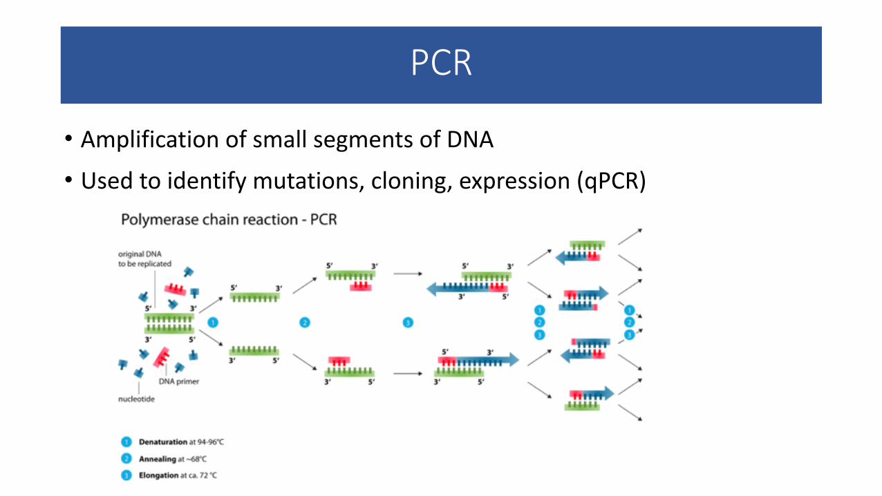
PCR
• Amplification of small segments of DNA
• Used to identify mutations, cloning, expression (qPCR)
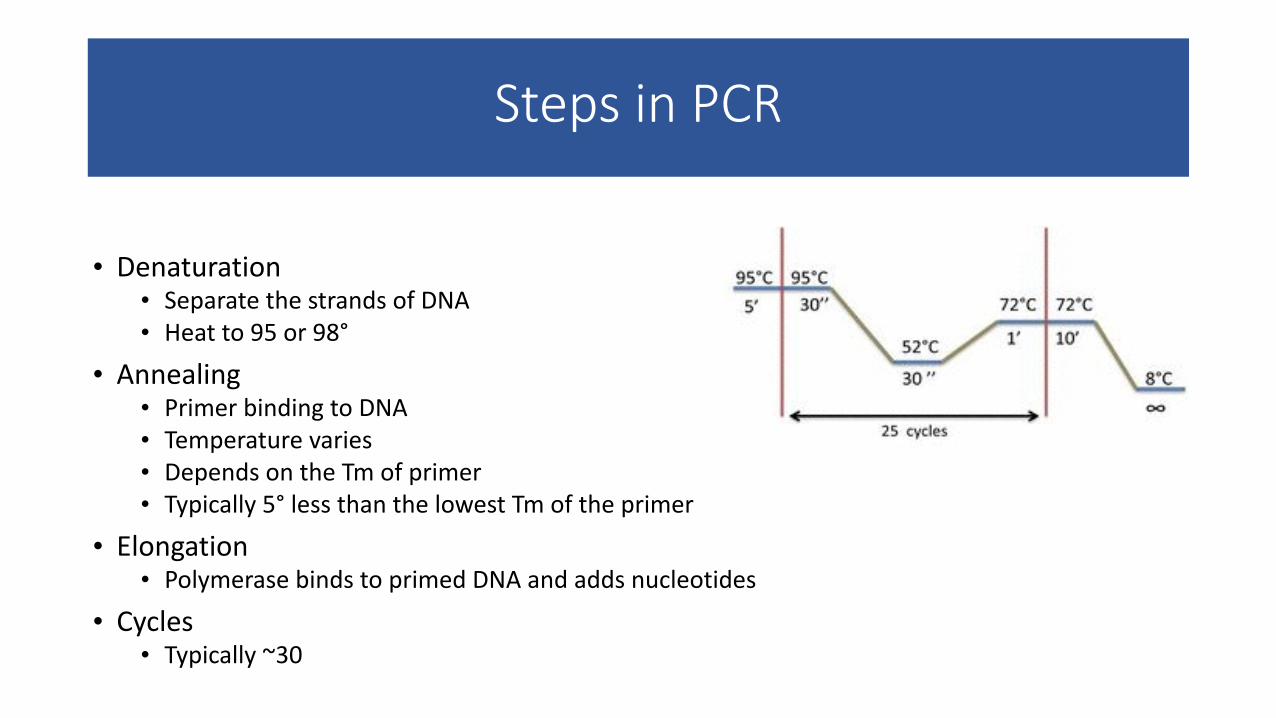
Steps in PCR
• Denaturation • Separate the strands of DNA • Heat to 95 or 98°
• Annealing • Primer binding to DNA • Temperature varies • Depends on the Tm of primer • Typically 5° less than the lowest Tm of the primer
• Elongation • Polymerase binds to primed DNA and adds nucleotides
• Cycles • Typically ~30
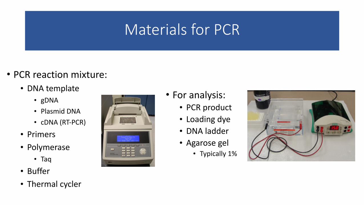
Materials for PCR
• PCR reaction mixture: • DNA template
• gDNA • Plasmid DNA • cDNA (RT-‐PCR)
• Primers • Polymerase
• Taq
• Buffer • Thermal cycler
• For analysis: • PCR product • Loading dye • DNA ladder • Agarose gel
• Typically 1%
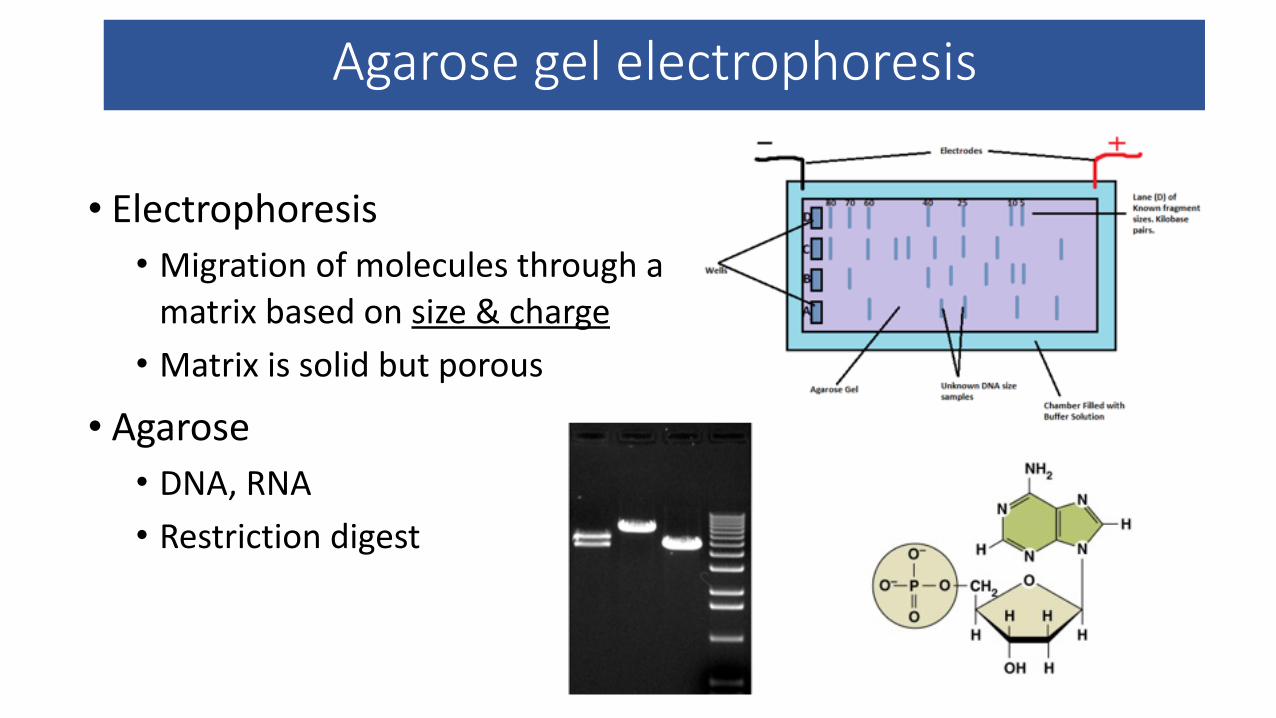
Agarose gel electrophoresis
• Electrophoresis • Migration of molecules through a matrix based on size & charge • Matrix is solid but porous
• Agarose • DNA, RNA • Restriction digest
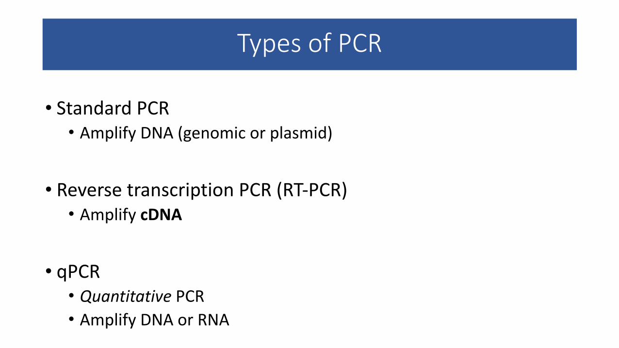
Types of PCR
• Standard PCR • Amplify DNA (genomic or plasmid)
!
• Reverse transcription PCR (RT-‐PCR) • Amplify cDNA
!
• qPCR • Quantitative PCR • Amplify DNA or RNA
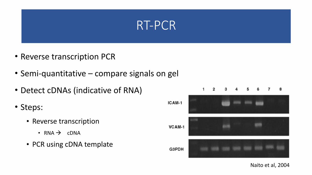
RT-‐PCR
• Reverse transcription PCR
• Semi-‐quantitative – compare signals on gel
• Detect cDNAs (indicative of RNA)
• Steps: • Reverse transcription
• RNA à cDNA
• PCR using cDNA template
Naito et al, 2004
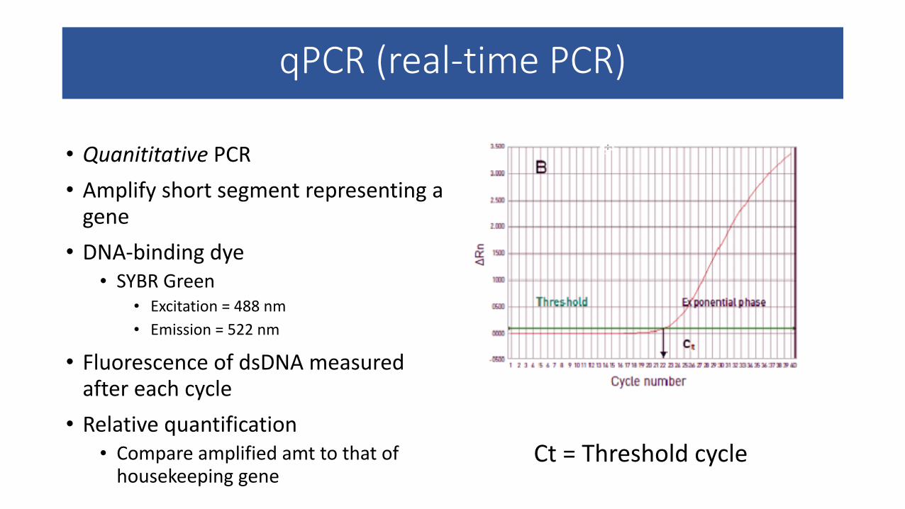
qPCR (real-‐time PCR)
• Quanititative PCR • Amplify short segment representing a gene • DNA-‐binding dye
• SYBR Green • Excitation = 488 nm • Emission = 522 nm
• Fluorescence of dsDNA measured after each cycle • Relative quantification
• Compare amplified amt to that of housekeeping gene
Ct = Threshold cycle
Quantitative RT-‐PCR
• Apply qPCR to cDNAs to quantify RNA levels • This replaces older RNA detection techniques such as northern blotting (which is semi-‐quantitative)
PCR experimental principles
• Controls • Amplification of housekeeping gene (RT-‐PCR & qPCR) • Positive – DNA template known to contain the correct sequence • Negative – Reaction mixture + water (no template)
• Quantification • Standard PCR is qualitative – presence/absence of band, sequence data • RT-‐PCR is semi-‐quantitative – band intensities can be compared based on equivalent control signal for each sample • qPCR is quantitative – Ct values can be compared

Western blot (WB)
• Allows comparison of levels of protein in samples • Ex: Is expression of a certain protein decreased when cells are treated with a drug?
!• Steps • Isolation of protein • Run lysates through SDS-‐PAGE gel • Transfer protein from gel to membrane • Incubate membrane with 1°/2° antibodies • Detect signal via film
Lee et al, 2011
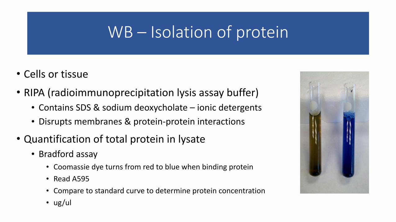
WB – Isolation of protein
• Cells or tissue • RIPA (radioimmunoprecipitation lysis assay buffer) • Contains SDS & sodium deoxycholate – ionic detergents • Disrupts membranes & protein-‐protein interactions
• Quantification of total protein in lysate • Bradford assay
• Coomassie dye turns from red to blue when binding protein • Read A595 • Compare to standard curve to determine protein concentration • ug/ul
WB –SDS-‐PAGE
• SDS = sodium dodecyl sulfate • Detergent
• PAGE = polyacrylamide gel electrophoresis !• Preparation of SDS-‐PAGE gel • Acrylamide:bisacrylamide (what polymerizes)
• 7.5% gel, 10% gel, etc • SDS (denaturant) • Buffer (maintains pH) • APS/TEMED (initiates polymerization)
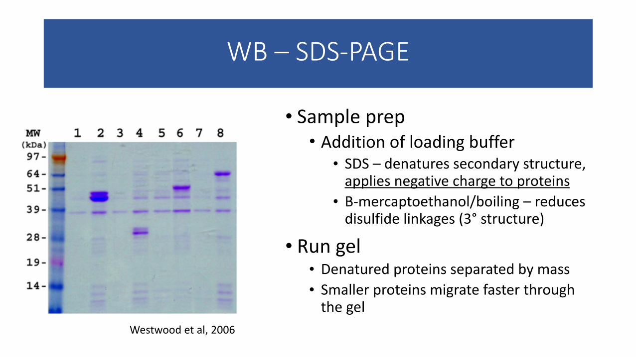
WB – SDS-‐PAGE
• Sample prep • Addition of loading buffer • SDS – denatures secondary structure, applies negative charge to proteins • Β-‐mercaptoethanol/boiling – reduces disulfide linkages (3° structure)
• Run gel • Denatured proteins separated by mass • Smaller proteins migrate faster through the gel
Westwood et al, 2006
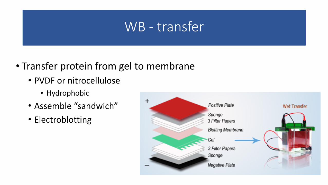
WB -‐ transfer
• Transfer protein from gel to membrane • PVDF or nitrocellulose
• Hydrophobic • Assemble “sandwich” • Electroblotting
WB – detection via 1°/2 ° antibodies
• Block membrane • Prevent non-‐specific binding of antibodies • BSA, non-‐fat milk
• 1° antibody • Specific for protein of interest
• 2° antibody • Specific for the species of the 1° Ab • Conjugated to a fluor or to HRP (horseradish peroxidase)
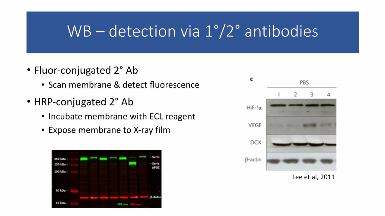
WB – detection via 1°/2° antibodies
• Fluor-‐conjugated 2° Ab • Scan membrane & detect fluorescence
• HRP-‐conjugated 2° Ab • Incubate membrane with ECL reagent • Expose membrane to X-‐ray film
Lee et al, 2011
WB experimental principles
• Controls • Immunoblotting (same membrane) for housekeeping gene (ex: GAPDH)
• Loading control
• Positive – Protein lysate from known positive sample • Negative – Dependent on experiment
• Ex: Treatment of control cells with PBS instead of drug • Ex: Treated cells at “0 h” (before drug can have an effect)
• Quantification • WB is semi-‐quantitative – band intensities can be compared based on equivalent control signal for each sample • In order to quantify, apply densitometry analysis
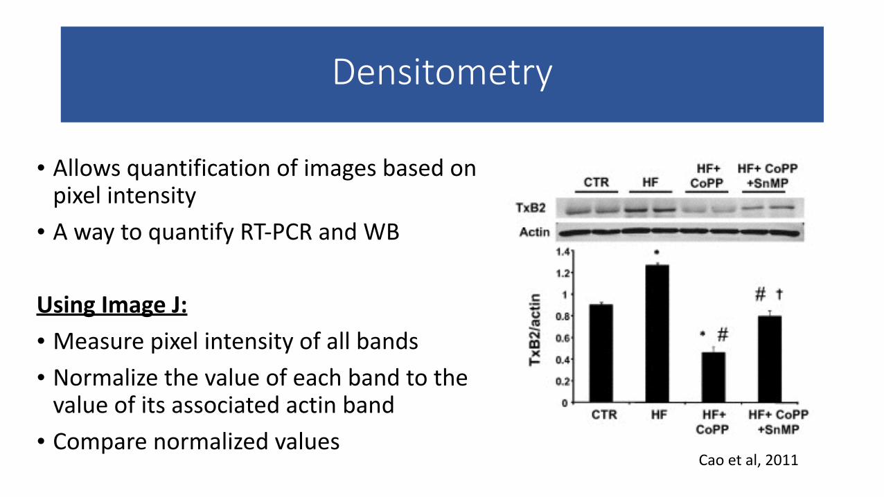
Densitometry
• Allows quantification of images based on pixel intensity • A way to quantify RT-‐PCR and WB !
Using Image J: • Measure pixel intensity of all bands • Normalize the value of each band to the value of its associated actin band • Compare normalized values
Cao et al, 2011
Cell culture
• 1900s – tissue culture • Harrison & Carrel • “a method for studying the behavior of animal cells free of systemic variations that might arise in the animal both during normal homeostasis and under the stress of an experiment”
• 1952 – development of continuous human tumor cell line • HeLa
• Uses of cultured cells: • Development of antiviral vaccines • Production of monoclonal antibodies • Production of cell products
• Insulin, HGH, interferon
• Understanding of neoplasia
Cell culture
• Benefits • Can carefully control environment
• Preservation
• Avoid using animals
• Rapid, relatively cheap
• Disadvantages/limitations • Expertise
• Identification of cell type
• Genetic & phenotypic instability
• Quantity
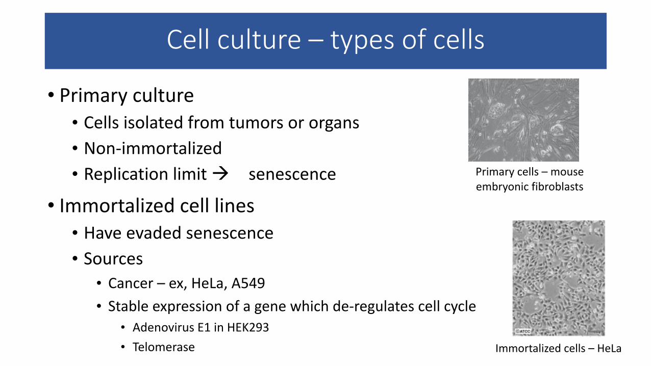
Cell culture – types of cells
• Primary culture • Cells isolated from tumors or organs • Non-‐immortalized • Replication limit à senescence
• Immortalized cell lines • Have evaded senescence • Sources
• Cancer – ex, HeLa, A549 • Stable expression of a gene which de-‐regulates cell cycle
• Adenovirus E1 in HEK293 • Telomerase
Primary cells – mouse embryonic fibroblasts
Immortalized cells – HeLa
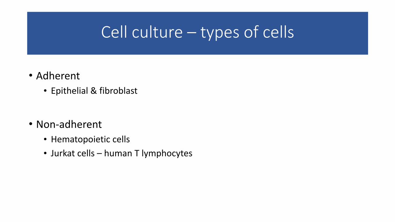
Cell culture – types of cells
• Adherent • Epithelial & fibroblast
!• Non-‐adherent • Hematopoietic cells • Jurkat cells – human T lymphocytes
Cell culture basics
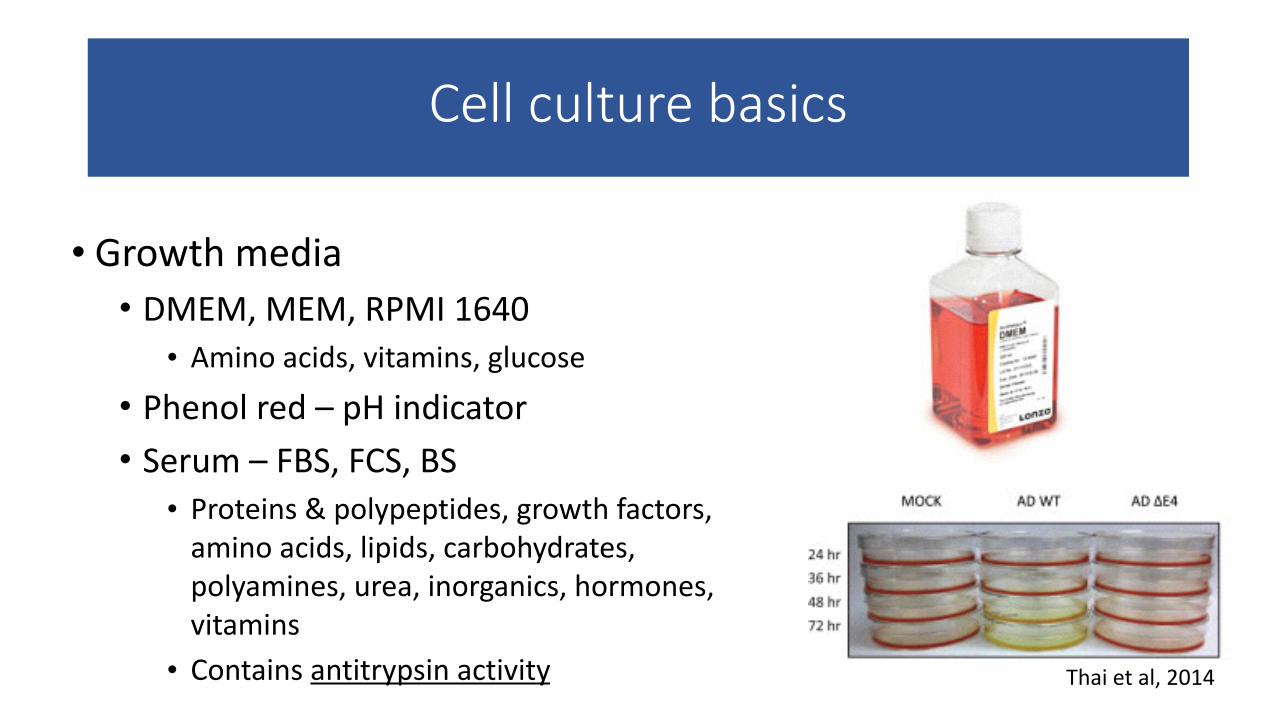
• Growth media • DMEM, MEM, RPMI 1640 • Amino acids, vitamins, glucose
• Phenol red – pH indicator • Serum – FBS, FCS, BS • Proteins & polypeptides, growth factors, amino acids, lipids, carbohydrates, polyamines, urea, inorganics, hormones, vitamins • Contains antitrypsin activity Thai et al, 2014
Cell culture basics – adherent cells
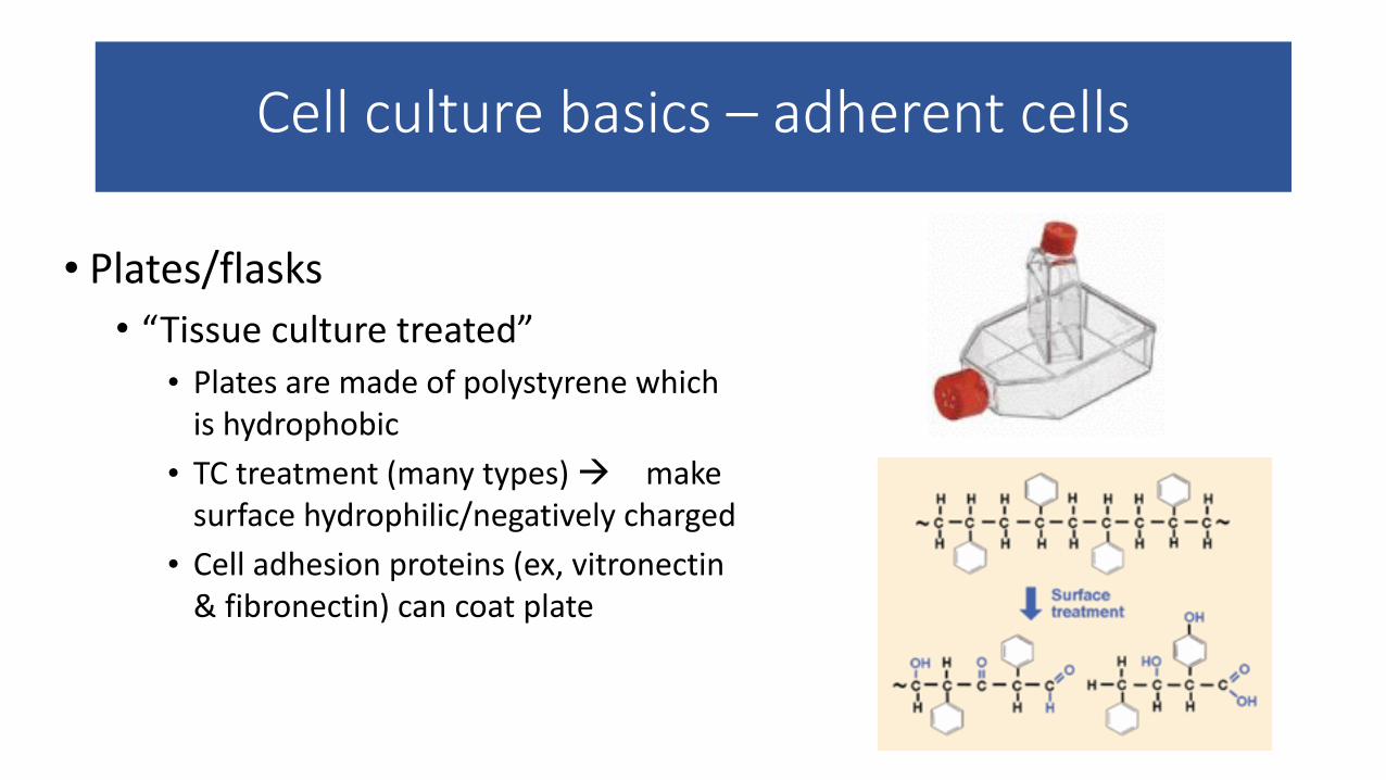
• Plates/flasks • “Tissue culture treated”
• Plates are made of polystyrene which is hydrophobic
• TC treatment (many types) à make surface hydrophilic/negatively charged
• Cell adhesion proteins (ex, vitronectin & fibronectin) can coat plate
Cell culture basics – adherent cells
• Subculturing • Trypsin/EDTA • Trypsin
• Serine protease • Cleaves adhesion proteins (integrins) • Optimal activity at 37°
• EDTA • Chelating agent – Calcium & Magnesium
• Neutralize with complete (serum-‐containing) medium
Aseptic technique
• Cell culture must be kept sterile
• Free of microorganisms • Bacteria • Fungi • Viruses
• Aseptic technique – designed to create a barrier between the sterile cell culture & microorganisms in the environment • Sterile work area • Good personal hygiene • Sterile reagents & media • Sterile handling
Summary -‐ I
• Cell & molecular techniques allow scientists to examine subcellular components
• Each experiment should include positive & negative controls
• Measurements can be qualitative or quantitative
• Technical replicates ensure consistency within an experiment; biological replicates provide confidence in experimental results
• DNA may be extracted using phenol/chloroform or (for plasmids) alkaline lysis
Summary -‐ II
• PCR allows us to amplify pieces of DNA or cDNA
• Western blotting allows detection of proteins within cells or tissue
• Densitometry enables quantification of otherwise semi-‐quantitative data through analysis of image pixel intensity
• Cell culture is a way to study cells in the laboratory • Aseptic technique is critical in cell culture

Questions?