Fig. 43-1 1.5 µm Chapter 43 The Immune System. Fig. 43-2 INNATE IMMUNITY Recognition of traits...
61
Fig. 43-1 1.5 µm Chapter 43 The Immune System
-
Upload
beverly-jackson -
Category
Documents
-
view
215 -
download
1
Transcript of Fig. 43-1 1.5 µm Chapter 43 The Immune System. Fig. 43-2 INNATE IMMUNITY Recognition of traits...

Fig. 43-1
1.5 µm
Chapter 43 The Immune System

Fig. 43-2
INNATE IMMUNITY
Recognition of traitsshared by broad rangesof pathogens, using asmall set of receptors
•
•Rapid response
•Recognition of traitsspecific to particularpathogens, using a vastarray of receptors
•Slower response
ACQUIRED IMMUNITY
Pathogens(microorganisms
and viruses)
Barrier defenses:SkinMucous membranesSecretions
Internal defenses:Phagocytic cellsAntimicrobial proteinsInflammatory responseNatural killer cells
Humoral response:Antibodies defend againstinfection in body fluids.
Cell-mediated response:Cytotoxic lymphocytes defendagainst infection in body cells.

Fig. 43-3
Microbes
PHAGOCYTIC CELL
Vacuole
Lysosomecontaining enzymes

Fig. 43-4Activates antimicrobial peptide by infecting with bacteria
Engineered to express the GFP gene upon activation of the innate immune response

• Can a single antimicrobial peptide protect fruit flies against infection?
• In 2002, Bruno Lematire et al., • To test the function of a single antimicrobial peptide.
– Drosomycin or defensin– A single antimicrobial peptide in the fly’s body
can provide an effective and specific immune defense against a particular pathogen.

Fig. 43-5RESULTS
% s
urv
ival
Wild type
Fruit fly survival after infection by N. crassa fungi
100
75
50
25
00 24 7248 96 120
100
75
50
25
00 24 7248 96 120
Hours post-infection
Fruit fly survival after infection by M. luteus bacteria
Hours post-infection
% s
urv
ival
Mutant + drosomycin
Mutant + defensinMutant
Wild type
Mutant +drosomycin
Mutant +defensin
Mutant
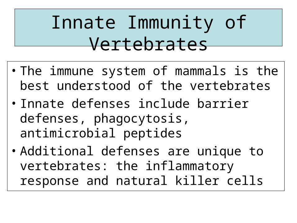
Innate Immunity of Vertebrates
• The immune system of mammals is the best understood of the vertebrates
• Innate defenses include barrier defenses, phagocytosis, antimicrobial peptides
• Additional defenses are unique to vertebrates: the inflammatory response and natural killer cells
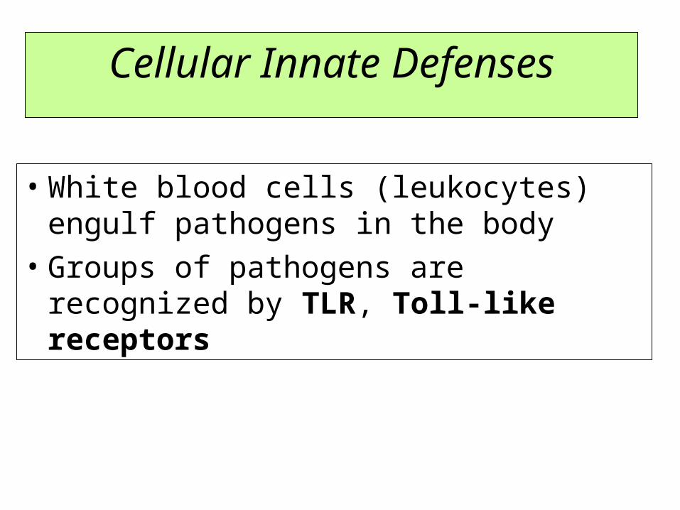
Cellular Innate Defenses
• White blood cells (leukocytes) engulf pathogens in the body
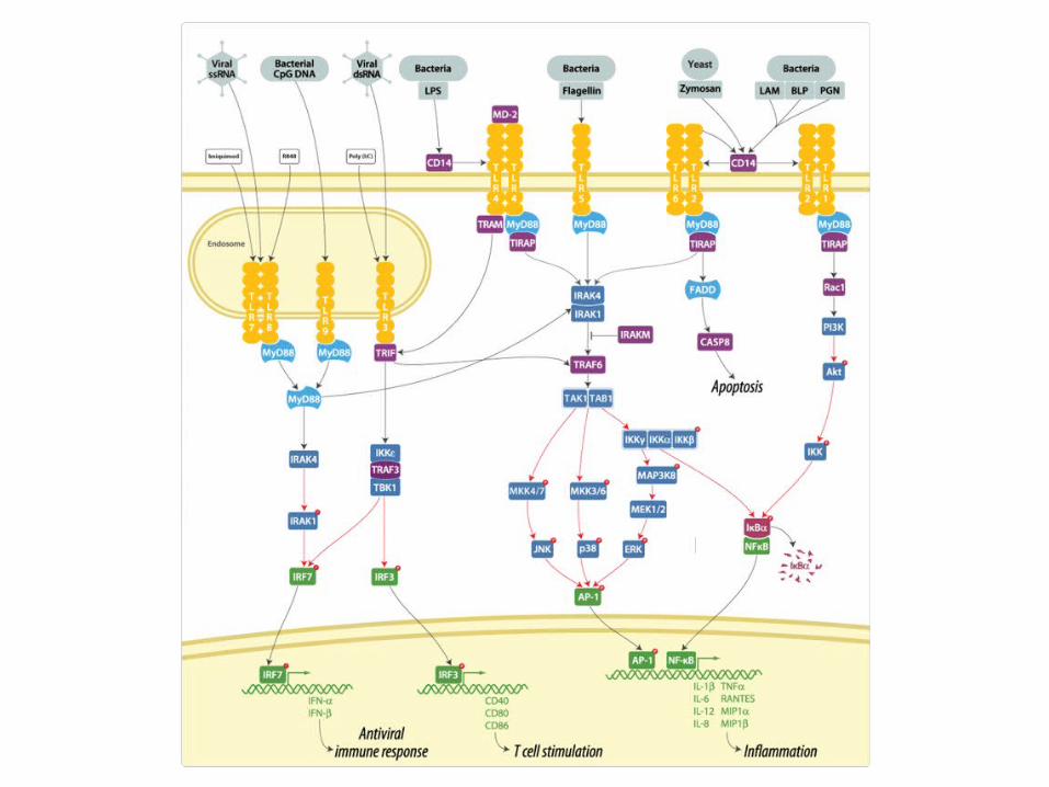
• Groups of pathogens are recognized by TLR, Toll-like receptors

Fig. 43-6
EXTRACELLULARFLUID Lipopolysaccharide
FlagellinTLR4
TLR5
Helperprotein
TLR9
TLR3
WHITEBLOODCELL
VESICLE
CpG DNA
ds RNA
Inflammatoryresponses
Toll-like receptor or TLR

Toll-like receptorFrom Wikipedia,
The curved leucine-rich repeat region of Toll-like receptors, represented
here by TLR3
Toll-like receptors (TLRs) are a class of proteins that play a key role in the innate immune system. They are single membrane-spanning non-catalytic receptors that recognize structurally conserved molecules derived from microbes. Once these microbes have breached physical barriers such as the skin or intestinal tract mucosa, they are recognized by TLRs which activates immune cell responses.
They receive their name from their similarity to the protein coded by the Toll gene identified in Drosophila in 1985 by Christiane Nüsslein-Volhard.[1]

Signaling pathway of Toll-like receptors.
Dashed grey lines represent unknown associations

• A white blood cell engulfs a microbe, then fuses with a lysosome to destroy the microbe
• There are different types of phagocytic cells:– Neutrophils engulf and destroy microbes– Macrophages are part of the lymphatic system and
are found throughout the body– Eosinophils discharge destructive enzymes– Dendritic cells stimulate development of acquired
immunity

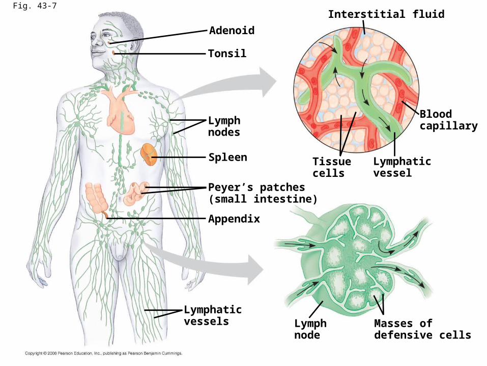
Fig. 43-7
Adenoid
Tonsil
Lymphnodes
Spleen
Peyer’s patches(small intestine)
Appendix
Lymphaticvessels Lymph
nodeMasses ofdefensive cells
Bloodcapillary
Lymphaticvessel
Tissuecells
Interstitial fluid
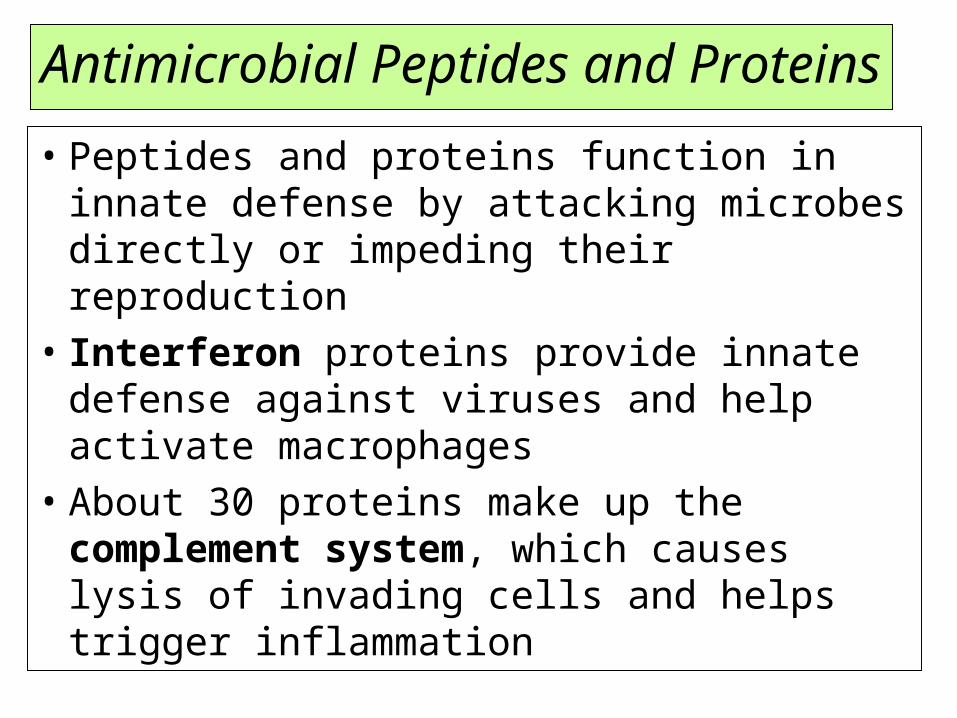
Antimicrobial Peptides and Proteins
• Peptides and proteins function in innate defense by attacking microbes directly or impeding their reproduction
• Interferon proteins provide innate defense against viruses and help activate macrophages
• About 30 proteins make up the complement system, which causes lysis of invading cells and helps trigger inflammation

Inflammatory Responses
• Following an injury, mast cells release histamine, which promotes changes in blood vessels; this is part of the inflammatory response
• These changes increase local blood supply and allow more phagocytes and antimicrobial proteins to enter tissues
• Pus, a fluid rich in white blood cells, dead microbes, and cell debris, accumulates at the site of inflammation

Fig. 43-8-1
Pathogen Splinter
Macrophage
Mast cell
Chemicalsignals
Capillary
Phagocytic cellRed blood cells
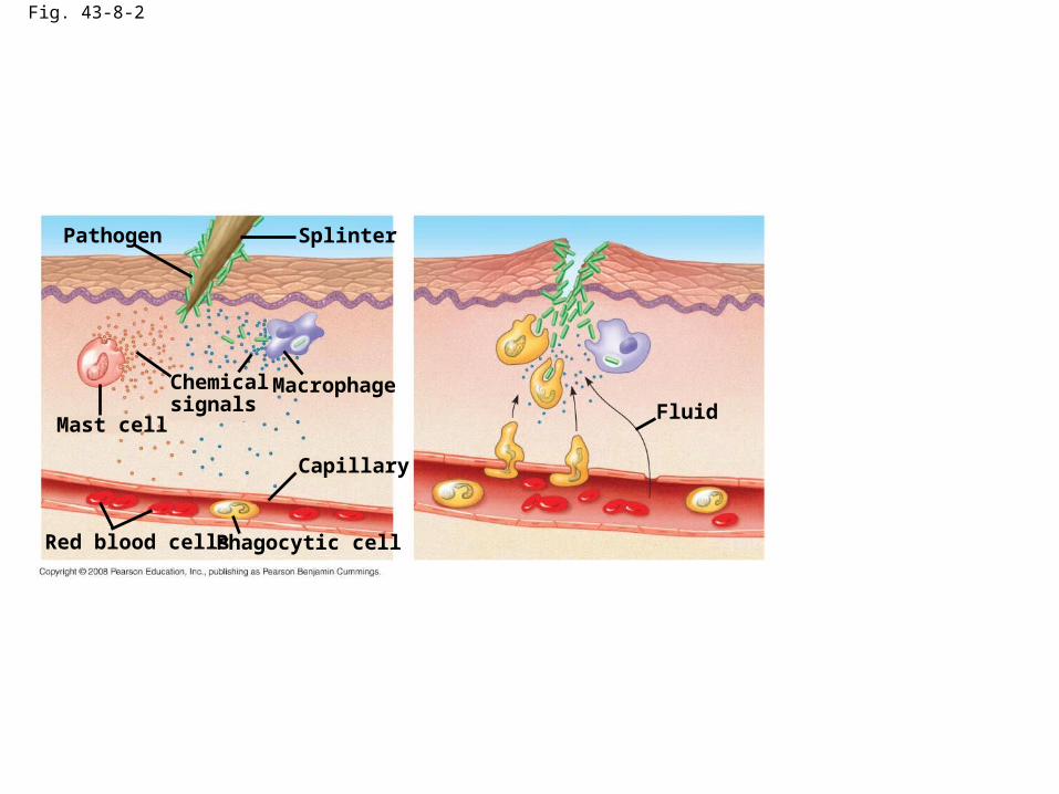
Fig. 43-8-2
Pathogen Splinter
Macrophage
Mast cell
Chemicalsignals
Capillary
Phagocytic cellRed blood cells
Fluid
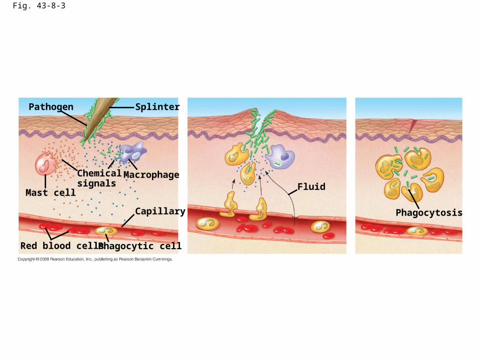
Fig. 43-8-3
Pathogen Splinter
Macrophage
Mast cell
Chemicalsignals
Capillary
Phagocytic cellRed blood cells
Fluid
Phagocytosis

Specific immunity

Fig. 43-9
Antigen-bindingsite
Antigen-binding site
Antigen-bindingsite
Disulfidebridge
Variableregions
Constantregions
Transmembraneregion
Plasmamembrane
Lightchain
Heavy chains
T cell
chain chain
Disulfide bridge
Cytoplasm of T cell
(b) T cell receptor
Cytoplasm of B cell
(a) B cell receptor
B cell
V
V
C C
V
V
C C C C
VV

Fig. 43-9a
Antigen-bindingsite
Antigen-binding site
Disulfidebridge
Variableregions
Constantregions
Transmembraneregion
Plasmamembrane
Lightchain
Heavy chains
Cytoplasm of B cell
(a) B cell receptor
B cell
V
V
C C
V
V
C C

Fig. 43-9b
Antigen-bindingsite
Variableregions
Constantregions
Transmembraneregion
Plasmamembrane
T cell
chain chain
Disulfide bridge
Cytoplasm of T cell
(b) T cell receptor
C C
VV

Fig. 43-10
Antigen-binding sites
Antigen-bindingsites
Epitopes(antigenicdeterminants)
Antigen
Antibody B
Antibody CAntibody A
CC
CV
V
V
V
C

• Class I MHC molecules are found on almost all nucleated cells of the body
• They display peptide antigens to cytotoxic T cells
• Class II MHC molecules are located mainly on dendritic cells, macrophages, and B cells
• Dendritic cells, macrophages, and B cells are antigen-presenting cells that display antigens to cytotoxic T cells and helper T cells

Fig. 43-11
Antigen
Top view: binding surfaceexposed to antigen receptors
Plasmamembrane ofinfected cell
AntigenClass I MHCmolecule
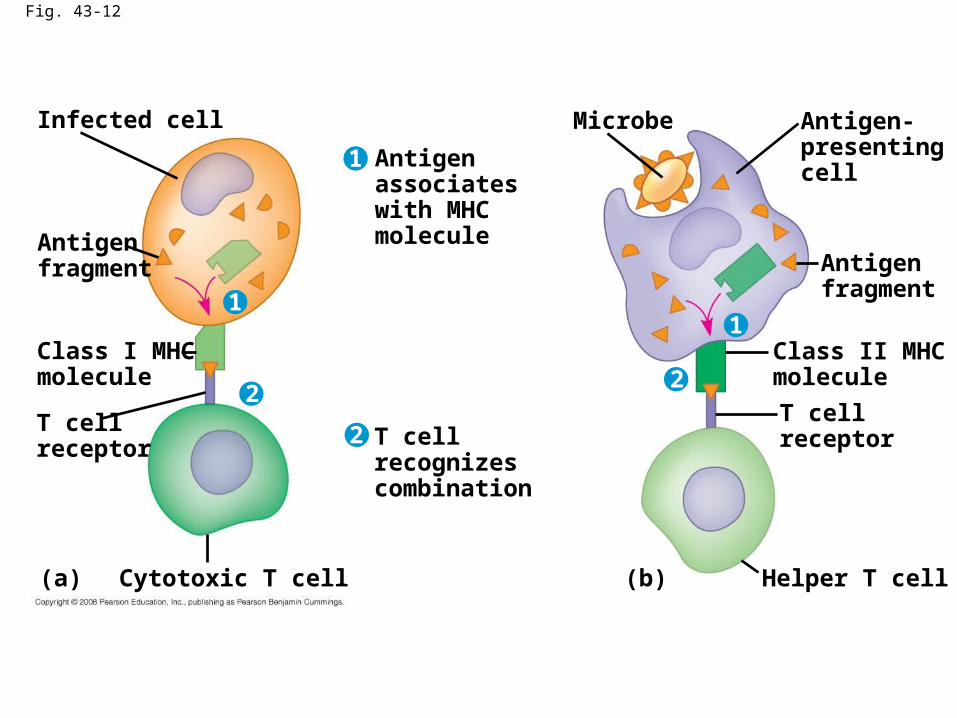
Fig. 43-12
Infected cell
Antigenfragment
Class I MHCmolecule
T cellreceptor
(a)
Antigenassociateswith MHCmolecule
T cellrecognizescombination
Cytotoxic T cell (b) Helper T cell
T cellreceptor
Class II MHCmolecule
Antigenfragment
Antigen-presentingcell
Microbe
1
11
2
22

Fig. 43-13
DNA of undifferentiated B cell
1
DNA of differentiated B cell
pre-mRNA
mRNA
Light-chain polypeptide
Variableregion
Constantregion
Translation
B cell
B cell receptor
RNA processing
Transcription
DNA deleted between randomly selected V and Jsegments
Functional gene
V37 V38 V39 V40 J1 J2 J3 J4 CJ5 Intron
V37 V38 V39 CJ5 Intron
V39 CJ5 Intron
V39 CJ5 Poly-A tailCap
CV
VV
V
V
C C
C C
2
3
4
Ig gene rearrangement

The mechanism of Ig diversity
1. DNA recombination
2. The joining V, D, and J domains is not always precise
3. The DNA coding for hypervariable region is hypermutation
CU

Origin of Self-Tolerance
• Antigen receptors are generated by random rearrangement of DNA
• As lymphocytes mature in bone marrow or the thymus, they are tested for self-reactivity
• Lymphocytes with receptors specific for the body’s own molecules are destroyed by apoptosis, or rendered nonfunctional

Fig. 43-14
B cells thatdiffer inantigen specificity
Antibodymolecules
Antigenreceptor
Antigen molecules
Clone of memory cells Clone of plasma cells
Conal selection of B calla
Amplifying Lymphocytes by Clonal Selection

Fig. 43-15
Antibodiesto A
Antibodiesto B
Secondary immune response toantigen A produces antibodies to A;primary immune response to antigenB produces antibodies to B.
Primary immune responseto antigen A producesantibodies to A.
An
tib
od
y co
nce
ntr
atio
n(a
rbit
rary
un
its)
Exposureto antigen A
Exposure toantigens A and B
Time (days)
104
103
102
101
100
0 7 14 21 28 35 42 49 56
The specifity of immunological memory

Fig. 43-16
Humoral (antibody-mediated) immune response
B cell
Plasma cells
Cell-mediated immune response
Key
Stimulates
Gives rise to
+
+
++
+
+
+Memory B cells
Antigen (1st exposure)
Engulfed by
Antigen-presenting cell
MemoryHelper T cells
Helper T cell Cytotoxic T cell
MemoryCytotoxic T cells
ActiveCytotoxic T cells
Antigen (2nd exposure)
Secretedantibodies
Defend against extracellular pathogens by binding to antigens,thereby neutralizing pathogens or making them better targetsfor phagocytes and complement proteins.
Defend against intracellular pathogensand cancer by binding to and lysing theinfected cells or cancer cells.
+
+ +

Fig. 43-16a
Key
Stimulates
Gives rise to
+
MemoryHelper T cells
Antigen-presenting cell
Helper T cell
Engulfed by
Antigen (1st exposure)
+
+
+
+ +
+
Defend against extracellular pathogens
MemoryB cells
Antigen (2nd exposure)
Plasma cells
B cell
Secretedantibodies
Humoral (antibody-mediated) immune response

Fig. 43-17
Antigen-presentingcell
Peptide antigen
Cell-mediatedimmunity (attack on
infected cells)
Class II MHC moleculeCD4
TCR (T cell receptor)
Helper T cell
Humoralimmunity
(secretion ofantibodies byplasma cells) Cytotoxic T cell
Cytokines
B cell
Bacterium
+
+ +
+
The central role of helper T cells in humoral and cell-mediated immune response

Fig. 43-16bCell-mediated immune response
Defend against intracellular pathogens
ActiveCytotoxic T cells
MemoryCytotoxic T cells
MemoryHelper T cells
Antigen-presenting cell
Antigen (2nd exposure)
Helper T cell
Engulfed by
Antigen (1st exposure)
Cytotoxic T cell
KeyStimulates
Gives rise to
+
+
+
+
+ +
+
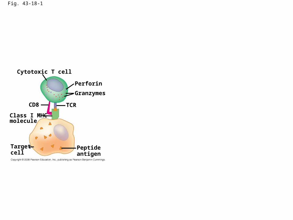
Fig. 43-18-1
Cytotoxic T cell
Perforin
Granzymes
TCRCD8
Class I MHCmolecule
Targetcell
Peptideantigen
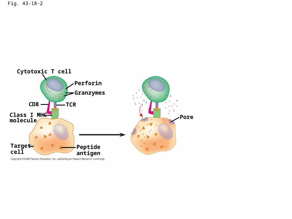
Fig. 43-18-2
Cytotoxic T cell
Perforin
Granzymes
TCRCD8
Class I MHCmolecule
Targetcell
Peptideantigen
Pore

Fig. 43-18-3
Cytotoxic T cell
Perforin
Granzymes
TCRCD8
Class I MHCmolecule
Targetcell
Peptideantigen
Pore
Released cytotoxic T cell
Dying target cell
The killing action of cytotoxic T cells
----A response to infected cells
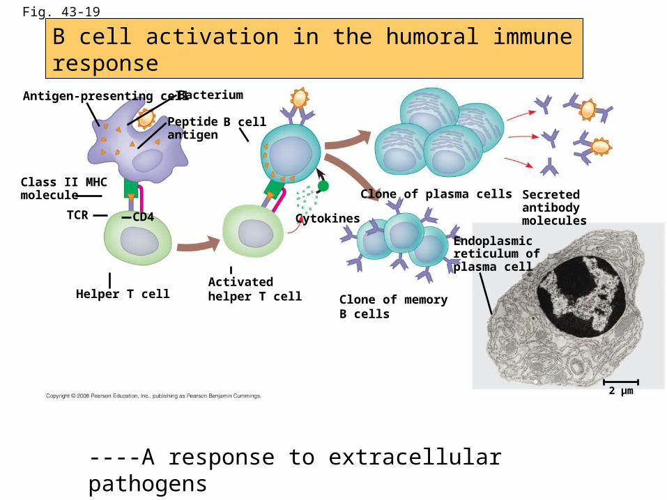
Fig. 43-19
Antigen-presenting cell
Endoplasmicreticulum ofplasma cell
Secretedantibodymolecules
Bacterium
B cellPeptideantigen
Class II MHCmolecule
TCR CD4
Helper T cellActivatedhelper T cell
Cytokines
Clone of memoryB cells
Clone of plasma cells
2 µm
+
B cell activation in the humoral immune response
----A response to extracellular pathogens
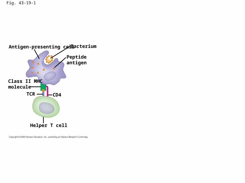
Fig. 43-19-1
Antigen-presenting cell Bacterium
Peptideantigen
Class II MHCmolecule
TCR CD4
Helper T cell

Fig. 43-19-2
Antigen-presenting cell Bacterium
Peptideantigen
Class II MHCmolecule
TCR CD4
Helper T cell
B cell
Activatedhelper T cell
Cytokines
+

Fig. 43-19-3
Antigen-presenting cell Bacterium
Peptideantigen
Class II MHCmolecule
TCR CD4
Helper T cell
B cell
Activatedhelper T cell
Cytokines
+ Secretedantibodymolecules
Clone of memoryB cells
Clone of plasma cells

Fig. 43-19a
Endoplasmicreticulum ofplasma cell
2 µm

Fig. 43-20Class of Immuno-
globulin (Antibody)
IgG(monomer)
IgM(pentamer)
J chain
IgA(dimer)
IgE(monomer)
IgD(monomer)
Trans-membraneregion
J chain
Secretorycomponent
Distribution Function
First Ig classproduced afterinitial exposure toantigen; then itsconcentration inthe blood declines
Promotes neutraliza-tion and cross-linking of antigens;very effective incomplement systemactivation
Present insecretions suchas tears, saliva,mucus, andbreast milk
Only Ig class thatcrosses placenta,thus conferringpassive immunityon fetus
Triggers release frommast cells andbasophils of hista-mine and otherchemicals that causeallergic reactions
Present primarilyon surface ofB cells that havenot been exposedto antigens
Acts as antigenreceptor in theantigen-stimulatedproliferation anddifferentiation ofB cells (clonalselection)
Most abundant Igclass in blood;also present intissue fluids
Promotes opsoniza-tion, neutralization,and cross-linking ofantigens; less effec-tive in activation ofcomplement systemthan IgM
Provides localizeddefense of mucousmembranes bycross-linking andneutralization of antigens
Presence in breastmilk conferspassive immunityon nursing infant
Present in bloodat low concen-trations
The five ab , or immunoglobulin (Ig) classes

Fig. 43-20a
DistributionClass of Immuno-
globulin (Antibody)
IgM(pentamer)
J chain
First Ig classproduced afterinitial exposure toantigen; then itsconcentration inthe blood declines
Promotes neutraliza-tion and cross-linking of antigens;very effective incomplement systemactivation
Function

Fig. 43-20b
Distribution FunctionClass of Immuno-
globulin (Antibody)
IgG(monomer)
Most abundant Igclass in blood;also present intissue fluids
Promotes opsoniza-tion, neutralization,and cross-linking ofantigens; less effec-tive in activation ofcomplement systemthan IgM
Only Ig class thatcrosses placenta,thus conferringpassive immunityon fetus

Fig. 43-20c
Distribution FunctionClass of Immuno-
globulin (Antibody)
IgA(dimer)
J chain
Secretorycomponent
Present insecretions suchas tears, saliva,mucus, andbreast milk
Provides localizeddefense of mucousmembranes bycross-linking andneutralization ofantigens
Presence in breastmilk conferspassive immunityon nursing infant

Fig. 43-20d
Distribution FunctionClass of Immuno-
globulin (Antibody)
IgE(monomer)
Present in bloodat low concen-trations
Triggers release frommast cells andbasophils of hista-mine and otherchemicals that causeallergic reactions

Fig. 43-20e
Distribution FunctionClass of Immuno-
globulin (Antibody)
IgD(monomer)
Trans-membraneregion
Present primarilyon surface ofB cells that havenot been exposedto antigens
Acts as antigenreceptor in theantigen-stimulatedproliferation anddifferentiation ofB cells (clonalselection)
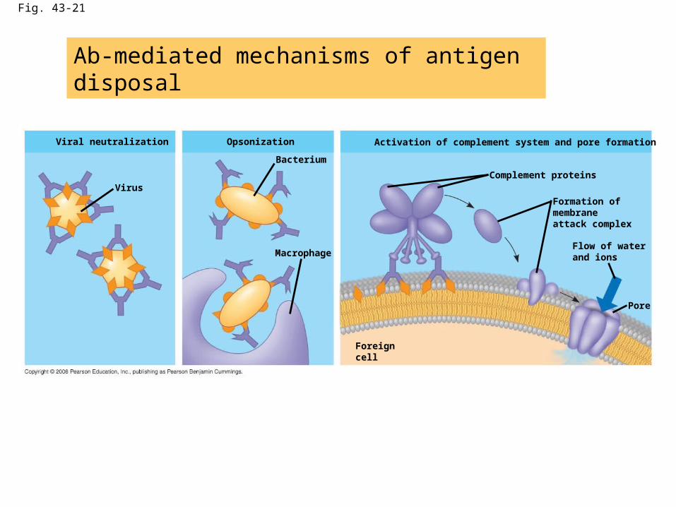
Fig. 43-21
Viral neutralization
Virus
Opsonization
Bacterium
Macrophage
Activation of complement system and pore formation
Complement proteins
Formation ofmembraneattack complex
Flow of waterand ions
Pore
Foreigncell
Ab-mediated mechanisms of antigen disposal

Fig. 43-21a
Viral neutralization
Virus

Fig. 43-21b
Opsonization
Bacterium
Macrophage
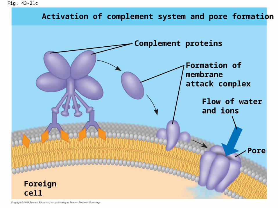
Fig. 43-21c
Activation of complement system and pore formation
Complement proteins
Formation ofmembraneattack complex
Flow of waterand ions
Pore
Foreigncell

Fig. 43-22

Allergen
IgE
Granule
Mast cell
Histamine
Impact on public health

Fig. 43-24
•include systemic lupus erythematosus,• rheumatoid arthritis,• insulin-dependent diabetes mellitus, and• multiple sclerosis
Autoimmune diseases

Acquired immune system evasion by pathogens
• Antigenic variation
• Latency
• Herpes simplex viruses: typeI and II– Sensory neurons express relatively few MHCI
molecules
• Attack on the immune system:HIV

Fig. 43-25
Weeks after infection
Mill
ion
s o
f p
aras
ites
per
mL
of
blo
od
Antibodies tovariant 1appear
Antibodies tovariant 2appear
Antibodies tovariant 3appear
Variant 3Variant 2Variant 1
25 26 27 280
0.5
1.0
1.5
A change in epitope expression, which are called antigenic variation
---- sleeping sickness (trypanosomiasis)

Fig. 43-26
Latency
Relative antibodyconcentration
AIDSH
elp
er T
cel
l co
nce
ntr
atio
nin
blo
od
(ce
lls/m
m3 )
Helper T cellconcentration
Relative HIVconcentration
Years after untreated infection0 1 2 3 4 5 6 7 8 9 10
0
200
400
600
800