Fibroblast migration and collagen deposition during …jas/paperpdfs/mcdougallwound.pdf ·...
21
Fibroblast migration and collagen deposition during dermal wound healing: mathematical modelling and clinical implications BY STEVEN MCDOUGALL 1 ,J OHN DALLON 2, * ,JONATHAN SHERRATT 3 AND PHILIP MAINI 4 1 Institute of Petroleum Engineering, and 3 Department of Mathematics, Heriot-Watt University, Edinburgh EH14 4AS, UK 2 Department of Mathematics, Brigham Young University, Provo, UT 84602-6539, USA 4 Centre for Mathematical Biology, Mathematical Institute, 24-29 St Giles’, Oxford OX1 3LB, UK The extent to which collagen alignment occurs during dermal wound healing determines the severity of scar tissue formation. We have modelled this using a multiscale approach, in which extracellular materials, for example collagen and fibrin, are modelled as continua, while fibroblasts are considered as discrete units. Within this model framework, we have explored the effects that different parameters have on the alignment process, and we have used the model to investigate how manipulation of transforming growth factor-b levels can reduce scar tissue formation. We briefly review this body of work, then extend the modelling framework to investigate the role played by leucocyte signalling in wound repair. To this end, fibroblast migration and collagen deposition within both the wound region and healthy peripheral tissue are considered. Trajectories of individual fibroblasts are determined as they migrate towards the wound region under the combined influence of collagen/fibrin alignment and gradients in a paracrine chemoattractant produced by leucocytes. The effects of a number of different physiological and cellular parameters upon the collagen alignment and repair integrity are assessed. These parameters include fibroblast concentration, cellular speed, fibroblast sensitivity to chemoattractant concentration and chemoattractant diffusion coefficient. Our results show that chemoattractant gradients lead to increased collagen alignment at the interface between the wound and the healthy tissue. Results show that there is a trade-off between wound integrity and the degree of scarring. The former is found to be optimized under conditions of a large chemoattractant diffusion coefficient, while the latter can be minimized when repair takes place in the presence of a competitive inhibitor to chemoattractants. Keywords: scar tissue; alignment; fibroblasts; chemoattractant; transforming growth factor-b Phil. Trans. R. Soc. A (2006) 364, 1385–1405 doi:10.1098/rsta.2006.1773 Published online 25 April 2006 One contribution of 15 to a Theme Issue ‘Biomathematical modelling II’. * Author for correspondence ([email protected]). 1385 q 2006 The Royal Society
Transcript of Fibroblast migration and collagen deposition during …jas/paperpdfs/mcdougallwound.pdf ·...
Fibroblast migration and collagen depositionduring dermal wound healing: mathematical
modelling and clinical implications
BY STEVEN MCDOUGALL1, JOHN DALLON
2,*, JONATHAN SHERRATT3
AND PHILIP MAINI4
1Institute of Petroleum Engineering, and 3Department of Mathematics,Heriot-Watt University, Edinburgh EH14 4AS, UK
2Department of Mathematics, Brigham Young University, Provo,UT 84602-6539, USA
4Centre for Mathematical Biology, Mathematical Institute, 24-29 St Giles’,Oxford OX1 3LB, UK
The extent to which collagen alignment occurs during dermal wound healing determinesthe severity of scar tissue formation. We have modelled this using a multiscale approach,in which extracellular materials, for example collagen and fibrin, are modelled ascontinua, while fibroblasts are considered as discrete units. Within this modelframework, we have explored the effects that different parameters have on the alignmentprocess, and we have used the model to investigate how manipulation of transforminggrowth factor-b levels can reduce scar tissue formation. We briefly review this body ofwork, then extend the modelling framework to investigate the role played by leucocytesignalling in wound repair. To this end, fibroblast migration and collagen depositionwithin both the wound region and healthy peripheral tissue are considered. Trajectoriesof individual fibroblasts are determined as they migrate towards the wound region underthe combined influence of collagen/fibrin alignment and gradients in a paracrinechemoattractant produced by leucocytes. The effects of a number of differentphysiological and cellular parameters upon the collagen alignment and repair integrityare assessed. These parameters include fibroblast concentration, cellular speed, fibroblastsensitivity to chemoattractant concentration and chemoattractant diffusion coefficient.Our results show that chemoattractant gradients lead to increased collagen alignment atthe interface between the wound and the healthy tissue. Results show that there is atrade-off between wound integrity and the degree of scarring. The former is found to beoptimized under conditions of a large chemoattractant diffusion coefficient, while thelatter can be minimized when repair takes place in the presence of a competitive inhibitorto chemoattractants.
Keywords: scar tissue; alignment; fibroblasts; chemoattractant;transforming growth factor-b
On
*A
Phil. Trans. R. Soc. A (2006) 364, 1385–1405
doi:10.1098/rsta.2006.1773
Published online 25 April 2006
e contribution of 15 to a Theme Issue ‘Biomathematical modelling II’.
uthor for correspondence ([email protected]).
1385 q 2006 The Royal Society

S. McDougall and others1386
1. Introduction
In dermal wound healing, after the skin is injured, several interacting eventsare initiated including inflammation, tissue formation, angiogenesis, tissuecontraction and tissue remodelling (Clark 1989). Crucial to all of theseevents is the interaction of the cells with the extracellular matrix (ECM).After the blood clot has formed during the inflammatory response, whiteblood cells invade the wound region by migrating through the ECM.Fibroblasts subsequently migrate into the region and begin to replace theblood clot with collagen. They biochemically alter the ECM by degrading thefibrin and producing collagen (Jennings & Hunt 1992). While new tissue isbeing generated, endothelial cells migrate into the region forming a newvasculature in the process known as angiogenesis (Clark 1996). The optimaloutcome is a healed wound with new tissue identical to that surrounding thewound but this is typically not the case. The new tissue is usuallycharacterized by a new architecture that differs from the original and isfrequently less functional than the original tissue. This remodelled tissue isknown as a scar.
The characteristics of scars are a result of altered structure andcomposition in the dermis. Scars typically have fewer blood vessels supplyingthe denser connective tissue which is less elastic. The most significantdifference between normal tissue and scar tissue seems to be the orientationof the fibrous matrix. In rodents, normal tissue has a reticular collagenpattern, whereas the collagen in scar tissue forms large parallel bundles atapproximately right angles to the basement membrane (Whitby & Ferguson1991). Human scar tissue is similar, with greater collagen density, largerfibres and more alignment than normal tissue, although the alignment isparallel to the skin (Ehrlich & Krummel 1996). Further structural differencesbetween scars and normal tissue include a different ratio of collagen typesand a lack of hair follicles and sweat glands. In the work discussed here wefocus on the fibre alignment in scar tissue.
First we describe the wound healing process, then review our previousmodelling work, and finally report new results. Our previous work will be dividedinto three parts. Our initial modelling of collagen alignment (Dallon et al. 1999)will be described in §3, followed by a description of a model for wound repair(Dallon et al. 2000) in §4, and in §5, a model explaining the effects oftransforming growth factor-b (TGFb) (Dallon et al. 2001) will be reviewed. In §6,we begin reporting the new results by describing the extension of the modeldesigned to examine the role of leucocyte signalling in wound repair, the role offibroblast distribution (§6a) and the effect of the activity of a diffusiblechemoattractant (§6b). Finally, we end with a discussion in §7.
2. Dermal wound healing
There are fundamental differences between foetal and adult wound healing,resulting in different outcomes. In adults, the end product is a scar. However,foetal tissue is repaired with reduced or absent scarring (McCallion & Ferguson
Phil. Trans. R. Soc. A (2006)

1387Modelling collagen during wound repair
1996) and the realization of this difference has generated a large volume ofresearch activity into mechanisms underlying scar formation.
The healing of full-thickness skin wounds involves a complex sequence ofoverlapping events. One of the first is the coagulation cascade (Jennings & Hunt1992) where fibrinogen, a soluble protein in the plasma, is converted to fibrinwhich polymerizes to form a blood clot. This clot stops bleeding and can dry toform a scab which covers the wound; it also forms a provisional matrix whichprovides a scaffold for the invasion of various cell types. Within 24–48 h (Forrest1983; Jennings & Hunt 1992), fibroblasts start to infiltrate the wound anddissolve the fibrin clot replacing it with a collagen matrix. In the final phase ofwound healing, the composition of the ECM is changed over a period of months,again by dermal fibroblasts.
Although several of the ECM–fibroblast interactions have been experimentallystudied, this area remains poorly understood (Hay 1991). This is partlybecause all interactions have not been found, but mainly because the processesinvolved interact in a complex manner with nonlinear feedback. Mathematicalmodelling is a powerful tool designed to address such complex feedbackmechanisms.
The following models assume a few well-known interactions which are brieflysummarized. First, fibroblast movement is directed by the orientation of thematrix, a phenomenon known as ‘contact guidance’ (Hsieh & Chen 1983; Clarket al. 1990; Guido & Tranquillo 1993). Second, the ECM affects the speed of thefibroblasts. It is known that the matrix composition influences the motility offibroblasts, which migrate more easily on fibronectin gels than on collagen gels(Wojciak-Stothard et al. 1997). Third, the composition of the ECM alters theproduction of different proteins by the fibroblasts (Clark et al. 1995; Xu & Clark1996). Fourth, the ECM in the wound region contains a plethora of growthfactors and cytokines which alter fibroblast behaviour. And finally, fibroblastsorganize the thin collagen fibrils into the fibrous structure seen in the dermis(Ehrlich & Krummel 1996).
Two of these interactions are key to our models and deserve furtherexplanation. Firstly, the phenomenon known as ‘contact guidance’ has beendemonstrated directly when fibroblasts placed on oriented collagen gelsinvade the gels in the direction of orientation (Guido & Tranquillo 1993).Further evidence of this behaviour is demonstrated when fibroblasts migratealong fibronectin fibrils (Hsieh & Chen 1983). The second key interactionrelates to the way in which collagen is produced by fibroblasts and thecomplex process by which the collagen polymerizes to form a fibrous networkof matrix (Alberts et al. 2002). The fibroblasts release procollagen moleculesvia secretory vesicles. The fusion of these vesicles with the cell membranecreates deep, narrow recesses in the fibroblast’s cell surface. It is in theserecesses that the collagen fibrils are formed. It is theorized that these deeprecesses give the fibroblast control over the micro-environment within whichthe collagen fibrils are forming and thus control over the structure of theresulting collagen matrix (Birk & Trelstad 1986). The collagen matrix isitself an essential framework, which the migrating fibroblasts use asscaffolding to crawl along. Thus, not only do the fibroblasts affect theorientation of the matrix, but the matrix orientation also influences thedirected movement of the fibroblasts.
Phil. Trans. R. Soc. A (2006)

S. McDougall and others1388
3. The orientation model
We began our modelling work with a multiscale model of a fibrous matrix andcells, which provides the foundation for our subsequent models. In this model weconsidered only one type of fibre, i.e. collagen, and one type of cell, fibroblasts.We represent the fibrous matrix as a continuum and the cells as discrete entitiesdue to the scales involved. A moving fibroblast can range greatly in its lengthwith typical values of the order 100 mm, and the diameters of the collagen fibresare of the order 1 mm (Doillon et al. 1985; Birk & Trelstad 1986). Additionally,normal dermal tissue is relatively sparsely populated with cells making a discreterepresentation more realistic, although during wound healing more cells arerecruited to the region.
In the orientation model, cell paths are denoted by f iðtÞ where the superscriptdenotes the particular cell. The fibrous matrix is denoted by cðx; tÞ where xrepresents the Cartesian coordinates of a point in the plane. The vectors c haveunit length and their direction indicates the predominant orientation of thefibrous matrix at the spatial location. The cells receive directional cues from thefibrous matrix (see equation (3.1)) and move in a direction determined by aweighted average of their previous direction of motion (persistence) and thedirection of the fibrous matrix at their current location. The persistence term isincorporated into the formulation since fibroblasts tend to form and maintain aleading edge (Friedl et al. 1998).
In all the models, we assume that fibroblasts produce collagen aligned withtheir direction of movement. In this orientation model, we assume thatproduction and degradation are balanced, so that the collagen density remainsthe same. However, the collagen orientation changes according to a weightedaverage of the velocities of nearby fibroblasts (equations (3.3) and (3.4)) asdescribed in equation (3.5).
Thus, the equations for the model which govern the cell motion are:
df iðtÞdt
h _fiðtÞZ s
viðtÞkviðtÞk ; ð3:1Þ
with
viðtÞZ ð1KrÞcðf iðtÞ; tÞCr_fiðtKtÞ
k _f iðtKtÞk; ð3:2Þ
where r and s are positive constants with s representing the cell speed and t atime lag. The equations which determine the fibrous matrix are:
f ðx; tÞZXNiZ0
wiðx; tÞ_fiðtKtÞ
k _f iðtKtÞk; ð3:3Þ
where t is again a time lag and N is the total number of cells. The weightfunction used here is defined by
wiðx; tÞZ a1a 2 with aj Zmax 1Kjf iðtÞjKxj j
L; 0
� �; ð3:4Þ
Phil. Trans. R. Soc. A (2006)

1389Modelling collagen during wound repair
where xZðx1; x 2Þ and LZ10 mm. Thus, the support of the function is a 20 by20 mm square. The square is used due to the lack of information in the modelabout the cell’s shape. The change in the collagen orientation is described by
dqðx; tÞdt
Z kkfksinðfKqÞ; ð3:5Þ
where q is the angle of c, the vector representing the collagen direction andfðx; tÞ is the angle of f ðx; tÞ. Thus, when the difference between the angles issmall the derivative is small, and when the directions are orthogonal the rate ofchange reaches a maximum. In addition, it is periodic so that when a fibre andcell are oriented in either the same direction or 1808 apart, the fibre does notchange direction.
The numerical algorithm consists of the following steps:
(i) Interpolate the direction of movement of the cell to the grid defining thefibres.
(ii) Orient the fibres with respect to the direction of the cell movement.(iii) Interpolate fibre direction to the location of the cell.(iv) Normalize the interpolated direction.(v) Change the direction of the fibres and normalize.(vi) Move the cell in the direction found in step (iii).
From this relatively simple model we can learn many things. Of the threeparameters in the model r, k and s, the overall alignment of the matrix is leastsensitive to r. In other words, the speed of the cells s and the relative rate atwhich they modify the fibrous structure k seem to be more important indetermining the overall alignment than the tendency of a cell to form andmaintain a leading edge (figure 1).
In addition to changing the parameters of the model, the initial conditions canbe varied. We examined variations in cell density, cell flux and the initial matrixorientation. Although increases in cell density do increase alignment, simulationslead us to conclude that cell density is less important in determining the overallalignment pattern than variations in the cell flux and the initial matrixorientation. A small region of alignment in the initial matrix can greatlyinfluence the final overall alignment pattern. When cells enter the wound regionfrom the same boundaries, the initial cells tend to form a region of alignmentwhich the subsequent cells will reinforce and enhance.
4. A model for dermal wound repair
In dermal wound healing, fibroblasts move from normal tissue into a fibrin clotand over a period of time replace the fibrin matrix with a collagen-based matrixthat forms the new tissue. We extend the orientation model to more closelymimic these processes, which are more complex than simple matrix orientation.This includes allowing for production and degradation of different ECMcomponents by cells and some cellular control by the different matrixcomponents (Dallon et al. 2000, 2001).
Phil. Trans. R. Soc. A (2006)

initial matrix k = 2.5 k = 5 k = 20
(a) (b) (c) (d )
Figure 1. The effect of altering the rate at which the fibroblasts change the fibre direction leads to abiphasic response of the structure inherent in the collagen orientation structure. That is, as k
increases the orientation initially becomes less random and then with further increases it becomesmore random. (a) Initial random fibrous matrix, (b) kZ2:5, (c) kZ5 and (d) kZ20. The collagenorientation is shown after 100 h of remodelling by the fibroblasts on a domain of 0.5 mm by1.0 mm. The cells have a speed of 15 mm hK1, rZ0, kZ0.15 h and the numerical grid for the vectorfield is 101 by 51.
S. McDougall and others1390
In this extension, the matrix is still modelled as a vector field but the vectorsare no longer unit vectors. The length of the vector represents the density of thefibrous protein at that location and we model both collagen and fibrin.
The model has the same framework as before and the same interactions ofcontact guidance and cells orienting the fibrous matrix. In this model, the matrixis composed of two vectors c and b for the collagen and the fibrin, respectively.The length of the vectors represents the density of each protein. The cellsproduce collagen at a constant rate and degrade collagen at a rate proportional tothe amount of collagen already present. They also degrade fibrin at a rateproportional to the amount present. In addition, the cell speed is regulated by thedensity of the proteins. The speed function is taken to be the product of a linearlyincreasing function of fibrin density (we assume that fibronectin is proportionalto the density of fibrin) and a decreasing function of collagen density.
The new model is described by the following equations:
uiðtÞZ ð1KaÞcðf iðtÞ; tÞCabðf iðtÞ; tÞ; ð4:1Þ
viðtÞZ ð1KrÞ uiðtÞkuiðtÞkCr
_fiðtKtÞ
k _f iðtKtÞk; ð4:2Þ
which allows the cells to be polarized (parametrized by r), a property we discusslater. The equation determining f i is given by
_fiðtÞZ sðkcðf iðtÞ; tÞk; kbðf iðtÞ; tÞkÞ viðtÞ
kviðtÞk ; ð4:3Þ
Phil. Trans. R. Soc. A (2006)

1391Modelling collagen during wound repair
f ðx; tÞZXMiZ0
wðf iðtÞKxÞ_fiðtKtÞ
k _f iðtKtÞk; ð4:4Þ
dqðtÞdt
Z kkfksinðfKqÞ; ð4:5Þ
where q is the direction of the collagen matrix at a point in space, k is aparameter which measures the ability of the fibroblasts to reorder the matrix andf is the direction of the vector f . Hence
dkckdt
Z ðpcKdckckÞXMiZ1
wðf iKxÞ; ð4:6Þ
dkbkdt
ZKdbkbkXMiZ1
wðf iKxÞ; ð4:7Þ
where pc, dc and db are positive constants.Simulations from this model indicate that changes in the production and
degradation rates of the proteins affect the alignment in an indirect manner. Bychanging these rates, the densities of the proteins are altered. This affects thespeed of the cells, which, as our previous model has already indicated,significantly influences the alignment pattern.
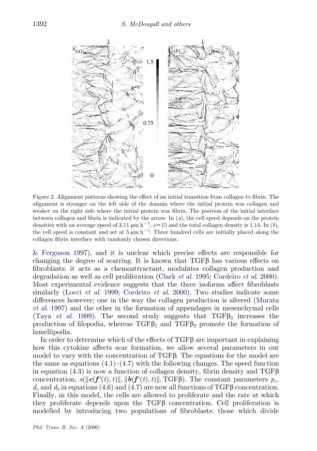
Examining transitions from a collagen-rich environment to one rich in fibringives some additional insight, since cells interact differently with each of theseproteins. Figure 2 shows that alignment in the collagen-rich region is significantlygreater than that observed in the initially fibrin-rich region. In addition, a layerof collagen oriented parallel to the initial protein boundary is evident.A simulation using constant cell speed exhibited similar effects.
The final orientation of the matrix is explained as follows. There are two waysin which the fibroblasts can interact with the fibrin and the collagen. Firstly, thecells reorient collagen but not fibrin, and secondly the cells move faster in fibrin.The fixed speed simulation (figure 2b) indicates that the difference in matrixorientation is not due to speed changes, but is due to the way in which cells reorientthe fibres. The fact that cells do not alter fibrin orientation means that its initialorientation (which was random in these simulations) will continue to affect thedirection of the cells until it is totally degraded. This has a randomizing effect andthe final orientation of the matrix is more random in the region that was initiallyfibrin-rich. The boundary layer is also a result of the randomizing influence of thefibrin, as the cells are more likely to turn when they enter the fibrin.
5. Modelling transforming growth factor-b in dermal wound repair
In this section, we describe an extension of the model designed to explore theeffects of transforming growth factor-b (TGFb) on wound healing (Dallon et al.2001). It has been demonstrated that either neutralizing TGFb1 and TGFb2 oradding TGFb3 will reduce the extent of scarring in a wound. TGFb alters thewound healing process in many ways however (Roberts & Sporn 1996; O’Kane
Phil. Trans. R. Soc. A (2006)
1.5
0.75
0
(a) (b)
Figure 2. Alignment patterns showing the effect of an initial transition from collagen to fibrin. Thealignment is stronger on the left side of the domain where the initial protein was collagen andweaker on the right side where the initial protein was fibrin. The position of the initial interfacebetween collagen and fibrin is indicated by the arrow. In (a), the cell speed depends on the proteindensities with an average speed of 3.11 mm hK1, sZ15 and the total collagen density is 1.14. In (b),the cell speed is constant and set at 5 mm hK1. Three hundred cells are initially placed along thecollagen fibrin interface with randomly chosen directions.
S. McDougall and others1392
& Ferguson 1997), and it is unclear which precise effects are responsible forchanging the degree of scarring. It is known that TGFb has various effects onfibroblasts: it acts as a chemoattractant, modulates collagen production anddegradation as well as cell proliferation (Clark et al. 1995; Cordeiro et al. 2000).Most experimental evidence suggests that the three isoforms affect fibroblastssimilarly (Locci et al. 1999; Cordeiro et al. 2000). Two studies indicate somedifferences however; one in the way the collagen production is altered (Murataet al. 1997) and the other in the formation of appendages in mesenchymal cells(Taya et al. 1999). The second study suggests that TGFb3 increases theproduction of filopodia, whereas TGFb1 and TGFb2 promote the formation oflamellipodia.
In order to determine which of the effects of TGFb are important in explaininghow this cytokine affects scar formation, we allow several parameters in ourmodel to vary with the concentration of TGFb. The equations for the model arethe same as equations (4.1)–(4.7) with the following changes. The speed functionin equation (4.3) is now a function of collagen density, fibrin density and TGFbconcentration, sðkcðf iðtÞ; tÞk; kbðf iðtÞ; tÞk;TGFbÞ. The constant parameters pc,dc and db in equations (4.6) and (4.7) are now all functions of TGFb concentration.Finally, in this model, the cells are allowed to proliferate and the rate at whichthey proliferate depends upon the TGFb concentration. Cell proliferation ismodelled by introducing two populations of fibroblasts: those which divide
Phil. Trans. R. Soc. A (2006)

1393Modelling collagen during wound repair
and those which do not. Dividing fibroblasts become non-dividing fibroblasts withtime. Each cell has a clock, not synchronized with the other cells, whichdetermines when it should divide. When a cell divides it loses its polarity and thedaughter cell is placed at a small distance (10 mm) from the mother cell.
Experimental evidence shows that the concentration of active TGFb peakswithin an hour after wounding, decreases to a level above normal within 1 day,peaks again 5 days after wounding and then decreases more gradually untilreaching approximately normal levels 14 days after wounding. Since fibroblastsdo not enter the wound until 24–48 h after wounding, we ignore the first peak inTGFb concentration and assume it to be a function of time as described earlier.
Simulations from this model suggest that the effect of TGFb on fibroblastspeed, proliferation and collagen production cannot explain its effect on scartissue formation (see figure 3a,b). However, the simulations suggest that theeffect on scarring could be explained if TGFb3 causes a cell to change directionmore frequently than TGFb1 and TGFb2. For example, if TGFb3 causes a cell tobe more responsive to matrix cues in determining its direction than TGFb1 andTGFb2, and if the three isoforms of TGFb bind competitively, our simulationsgive an explanation of the anti-scarring properties of TGFb3 which is consistentwith the experimental data.
6. Extending the model to include a diffusible chemoattractant
The above models of Dallon et al. consider the wound space as an isolated region,with fibroblasts entering through the base and the sides in a predetermined way.In reality, the wound resides within intact, uninjured skin, and the fibroblastsenter the wound by migrating from this surrounding tissue. It is relativelystraightforward to extend the model to include a band of normal tissue aroundthe wound. One simply makes the computational domain larger, and specifies aninitial region as wounded (fibrin-based ECM and no fibroblasts), with theremaining tissue unwounded (collagen-based ECM, with a resident fibroblastpopulation). However, numerical solutions of the model on such an enlargeddomain do not predict healing on a reasonable time-scale (figure 4). Themovement of fibroblasts into the wound region is extremely slow—many timesslower than the rates imposed by Dallon et al. As a result, the repair processoccurs at a rate many times slower than that observed in practice.
The slow progress of repair in simulations such as that shown in figure 4 occursbecause fibroblasts only enter the wound region as part of their intrinsicmovement in normal tissue unbiased by chemoattractants present in woundhealing. In reality, fibroblast movement is actively directed towards the woundduring healing by a range of chemical cues. For example, PDGF, IL-1b and TNF-a are all produced by white blood cells during the inflammatory phase of healing,and are established chemoattractants for fibroblasts (Kim et al. 1999; Sasakiet al. 2000).
To incorporate this directed movement into our model, we assume that asingle generic chemoattractant is produced in the wound and diffuses intosurrounding tissue; for simplicity, we assume the production rate to be uniformin the wound, with a decay rate that is the same in the wound and in thesurrounding tissue. This gives a simple equation for the chemoattractant
Phil. Trans. R. Soc. A (2006)

normal no TGFb anti-TGFb1
(a) (b) (c)
Figure 3. The collagen matrix for a simulation of normal wound healing with TGFb, resulting in ascar, is shown in (a). In (b), we show a simulation of a wound treated with an antibody for TGFbassuming TGFb alters cell proliferation, cell motility and flux, and collagen production. Thesimulation suggests that these effects do not explain the effects on scar tissue formation of TGFb.In (c), we show the results for a numerical experiment simulating anti-TGFb1 treatment, showinga more disordered collagen fibre pattern that is highly reminiscent of experimental data. The moredisordered matrix pattern is a key component of reduced scarring. The parameters for thissimulation are identical to (b), except that the cells reorient approximately every 9 min instead ofevery 27 min. The ability of a cell to change direction depends on the time discretization used whenintegrating equation (4.3). The collagen matrix is represented after 240 h of simulationcorresponding to about 12 days after wounding (the fibroblasts enter the wound region between24 and 48 h after wounding). Red indicates high density and dark blue indicates low collagen. Theline segments are streamlines for the collagen vector field. The region represents a cross-section of aslash wound with the surface of the skin at the top, normal tissue to the right and left and thebasement membrane at the bottom. When combining the three effects of TGFb, we assume thatthe cell proliferation increases at 90 pg mmK3 of TGFb and then decreases for higherconcentrations; for the motility we combine the decreased cell speed and the elevated flux at90 pg mmK3 of TGFb which decreases with increased concentrations of TGFb.
S. McDougall and others1394
concentration Aðx; tÞ of the form
vA=vt ZDAV2AKk1AC
k2 in the wound;
0 in the surrounding tissue:
(ð6:1Þ
Chemoattractant production begins during the inflammation phase of repair,several days earlier than the fibroblast activity at the centre of our model.Within this period, equation (6.1) effectively reaches an equilibrium, and weassume that the chemoattractant profile remains at this equilibrium throughoutthe dermal healing phase. A typical chemical profile is illustrated in figure 5.
We modify the cell movement rules to take account of this chemoattractant.We assume that both the speed and direction of the cell vary with the gradient
Phil. Trans. R. Soc. A (2006)

Figure 4. Solution of the model of Dallon et al. on an enlarged domain, which includes a region ofunwounded tissue around the wound. The solution shown is after 100 h of the proliferative phase ofhealing, which corresponds to about 7 days post-wounding. In the simulation, very little healinghas occurred, whereas in reality the wound would be significantly repaired by this time. Theseresults indicate that the model must be amended to include the directed movement of fibroblastsinto the wound. The model details and parameter values are as in §4, with aZ0:5, rZ0:5,sZ15 mm hK1, kZ5, pcZ0:64, dcZ0:44, dbZ0:6 and tZ0:15 h. Eight hundred fibroblasts areinitially resident in the unwounded dermis, and mitosis occurs on average every 18 h.
1395Modelling collagen during wound repair
VA of the chemoattractant. For the cell speed, we alter the form to
snew Z soldðkck; kbkÞð1CkVAk=kVAkmaxÞ:Here, sold is the speed assumed in the previous model, given in equation (4.3), andkVAkmax is the maximum value of kVAk across the domain. Thus, cell speed is anincreasing function of chemoattractant gradient. We assume that cell direction ismodified by the chemical gradient in a manner that is directly analogous to theaffects of collagen and fibrin densities; thus the cell direction is given by
vnew Zð1Kr2ÞvoldCr2VA=kVAk
k½ð1Kr2ÞvoldCr2VA=kVAk�k:
Here, the unit vector vold is the cell direction assumed in the previous model,given in equation (4.2). The quantity r2 reflects the dependence of cell directionon chemoattractant gradient, and we assume that it is an increasing, saturatingfunction of this gradient, with a Hill function form
r2 Z kVAka=½kVAkacritCkVAka�:Here, a and kVAkcrit are positive parameters.
Phil. Trans. R. Soc. A (2006)

Figure 5. A typical chemoattractant profile, as predicted by equation (6.1). The chemoattractantconcentration is greatest in the wound, and gradually decreases in the wound periphery. Theresultant chemical gradient directs fibroblasts into the wound region. The parameter values areDAZ0:5, k1Z2:0!10K4, k2Z0:025.
S. McDougall and others1396
One final change to the model is required. Dallon et al. assume that themajority of the fibroblasts enter the wound through its base, with only a minorityentering through the sides. This assumption is based on cell staining experimentsof Adams (1997), and reflects the significant increase in fibroblast density withdepth in unwounded dermis. Thus, the upper part of the dermis, near theepidermis, is relatively acellular, whereas the part near the fascia has a highdensity of fibroblasts. In the model, we simply impose a linear variation infibroblast density as an initial condition; the limited fibroblast movement withinnormal tissue means that the density gradient is maintained over the time-scaleof wound repair. The initial linear density gradient is achieved by inverting alinear probability distribution function of fibroblast density as a function ofdepth from the skin surface. Assuming no fibroblasts at the skin surface leads tothe following equation for determining the vertical coordinate ðyFBÞ of a givenfibroblast: yFBZymaxð1K
ffiffiffiffiffiffiffiffiffiffiffiffiffiffiffi1Krnd
pÞ where ymax corresponds to the base of the
simulation domain and rnd is a random number between 0 and 1.Figure 6 shows a typical solution of the extended model. We plot the collagen
profile both during the early stages of repair and after 100 h of the proliferativephase of healing (this corresponds to about 7 days post-wounding). A steadygradient of a chemoattractant is rapidly established, and this directs fibroblastsinto the wound, so that healing proceeds at a similar rate to that observed inpractice. As in the previous model, the fibroblasts break down the initial fibrin-based matrix and replace it with collagen. Initially, collagen fibres are laid down
Phil. Trans. R. Soc. A (2006)

(a) (b)
(c) (d)
(e) ( f )
Figure 6. (Caption overleaf.)
1397Modelling collagen during wound repair
Phil. Trans. R. Soc. A (2006)

Figure 6. (Overleaf.) A typical simulation of the extended model, showing wound repairorchestrated by a chemoattractant for fibroblasts, produced within the wound space. We show thecollagen (a,d), fibroblast (b,e) and fibrin (c,f ) profiles at (a–c) 25 and (d–f ) 100 h after the start ofthe proliferative phase of healing (which itself begins about 3 days after injury). The fibroblastsmove rapidly into the wound in response to the chemical gradient, break down the fibrin matrix,and replace it with collagen fibres whose alignment reflects the direction of fibroblast migration.The parameter values are DAZ0:5, k1Z2:0!10K4, k2Z0:025, kVAkcritZ0:5kVAkmax, aZ10.Other model details and parameter values are as in figure 3.
Figure 7. An illustration of the change in collagen profile with the steepness of the chemoattractantgradient. (a) A highly diffuse chemoattractant causes fibroblasts to move towards the wound, andthus reorient the collagen matrix, over a large region of unwounded tissue. (b) Conversely, a highlylocalized chemoattractant only affects the movement of fibroblasts close to the wound periphery.Therefore, in (a) the wound and normal tissue matrices are highly interwoven, whereas in (b), theyare relatively distinct, resulting in a much greater risk of rupture. The parameter values are as infigure 6 except for (a) DAZ2:0 and (b) DAZ0:1.
S. McDougall and others1398
in the direction of fibroblast movement, and this orientation pattern is reinforcedby the tendency of successive fibroblasts to move along the existing fibres. In thisextended model, fibroblast direction is also strongly influenced by thechemoattractant profile, and as a result the collagen fibre orientation patternreflects the gradient of chemoattractant. In figure 6, we also show the fibroblastdistribution. This illustrates the gradient of fibroblast density in unwoundedskin, and shows that this is reflected in the wound, with significantly higher cellnumbers at the base of the wound compared to the top.
Our extended model demonstrates that the rapid influx of fibroblasts into thewound space can be explained simply as a result of a chemoattractant gradient,coupled with the variation of fibroblast density with depth in unwounded dermis.The new model also enables the study of an important feature of wound repairthat is outside the scope of the model of Dallon et al., namely the extent to whichthe collagen matrix around the wound is altered during healing. In figure 6, there
Phil. Trans. R. Soc. A (2006)

Figure 8. An illustration of the dependence of wound collagen profile on the variation of fibroblastdensity with depth in unwounded dermis. In (a), we show the effect of assuming uniform fibroblastdensity in the dermis: there is a much more uniform deposition of collagen than in the referencesolution (figure 6). In (b), we use a fibroblast density that is heavily skewed towards the base of thedermis; in this case the collagen fibres in the wound are predominantly oriented perpendicular tothe skin surface because most fibroblasts enter the wound through its base. The model details andparameter values are as in figure 6, except for the fibroblast distribution. The total number ofdermal fibroblasts is the same in (a), (b) and in figure 6; in (b), the distribution of fibroblasts has20% of fibroblasts above the wound base, distributed as a linear function of depth in the dermis,and 80% below the wound base.
1399Modelling collagen during wound repair
is a clear reorientation of the collagen fibres around the wound, over a distance ofabout half the wound width (0.25 mm). This is caused by the fibroblastsremodelling the matrix as they move towards the wound. This reorientation ofthe existing collagen matrix results in an interdigitation of collagen fibres in
Phil. Trans. R. Soc. A (2006)

Figure 9. The effects of neglecting the dependence of fibroblast speed on chemoattractant gradient.Fewer fibroblasts enter the wound in comparison to the reference case (figure 6), and as a result therate of collagen deposition is lower, but the orientation pattern remains broadly similar. The modeldetails and parameter values are as in figure 6 except that the cell speed is given by soldðkck; kbkÞ.
S. McDougall and others1400
the scar and the surrounding tissue, which is a crucial factor for wound strength.By varying the parameters associated with the chemoattractant in our model, wehave found that the degree of interdigitation depends critically on the spread ofchemoattractant into the tissue around the wound. A very widely dispersedchemoattractant profile leads to fibroblast recruitment into the wound from alarge area of surrounding tissue, and a consequently high degree ofinterdigitation (figure 7). Similarly, a highly localized chemoattractant profilecauses the scar and the surrounding tissue to be poorly linked (figure 7).Therefore, our model makes the novel prediction that wound strength could beimproved by manipulation of the chemical gradients set up during theinflammatory phase of repair. Moreover, experimental measurements on theextent of interdigitation could be used to better constrain some of our modelparameters.
(a ) The role of fibroblast distribution
In figure 6, we have imposed a variation in fibroblast density with unwoundeddermis, with higher cell densities at greater depths. Such a variation occurs inpractice, but we are not aware of any quantitative data from which anappropriate functional form could be estimated. This makes it important tostudy the way in which the healing profile changes with the assumed fibroblastdistribution. In figure 8a, we show the effects of neglecting the cell density
Phil. Trans. R. Soc. A (2006)

Figure 10. The effects of neglecting the dependence of fibroblast direction on chemoattractantgradient. Significantly fewer fibroblasts enter the wound in comparison to the reference case(figure 6), and as a result the rate of collagen deposition is much lower. In addition, the orientationpattern is significantly altered, with a much more disordered appearance that is more reminiscentof unwounded dermis. The model details and parameter values are as in figure 6 except that the celldirection is given by vold.
1401Modelling collagen during wound repair
gradient and assuming a uniform distribution of fibroblasts in the dermis. Incomparison to the reference solution in figure 6, there is a much more uniformdeposition of collagen in the wound, and a higher degree of fibre alignmentparallel to the skin surface. This latter effect is a result of more cells entering thewound through the sides rather than the base. This alignment is greatly reducedif we assume that the fibroblast distribution is heavily skewed towards the lowerparts of the dermis (figure 8b). In this case, most of the cells entering the wounddo so through the base, simply because of the high cell number below the wound,and as a result the wound collagen at later times has a predominant orientationperpendicular to the skin surface.
(b ) The activity of the chemoattractant
The chemoattractant plays a key role in orchestrating fibroblast movement intothe wound. In our model, we assume that both the speed and direction of cellmovement are regulated by the chemoattractant gradient, and it is important tounderstand how these two separate effects conspire to produce the observed healingpattern. In figure 9, we show the collagen pattern given by neglecting thedependence of cell speed on chemoattractant gradient, so that only cell direction is
Phil. Trans. R. Soc. A (2006)
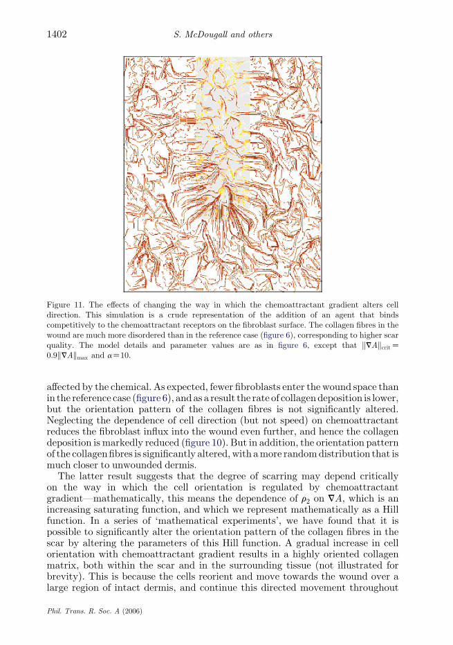
Figure 11. The effects of changing the way in which the chemoattractant gradient alters celldirection. This simulation is a crude representation of the addition of an agent that bindscompetitively to the chemoattractant receptors on the fibroblast surface. The collagen fibres in thewound are much more disordered than in the reference case (figure 6), corresponding to higher scarquality. The model details and parameter values are as in figure 6, except that kVAkcritZ0:9kVAkmax and aZ10.
S. McDougall and others1402
affected by the chemical. As expected, fewer fibroblasts enter the wound space thanin the reference case (figure 6), andas a result the rate of collagendeposition is lower,but the orientation pattern of the collagen fibres is not significantly altered.Neglecting the dependence of cell direction (but not speed) on chemoattractantreduces the fibroblast influx into the wound even further, and hence the collagendeposition is markedly reduced (figure 10). But in addition, the orientation patternof the collagen fibres is significantly altered,with amore randomdistribution that ismuch closer to unwounded dermis.
The latter result suggests that the degree of scarring may depend criticallyon the way in which the cell orientation is regulated by chemoattractantgradient—mathematically, this means the dependence of r2 on VA, which is anincreasing saturating function, and which we represent mathematically as a Hillfunction. In a series of ‘mathematical experiments’, we have found that it ispossible to significantly alter the orientation pattern of the collagen fibres in thescar by altering the parameters of this Hill function. A gradual increase in cellorientation with chemoattractant gradient results in a highly oriented collagenmatrix, both within the scar and in the surrounding tissue (not illustrated forbrevity). This is because the cells reorient and move towards the wound over alarge region of intact dermis, and continue this directed movement throughout
Phil. Trans. R. Soc. A (2006)

1403Modelling collagen during wound repair
the wound. Conversely, if the chemical gradient has little effect on the cellorientation when it is small, with a steep increase in the cell orientationresponse at higher gradients, the collagen pattern in the wound is much moredisordered (figure 11), corresponding to high scar quality. In figure 11, thefibroblasts close to the wound edge are directed into the wound by the highchemoattractant gradients at the wound edge. However, the chemical has littleeffect on fibroblast direction either far from the wound, or within thewound—because the chemical gradient is relatively low at both locations.Once in the wound, fibroblast movement is regulated mainly by ECMcomposition and orientation, and this results in a disordered scar matrix,such as that shown in figure 11.
7. Discussion
We have presented a multiscale modelling framework which allows us toanalyse the effects of different factors on collagen alignment and scarformation during dermal wound healing. From a therapeutic point of view,these results suggest an anti-scarring treatment that decreases the sensitivityof fibroblast reorientation to chemoattractant gradients. This could beachieved in practice by adding an agent that binds preferentially to thechemoattractant receptors on the cell surface. The anti-scarring agentmannose-6-phosphate acts via exactly this type of competitive inhibition,binding preferentially to TGFb receptors, thereby reducing the effectiveconcentration of TGFb (Ferguson & O’Kane 2004). It has been proposed thatthe lower degree of scarring that results from this treatment is due to changesin fibroblast motility (Taya et al. 1999; Dallon et al. 2001). Our results suggestthat in fact, changes in effective chemotactic activity of TGFb may also be animportant contribution to the anti-scarring effect. Moreover, we suggest thatcompetitive inhibition of the binding of other chemoattractants to dermalfibroblasts may represent a novel alternative or supplementary approach toanti-scarring therapy.
Editors’ note
Please see also related communications in this focussed issue by Byrne et al.(2006) and Lu et al. (2006).
J.A.S. was supported in part by an Advanced Research Fellowship from EPSRC.
References
Adams, J. J. 1997 The cell kinetics of murine incisional wound healing. Ph.D. thesis, University ofManchester.
Alberts, B., Johnson, A., Lewis, J., Raff, M., Roberts, K. & Walter, P. 2002 Cell junctions, celladhesion, and the extracellular matrix. In Molecular biology of the cell, pp. 978–986, 4th edn.New York: Garland Publishing.
Birk, D. E. & Trelstad, R. L. 1986 Extracellular compartments in tendon morphogenesis:collagen, fibril, bundle, and macroaggregate formation. J. Cell Biol. 103, 231–240. (doi:10.1083/jcb.103.1.231)
Phil. Trans. R. Soc. A (2006)

S. McDougall and others1404
Byrne, H. M., Alarcon, T., Owen, M. R., Webb, S. D. & Maini, P. K. 2006 Modelling aspects of
cancer dynamics: a review. Phil. Trans. R. Soc. A 364, 1563–1578. (doi:10.1098/rsta.2006.1786)
Clark, R. A. F. 1989 Wound repair. Curr. Opin. Cell Biol. 1, 1000–1008. (doi:10.1016/0955-
0674(89)90072-0)
Clark, R. A. F. 1996 Wound repair overview and general considerations. In The molecular and
cellular biology of wound repair (ed. R. A. F. Clark), pp. 3–50, 2nd edn. New York: Plenum
Press.
Clark, P., Connolly, P., Curtis, A. S. G. & Wilkinson, C. C. W. 1990 Topographical control of cell
behaviour. II. Multiple grooved substrata. Development 108, 635–644.
Clark, R. A. F., Nielsen, L. D., Welch, M. P. & McPherson, J. M. 1995 Collagen matrices attenuate
the collagen-synthetic response of cultured fibroblasts to TGF-b. J. Cell Sci. 108, 1251–1261.
Cordeiro, M. F., Bhattacharya, S. S., Schultz, G. S. & Khaw, P. T. 2000 Tgf-b1, -b2, and -b3
in vitro: biphasic effects on tenon’s fibroblast contraction, proliferation, and migration. Invest.
Ophthalmol. Vis. Sci. 41, 756–763.
Dallon, J. C., Sherratt, J. A. & Maini, P. K. 1999 Mathematical modelling of extracellular matrix
dynamics using discrete cells: fiber orientation and tissue regeneration. J. Theor. Biol. 199,
449–471. (doi:10.1006/jtbi.1999.0971)
Dallon, J. C., Sherratt, J. A., Maini, P. K. & Ferguson, M. W. J. 2000 Biological implications of a
discrete mathematical model for collagen deposition and alignment in dermal wound repair.
IMA J. Math. Appl. Med. Biol. 17, 379–393.
Dallon, J. C., Sherratt, J. A. & Maini, P. K. 2001 Modeling the effects of transforming growth
factor-b on extracellular matrix alignment in dermal wound repair. Wound Repair Regen. 9,
278–286. (doi:10.1046/j.1524-475X.2001.00278.x)
Doillon, C. J., Dunn, M. G., Bert, R. A. & Silver, F. H. 1985 Collagen deposition during wound
repair. Scanning Electron Microsc. 2, 897–903.
Ehrlich, P. H. & Krummel, T. M. 1996 Regulation of wound healing from a connective tissue
perspective. Wound Repair Regen. 4, 203–210. (doi:10.1046/j.1524-475X.1996.40206.x)
Ferguson, M. W. J. & O’Kane, S. 2004 Scar-free healing: from embryonic mechanisms to adult
therapeutic intervention. Phil. Trans. R. Soc. B 359, 839–850. (doi:10.1098/rstb.2004.1475)
Forrest, L. 1983 Current concepts in soft connective tissue wound healing. Br. J. Surg. 70, 133–140.
Friedl, P., Zanker, K. S. & Brocker, E. B. 1998 Cell migration strategies in 3-d extracellular
matrix: differences in morphology, cell matrix interactions, and integrin function. Microsc. Res.
Tech. 43, 369–378. (doi:10.1002/(SICI)1097-0029(19981201)43:5!369::AID-JEMT3O3.0.CO;2-6)
Guido, S. & Tranquillo, R. T. 1993 A methodology for the systematic and quantitative study of cell
contact guidance in oriented collagen gels. J. Cell Sci. 105, 317–331.
Hay, E. D. (ed.) 1991 Cell biology of extacellular matrix, 2nd edn. New York: Plenum Press.
Hsieh, P. & Chen, L. B. 1983 Behavior of cells seeded on isolated fibronectin matrices. J. Cell Biol.
96, 1208–1217. (doi:10.1083/jcb.96.5.1208)
Jennings, R. W. & Hunt, T. K. 1992 Overview of postnatal wound healing. In Fetal wound healing
(ed. N. S. Adzick & M. T. Longaker), pp. 25–52. New York: Elsevier.
Kim, W., Mohan, R., Mohan, R. & Wilson, S. 1999 Effect of pdgf, il-1 alpha, and bmp2/4 on
corneal fibroblast chemotaxis: expression of the platelet-derived growth factor system in the
cornea. Invest. Ophthalmol. Vis. Sci. 40, 1364–1372.
Locci, P., Baroni, T., Lilli, C., Martinese, D., Marinucci, L., Bellocchio, S., Calvitti, M. &
Becchetti, E. 1999 TGFb and TGFa, antagonistic effect in vitro on extracellular matrix
accumulation by chick skin fibroblasts at two distinct embryonic stages. Int. J. Dev. Biol. 43,
157–165.
Lu, Y., Parker, K. H. & Wang, W. 2006 Effects of osmotic pressure in the extracellular matrix on
tissue deformation. Phil. Trans. R. Soc. A 364, 1407–1422. (doi:10.1098/rsta.2006.1778)
McCallion, R. L. & Ferguson, M. W. J. 1996 Fetal wound healing and the development of
antiscarring therapies for adult wound healing. In The molecular and cellular biology of wound
repair (ed. R. A. F. Clark), pp. 561–600, 2nd edn. New York: Plenum Press.
Phil. Trans. R. Soc. A (2006)

1405Modelling collagen during wound repair
Murata, H., Zhou, L., Ochoa, S., Hasan, A., Badiavas, E. & Falanga, V. 1997 TGF-b3 stimulatesand regulates collagen synthesis through TGF-b1-dependent and independent mechanisms.J. Invest. Dermatol. 108, 258–262. (doi:10.1111/1523-1747.ep12286451)
O’Kane, S. & Ferguson, M. W. J. 1997 Transforming growth factor bs and wound healing. Int.J. Biochem. Cell Biol. 29, 63–78. (doi:10.1016/S1357-2725(96)00120-3)
Roberts, A. B. & Sporn, M. B. 1996 Transforming growth factor-b. In The molecular and cellularbiology of wound repair (ed. R. A. F. Clark), pp. 275–308, 2nd edn. New York: Plenum Press.
Sasaki, M., Kashima, M., Ito, T., Watanabe, A., Izumiyama, N., Sano, M., Kagaya, M., Shioya, T.& Miura, M. 2000 Differential regulation of metalloproteinase production, proliferation andchemotaxis of human lung fibroblasts by pdgf, interleukin-1 beta and TNF-alpha. MediatorsInflamm. 9, 155–160. (doi:10.1080/09629350020002895)
Taya, Y., O’Kane, S. & Ferguson, M. W. J. 1999 Pathogenesis of cleft palate in TGF-b3 knockoutmice. Development 126, 3869–3879.
Whitby, D. J. & Ferguson, M. W. J. 1991 The extracellular matrix of lip wounds in fetal, neonataland adult mice. Development 112, 651–668.
Wojciak-Stothard, B., Denyer, M., Mishra, M. & Brown, R. A. 1997 Adhesion orientation, andmovement of cells cultured on ultrathin fibronectin fibers. In Vitro Cell. Dev. Biol. 33, 110–117.
Xu, J. & Clark, R. A. F. 1996 Extracellular matrix alters PDGF regulation of fibroblast integrins.J. Cell Biol. 132, 239–249. (doi:10.1083/jcb.132.1.239)
Phil. Trans. R. Soc. A (2006)