Fibroblast growth factor receptor influences primary ... · Fibroblast growth factor receptor...
10
Fibroblast growth factor receptor influences primary cilium length through an interaction with intestinal cell kinase Michaela Kunova Bosakova a , Alexandru Nita a , Tomas Gregor b , Miroslav Varecha a,c , Iva Gudernova a , Bohumil Fafilek a,c , Tomas Barta d , Neha Basheer a , Sara P. Abraham a , Lukas Balek a , Marketa Tomanova a , Jana Fialova Kucerova a , Juraj Bosak a , David Potesil b , Jennifer Zieba e , Jieun Song f , Peter Konik g , Sohyun Park h , Ivan Duran e , Zbynek Zdrahal b , David Smajs a , Gert Jansen i , Zheng Fu h , Hyuk Wan Ko f , Ales Hampl c,d , Lukas Trantirek b , Deborah Krakow e,j,k,1 , and Pavel Krejci a,c,l,1 a Department of Biology, Faculty of Medicine, Masaryk University, 62500 Brno, Czech Republic; b Central European Institute of Technology, Masaryk University, 62500 Brno, Czech Republic; c International Clinical Research Center, St. Anne’s University Hospital, 65691 Brno, Czech Republic; d Department of Histology and Embryology, Faculty of Medicine, Masaryk University, 62500 Brno, Czech Republic; e Department of Orthopaedic Surgery, David Geffen School of Medicine University of California, Los Angeles, CA 90095; f Department of Biochemistry, College of Life Science and Biotechnology, Yonsei University, 03722 Seoul, Korea; g Institute of Chemistry and Biochemistry, Faculty of Science, University of South Bohemia, 37005 Ceske Budejovice, Czech Republic; h Department of Pharmacology, University of Virginia School of Medicine, Charlottesville, VA 22908; i Department of Cell Biology, Erasmus Medical Center, 3000 CA Rotterdam, The Netherlands; j Human Genetics, David Geffen School of Medicine, University of California, Los Angeles, CA 90095; k Obstetrics and Gynecology, David Geffen School of Medicine, University of California, Los Angeles, CA 90095; and l Institute of Animal Physiology and Genetics, Czech Academy of Sciences, 60200 Brno, Czech Republic Edited by Roeland Nusse, Stanford University School of Medicine, Stanford, CA, and approved January 22, 2019 (received for review January 8, 2018) Vertebrate primary cilium is a Hedgehog signaling center but the extent of its involvement in other signaling systems is less well understood. This report delineates a mechanism by which fibro- blast growth factor (FGF) controls primary cilia. Employing pro- teomic approaches to characterize proteins associated with the FGF-receptor, FGFR3, we identified the serine/threonine kinase intestinal cell kinase (ICK) as an FGFR interactor. ICK is involved in ciliogenesis and participates in control of ciliary length. FGF signaling partially abolished ICK’s kinase activity, through FGFR- mediated ICK phosphorylation at conserved residue Tyr15, which interfered with optimal ATP binding. Activation of the FGF signaling pathway affected both primary cilia length and function in a manner consistent with cilia effects caused by in- hibition of ICK activity. Moreover, knockdown and knockout of ICK rescued the FGF-mediated effect on cilia. We provide con- clusive evidence that FGF signaling controls cilia via interaction with ICK. fibroblast growth factor | FGFR | intestinal cell kinase | ICK | cilia length I n vertebrates, the signaling of Hedgehog (Hh) morphogens depends entirely on primary cilium. The cilia provide a struc- tural and functional compartment that integrates a series of in- tricate molecular mechanisms allowing cells to process Hh-target transcriptional regulators and alter gene-expression programs in response to Hh (1). Giving the rapidly growing importance of primary cilia in the regulation of physiologic and pathologic cellular functions (2), many other signaling systems are antici- pated to work through the cilia. Several recent lines of evidence demonstrate that fibroblast growth factors (FGF) regulate primary cilia. Inactivation of the FGF-receptor Fgfr1 or its FGF ligands lead to shorter cilia in zebrafish and Xenopus (3). In mammals, FGF signaling regu- lates the length of primary cilia in skin fibroblasts, lung, kidney, and liver cells, human embryonic stem cells and human induced pluripotent stem cells, embryonal fibroblasts, and mesenchymal cells (4). In addition, the human skeletal dysplasias caused by activating FGFR3 mutations, such as achondroplasia, manifest by abnormal cilia (4, 5). Evidence strongly suggests that FGF signaling integrates cilia into the canonical FGF signaling pathway. However, the mechanism through which FGFs regu- late primary cilia is not known. Several serine/threonine kinases control ciliogenesis or other specific functions of primary cilia. These “ciliary kinases” include TTBK2 and GSK3β, involved in initiation of ciliogenesis and assembly of the ciliary membrane (6, 7), NEK2, which regu- lates cilia disassembly (8), and CK1 and GRK2, which are important for Smoothened (SMO) translocation into the cilia (9). The MAP-kinase superfamily kinase intestinal cell kinase (ICK) is another well-known regulator of primary cilia, con- served in this function from single-cell organisms to mammals. Deletion of ICK or its homologs increases the cilia length in green algae, protists, and nematodes in vivo (10–12). In cul- tured mammalian cells, down-regulations of ICK kinase ac- tivity lead to extended and abnormal cilia, demonstrating that ICK is an essential regulator of the length of primary cilia (13–16). Significance A properly functioning primary cilium is prerequisite for both normal development and aging of all ciliated organisms, in- cluding humans. In vertebrates, the signaling of Hedgehog family morphogens depends entirely on primary cilium. Re- cently, we reported that fibroblast growth factors (FGF) sig- naling interacts with that of Hedgehog, and that this is a consequence of FGF regulating length of the cilium and speed of processes that happen therein. In this report, we provide a molecular mechanism of such interaction, identifying intestinal cell kinase as a mediator of the FGF-induced changes in the ciliary morphology and function. This expands our un- derstanding how FGF signaling regulates intracellular pro- cesses, and how aberrant FGF signaling contributes to diseases, such as achondroplasia and cancer. Author contributions: M.K.B. and P. Krejci designed research; M.K.B., A.N., T.G., M.V., I.G., B.F., T.B., N.B., S.P.A., L.B., M.T., J.F.K., J.B., D.P., J.Z., J.S., P. Konik, S.P., I.D., Z.Z., D.S., G.J., Z.F., H.W.K., A.H., and L.T. performed research; and M.K.B., D.K., and P. Krejci wrote the paper. The authors declare no conflict of interest. This article is a PNAS Direct Submission. This open access article is distributed under Creative Commons Attribution-NonCommercial- NoDerivatives License 4.0 (CC BY-NC-ND). 1 To whom correspondence may be addressed. Email: [email protected] or [email protected]. This article contains supporting information online at www.pnas.org/lookup/suppl/doi:10. 1073/pnas.1800338116/-/DCSupplemental. Published online February 19, 2019. 4316–4325 | PNAS | March 5, 2019 | vol. 116 | no. 10 www.pnas.org/cgi/doi/10.1073/pnas.1800338116 Downloaded by guest on January 6, 2020
Transcript of Fibroblast growth factor receptor influences primary ... · Fibroblast growth factor receptor...

Fibroblast growth factor receptor influences primarycilium length through an interaction with intestinalcell kinaseMichaela Kunova Bosakovaa, Alexandru Nitaa, Tomas Gregorb, Miroslav Varechaa,c, Iva Gudernovaa, Bohumil Fafileka,c,Tomas Bartad, Neha Basheera, Sara P. Abrahama, Lukas Baleka, Marketa Tomanovaa, Jana Fialova Kucerovaa,Juraj Bosaka, David Potesilb, Jennifer Ziebae, Jieun Songf, Peter Konikg, Sohyun Parkh, Ivan Durane, Zbynek Zdrahalb,David Smajsa, Gert Janseni, Zheng Fuh, Hyuk Wan Kof, Ales Hamplc,d, Lukas Trantirekb, Deborah Krakowe,j,k,1,and Pavel Krejcia,c,l,1
aDepartment of Biology, Faculty of Medicine, Masaryk University, 62500 Brno, Czech Republic; bCentral European Institute of Technology, MasarykUniversity, 62500 Brno, Czech Republic; cInternational Clinical Research Center, St. Anne’s University Hospital, 65691 Brno, Czech Republic; dDepartmentof Histology and Embryology, Faculty of Medicine, Masaryk University, 62500 Brno, Czech Republic; eDepartment of Orthopaedic Surgery, David GeffenSchool of Medicine University of California, Los Angeles, CA 90095; fDepartment of Biochemistry, College of Life Science and Biotechnology, YonseiUniversity, 03722 Seoul, Korea; gInstitute of Chemistry and Biochemistry, Faculty of Science, University of South Bohemia, 37005 Ceske Budejovice, CzechRepublic; hDepartment of Pharmacology, University of Virginia School of Medicine, Charlottesville, VA 22908; iDepartment of Cell Biology, ErasmusMedical Center, 3000 CA Rotterdam, The Netherlands; jHuman Genetics, David Geffen School of Medicine, University of California, Los Angeles, CA90095; kObstetrics and Gynecology, David Geffen School of Medicine, University of California, Los Angeles, CA 90095; and lInstitute of Animal Physiologyand Genetics, Czech Academy of Sciences, 60200 Brno, Czech Republic
Edited by Roeland Nusse, Stanford University School of Medicine, Stanford, CA, and approved January 22, 2019 (received for review January 8, 2018)
Vertebrate primary cilium is a Hedgehog signaling center but theextent of its involvement in other signaling systems is less wellunderstood. This report delineates a mechanism by which fibro-blast growth factor (FGF) controls primary cilia. Employing pro-teomic approaches to characterize proteins associated with theFGF-receptor, FGFR3, we identified the serine/threonine kinaseintestinal cell kinase (ICK) as an FGFR interactor. ICK is involvedin ciliogenesis and participates in control of ciliary length. FGFsignaling partially abolished ICK’s kinase activity, through FGFR-mediated ICK phosphorylation at conserved residue Tyr15,which interfered with optimal ATP binding. Activation of theFGF signaling pathway affected both primary cilia length andfunction in a manner consistent with cilia effects caused by in-hibition of ICK activity. Moreover, knockdown and knockout ofICK rescued the FGF-mediated effect on cilia. We provide con-clusive evidence that FGF signaling controls cilia via interactionwith ICK.
fibroblast growth factor | FGFR | intestinal cell kinase | ICK | cilia length
In vertebrates, the signaling of Hedgehog (Hh) morphogensdepends entirely on primary cilium. The cilia provide a struc-
tural and functional compartment that integrates a series of in-tricate molecular mechanisms allowing cells to process Hh-targettranscriptional regulators and alter gene-expression programs inresponse to Hh (1). Giving the rapidly growing importance ofprimary cilia in the regulation of physiologic and pathologiccellular functions (2), many other signaling systems are antici-pated to work through the cilia.Several recent lines of evidence demonstrate that fibroblast
growth factors (FGF) regulate primary cilia. Inactivation of theFGF-receptor Fgfr1 or its FGF ligands lead to shorter cilia inzebrafish and Xenopus (3). In mammals, FGF signaling regu-lates the length of primary cilia in skin fibroblasts, lung, kidney,and liver cells, human embryonic stem cells and human inducedpluripotent stem cells, embryonal fibroblasts, and mesenchymalcells (4). In addition, the human skeletal dysplasias caused byactivating FGFR3 mutations, such as achondroplasia, manifestby abnormal cilia (4, 5). Evidence strongly suggests that FGFsignaling integrates cilia into the canonical FGF signalingpathway. However, the mechanism through which FGFs regu-late primary cilia is not known.Several serine/threonine kinases control ciliogenesis or other
specific functions of primary cilia. These “ciliary kinases” include
TTBK2 and GSK3β, involved in initiation of ciliogenesis andassembly of the ciliary membrane (6, 7), NEK2, which regu-lates cilia disassembly (8), and CK1 and GRK2, which areimportant for Smoothened (SMO) translocation into the cilia(9). The MAP-kinase superfamily kinase intestinal cell kinase(ICK) is another well-known regulator of primary cilia, con-served in this function from single-cell organisms to mammals.Deletion of ICK or its homologs increases the cilia length ingreen algae, protists, and nematodes in vivo (10–12). In cul-tured mammalian cells, down-regulations of ICK kinase ac-tivity lead to extended and abnormal cilia, demonstrating thatICK is an essential regulator of the length of primary cilia(13–16).
Significance
A properly functioning primary cilium is prerequisite for bothnormal development and aging of all ciliated organisms, in-cluding humans. In vertebrates, the signaling of Hedgehogfamily morphogens depends entirely on primary cilium. Re-cently, we reported that fibroblast growth factors (FGF) sig-naling interacts with that of Hedgehog, and that this is aconsequence of FGF regulating length of the cilium and speedof processes that happen therein. In this report, we provide amolecular mechanism of such interaction, identifying intestinalcell kinase as a mediator of the FGF-induced changes in theciliary morphology and function. This expands our un-derstanding how FGF signaling regulates intracellular pro-cesses, and how aberrant FGF signaling contributes to diseases,such as achondroplasia and cancer.
Author contributions: M.K.B. and P. Krejci designed research; M.K.B., A.N., T.G., M.V., I.G.,B.F., T.B., N.B., S.P.A., L.B., M.T., J.F.K., J.B., D.P., J.Z., J.S., P. Konik, S.P., I.D., Z.Z., D.S., G.J.,Z.F., H.W.K., A.H., and L.T. performed research; and M.K.B., D.K., and P. Krejci wrotethe paper.
The authors declare no conflict of interest.
This article is a PNAS Direct Submission.
This open access article is distributed under Creative Commons Attribution-NonCommercial-NoDerivatives License 4.0 (CC BY-NC-ND).1To whom correspondence may be addressed. Email: [email protected] [email protected].
This article contains supporting information online at www.pnas.org/lookup/suppl/doi:10.1073/pnas.1800338116/-/DCSupplemental.
Published online February 19, 2019.
4316–4325 | PNAS | March 5, 2019 | vol. 116 | no. 10 www.pnas.org/cgi/doi/10.1073/pnas.1800338116
Dow
nloa
ded
by g
uest
on
Janu
ary
6, 2
020

As the activity of kinases is frequently modulated bytransphosphorylation by unrelated kinases, the ciliary kinasesrepresent potential sites of interaction of primary cilia withother signaling systems. In this study, we describe onesuch mechanism. We unravel how FGF signaling regulatesprimary cilia length, leading to direct downstream conse-
quences. Using proteomics to characterize the FGFR3 inter-actome in cells, we identified ICK as an FGFR interactor (17).Here, we demonstrate that FGFRs phosphorylate ICKand partially suppress ICK kinase activity and thus employICK to regulate the length and function of primary ciliain cells.
Fig. 1. FGFRs interact with ICK, MAK, and CCRK. (A) IP of FLAG-tagged ICK with V5-tagged wild-type (WT) FGFR3 or activating FGFR3 mutant K650M in293T cells, or (B and C) FLAG-tagged MAK or CCRK with V5-tagged wild-type FGFR3 or FGFR3-K650M in 293T cells. Actin serves as a loading control. (D) IP ofICK with FGFR1, FGFR2, and FGFR4 demonstrating the ICK association with FGFR1 and FGFR4 but not FGFR2. (E) Wild-type NIH 3T3 cells were transfected withFLAG-tagged ICK together with V5-tagged FGFR3; IckFlag NIH 3T3 cells were transfected only with V5-tagged FGFR3. The antibodies against protein tags wereused in the PLA (red); FGFR3 antibody was used to counterstain the transfected cells (green). As a negative control, cells were transfected with FGFR3 and anempty vector (WT), or by GFP (WT and IckFlag). Numbers of PLA dots per cell were calculated and plotted (Student’s t test, ***P < 0.001). (Scale bars, 10 μm.)Two clones of IckFlag NIH 3T3 cells, B11, and E5, were analyzed. (F) IP of endogenous FLAG-tagged ICK with endogenous FGFR1 in IckFlag NIH 3T3 cells; actinserves as a loading control. (G–I) Endogenous ICK forms a complex with endogenous FGFR1 in NIH 3T3 cells. (G) Scheme of the procedure, comprising ul-tracentrifugation, BN-PAGE, SDS/PAGE, and Western blot. (H) Cofractionalization of FGFR1 and ICK-FLAG in IckFlag(B11) NIH 3T3 cells (*). WT NIH 3T3 cells wereused as a control. (I) Native complexes (fractions #6 and/or #7) were separated using BN-PAGE, followed by second dimension SDS/PAGE. Orange box showsseparation of the ∼669-kDa complex containing FGFR1 and ICK-FLAG (arrow).
Kunova Bosakova et al. PNAS | March 5, 2019 | vol. 116 | no. 10 | 4317
CELL
BIOLO
GY
Dow
nloa
ded
by g
uest
on
Janu
ary
6, 2
020

Results and DiscussionFGFR1, -3, and -4, but Not FGFR2, Interacts with ICK. Tandem mass-spectrometry (MS) was used to identify novel FGFR3 interactorsamong proteins coimmunoprecipitated (co-IP) with FGFR3from cells, or among phosphotyrosine proteins isolated fromcells with activated FGFR3 signaling. In a total of 26 experi-ments carried out in 293T cells overexpressing FGFR3, ICKand its homolog male germ cell-associated kinase (MAK) werefound in 10 (38%) and 12 (46%) of experiments, respectively(17). Additionally, the ICK-activating kinase, CCRK (18), wasidentified in 10 (38%) experiments. The ICK association withFGFR3 was confirmed by co-IPs of wild-type FGFR3 and ICKexpressed in 293T cells (Fig. 1A). The active FGFR3 mutantK650M that associates with thanatophoric dysplasia (19) alsocoimmunoprecipitates with ICK. FGFR3 coimmunoprecipitateswith MAK and CCRK (Fig. 1 B and C). ICK coimmunoprecipi-tates with FGFR1 and FGFR4; no association with FGFR2 wasfound (Fig. 1D).Because 293T cells do not form cilia, we asked if ICK interacts
with FGFR3 in ciliated NIH 3T3 cells. Expressed V5-taggedFGFR3 and FLAG-tagged ICK interacted in intact NIH 3T3cells by proximity ligation assay (PLA) (Fig. 1E). Next, we usedCRISPR/Cas9 to insert FLAG epitope into the Ick locus inNIH 3T3 cells, to generate cells expressing C-terminally 3xFLAG-tagged endogenous ICK (IckFlag cells). PLA showed interaction ofendogenous ICK with expressed FGFR3 in two independentIckFlag clones (Fig. 1E). Importantly, IP of endogenous FGFR1from IckFlag cells demonstrated that endogenous ICK interactswith endogenous FGFR1 (Fig. 1F). In native lysates of IckFlag
cells separated at 5–25% sucrose gradients, a cofractionation ofFGFR1 with ICK was observed (Fig. 1 G and H). Fractions richin both FGFR1 and ICK were resolved by blue-native (BN)-PAGE to separate protein complexes, which were then ana-lyzed by second-dimension SDS/PAGE to obtain their indi-vidual components. Immunoblotting revealed an ∼669-kDa proteincomplex containing FGFR1 and ICK (Fig. 1I).To further characterize the FGFR–ICK interaction, we gen-
erated a series of FGFR3 constructs with truncations in theirintracellular domain (Fig. 2A). Mutated variants of FGFR3 werealso prepared, by targeting Y724 in the tyrosine kinase (TK)domain, and Y760 and Y770 both located in the C-terminal tail.The Y724/Y760/Y770 mediate interaction with signaling inter-mediates SH2-βB, p85 PI3K, PLCγ, and GRB14 (20–22). Thebinding site for the FRS2 adapter was also targeted, either byreplacing P418, L419, R425, and V427 with alanines, or by re-moving the entire region implicated in the interaction (aminoacids 406–427) (23). FGFR3 variants were coexpressed with ICKin 293T cells, and analyzed by co-IP. All FGFR3 variants withdeleted C terminus did not interact with ICK (Fig. 2B). Similarly,FGFR3-Y724F and -3YF (containing triple substitution Y724F/Y760F/Y770F) did not interact, despite having intact C termini.Deletion of FRS2 binding site had no effect on FGFR3 in-teraction with ICK, similar to Y760F or Y770F substitutions,which coimmunoprecipitated with ICK normally.Fig. 2 shows that both the C terminus of FGFR3 and Y724
are required for ICK binding. To characterize in detail theFGFR3 epitopes involved in interaction with ICK, a peptidemicroarray technology was used. Short peptides (7–22 aa)
Fig. 2. FGFR3 interacts with ICK via C terminus andY724. (A) Truncated or mutated FGFR3 variantsprepared for this study. Positions of point-mutatedresidues are indicated in red. (B) IP of FGFR3 withFLAG-tagged ICK in 293T cells. Actin serves as load-ing control. FGFR3 variants lacking C terminus orcarrying the Y724F mutation did not co-IP with ICK.Arrows indicate FGFR3.
4318 | www.pnas.org/cgi/doi/10.1073/pnas.1800338116 Kunova Bosakova et al.
Dow
nloa
ded
by g
uest
on
Janu
ary
6, 2
020

covering the intracellular part of FGFR3 were synthetized andimmobilized on a glass slide, incubated with recombinant ICK, andthe peptide–ICK interaction was analyzed as detailed in Materialsand Methods. Strong ICK binding was obtained for the FGFR3peptide 740TFKQLVEDLDRVLTVTSTDEY760, located at theboundary between the TK domain and the C terminus (SI Appendix,Fig. S1A). Interestingly, shorter versions of this peptide, specifically748LDRVLTVTSTDEY760 and 740TFKQLVEDLDRVLTV754,did not show any ICK binding (Fig. 3A and SI Appendix, Fig. S1B).Crystal structure of the FGFR3 TK domain (PDB ID code 4K33)shows that 740TFKQLVEDLDRVLTV754 forms an α-helix fol-lowed by an intrinsically disordered 755TSTDEY760 sequence (Fig.3B). It is thus possible that ICK binds only to a correctly as-sembled secondary structure in FGFR3, and not to the pep-tides lacking either the helical or the unstructured parts of the740TFKQLVEDLDRVLTVTSTDEY760 motif. This is supportedby the IP data, where the C-lobe of the TK domain alone or the Cterminus alone did not interact with ICK (Fig. 2B) (FGFR3-ΔC-tand FGFR3-ΔTK). Next, we asked whether differences in the se-quence of FGFR3 motif involved in ICK interaction could accountfor the lack of FGFR2 association with ICK, observed in co-IPexperiments (Fig. 1D). We replaced the 751VLTVTSTDEY760
in FGFR3 with homologous FGFR2 sequence 760ILTLTTNEEY769,and determined the interaction of chimeric FGFR3 (FGFR3-R2-C-t) with ICK using co-IP. Fig. 3C demonstrates that FGFR3-R2-C-t capacity to co-IP with ICK diminished by ∼40%, com-pared with the wild-type FGFR3.Our data indicate that the Y724 and C terminus of the FGFR3 are
both essential for ICK binding; FGFR3-Y724F has an intact C ter-minus but does not bind ICK. Similarly, the FGFR3 constructs with adeleted C terminus did not bind ICK, despite having the Y724 intact(Fig. 2B). Thus, the ICK binding to either site is rather weak andcooperativity between these two sites is required in the context of the3D FGFR3 structure (Fig. 3B). Alternatively, the Y724 mediatesICK binding indirectly, acting as an allosteric element controllingaccessibility of the C terminus for ICK.
FGF Signaling Triggers Cytoplasmic Accumulation of ICK. The asso-ciation of wild-type FGFR3 and FGFR3-K650M with ICK wasconfirmed by PLA, carried out with 293T cells expressing
V5-tagged FGFR3 and FLAG-tagged ICK (Fig. 4A). Immu-nocytochemistry revealed an overlap of FGFR3 signal withICK in the cytoplasm, which contrasted with predominantnuclear ICK localization in cells not expressing FGFR3 (Fig.4B), suggesting that FGFRs could cause ICK’s retention inthe cytoplasm. We tested this hypothesis by determining theICK localization in 293T cells, where the signaling of endog-enous FGFR1–4 (24) was activated by addition of FGFR li-gand FGF2. Progressive cytoplasmic accumulation of ICK wasfound in cells treated with FGF2 (Fig. 4 C and D). Expressionof active FGFR3-K650E or -K650M also retained ICK in thecytoplasm (Fig. 4E). Thus, the activation of FGF signalingalters subcellular localization of ICK, causing its cytoplasmicaccumulation.
FGFRs Phosphorylate ICK and Inhibit ICK Kinase Activity. ICK shut-tles between the cytoplasm and nucleus (25) and this is affectedby kinase activity, as demonstrated by cytoplasmic accumulationof the partially inactive ICK mutant R272Q (26). Because FGFsignaling altered ICK subcellular localization (Fig. 4), we askedwhether it affected ICK activity through phosphorylation. Kinaseassays utilizing recombinant FGFR3 and ICK revealed thatFGFR3 phosphorylated ICK at tyrosine residues (Fig. 5A, lane4). Kinase assays carried out with recombinant MAK and CCRKyielded similar results (Fig. 5 B and C, lane 4). Phosphotyrosinemapping identified several ICK tyrosines phosphorylated byFGFR3, among which Y15, Y156, Y495, and Y555 are con-served in ICK and MAK in human, mouse, chick, Xenopus, andzebrafish (Fig. 5D). Y495 and Y555 localized to an unstructuredregulatory region of ICK (amino acids 320–632 for human ICK),making it impossible to predict the effect of their phosphoryla-tion on ICK function. In contrast, the Y15 lies within the highlystructured ICK kinase domain. In silico modeling revealed thatphosphorylation at Y15 positions a negatively charged phosphatemoiety in immediate proximity to the pocket used for binding ofan ATP phosphate group, suggesting that phosphorylation atY15 down-regulates ICK kinase activity via interference withoptimal ATP binding (Fig. 5D). We tested this prediction bydetermining the kinase activity of ICK, immunopurified from293T cells in which endogenous FGFR was activated by treatment
Fig. 3. The 751VLTVTSTDEY760 motif in FGFR3 is required for the interaction with ICK. (A) Averaged fluorescence intensities from three replicates of the peptidemicroarray involving peptides from FGFR3 C-terminal region. (B) Ribbon and surface representations of the crystal structure of the TK domain of FGFR3 (PDB IDcode 4K33). Residue Y724 and C-terminal region implicated in ICK binding by co-IP experiments and peptide microarray analysis are highlighted in blue andorange/green, respectively. Orange, α-helix; green, the residue T755 (unstructured). Note that the absence of structural information for residues 756STDEY760 issuggestive of structural disorder. (C) The putative ICK interacting motif on FGFR3 (751VLTVTSTDEY760) was replaced by the analogous sequence from FGFR2(760ILTLTTNEEY769), creating the FGFR3-R2-C-t chimera. The co-IP of FGFR3-R2-C-t with ICK compared with wild-type FGFR3 (Student’s t test; ***P < 0.001).
Kunova Bosakova et al. PNAS | March 5, 2019 | vol. 116 | no. 10 | 4319
CELL
BIOLO
GY
Dow
nloa
ded
by g
uest
on
Janu
ary
6, 2
020

with FGF2. FGF2 induced accumulation of expressed ICK, but itskinase activity diminished by ∼30% at the same time, as determinedin a kinase assay utilizing myelin basic protein (MBP) as a substrateand P32-ATP to visualize ICK phosphorylation (Fig. 5E). Based onFGF treatment producing accumulated cytoplasmic ICK, it predictsthat the degree of ICK accumulation should induce MBP phos-phorylation at commensurate levels. However, the results show littleinduction of MBP phosphorylation after FGF2 treatment, sug-gesting partial inhibition of ICK activity by FGF (Fig. 5E). Becausethe MBP kinase assay is a cell-free experiment, we tested whetherthe FGF signaling inhibited ICK activity in cells. In 293T cellscoexpressing ICK and mammalian target of rapamycin (mTOR)complex 1 protein Raptor, the levels of ICK kinase activity weredetermined by detecting previously established ICK-mediatedRaptor phosphorylation at T908 (27). Treatment with FGF2caused accumulation of ICK, but the pRaptor(T908) levelsincreased only weakly, corresponding to 51–55% inhibition ofrelative ICK activity (Fig. 5F). These experiments demonstratethat interaction with FGFRs stabilizes cytoplasmic ICK whilepartially downregulating its kinase activity.
FGF Signaling Regulates the Cilia Length via ICK. Because both ICKand FGF regulate cilia length (4, 15), we asked whether FGFsignaling regulates cilia via ICK. NIH 3T3 cells were serum-starved for 12 h to produce primary cilia, treated with FGF2for up to 24 h, and cilia were visualized by immunostainings forthe axoneme (acetylated tubulin), ciliary membrane (ARL13B),and centrioles (pericentrin). FGF2 triggered progressive increasein cilia length peaking at 12 h (3.13 ± 0.05 μm vs. 2.33 ± 0.04 μmin controls) (Fig. 6A). Transfection of two independent Ick short-hairpin (sh)RNAs resulted in ∼20–40% knockdown of Ickexpression in NIH 3T3 cells, with corresponding (11–18%) ex-tension of primary cilia length, compared with nontransfectedcontrols or cells transfected with scrambled shRNA (Fig. 6B).Importantly, the cilia in Ick shRNA cells were resistant to FGF2-
mediated elongation, in contrast to scramble shRNA or controlcells, which responded to FGF2 with cilia elongation (31–33%).Next, we down-regulated ICK in IckFlag(B11) NIH 3T3 cells by stabletransfection of doxycycline (DOX)-inducible lentiviral shRNAconstruct. DOX caused ∼67% down-regulation of the ICKprotein. DOX-induced cells were resistant to FGF2-mediatedcilia elongation, in contrast to controls, which responded toFGF2 with cilia elongation (Fig. 6C). Finally, we tested whetherchemical inhibition of ICK kinase activity affects cilia length.According to the DrugKiNET database (www.drugkinet.ca), fla-vopiridol, AT7519, and lestaurtinib act as chemical ICK inhibitors,with KD values of 0.69 nM, 8.3 nM, and 39 nM, respectively (28).NIH 3T3 cells treated with flavopiridol, AT7519 or lestaurtinibshowed concentration-dependent deregulation of cilia length.Importantly, FGF2 failed to elongate these cilia (SI Appendix,Fig. S2).Next, the Ick locus in NIH 3T3 cells was inactivated by
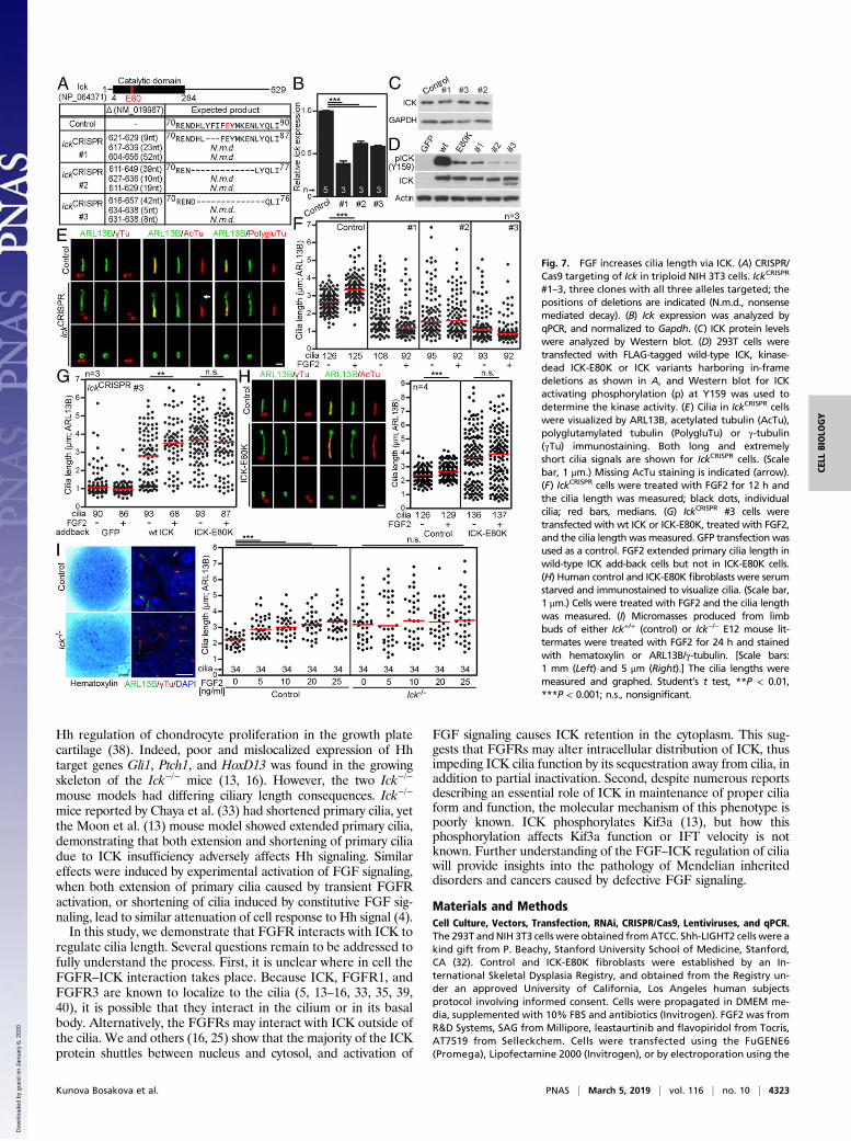
CRISPR/Cas9. A sequence corresponding to Glu80 in ICK wastargeted, because it localizes to the ATP binding pocket and itssubstitution with Lys abolishes ICK kinase activity (16). Wefailed to generate IckE80K cells, but nevertheless produced cloneswith disrupted Ick. Interestingly, none of the 302 clones obtainedin three rounds of CRISPR targeting possessed complete in-activation of all three Ick loci in triploid NIH 3T3 cells (29),suggesting that some level of ICK protein is essential for NIH3T3 growth. Three clones were selected for further analyses, inwhich the two Ick alleles were inactivated, and the remaining oneallele contained in frame deletions ranging from 3 to 14 residuessurrounding the Glu80 (Fig. 7A). IckmRNA was down-regulatedto about 40–60% among the selected clones, but the proteinlevels remained unchanged (Fig. 7 B and C), again suggestingthat presence of the ICK protein is necessary for NIH3T3 survival. The ICK deletions generated by CRISPR wereintroduced into the wild-type ICK via site-directed mutagene-sis, and the resulting ICK variants were evaluated for kinase
Fig. 4. FGF signaling alters ICK’s subcellular distribution. (A) 293T cells were transfected with FLAG-tagged ICK together with V5-tagged wild-type FGFR3 orits active mutant K650M. The antibodies against protein tags were used in the PLA (red); FGFR3 antibody was used to counterstain the transfected cells(green). As a negative control, cells were transfected with FGFR3 and an empty vector. Numbers of PLA dots per cell were calculated and plotted (Student’st test, ***P < 0.001). (Scale bar, 10 μm.) (B) Increased cytosolic localization of transfected ICK in a 293T cell cotransfected with FGFR3-K650E, determined by ICKand FGFR3 immunocytochemistry (Scale bar, 20 μm.). (C and D) Altered ICK subcellular distribution in 293T cells expressing FLAG-tagged ICK, treated withFGF2; ICK was visualized by FLAG immunocytochemistry. (C) Typical localization patterns of ICK (DIC, differential interference contrast). (Scale bar, 20 μm.) (D)Percentages of cells in each category of ICK localization (Student’s t test, **P < 0.01). (E) 293T cells were transfected with FLAG-tagged wild-type FGFR3, activeFGFR3-K650E, or K650M, or empty vector, and immunoblotted for phosphorylated (p) FGFR3. ICK and FGFR3 were visualized by immunocytochemistry, andICK subcellular localization was determined.
4320 | www.pnas.org/cgi/doi/10.1073/pnas.1800338116 Kunova Bosakova et al.
Dow
nloa
ded
by g
uest
on
Janu
ary
6, 2
020

activity in 293T cells. We observed a dramatic reduction in ICKactivating autophosphorylation at the Y159 (25) in all threeCRISPR variants, compared with wild-type ICK, demonstrating thatIckCRISPR cells express normal levels of kinase-dead ICK (Fig. 7D).Severe abnormalities were found in the cilia of IckCRISPR cells,
which we termed a “cilia disaster” phenotype. These cilia werehighly variable in length (0.4–6.9 μm; CV 65.14%; n = 296),compared with relatively narrow range in control cells (1.2–4.4 μm; 22.05%; n = 409) (SI Appendix, Fig. S3). The IckCRISPR
cilia displayed abnormal morphology, often manifested as rudi-ments negative for acetylated and polyglutamylated tubulin (Fig.7E, Bottom). Long and twisted cilia in some IckCRISPR cells alsoappeared less stable than wild-type cilia, as suggested by lesseraxoneme staining for acetylated tubulin (30) (Fig. 7E, arrow).IckCRISPR cells showed profound disruption of Hh signaling,manifested as failure to process the GLI3 transcriptional regu-lator in response to Hh agonist SAG (SMO agonist) (31), and toinduce expression of Hh target genes Gli1 and Ptch1 (SI Ap-pendix, Fig. S4 A–C). Mislocalization of GLI3 and the compo-nents of the cilia transport BBS8 and intraflagellar transport(IFT)172 was found in the IckCRISPR cilia tips (SI Appendix, Fig.S4D). Finally, the Hh coreceptor SMO localized into the cilia inSAG-naïve IckCRISPR cells, in contrast to wild-type controls,which localized SMO to cilia only when treated with SAG
(SI Appendix, Fig. S4E), suggesting increased permeabilityin IckCRISPR cilia.FGF2 did not elongate cilia in any of the three IckCRISPR
clones; however, the informative value of this data may be af-fected by the profound ciliary dysregulation (Fig. 7F). Additionof wild-type ICK into the IckCRISPR background partially re-versed the cilia disaster phenotype, resulting in formation ofmany cilia of normal length, which responded to FGF2 withusual elongation (Fig. 7G). Addition of kinase-dead ICK-E80Kalso rescued the cilia disaster; however, these cilia were longerthan those in wild-type ICK add-back cells and, importantly,were resistant to FGF2-mediated elongation (Fig. 7G).We next evaluated the effect of FGF2 on cilia length in fibroblasts
established from an individual with lethal short rib polydactyly syn-drome due to an inactivating E80K mutation in ICK (16). Com-pared with control fibroblasts, ICK-E80K cells exhibited greaterrange in their cilia length distribution, with many long and twistedcilia (Fig. 7H). Treatment with FGF2 caused statistically significantcilia elongation in control fibroblasts but not in ICK-E80K cells.Finally, the limb bud micromass cultures were used to evaluate
the FGF2 effect on cilia. FGF2 caused primary cilia extension inmicromasses established from embryonic day 12 (E12) mouselimb buds isolated from wild-type or Ick+/− mice (Fig. 7I) (4).Importantly, this effect depended on ICK, as cells derived fromIck−/− mouse limb buds (13) were insensitive to FGF2-mediated
Fig. 5. FGFRs phosphorylate and partially inactivate ICK. (A–C) Tyrosine phosphorylation of ICK, MAK, or CCRK in cell-free kinase assay with recombinantFGFR3 and recombinant ICK, MAK or CCRK, detected by Western blot with pY antibody. (D) FGFR3-mediated tyrosine phosphorylation of ICK in cell-freekinase assay analyzed by MS. Footnotes: (1) Tyrosine position relative to human ICK sequence (NP_055735.1); (2) number of experiments varies due to limitedsequence coverage in some MS samples; (3) conservation in Homo sapiens ICK/MAK, Mus musculus Ick/Mak, Gallus gallus ICK/MAK, Xenopus laevis ick/mak,Danio reriomak, Drosophila melanogaster DmeI_CG42366; (4) conservation in H. sapiens,M. musculus, G. gallus, X. laevis, D. rerio but not in D. melanogaster;(5) representative peptide sequence found in MS, pY underlined; (6) ICK activating dual phosphorylation motif T157-D-Y159; (7) conservation only in ICK, not inMAK. (Middle) Sequence alignment of N-terminal region of ICK, conserved residues in gray; Y15 in red. (Bottom Left) Three-dimensional homology model ofICK kinase domain showing ATP binding site, activation loop, and substrate binding site in yellow, green, and purple, respectively, Y15 in red. (Bottom Right)Electrostatic representation of ATP binding site with bound ATP. Electronegative and electropositive sites are in blue and red, respectively. (E) The kinaseactivity of ICK immunoprecipitated from FGF2-treated 293T cells, measured using MBP as a substrate, in the presence of [32P]-ATP. ICK activity is presentedas a relative [32P]MBP/MBP ratio (“Obtained”), and compared with the expected values based on ICK amounts entering the kinase assay quantified usingCoomassie stained gel (ICK Coomassie) or ICK immunoblot (ICK IP) (Student’s t test, ***P < 0.001). The percentages express the extent of inhibition of the ICKkinase activity in FGF2-treated cells. (F) ICK kinase activity in 293T cells treated with FGF2, determined as a degree of ICK-mediated Thr908 phosphorylation ofexpressed Raptor. The ICK activity is presented as a relative pRaptor/Raptor ratio (“Obtained”) and compared with the expected values based on ICK levels.The percentages express the extent of inhibition of the ICK kinase activity in FGF2-treated cells.
Kunova Bosakova et al. PNAS | March 5, 2019 | vol. 116 | no. 10 | 4321
CELL
BIOLO
GY
Dow
nloa
ded
by g
uest
on
Janu
ary
6, 2
020

cilia elongation. Taken together, the ICK deletion or down-regulation of ICK kinase activity rescued the FGF-mediatedelongation of primary cilia. These findings establish that FGFsignaling regulates cilia length via ICK.Because ICK inactivation inhibits Hh signaling, we analyzed
the Hh activity in several models to ICK inhibition. The Sonichedgehog (Shh)-LIGHT2 NIH 3T3 cells stably express the GLI-driven Firefly luciferase reporter capable of monitoring the Hhpathway activity (32). We inhibited endogenous Ick activity bytreatment with FGF2 in Shh-LIGHT2 cells, and analyzed thetransactivation of the Hh reporter. At the maximal inductionobtained with 100 nM SAG, FGF2 inhibited the luciferaseactivity by 37% (SI Appendix, Fig. S5A). Similarly, inhibitionof Ick activity in NIH 3T3 cells using flavopiridol, AT7519, orlestaurtinib abolished the SAG-mediated GLI1 up-regulation,as did FGF2 (SI Appendix, Fig. S5B). Finally, micromassesderived from murine Ick−/− limb buds failed to efficiently up-
regulate Gli1 and Ptch1 expression and ciliary GLI2 localizationupon SAG, similarly to the FGF2-treated control micromasses (SIAppendix, Fig. S5 C and D).
Regulation of Primary Cilia by FGF–ICK Pathway. The regulation ofprimary cilia length and function by FGF signaling has recentlyemerged as a new paradigm in cell biology (3–5). However, themolecular mechanism by which FGFs regulate cilia remainsunclear. In this article, we uncover that FGFRs interact with andphosphorylate an important and conserved ciliary kinase, ICK,leading to partial inhibition of its kinase activity and alteredsubcellular localization (Figs. 1–5). Modulations of FGF signal-ing regulate primary cilia length, IFT velocity, and Hh signalingconsistent with effects caused by its inhibition of ICK activity (3,4, 15, 16, 33). Moreover, ICK removal or down-regulationabolished the FGF-mediated effect on ciliary length (Figs. 6and 7), demonstrating that FGF signaling regulates primary ciliavia ICK. In the following section, we describe known cilia phe-notypes regulated by FGF signaling, and explain these pheno-types on the basis of the FGF–ICK interaction (Fig. 8).First, the ICK is a sensitive regulator of primary cilia length.
Deletion of ICK homologs LF4, LmxMPK9, and DYF-5 increasedthe length of cilia in green algae (Chlamydomonas reinhardtii),protists (Leishmania mexicana), and worms (Caenorhabditis elegans)(10–12). Similarly, Ick−/− mice or humans carrying partially orcompletely inactivating ICK mutations R272Q and E80K showedelongated primary cilia (13, 16, 34). Expression of these variants andother inactive ICK mutants in cultured cells, or partial down-regulation of Ick expression via RNA interference also elongatedprimary cilia (13–16, 35). Because FGFRs phosphorylate and in-activate ICK (Fig. 5), a down-regulation of FGF signaling shouldrelieve this inhibition, resulting in elevated ICK activity and shortercilia (Fig. 8B). Indeed, mice treated with chemical inhibitors ofFGFR activity showed shorter cilia in the biliary ducts, kidney, andlung (4). This is corresponding to the evidence obtained in zebrafishand Xenopus, where inactivation of Fgf signaling led to shorter ciliain multiple tissues (3). Similar to ICK inactivation, stimulation ofFGF signaling elongated primary cilia in NIH 3T3 cells, primarymouse embryonic and human fibroblasts, epithelial IMCD3 cells,and mouse limb bud mesenchymal cells (4), but failed to do so inIck−/− background or in cells endogenously expressing inactive ICK(Fig. 7). Similarly, cells with diminished Ick expression or activitywere insensitive to the FGF-mediated cilia elongation (Fig. 6),demonstrating that FGF signaling elongates primary cilia via in-hibition of ICK kinase activity (Fig. 8C).While inhibition of ICK activity extended primary cilia, experi-
mental up-regulation of ICK activity produced shorter cilia (11–13,16, 33). Interestingly, ICK inactivation also led to cilia shortening inat least two instances, in the neural tube and embryonic fibroblastsisolated from an Ick−/− mice (33), and in the NIH 3T3 IckCRISPR
cells reported here (Fig. 7 A–G and SI Appendix, Fig. S3) (the ciliadisaster phenotype). In our previous work, we showed that FGFsignaling can also shorten primary cilia (4, 5), as observed in cellsexpressing constitutively active FGFR3-K650E or -K650Mmutants. Interestingly, these FGFR3 mutants interacted withICK (Fig. 1). Thus, the FGF-mediated ICK inactivation maylead to profound dysregulation of ciliogenesis manifested ascilia shortening or extension, depending on the strength andduration of the FGF stimulus (Fig. 8D).Ick−/− mouse models described by both Chaya et al. (33) and
Moon et al. (13) displayed preaxial polydactyly and dwarfism, asimilar phenotype to human short-rib polydactyly syndrome as-sociated with an inactivating ICK mutation (16), or to mice andhumans with endocrine-cerebro-osteodysplasia syndrome cou-pled with mildly inactivating ICK mutations (26, 35, 36). Thesephenotypes suggest impaired Hh signaling, as polydactyly stemsfrom defective Hh function in early limb patterning (37), whilethe shortened appendicular skeletons likely result from disrupted
Fig. 6. FGF regulates the length of primary cilia via ICK. (A) Primary cilialength extension in NIH 3T3 cells treated with FGF2. Cilia were visualized byARL13B, acetylated tubulin (AcTu) and pericentrin immunostaining, mea-sured in 3D and plotted. Black dots, individual cilia; red bars, medians. [Scalebars: 5 μm (cells) and 1 μm (cilia).] (B) Rescue of FGF2-mediated cilia exten-sion with two independent Ick shRNAs (Ick #1 and Ick #2). Ick transcript levelswere monitored by qPCR at 24 h (beginning of serum starvation) and 36 h(FGF2 treatment) after transfection, and normalized to Gapdh expression.The columns show Ick expression levels relative to the scrambled control (reddashed line). Cilia length was measured 48 h after transfection and graphed.(C) Rescue of FGF2-mediated cilia extension in IckFlag(B11) NIH 3T3 cells stablytransfected with DOX-inducible shRNA-expressing construct targeting ICKexpression. ICK protein levels were monitored by immunoblot after 4 d withDOX (beginning of FGF2 treatment), normalized to actin, and plotted. Cellsexpressing scrambled (Scr.) shRNA upon DOX were used as a control. Stu-dent’s t test, **P < 0.01, ***P < 0.001; n.s., not significant.
4322 | www.pnas.org/cgi/doi/10.1073/pnas.1800338116 Kunova Bosakova et al.
Dow
nloa
ded
by g
uest
on
Janu
ary
6, 2
020
Hh regulation of chondrocyte proliferation in the growth platecartilage (38). Indeed, poor and mislocalized expression of Hhtarget genes Gli1, Ptch1, and HoxD13 was found in the growingskeleton of the Ick−/− mice (13, 16). However, the two Ick−/−
mouse models had differing ciliary length consequences. Ick−/−
mice reported by Chaya et al. (33) had shortened primary cilia, yetthe Moon et al. (13) mouse model showed extended primary cilia,demonstrating that both extension and shortening of primary ciliadue to ICK insufficiency adversely affects Hh signaling. Similareffects were induced by experimental activation of FGF signaling,when both extension of primary cilia caused by transient FGFRactivation, or shortening of cilia induced by constitutive FGF sig-naling, lead to similar attenuation of cell response to Hh signal (4).In this study, we demonstrate that FGFR interacts with ICK to
regulate cilia length. Several questions remain to be addressed tofully understand the process. First, it is unclear where in cell theFGFR–ICK interaction takes place. Because ICK, FGFR1, andFGFR3 are known to localize to the cilia (5, 13–16, 33, 35, 39,40), it is possible that they interact in the cilium or in its basalbody. Alternatively, the FGFRs may interact with ICK outside ofthe cilia. We and others (16, 25) show that the majority of the ICKprotein shuttles between nucleus and cytosol, and activation of
FGF signaling causes ICK retention in the cytoplasm. This sug-gests that FGFRs may alter intracellular distribution of ICK, thusimpeding ICK cilia function by its sequestration away from cilia, inaddition to partial inactivation. Second, despite numerous reportsdescribing an essential role of ICK in maintenance of proper ciliaform and function, the molecular mechanism of this phenotype ispoorly known. ICK phosphorylates Kif3a (13), but how thisphosphorylation affects Kif3a function or IFT velocity is notknown. Further understanding of the FGF–ICK regulation of ciliawill provide insights into the pathology of Mendelian inheriteddisorders and cancers caused by defective FGF signaling.
Materials and MethodsCell Culture, Vectors, Transfection, RNAi, CRISPR/Cas9, Lentiviruses, and qPCR.The 293T andNIH 3T3 cells were obtained fromATCC. Shh-LIGHT2 cells were akind gift from P. Beachy, Stanford University School of Medicine, Stanford,CA (32). Control and ICK-E80K fibroblasts were established by an In-ternational Skeletal Dysplasia Registry, and obtained from the Registry un-der an approved University of California, Los Angeles human subjectsprotocol involving informed consent. Cells were propagated in DMEM me-dia, supplemented with 10% FBS and antibiotics (Invitrogen). FGF2 was fromR&D Systems, SAG from Millipore, leastaurtinib and flavopiridol from Tocris,AT7519 from Selleckchem. Cells were transfected using the FuGENE6(Promega), Lipofectamine 2000 (Invitrogen), or by electroporation using the
Fig. 7. FGF increases cilia length via ICK. (A) CRISPR/Cas9 targeting of Ick in triploid NIH 3T3 cells. IckCRISPR
#1–3, three clones with all three alleles targeted; thepositions of deletions are indicated (N.m.d., nonsensemediated decay). (B) Ick expression was analyzed byqPCR, and normalized to Gapdh. (C) ICK protein levelswere analyzed by Western blot. (D) 293T cells weretransfected with FLAG-tagged wild-type ICK, kinase-dead ICK-E80K or ICK variants harboring in-framedeletions as shown in A, and Western blot for ICKactivating phosphorylation (p) at Y159 was used todetermine the kinase activity. (E) Cilia in IckCRISPR cellswere visualized by ARL13B, acetylated tubulin (AcTu),polyglutamylated tubulin (PolygluTu) or γ-tubulin(γTu) immunostaining. Both long and extremelyshort cilia signals are shown for IckCRISPR cells. (Scalebar, 1 μm.) Missing AcTu staining is indicated (arrow).(F) IckCRISPR cells were treated with FGF2 for 12 h andthe cilia length was measured; black dots, individualcilia; red bars, medians. (G) IckCRISPR #3 cells weretransfected with wt ICK or ICK-E80K, treated with FGF2,and the cilia length was measured. GFP transfection wasused as a control. FGF2 extended primary cilia length inwild-type ICK add-back cells but not in ICK-E80K cells.(H) Human control and ICK-E80K fibroblasts were serumstarved and immunostained to visualize cilia. (Scale bar,1 μm.) Cells were treated with FGF2 and the cilia lengthwas measured. (I) Micromasses produced from limbbuds of either Ick+/+ (control) or Ick−/− E12 mouse lit-termates were treated with FGF2 for 24 h and stainedwith hematoxylin or ARL13B/γ-tubulin. [Scale bars:1 mm (Left) and 5 μm (Right).] The cilia lengths weremeasured and graphed. Student’s t test, **P < 0.01,***P < 0.001; n.s., nonsignificant.
Kunova Bosakova et al. PNAS | March 5, 2019 | vol. 116 | no. 10 | 4323
CELL
BIOLO
GY
Dow
nloa
ded
by g
uest
on
Janu
ary
6, 2
020

Neon Transfection System (Invitrogen). The following vectors were used:FGFR1–4 (41, 42); pmaxGFP (Lonza); Raptor (43); ICK, MAK, and CCRK (Ori-gene). The ICK mutation E80K was described previously (16); the ICK mutantswere generated by site-directed mutagenesis (Agilent Technologies). Trun-cated and mutated FGFR3 variants were generated by PCR mutagenesis.SureSilencing shRNA plasmid (KM37622G, clones #1 and #2; Qiagen) andscrambled control NEG4-G were used for RNAi; the GFP expression was used toidentify shRNA-expressing cells. CRISPR/Cas9 was used as described previously(44). A pair of SpCas9n (D10A) nickases targeting 5′-ACTCGAAGATAAA-GTAAAGATGG-3′, 5′-CTTTATCTTCGAGTACATGAAGG-3′ sites in the fourthexon of Ick were used, and cotransfected with pmaxGFP. GFP+ cells were pickedto grow single-cell clones. Targeting was confirmed by PCR sequencing. Togenerate IckFlag cells, NIH 3T3 cells were transfected with a pair of nickasestargeting 5′-AGTACCCATCCCGGCGGTGA-3′ and 5′-GCGGTGACTGTCCCGGC-CA-3′ sites at the end of the last exon together with synthetic DNA (gBlock,IDT) containing 3xFlag sequence surrounded with 451- and 491-bp longhomology arms at left and right. Single-cell colonies which were screened byWestern blot for the presence of Flag tag at Ick. Targeting was verified byPCR sequencing. A lentiviral vector containing DOX-inducible U6 promoterand TetRep-P2A-Puro-P2A-mCherry (45) was modified to express shRNA byintroducing oligonucleotides (shIck forward: ccggcacaaccacgaggcggtgtaa-ctcgagttacaccgcctcgtggttgtgtttttg, reverse: attcaaaaacacaaccacgaggcg-gtgtaactcgagttacaccgcctcgtggttgtg; Scrambled forward: ccgggcgta-ccaaccggaactgagactcgagtctcagttccggttggtacgtttttg; reverse: aattcaaaa-agcgtaccaaccggaactgagactcgagtctcagttccggttggtacgc). Lentiviral particles weregenerated as described previously (46) using pMD2.G (Addgene #12259) andpsPAX2 (Addgene #12260) (gift from Didier Trono, École Polytechnique Féd-érale de Lausanne, Lausanne, Switzerland). IckFlag NIH 3T3 cells were trans-duced using generated lentiviral particles and mCherry+ cells were sorted usingBD FACSAria (BD Biosciences). Cells were treated with 1 μg/mL DOX (Sigma-Aldrich) to induce the shRNA expression for 4 d. Total RNA was isolated usingRNeasy Mini Kit (Qiagen) and reverse-transcribed by First Strand cDNA Syn-thesis Kit (Roche). For qPCR, the following QuantiTect Primers were used(Qiagen): Mm_Ick_1_SG (QT01037729), Mm_Gapdh_3_SG (QT01658692),Mm_Ptch1_1_SG (QT00149135), and Mm_Gli1_1_SG (QT00173537). Formicromasses, the following primers were used in qPCR: Gli1 (5′-CTTGTGGTGGAGT-CATTGGA-3′, 5′-GAGGTTGGGATGAAGAAGCA-3′), Ptch1 (5′-AATTCTC-GACTCACTCGTCCA-3′, 5′-CTCCTCATATTTGGGGCCTT-3′), Gapdh (5′-CCT-GTTGCTGTAGCCGTATT-3′, 5′-AACAGCAACTCCCACTCTTC-3′).
Gradient Ultracentrifugation, BN-PAGE, Western Blot, IP, Kinase Assays, andPeptide Microarrays. Native cell lysates (50 mM Tris·HCl pH 7.4, 150 mMNaCl, 0.5% Igepal CA-630, 1 mM EDTA pH 8, 0.25% sodium deoxycholate,1 mM Na2VO4, proteinase inhibitors) were cleared, loaded on 5–25% sucrosegradient (1 mM Tris·HCl pH 7.4, 150 mM NaCl, 3 mM MgCl2, proteinase in-hibitors), and centrifuged at 40,000 rpm/4 °C/16 h using SW 40 Ti swingingbucket rotor (Beckman Coulter). Gradient fractions were either precipitatedwith 10% TCA or used for BN-PAGE. For BN-PAGE, fractions were mixed withsolubilization buffer (50 mM NaCl, 50 mM imidazole/HCl, 1 mM EDTA, pH 7)and concentrated using Spin-X UF 500 (cut-off 30 kDa; Corning). Beforesample loading, the wells of the 4–15% native tricine-imidazole gel werewashed with cathode buffer (50 mM tricine, 7.5 mM imidazole, 0.02%Coomassie blue G250, pH ∼ 7), and the gel run at constant 15 mA/4 °C. The29- to 669-kDa native protein ladder was from Sigma. Individual lanes werecut from the gel, placed over the SDS gel, and protein complexes wereseparated using second dimension SDS/PAGE, and analyzed by Western blot.For Western blot, lysates were resolved by SDS/PAGE, transferred onto aPVDF membrane and visualized by chemiluminiscence (Thermo). The fol-lowing antibodies were used: GAPDH (5174), GLI1 (2643), FGFR1 (9740), actin(3700; Cell Signaling); HA (sc-805), FGFR3 (sc-123), pFGFR3Y724 (sc-33041;Santa Cruz Biotechnology); FLAG (F1804), GST (G1160; Sigma); V5 (R960-25; Invitrogen), 4G10 (05-321; Millipore), pICKY159 (ab138435; Abcam), GLI3(AF3690; RnD Systems), ICK (43), and pRaptorT908 (27). For IP, cells wereextracted in buffer containing 50 mM Tris·HCl pH 7.4, 150 mM NaCl, 0.5%Nonidet P-40, 0.1% sodium deoxycholate, 2 mM EDTA pH 8.0, 0.5 mM DTT,proteinase inhibitors; immunocomplexes were collected on protein A/Gagarose (Santa Cruz). Kinase assays were performed using recombinantFGFR3 together with ICK, MAK, or CCRK (SignalChem), in kinase buffer(60 mM Hepes pH 7.5, 3 mM MgCl2, 3 mM MnCl2, 3 μM Na3VO4, 1.2 mM DTT)in the presence of 10 μM ATP. Tyrosine phosphorylation was determined byWestern blot with 4G10 antibody (Millipore). [32P]-ATP kinase assays were carriedout with IP ICK and 4 μg of recombinant MBP (Sigma) as a substrate, in a kinasebuffer (50 mM Hepes pH 7.5, 10 mM MnCl2, 10 mM MgCl2, 8 mM β-glycer-ophosphate, 1 mMDTT, 0.1 mMNa3VO4, 0.1 mM PMSF), in the presence of 1 μCi[32P]-ATP (Izotop). Samples were resolved by SDS/PAGE and visualized by auto-radiography. Band intensities were quantified in ImageJ. Peptide libraries cor-responding to FGFR3 intracellular domain were synthetized and immobilized ona glass slide via hydrophilic linker (Pepstar microarray; JPT Peptide Technologies).The library contained overlapping peptides (13-aa long with 10-aa overlap)covering disordered parts of FGFR3 in the juxtamembrane and C-terminal re-gions, and nonoverlapping peptides of varying length that emulated elementsof the secondary structure exposed on the surface of FGFR3 TK domain (FGFR3:4K33) (SI Appendix, Fig. S1B). Microarrays were incubated with recombinant ICK(I01-10G; SignalChem), labeled by DyLigh 650 (ThermoFisher Scientific). Fluores-cence intensities were obtained using laser scanner Axon Genepix Scanner 4300SL50, and quantified using GenePix (Molecular Devices). The peptides withfluorescence intensity at least 10-fold above nonspecific were considered aspotential binding sites. Microarray preparation, data acquisition, and analysiswere described previously (47).
Immunocytochemistry, PLA, and Cilia Length Measurements. Cells were fixed inparaformaldehyde and incubated with the following antibodies: V5 (R960-25), acetylated α-tubulin (32-2700; Invitrogen), FLAG (F1804; Sigma),ARL13B (17711-1-AP; Proteintech), γ-tubulin (ab11316), pericentrin (ab4448;Abcam), polyglutamylated tubulin (AG-20B-0020-C100; AdipoGen), SMO (sc-166685), BBS8 (sc-271009), IFT172 (sc-398393; Santa Cruz), and GLI3 (AF3690;RnD Systems). Duolink PLA (Sigma) was used for PLA, with V5 (sc-83849;Santa Cruz) and FLAG (F1804; Sigma) antibodies; FGFR3 (sc-123; SantaCruz) was used to counterstain the transfected cells. AlexaFluor488/594 sec-ondary antibodies were from Invitrogen. PLA analysis was done in Fiji (fiji.sc/)using maximum projections of z-stacks. Cilia length in 3D were determinedas described previously (16).
MS, Modeling, Animal Experiments, Immunohistochemistry, and Statistics. Ki-nase reactions containing FGFR3 and ICK were subjected to reduction, al-kylation, and in-solution digestion by trypsin. Samples were analyzed usingnanoscale liquid chromatography connected to the tandem mass spec-trometer (RSLCnano connected to Orbitrap Elite; Thermo Fisher). High-resolution HCD or ETD MS/MS spectra were acquired in the Orbitrapanalyzer. The analysis of the MS RAW data files was carried out using theProteome Discoverer software (v.1.4; Thermo) with a Mascot search engine(v.2.4.1; Matrix Science). Quantitative information assessment and phos-phopeptide signal validation was done in Skyline. The 3D ICK model wasobtained via template-based homology modeling using the PHYRE software(48). The ICK-specific functional elements, predicted using the National
Fig. 8. Model describing regulation of primary cilia length by the FGF–ICKpathway. (A) Under basal conditions, endogenous FGF signaling restricts ICKactivity to the level required for optimal length of the cilia. (B) Experimentalup-regulation of ICK activity, by increased expression of active ICK, shortensthe primary cilia (12, 15, 16). Similar effect is achieved in cells with abolishedFGF signaling, which are unable to inhibit ICK, leading to up-regulation ofICK kinase activity and ciliary shortening (3, 4). (C) Down-regulation of ICKactivity, either by transient increase in activity of FGF signaling, or by smallchemicals, expression of kinase-inactive ICK mutants (*), Ick knockout orknockdown, extends primary cilia (4, 12, 13, 15, 16, 33). (D) Under certainconditions, inactivation of ICK results in profound dysregulation of primarycilia (cilia disaster), manifested by low cilia stability and extreme length vari-ability (10, 33). A similar phenotype is achieved by strong activation of FGFsignaling via disease-associated FGFR3 mutations (*) (4, 5), suggesting that thestrength of the FGF stimulus regulates optimal ciliary length through ICK.
4324 | www.pnas.org/cgi/doi/10.1073/pnas.1800338116 Kunova Bosakova et al.
Dow
nloa
ded
by g
uest
on
Janu
ary
6, 2
020

Center for Biotechnology Information Conserved Domain Database (49), weremapped onto a 3D model of the ICK using the CHIMERA software (50). Ho-mology modeling (PDB ID codes 3C4W and 1JNK) was employed to dock ATPinto the ICK ATP binding site. Ick−/− mice were described previously (16). Ick+/−
mice were maintained in a C57/BL6N background and used as controls. Formicromasses, the primary mesenchymal cells were harvested from the limbbuds of E12 mouse embryos, digested with dispase II, and spotted in 10-μLaliquots at 2 × 10⁷ cells/mL. Cells were allowed to adhere before differenti-ating media (60% F12/40% DMEM, 10% FBS, 50 μg/mL ascorbic acid, 10 mMβ-glycerol phosphate, 1% L-glutamine) was added. Micromasses were grownfor 1 d in media supplemented with FGF2 (Sigma), fixed with methanol andparaformaldehyde, and immunolabeled by ARL13B and γ-tubulin (T6557;Sigma) antibodies. For GLI2 detection, micromasses were serum-starved for12 h, treated with FGF2 and SAG for additional 12 h, and immunostainedusing GLI2 and ARL13B antibodies (13). Animal experiments were approved bythe Institutional Animal Care and Use Committee of Dongguk University,Korea (IACUC-2016-016-1). All experiments were performed at least in tripli-cate unless stated otherwise. The n values express the number of independentbiological experiments. Data are presented as mean ± SEM. Two-tailed Stu-dent’s t test was used for statistical analysis of data. Brightness and contrastwere adjusted in microphotographs, homogenously throughout each panel.
ACKNOWLEDGMENTS. We thank Mikael Altun and Johan Boström for pro-viding the U6 vector. This work was supported by the Ministry of Education,Youth and Sports of the Czech Republic (Grant KONTAKT II LH15231, Na-tional Program of Sustainability II Projects LQ1605 and LQ1601); the Agencyfor Healthcare Research of the Czech Republic (Grants 15-33232A, 15-34405A, and NV18-08-00567); and the Czech Science Foundation (GrantsGA17-09525S, 14-31540S, and GJ16-24004Y). D.K. is supported by NIH GrantsR01AR062651, R01AR066124, and R01DE019567, and by NIH/National Centerfor Advancing Translational Science University of California, Los AngelesClinical and Translational Science Institute Grant UL1TR000124. Z.F. is sup-ported by NIH Grant NIH-GM127690. H.W.K. is supported by Korea MousePhenotyping Project NRF2014M3A9D5A01073969 of the Ministry of Sci-ence, Information, and Communication Technology and Future Planningthrough the National Research Foundation. Czech Infrastructure for Inte-grative Structural Biology Research Infrastructure Project LM2015043funded by the Ministery of Education, Youth and Sports of the Czech Re-public is gratefully acknowledged for the financial support of the measure-ments at the CEITEC Proteomics Core Facility. M.K.B. was supported by thefunds from Masaryk University (Grant MUNI/E/0563/2018). M.K.B. and B.F. aresupported by a Junior Researcher award from the Faculty of Medicine, MasarykUniversity.
1. Bangs F, Anderson KV (2017) Primary cilia and mammalian hedgehog signaling. ColdSpring Harb Perspect Biol 9:a028175.
2. Reiter JF, Leroux MR (2017) Genes and molecular pathways underpinning ciliopathies.Nat Rev Mol Cell Biol 18:533–547.
3. Neugebauer JM, Amack JD, Peterson AG, Bisgrove BW, Yost HJ (2009) FGF signalling duringembryo development regulates cilia length in diverse epithelia. Nature 458:651–654.
4. Kunova Bosakova M, et al. (2018) Regulation of ciliary function by fibroblast growthfactor signaling identifies FGFR3-related disorders achondroplasia and thanatophoricdysplasia as ciliopathies. Hum Mol Genet 27:1093–1105.
5. Martin L, et al. (2018) Constitutively-active FGFR3 disrupts primary cilium length andIFT20 trafficking in various chondrocyte models of achondroplasia. Hum Mol Genet 27:1–13.
6. �Cajánek L, Nigg EA (2014) Cep164 triggers ciliogenesis by recruiting Tau tubulin ki-nase 2 to the mother centriole. Proc Natl Acad Sci USA 111:E2841–E2850.
7. Zhang B, et al. (2015) GSK3β-Dzip1-Rab8 cascade regulates ciliogenesis after mitosis.PLoS Biol 13:e1002129.
8. Kim S, Lee K, Choi J-H, Ringstad N, Dynlacht BD (2015) Nek2 activation ofKif24 ensures cilium disassembly during the cell cycle. Nat Commun 6:8087.
9. Chen Y, et al. (2011) Sonic hedgehog dependent phosphorylation by CK1α and GRK2 isrequired for ciliary accumulation and activation of smoothened. PLoS Biol 9:e1001083.
10. Berman SA, Wilson NF, Haas NA, Lefebvre PA (2003) A novel MAP kinase regulatesflagellar length in Chlamydomonas. Curr Biol 13:1145–1149.
11. Bengs F, Scholz A, KuhnD, Wiese M (2005) LmxMPK9, a mitogen-activated protein kinasehomologue affects flagellar length in Leishmania mexicana.MolMicrobiol 55:1606–1615.
12. Burghoorn J, et al. (2007) Mutation of the MAP kinase DYF-5 affects docking andundocking of kinesin-2 motors and reduces their speed in the cilia of Caenorhabditiselegans. Proc Natl Acad Sci USA 104:7157–7162.
13. Moon H, et al. (2014) Intestinal cell kinase, a protein associated with endocrine-cerebro-osteodysplasia syndrome, is a key regulator of cilia length and Hedgehogsignaling. Proc Natl Acad Sci USA 111:8541–8546.
14. Yang Y, Roine N, Mäkelä TP (2013) CCRK depletion inhibits glioblastoma cell pro-liferation in a cilium-dependent manner. EMBO Rep 14:741–747.
15. Broekhuis JR, Verhey KJ, Jansen G (2014) Regulation of cilium length and intraflagellartransport by the RCK-kinases ICK and MOK in renal epithelial cells. PLoS One 9:e108470.
16. Paige Taylor S, et al.; University of Washington Center for Mendelian Genomics (2016)An inactivating mutation in intestinal cell kinase, ICK, impairs hedgehog signallingand causes short rib-polydactyly syndrome. Hum Mol Genet 25:3998–4011.
17. Balek L, et al. (2018) Proteomic analyses of signalling complexes associated with re-ceptor tyrosine kinase identify novel members of fibroblast growth factor receptor3 interactome. Cell Signal 42:144–154.
18. Fu Z, et al. (2006) Identification of yin-yang regulators and a phosphorylation consensusfor male germ cell-associated kinase (MAK)-related kinase. Mol Cell Biol 26:8639–8654.
19. Naski MC, Wang Q, Xu J, Ornitz DM (1996) Graded activation of fibroblast growthfactor receptor 3 by mutations causing achondroplasia and thanatophoric dysplasia.Nat Genet 13:233–237.
20. Kong M, Wang CS, Donoghue DJ (2002) Interaction of fibroblast growth factor receptor3 and the adapter protein SH2-B. A role in STAT5 activation. J Biol Chem 277:15962–15970.
21. Salazar L, et al. (2009) A novel interaction between fibroblast growth factor receptor3 and the p85 subunit of phosphoinositide 3-kinase: Activation-dependent regulationof ERK by p85 in multiple myeloma cells. Hum Mol Genet 18:1951–1961.
22. Brewer JR, Molotkov A, Mazot P, Hoch RV, Soriano P (2015) Fgfr1 regulates develop-ment through the combinatorial use of signaling proteins. Genes Dev 29:1863–1874.
23. Ong SH, et al. (2000) FRS2 proteins recruit intracellular signaling pathways by bindingto diverse targets on fibroblast growth factor and nerve growth factor receptors.MolCell Biol 20:979–989.
24. Gudernova I, et al. (2016) Multikinase activity of fibroblast growth factor receptor(FGFR) inhibitors SU5402, PD173074, AZD1480, AZD4547 and BGJ398 compromisesthe use of small chemicals targeting FGFR catalytic activity for therapy of short-stature syndromes. Hum Mol Genet 25:9–23.
25. Fu Z, et al. (2005) Activation of a nuclear Cdc2-related kinase within a mitogen-activated protein kinase-like TDY motif by autophosphorylation and cyclin-dependent protein kinase-activating kinase. Mol Cell Biol 25:6047–6064.
26. Lahiry P, et al. (2009) A multiplex human syndrome implicates a key role for intestinalcell kinase in development of central nervous, skeletal, and endocrine systems. Am JHum Genet 84:134–147.
27. Wu D, et al. (2012) Intestinal cell kinase (ICK) promotes activation of mTOR complex 1(mTORC1) through phosphorylation of Raptor Thr-908. J Biol Chem 287:12510–12519.
28. Davis MI, et al. (2011) Comprehensive analysis of kinase inhibitor selectivity. NatBiotechnol 29:1046–1051.
29. Todaro GJ, Green H (1963) Quantitative studies of the growth of mouse embryo cellsin culture and their development into established lines. J Cell Biol 17:299–313.
30. Janke C, Bulinski JC (2011) Post-translational regulation of the microtubule cyto-skeleton: Mechanisms and functions. Nat Rev Mol Cell Biol 12:773–786.
31. Chen JK, Taipale J, Young KE, Maiti T, Beachy PA (2002) Small molecule modulation ofSmoothened activity. Proc Natl Acad Sci USA 99:14071–14076.
32. Taipale J, et al. (2000) Effects of oncogenic mutations in Smoothened and patchedcan be reversed by cyclopamine. Nature 406:1005–1009.
33. Chaya T, Omori Y, Kuwahara R, Furukawa T (2014) ICK is essential for cell type-specificciliogenesis and the regulation of ciliary transport. EMBO J 33:1227–1242.
34. Okamoto S, et al. (2017) Ick ciliary kinase is essential for planar cell polarity formationin inner ear hair cells and hearing function. J Neurosci 37:2073–2085.
35. Oud MM, et al. (2016) A novel ICK mutation causes ciliary disruption and lethalendocrine-cerebro-osteodysplasia syndrome. Cilia 5:8.
36. Ding M, et al. (2018) A murine model for human ECO syndrome reveals a critical roleof intestinal cell kinase in skeletal development. Calcif Tissue Int 102:348–357.
37. te Welscher P, et al. (2002) Progression of vertebrate limb development through SHH-mediated counteraction of GLI3. Science 298:827–830.
38. Vortkamp A, et al. (1996) Regulation of rate of cartilage differentiation by Indianhedgehog and PTH-related protein. Science 273:613–622.
39. Honda A, et al. (2018) FGFR1-mediated protocadherin-15 loading mediates cargospecificity during intraflagellar transport in inner ear hair-cell kinocilia. Proc NatlAcad Sci USA 115:8388–8393.
40. Ding VMY, et al. (2011) Tyrosine phosphorylation profiling in FGF-2 stimulated hu-man embryonic stem cells. PLoS One 6:e17538.
41. Krejci P, et al. (2007) Bisindolylmaleimide I suppresses fibroblast growth factor-mediatedactivation of Erk MAP kinase in chondrocytes by preventing Shp2 association with theFrs2 and Gab1 adaptor proteins. J Biol Chem 282:2929–2936.
42. Krejci P, et al. (2012) Receptor tyrosine kinases activate canonical WNT/β-cateninsignaling via MAP kinase/LRP6 pathway and direct β-catenin phosphorylation. PLoSOne 7:e35826.
43. Fu Z, Kim J, Vidrich A, Sturgill TW, Cohn SM (2009) Intestinal cell kinase, a MAP kinase-related kinase, regulates proliferation and G1 cell cycle progression of intestinal ep-ithelial cells. Am J Physiol Gastrointest Liver Physiol 297:G632–G640.
44. Ran FA, et al. (2013) Genome engineering using the CRISPR-Cas9 system. Nat Protoc 8:2281–2308.
45. Eshtad S, et al. (2016) hMYH and hMTH1 cooperate for survival in mismatch repairdefective T-cell acute lymphoblastic leukemia. Oncogenesis 5:e275.
46. Barta T, Peskova L, Hampl A (2016) miRNAsong: A web-based tool for generation andtesting of miRNA sponge constructs in silico. Sci Rep 6:36625.
47. Harno�s J, et al. (2018) Analysis of binding interfaces of the human scaffold proteinAXIN1 by peptide microarrays. J Biol Chem 293:16337–16347.
48. Kelley LA, Mezulis S, Yates CM, Wass MN, Sternberg MJE (2015) The Phyre2 webportal for protein modeling, prediction and analysis. Nat Protoc 10:845–858.
49. Marchler-Bauer A, et al. (2011) CDD: A conserved domain database for the functionalannotation of proteins. Nucleic Acids Res 39:D225–D229.
50. Pettersen EF, et al. (2004) UCSF Chimera—A visualization system for exploratory re-search and analysis. J Comput Chem 25:1605–1612.
Kunova Bosakova et al. PNAS | March 5, 2019 | vol. 116 | no. 10 | 4325
CELL
BIOLO
GY
Dow
nloa
ded
by g
uest
on
Janu
ary
6, 2
020