Fibroblast Growth Factor Receptor 3 Signaling Regulates ...
12
Fibroblast Growth Factor Receptor 3 Signaling Regulates the Onset of Oligodendrocyte Terminal Differentiation Luke Y. S. Oh, 1 Adam Denninger, 1 Jennifer S. Colvin, 3 Aditee Vyas, 2 Shubha Tole, 2 David M. Ornitz, 3 and Rashmi Bansal 1 1 Department of Neuroscience, University of Connecticut Medical School, Farmington, Connecticut 06030, 2 Department of Biological Sciences, Tata Institute of Fundamental Research, Mumbai, 400 005 India, and 3 Department of Molecular Biology and Pharmacology, Washington University School of Medicine, St. Louis, Missouri 63110 Fibroblast growth factor receptor (FGFR) signaling is essential for nervous system development. We have shown that, in the normal postnatal brain, the spatial and temporal expression pattern of FGFR3 parallels the appearance of differentiated oligodendrocytes and that in culture FGFR3 is expressed maximally at the critical stage in the lineage at which oligodendrocyte late progenitors (Pro-OLs) enter terminal differentiation. Therefore, FGFR3 expression is positioned ideally to have an impact on oligodendrocyte differentiation. In support of this we show that, during the onset and active phase of myelination in FGFR3-deficient mice, there are reduced numbers of differentiated oligodendrocytes in the forebrain, cerebellum, hindbrain, and spinal cord. Furthermore, myelination is delayed in parallel. Delay of oligodendrocyte differentiation also is observed in primary cell culture from this mutant. On the other hand, no differences are observed in the survival or proliferation of oligodendrocyte progenitors. This suggests that the decrease in the number of differentiated oligodendrocytes is attributable to a delay in the timing of their differentiation process. Astrocytes also express FGFR3, and in mice lacking FGFR3 there is an enhancement of the astrocytic marker glial fibrillary acidic protein expression in a region-specific manner. Thus our findings suggest that there are cell type- and region-specific functions for FGFR3 signaling and in particular emphasize a prominent role for FGFR3 as part of a system regulating the onset of oligodendrocyte terminal differentiation. Key words: oligodendrocyte; myelin; FGF; astrocyte; cerebellar neuron; FGF receptor Fibroblast growth factors (FGFs), a family of 23 known members, play central roles in nervous system development (Vaccarino et al., 1999; Ford-Perriss et al., 2001; Ornitz and Itoh, 2001). FGFs signal through four high-affinity tyrosine kinase receptors (FGFR1–FGFR4) (Johnson and Williams, 1993). FGFRs are ex- pressed differentially during development and in the adult brain (Orr-Urtreger et al., 1991; Peters et al., 1992, 1993; Asai et al., 1993; Yazaki et al., 1994; Miyake et al., 1996; Ford-Perriss et al., 2001). FGFs modulate a variety of biological activities including proliferation, migration, and survival of neurons and glial cells (Vaccarino et al., 1999; Ford-Perriss et al., 2001). Furthermore, FGFs influence specification of neuronal and glial cell fate, re- gional patterning of neocortex and midbrain– hindbrain bound- aries, cerebellar development, and cerebral cortex size (Crossley et al., 1996; Qian et al., 1997; Ye et al., 1998; Fukuchi-Shimogori and Grove, 2001). For example, mice lacking FGF-2 exhibit de- creased numbers of neurons and glia, whereas the injection of FGF-2 into the embryonic subventricular zone produces the op- posite effect (Vaccarino et al., 1999). In mice lacking both FGF-17 and one allele of FGF-18 the anterior lobe of the cerebellar vermis does not develop (Xu et al., 2000), whereas in mice lacking FGF-14 recent studies have identified a function for this molecule in neuronal signaling in the adult brain (Wang et al., 2002). How- ever, the role of FGFs in early postnatal development is less well understood, especially with respect to oligodendrocyte and astro- cyte differentiation. Oligodendrocyte (OL) progenitors originate at specific loca- tions in the ventral ventricular zones. As they migrate to their final destinations, they mature through a series of stages that include proliferative and migratory early progenitors (EPs) and proliferative and nonmigratory late progenitor or pro- oligodendrocytes (Pro-OLs). Ultimately, the Pro-OLs become postmitotic and enter terminal differentiation, leading to myeli- nation (Warrington and Pfeiffer, 1992; Pfeiffer et al., 1993; Miller, 1996; Woodruff et al., 2001). OL development is regulated by a number of growth factors, including FGF-2 (McMorris and McKinnon, 1996). FGF-2 affects multiple responses including proliferation and migration of OL progenitors and the conver- sion of mature OLs to a novel phenotype (for review, see Oh and Yong, 1996; Bansal and Pfeiffer, 1997; Bansal, 2002). During OL maturation FGF receptor transcripts are expressed in a develop- mentally regulated manner that could account at least in part for the variety of responses of OL lineage cells to FGF-2. Specifically, although FGFR1 is expressed at all stages, FGFR2 appears only in differentiated OLs, and FGFR3 expression peaks in Pro-OLs and then is downregulated as cells enter terminal differentiation (Bansal et al., 1996). On the basis of the temporal expression pattern of FGFR3, we Received July 16, 2002; revised Oct. 31, 2002; accepted Nov. 1, 2002. This work was supported by National Institutes of Health Grant NS 38878. We thank Drs. W. D. Richardson and N. P. Pringle (University College, London, UK) for communicating their unpublished data, for a critical reading of this manuscript, and for the PDGFR and FGFR3 cRNA probe; we also thank Dr. W. B. Macklin (Cleveland Clinic, Cleve- land, OH) for the generous gift of the PLP cRNA probe. We are pleased to acknowledge the contributions to cell culture by S. Winkler, manuscript processing by J. Seagren, and insightful manuscript reviewing by Drs. S. E. Pfeiffer, M. Rasband, and K. Morest (University of Connecticut Medical School). We especially appreciate the valuable advice and encouragement of Dr. Morest during the course of this work. Correspondence should be addressed to Dr. Rashmi Bansal, Department of Neuroscience, University of Connect- icut Medical School, 263 Farmington Avenue, Farmington, CT 06030-3401. E-mail: [email protected]. Copyright © 2003 Society for Neuroscience 0270-6474/03/230883-12$15.00/0 The Journal of Neuroscience, February 1, 2003 • 23(3):883– 894 • 883
Transcript of Fibroblast Growth Factor Receptor 3 Signaling Regulates ...
Fibroblast Growth Factor Receptor 3 Signaling Regulates theOnset of Oligodendrocyte Terminal Differentiation
Luke Y. S. Oh,1 Adam Denninger,1 Jennifer S. Colvin,3 Aditee Vyas,2 Shubha Tole,2 David M. Ornitz,3 andRashmi Bansal1
1Department of Neuroscience, University of Connecticut Medical School, Farmington, Connecticut 06030, 2Department of Biological Sciences, Tata Instituteof Fundamental Research, Mumbai, 400 005 India, and 3Department of Molecular Biology and Pharmacology, Washington University School of Medicine,St. Louis, Missouri 63110
Fibroblast growth factor receptor (FGFR) signaling is essential for nervous system development. We have shown that, in the normalpostnatal brain, the spatial and temporal expression pattern of FGFR3 parallels the appearance of differentiated oligodendrocytes andthat in culture FGFR3 is expressed maximally at the critical stage in the lineage at which oligodendrocyte late progenitors (Pro-OLs) enterterminal differentiation. Therefore, FGFR3 expression is positioned ideally to have an impact on oligodendrocyte differentiation. Insupport of this we show that, during the onset and active phase of myelination in FGFR3-deficient mice, there are reduced numbers ofdifferentiated oligodendrocytes in the forebrain, cerebellum, hindbrain, and spinal cord. Furthermore, myelination is delayed in parallel.Delay of oligodendrocyte differentiation also is observed in primary cell culture from this mutant. On the other hand, no differences areobserved in the survival or proliferation of oligodendrocyte progenitors. This suggests that the decrease in the number of differentiatedoligodendrocytes is attributable to a delay in the timing of their differentiation process. Astrocytes also express FGFR3, and in micelacking FGFR3 there is an enhancement of the astrocytic marker glial fibrillary acidic protein expression in a region-specific manner.Thus our findings suggest that there are cell type- and region-specific functions for FGFR3 signaling and in particular emphasize aprominent role for FGFR3 as part of a system regulating the onset of oligodendrocyte terminal differentiation.
Key words: oligodendrocyte; myelin; FGF; astrocyte; cerebellar neuron; FGF receptor
Fibroblast growth factors (FGFs), a family of 23 known members,play central roles in nervous system development (Vaccarino etal., 1999; Ford-Perriss et al., 2001; Ornitz and Itoh, 2001). FGFssignal through four high-affinity tyrosine kinase receptors(FGFR1–FGFR4) (Johnson and Williams, 1993). FGFRs are ex-pressed differentially during development and in the adult brain(Orr-Urtreger et al., 1991; Peters et al., 1992, 1993; Asai et al.,1993; Yazaki et al., 1994; Miyake et al., 1996; Ford-Perriss et al.,2001). FGFs modulate a variety of biological activities includingproliferation, migration, and survival of neurons and glial cells(Vaccarino et al., 1999; Ford-Perriss et al., 2001). Furthermore,FGFs influence specification of neuronal and glial cell fate, re-gional patterning of neocortex and midbrain– hindbrain bound-aries, cerebellar development, and cerebral cortex size (Crossleyet al., 1996; Qian et al., 1997; Ye et al., 1998; Fukuchi-Shimogoriand Grove, 2001). For example, mice lacking FGF-2 exhibit de-creased numbers of neurons and glia, whereas the injection ofFGF-2 into the embryonic subventricular zone produces the op-posite effect (Vaccarino et al., 1999). In mice lacking both FGF-17
and one allele of FGF-18 the anterior lobe of the cerebellar vermisdoes not develop (Xu et al., 2000), whereas in mice lackingFGF-14 recent studies have identified a function for this moleculein neuronal signaling in the adult brain (Wang et al., 2002). How-ever, the role of FGFs in early postnatal development is less wellunderstood, especially with respect to oligodendrocyte and astro-cyte differentiation.
Oligodendrocyte (OL) progenitors originate at specific loca-tions in the ventral ventricular zones. As they migrate to theirfinal destinations, they mature through a series of stages thatinclude proliferative and migratory early progenitors (EPs) andproliferative and nonmigratory late progenitor or pro-oligodendrocytes (Pro-OLs). Ultimately, the Pro-OLs becomepostmitotic and enter terminal differentiation, leading to myeli-nation (Warrington and Pfeiffer, 1992; Pfeiffer et al., 1993;Miller, 1996; Woodruff et al., 2001). OL development is regulatedby a number of growth factors, including FGF-2 (McMorris andMcKinnon, 1996). FGF-2 affects multiple responses includingproliferation and migration of OL progenitors and the conver-sion of mature OLs to a novel phenotype (for review, see Oh andYong, 1996; Bansal and Pfeiffer, 1997; Bansal, 2002). During OLmaturation FGF receptor transcripts are expressed in a develop-mentally regulated manner that could account at least in part forthe variety of responses of OL lineage cells to FGF-2. Specifically,although FGFR1 is expressed at all stages, FGFR2 appears only indifferentiated OLs, and FGFR3 expression peaks in Pro-OLs andthen is downregulated as cells enter terminal differentiation(Bansal et al., 1996).
On the basis of the temporal expression pattern of FGFR3, we
Received July 16, 2002; revised Oct. 31, 2002; accepted Nov. 1, 2002.This work was supported by National Institutes of Health Grant NS 38878. We thank Drs. W. D. Richardson and
N. P. Pringle (University College, London, UK) for communicating their unpublished data, for a critical reading of thismanuscript, and for the PDGFR� and FGFR3 cRNA probe; we also thank Dr. W. B. Macklin (Cleveland Clinic, Cleve-land, OH) for the generous gift of the PLP cRNA probe. We are pleased to acknowledge the contributions to cellculture by S. Winkler, manuscript processing by J. Seagren, and insightful manuscript reviewing by Drs. S. E. Pfeiffer,M. Rasband, and K. Morest (University of Connecticut Medical School). We especially appreciate the valuable adviceand encouragement of Dr. Morest during the course of this work.
Correspondence should be addressed to Dr. Rashmi Bansal, Department of Neuroscience, University of Connect-icut Medical School, 263 Farmington Avenue, Farmington, CT 06030-3401. E-mail: [email protected] © 2003 Society for Neuroscience 0270-6474/03/230883-12$15.00/0
The Journal of Neuroscience, February 1, 2003 • 23(3):883– 894 • 883
hypothesize that FGFR3 transduces signals important for the reg-ulation of the critical interface between proliferation and differ-entiation. We report here that, in mice lacking FGFR3, the ap-pearance of terminally differentiated OLs is retarded initially,whereas the expression of the astrocytic marker glial fibrillaryacidic protein is enhanced. Because neither survival nor prolifer-ation of OL progenitors is altered, we conclude that FGFR3 sig-naling is involved in the regulation of the onset of terminal dif-ferentiation of Pro-OLs and in the negative regulation ofastrocytic differentiation and/or function.
Materials and MethodsFGFR3 null miceFGFR3 null mice exhibit a characteristic phenotype including crookedtail, curvature of vertebrae, and abnormal long bone growth (Colvin etal., 1996; Deng et al., 1996). FGFR3 null and wild-type mice were ob-tained from heterozygous crosses of breeders received from Dr. DavidOrnitz (Washington University, St. Louis, MO). Genotypes were deter-mined by PCR of tail DNA by using three sets of primers: (1) 5�-GGGCTCCTTATTGGACTCGC-3�, (2) 5�-AGGTATAGTTGCCACC-ATCGGAGGG-3�, and (3) 5�-TGCTAAAGCGCATGCTCCAGACTGC-3�. Products of 322 bp (wild-type gene) and 221 bp (homozygote gene)were amplified by using primers 1 and 2 and 1 and 3, respectively (35cycle PCR reactions: 2 min at 94°C, 30 sec at 94°C, 30 sec at 55°C, and 10sec at 72°C). Initially, three groups of mice were studied: wild types(�/�), heterozygotes (�/�), and FGFR3 null mice (�/�). However, nodifferences were observed between �/� and �/�, in agreement withprevious studies (Colvin et al., 1996).
In situ hybridizationMice older than postnatal day 7 (P7) were perfused with 4% paraformal-dehyde (PFA), and the brains were removed, postfixed in 4% PFA at 4°Covernight, and cryoprotected by consecutive incubations in 10% sucroseand 30% sucrose overnight at 4°C (mice at P2 and P7 were not perfusedbut followed the same postfixation procedure). Brains were immersed incryo-embedding media and were quick frozen at – 80°C. Using a cryostat,we cut whole brain sections (with a small portion of the cervical spinalcord attached) parasagittally (15 �m thick) and collected them onRNase-free Superfrost glass slides (Fisher Scientific, Pittsburgh, PA),stored them at –20°C, and used them for in situ hybridization andimmunohistochemistry.
A riboprobe specific for proteolipid protein (PLP) mRNA was de-signed to cover the entire coding region (a gift from B. Fuss and W. B.Macklin, Cleveland, OH). A platelet-derived growth factor receptor �(PDGF-R�) mRNA probe was transcribed from a 1637 bp EcoRI cDNAfragment encoding most of the extracellular domain of mouse PDGF-R�, and a FGFR3 probe was transcribed from a 900 bp EcoRI cDNAfragment encoding the extracellular domain and a part of intracellulartyrosine kinase domain (a gift from Bill Richardson and Nigel Pringle,London, UK). Hybridization for PLP mRNA was performed as describedpreviously (Bansal et al., 1999). Briefly, sections were dried for 2 hr atroom temperature and fixed with 3% PFA for 30 min. The sections weretreated with 0.1 M HCl (5 min), followed by acetylation with 0.1 M trieth-anolamine, pH 8, in acetic anhydride (10 min). After the sections werewashed in 2� SSC and air-dried, hybridization was performed overnightat 50°C by using digoxigenin-labeled sense and antisense cRNA probes inhybridization solution [containing 50% formamide and (in �M) 350NaCl, 10 dithiothreitol, 20 Tris-Cl, pH 7.5, 1 EDTA plus 1� Denhardt’s,500 �g/ml tRNA, and 100 �g/ml poly(A) RNA]. Then the sections werewashed in 2� SSC three times and digested in RNase solution (20 �g/mlRNase and 1 U RNase T1 at 37°C for 30 min), followed by washing in0.2� SSC at 50°C (5 min) and at room temperature (5 min). After equil-ibration in 100 mM Tris-HCl plus 150 mM NaCl (10 min) and blockingfor nonspecific binding in 1% blocking buffer (Boehringer Mannheim,Indianapolis, IN) and 0.5% bovine serum albumin (BSA; 1 hr), thebound cRNA was detected via an alkaline phosphatase-coupled anti-digoxigenin antibody (1:1000 for 2 hr; Boehringer Mannheim). After awashing in equilibrium buffer containing (in mM) 100 Tris-HCl, pH 9.5,
100 NaCl, and 50 MgCl2, color development in the presence of 4-nitrobluetetrazolium chloride, 5-bromo-4-chloro-3-indolylphosphate, and levami-sole was performed in the dark at room temperature. The sections werewashed in PBS and incubated in Hoechst blue dye 33342 (1 �g/ml; Sigma, St.Louis, MO) to counterstain the nuclei, were air-dried, and were mountedwith 90% glycerol.
In situ hybridization for PDGFR-� or FGFR3 mRNAs was performedwith a slight modification. Briefly, air-dried and PFA-fixed sections werehybridized directly with these probes overnight at 65°C. Then the slideswere rinsed and incubated in preheated wash buffer (1� SSC, 50% for-mamide, 0.1% Tween 20) at 65°C (15 min), washed twice with washbuffer at room temperature (30 min) and twice with MABT solution (30min) [100 mM maleic acid, pH 7.5, 150 mM NaCl, and 0.1% (v/v) Tween20], blocked in MABT containing 2% blocking reagent (BoehringerMannheim) and 1% BSA (1 hr), incubated in alkaline phosphatase-coupled anti-digoxigenin antibody, and developed as described above.To increase sensitivity, we included 50% (w/v) polyvinyl alcohol in thefinal color reaction and developed it overnight at 37°C.
ImmunohistochemistryTissue preparation and sectioning were performed as described above.For myelin basic protein (MBP) and glial fibrillary acidic protein (GFAP)immunolabeling, whole brain parasagittal cryosections were permeabil-ized in 100% ethanol (10 min), washed in PBS, and blocked for 1 hr in abuffer consisting of 10% normal goat serum (NGS), 5% BSA, 0.05%NaN3, and 0.1% gelatin in PBS. For NG2 immunolabeling the sectionswere blocked in 3% NGS/0.1% Triton X-100 in PBS. Sections were incu-bated overnight at 4°C in polyclonal rabbit anti-MBP (1:3000; Dr. S. E.Pfeiffer, Farmington, CT), monoclonal anti-rat GFAP (1:50; Dr. VirginiaLees, University of Pennsylvania, Philadelphia, PA), or polyclonal rabbitanti-NG2 (1:100; Chemicon, Temecula, CA). After being washed in PBS,the sections were incubated for 1 hr with either goat anti-rabbit IgGconjugated to Oregon green (1:100; Molecular Probes, Eugene, OR) orgoat anti-rat IgG conjugated to fluorescein (1:100; Chemicon), washed inPBS, mounted in DABCO [1,4-diazobicyclo-(2,2,2)-octane in glycerol],and analyzed with an epifluorescent microscope (Axiovert microscope,Carl Zeiss, Thornwood, NY).
Detection of cell proliferationSo that we could identify cells that were in the S phase of the cell cycle,mice received an intraperitoneal injection of bromodeoxyuridine (BrdU;100 �g/gm body weight) for incorporation into newly synthesized DNAand were killed 3 hr later. After perfusion, postfixation, and sectioning asdescribed above, the sections were rinsed in PBS for 10 min, fixed withacid alcohol (95% ethanol/5% acetic acid, 2 min, –20°C), washed in PBS,denatured with 2N HCl (10 min), neutralized with 0.1 M sodium boratebuffer, pH 8.5 (10 min), blocked with 3% NGS/PBS (1 hr), incubated inmouse monoclonal anti-BrdU antibody (overnight at 4°C; 1:50; BectonDickinson, Lincoln Park, NJ), and then washed in PBS, followed byincubation in goat anti-mouse IgG conjugated to Cy3 (1:500; JacksonImmunoResearch, West Grove, PA) and Hoechst Blue 33342 (1:1000; 1hr). Slides were washed and mounted in DABCO. In double-labelingexperiments the sections were immunolabeled first with anti-NG2 asdescribed above, followed by anti-BrdU labeling.
Detection of apoptotic cellsApoptotic cells were detected by using terminal deoxynucleotidyltransferase-mediated dUTP-biotin nick end labeling (TUNEL) assay(Apoptag kit; Intergen, Purchase, NY) according to the manufacturer’sprotocol. In brief, brain cryosections were incubated in 4% PFA for 30min, treated with 0.3% H2O2 to quench endogenous peroxidase, washedin equilibrium buffer, and incubated in reaction buffer containingdigoxygenin-dNTP and terminal deoxynucleotidyl transferase (30 min,37°C). The sections were washed and incubated with anti-digoxygenin–peroxidase conjugate for 30 min. The TUNEL � cells were identified byreaction with 3,3-diaminobenzidine (DAB; Research Genetics, Hunts-ville, AL) and analyzed by epifluorescence microscopy.
ImmunoblottingForebrains, cerebella, hindbrains, and spinal cords were harvested andstored at �80°C before analysis. Mixed primary cultures were harvested
884 • J. Neurosci., February 1, 2003 • 23(3):883– 894 Oh et al. • FGFR3 Signaling in Oligodendrocyte Development
in RIPA buffer (10 mM Tris-HCl, 150 mM NaCl, 0.1% SDS, 1% deoxy-cholate, and 1% NP-40, pH 7.4) with protease inhibitors (2 mM PMSF, 2�g/ml leupeptin, 2 �g/ml aprotinin) on ice and were cup sonicated (30sec; 4°C). Tissue samples were homogenized similarly. Then the homog-enates were incubated (30 min, on ice) and centrifuged (15,000 � g for 10min at 4°C). The protein concentration was assayed with the DC ProteinAssay kit (Bio-Rad, Hercules, CA). Aliquots of total protein were electro-phoresed on 12% SDS polyacrylamide gels and transferred onto polyvi-nylidene difluoride membranes. The membranes were blocked for 1 hr(Tris-buffered saline, 5% powder milk, 0.2% Tween 20) and incubatedfor 1 hr in monoclonal anti-myelin oligodendrocyte glycoprotein (MOG;1:5000; Dr. C. Linington, Max Planck Institute, Munich, Germany),polyclonal anti-MBP (1:10,000), polyclonal anti-2�,3�-cyclic nucleotide3�-phosphodiesterase (CNP; 1:5000; Dr. S. Pfeiffer, Farmington, CT),polyclonal anti-FGFR3 (1:1000; Dr. D. Ornitz, Washington University,St. Louis, WA), or polyclonal anti-GFAP (1:5000; Dako, Carpinteria,CA). Then the membranes were incubated for 30 min in either anti-rabbit IgG (1:10,000; Santa Cruz Biotech, Santa Cruz, CA) or anti-mouseIgG (1:10,000; Transduction Laboratories, Lexington, KY), both conju-gated to horseradish peroxidase. The membranes were developed withthe ECL Plus kit (Amersham, Arlington Heights, IL). The NIH Imageanalysis program (Bethesda, MD) was used for quantification of thebands.
Electron microscopyAnimals were anesthetized and perfused with 2% PFA/2.5% glutaralde-hyde in 100 mM sodium phosphate buffer, pH 7.4. The brains were re-moved and postfixed overnight in the same fixative. The medial parts ofcorpus callosum were dissected, treated with 1% osmium tetroxide,stained in block in 0.5% uranyl acetate, dehydrated in ethanol, cleared inpropylene oxide, and embedded in Polybed (Polysciences, Warrington,PA). The sections were stained in uranyl acetate and lead citrate and thenmounted on a 200 mesh grid; fields were sampled randomly, and axonswithin these fields were chosen randomly for analysis. Axon diameterswere determined by tracing the parameters, using the NIH Image pro-gram; myelin thickness was measured similarly and quantified.
Cell cultureMixed primary cultures. Mixed primary cultures of neonatal (P2) micetelencephala were prepared as described previously (Bansal et al., 1999).Briefly, dissociated cells were plated in 10% fetal calf serum in DMEM(FCS/DMEM) at a density of 2.5 � 10 5 cells/cm 2 into poly-L-lysine-coated (50 �g/ml; Sigma) 35 mm tissue culture plates for protein isola-tion. After 1 d the cultures were changed to defined medium mN2[DMEM with 4.5 gm/l D-glucose, 50 �g/ml human transferrin, 5 �g/mlbovine pancreatic insulin, and (in nM) 15 3,3,5-triiodo-L-thyronine, 30sodium selenium, 10 D-biotin, 10 hydrocortisone, plus 0.11 mg/ml so-dium pyruvate, penicillin/streptomycin (10 IU/ml and 100 �g/ml, re-spectively), and 0.1% BSA (all ingredients from Sigma) plus 1% FCS and1% horse serum].
Purified populations. Purified populations of developmentally syn-chronized OL lineage cells were prepared at three stages: EPs, Pro-OLs,and terminally differentiated OLs; we characterized the purity and phe-notype of each population extensively by immunolabeling the cells witha panel of antibodies (Pfeiffer et al., 1993; Bansal et al., 1996). Briefly,progenitors were obtained from mixed primary cultures from neonatalrat telencephalon by overnight shaking, followed by differential adhesionand complement lysis with anti-galactocerebroside to remove astrocytes,macrophages, and terminally differentiated OLs. Cells were plated in 5%FCS/DMEM in tissue culture plates coated with 50 �g/ml poly-D,L-ornithine (Sigma). After cell attachment for 2–3 hr the medium waschanged to serum-free defined medium mN2 (see above). Cultures weregrown for 7 d to produce terminally differentiated OLs or were expandedand arrested at either the EP stage by growth in PDGF-BB plus FGF-2 (10ng/ml each; Upstate Biotechnology, Lake Placid, NY) for 2 d or at thePro-OL stage by growth in FGF-2. For some experiments the progenitorsalso were prepared without growth factor treatment (similar results wereobtained with cells grown in both conditions).
Astrocyte cultures. Astrocyte cultures prepared from monolayer cul-
tures remaining after releasing OL progenitors from mixed primary cul-tures (see above; Bansal et al., 1996) were 99% positive for the astrocyticmarker GFAP.
Comparative analyses of wild-type and mutant mice. These assays weredone as described previously (Bansal et al., 1999). Briefly, three to ninemice from at least three separate litters were analyzed from both controland mutant groups at each time point. Whole brains (plus the mostrostral portion of the cervical spinal cord) were cut parasagittally (15-�m-thick sections) in the sampling plane, which was located �300 �mfrom the midline. Wild-type and FGFR3 null sections were matched bycomparing landmarks observed by counterstaining with Hoechst dye sothat they were equidistant from the midline and in the hippocampalregion. Cell counts were obtained from sections from the sampling planethrough the entire brain region (such as cortex, corpus callosum, cere-bellar white matter, or cerebellar cortex), and all of the cells in each areawere counted in these sections, except in cases for which specific matchedsubregions are specified. Cells in the sections were counted systematicallywith a grid and 20� objective. Cell counts and observations were madeby double-blind analysis by at least two independent investigators.
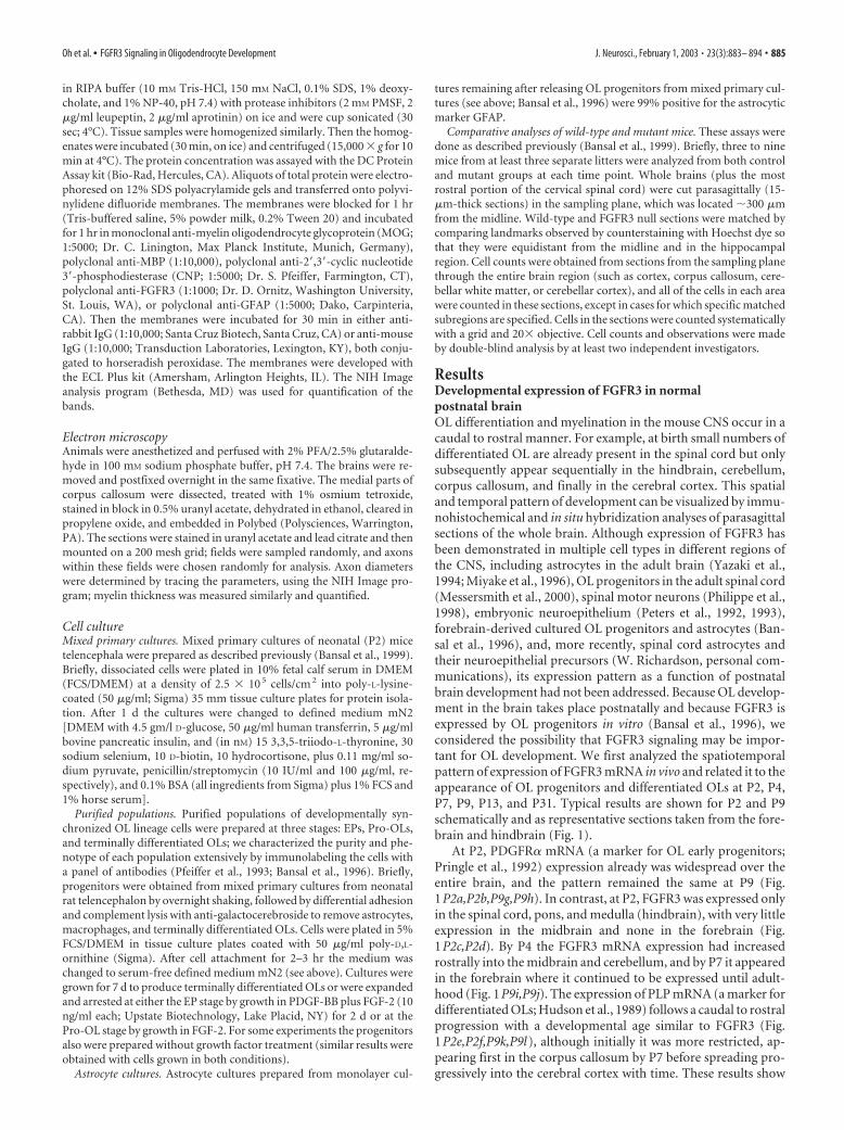
ResultsDevelopmental expression of FGFR3 in normalpostnatal brainOL differentiation and myelination in the mouse CNS occur in acaudal to rostral manner. For example, at birth small numbers ofdifferentiated OL are already present in the spinal cord but onlysubsequently appear sequentially in the hindbrain, cerebellum,corpus callosum, and finally in the cerebral cortex. This spatialand temporal pattern of development can be visualized by immu-nohistochemical and in situ hybridization analyses of parasagittalsections of the whole brain. Although expression of FGFR3 hasbeen demonstrated in multiple cell types in different regions ofthe CNS, including astrocytes in the adult brain (Yazaki et al.,1994; Miyake et al., 1996), OL progenitors in the adult spinal cord(Messersmith et al., 2000), spinal motor neurons (Philippe et al.,1998), embryonic neuroepithelium (Peters et al., 1992, 1993),forebrain-derived cultured OL progenitors and astrocytes (Ban-sal et al., 1996), and, more recently, spinal cord astrocytes andtheir neuroepithelial precursors (W. Richardson, personal com-munications), its expression pattern as a function of postnatalbrain development had not been addressed. Because OL develop-ment in the brain takes place postnatally and because FGFR3 isexpressed by OL progenitors in vitro (Bansal et al., 1996), weconsidered the possibility that FGFR3 signaling may be impor-tant for OL development. We first analyzed the spatiotemporalpattern of expression of FGFR3 mRNA in vivo and related it to theappearance of OL progenitors and differentiated OLs at P2, P4,P7, P9, P13, and P31. Typical results are shown for P2 and P9schematically and as representative sections taken from the fore-brain and hindbrain (Fig. 1).
At P2, PDGFR� mRNA (a marker for OL early progenitors;Pringle et al., 1992) expression already was widespread over theentire brain, and the pattern remained the same at P9 (Fig.1P2a,P2b,P9g,P9h). In contrast, at P2, FGFR3 was expressed onlyin the spinal cord, pons, and medulla (hindbrain), with very littleexpression in the midbrain and none in the forebrain (Fig.1P2c,P2d). By P4 the FGFR3 mRNA expression had increasedrostrally into the midbrain and cerebellum, and by P7 it appearedin the forebrain where it continued to be expressed until adult-hood (Fig. 1P9i,P9j). The expression of PLP mRNA (a marker fordifferentiated OLs; Hudson et al., 1989) follows a caudal to rostralprogression with a developmental age similar to FGFR3 (Fig.1P2e,P2f,P9k,P9l), although initially it was more restricted, ap-pearing first in the corpus callosum by P7 before spreading pro-gressively into the cerebral cortex with time. These results show
Oh et al. • FGFR3 Signaling in Oligodendrocyte Development J. Neurosci., February 1, 2003 • 23(3):883– 894 • 885
that there is a wave of FGFR3 mRNA ex-pression in the normal postnatal brainthat spreads from the hindbrain to theforebrain in parallel to markers for termi-nally differentiated OLs rather than forOL early progenitors.
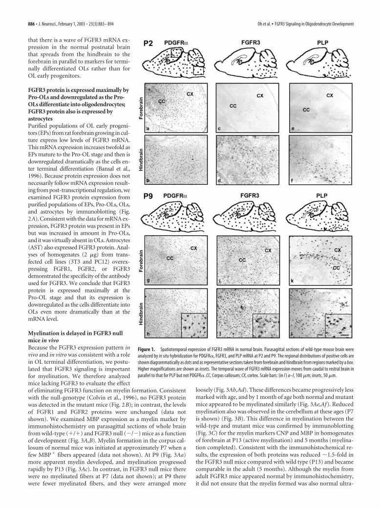
FGFR3 protein is expressed maximally byPro-OLs and downregulated as the Pro-OLs differentiate into oligodendrocytes;FGFR3 protein also is expressed byastrocytesPurified populations of OL early progeni-tors (EPs) from rat forebrain growing in cul-ture express low levels of FGFR3 mRNA.This mRNA expression increases twofold asEPs mature to the Pro-OL stage and then isdownregulated dramatically as the cells en-ter terminal differentiation (Bansal et al.,1996). Because protein expression does notnecessarily follow mRNA expression result-ing from post-transcriptional regulation, weexamined FGFR3 protein expression frompurified populations of EPs, Pro-OLs, OLs,and astrocytes by immunoblotting (Fig.2A). Consistent with the data for mRNA ex-pression, FGFR3 protein was present in EPsbut was increased in amount in Pro-OLs,and it was virtually absent in OLs. Astrocytes(AST) also expressed FGFR3 protein. Anal-yses of homogenates (2 �g) from trans-fected cell lines (3T3 and PC12) overex-pressing FGFR1, FGFR2, or FGFR3demonstrated the specificity of the antibodyused for FGFR3. We conclude that FGFR3protein is expressed maximally at thePro-OL stage and that its expression isdownregulated as the cells differentiate intoOLs even more dramatically than at themRNA level.
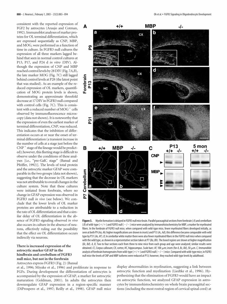
Myelination is delayed in FGFR3 nullmice in vivoBecause the FGFR3 expression pattern invivo and in vitro was consistent with a rolein OL terminal differentiation, we postu-lated that FGFR3 signaling is importantfor myelination. We therefore analyzedmice lacking FGFR3 to evaluate the effectof eliminating FGFR3 function on myelin formation. Consistentwith the null-genotype (Colvin et al., 1996), no FGFR3 proteinwas detected in the mutant mice (Fig. 2B); in contrast, the levelsof FGFR1 and FGFR2 proteins were unchanged (data notshown). We examined MBP expression as a myelin marker byimmunohistochemistry on parasagittal sections of whole brainfrom wild-type (�/�) and FGFR3 null (�/�) mice as a functionof development (Fig. 3A,B). Myelin formation in the corpus cal-losum of normal mice was initiated at approximately P7 when afew MBP� fibers appeared (data not shown). At P9 (Fig. 3Aa)more apparent myelin developed, and myelination progressedrapidly by P13 (Fig. 3Ac). In contrast, in FGFR3 null mice therewere no myelinated fibers at P7 (data not shown); at P9 therewere fewer myelinated fibers, and they were arranged more
loosely (Fig. 3Ab,Ad). These differences became progressively lessmarked with age, and by 1 month of age both normal and mutantmice appeared to be myelinated similarly (Fig. 3Ae,Af). Reducedmyelination also was observed in the cerebellum at these ages (P7is shown) (Fig. 3B). This difference in myelination between thewild-type and mutant mice was confirmed by immunoblotting(Fig. 3C) for the myelin markers CNP and MBP in homogenatesof forebrain at P13 (active myelination) and 5 months (myelina-tion completed). Consistent with the immunohistochemical re-sults, the expression of both proteins was reduced �1.5-fold inthe FGFR3 null mice compared with wild type (P13) and becamecomparable in the adult (5 months). Although the myelin fromadult FGFR3 mice appeared normal by immunohistochemistry,it did not ensure that the myelin formed was also normal ultra-
Figure 1. Spatiotemporal expression of FGFR3 mRNA in normal brain. Parasagittal sections of wild-type mouse brain wereanalyzed by in situ hybridization for PDGFR�, FGFR3, and PLP mRNA at P2 and P9. The regional distributions of positive cells areshown diagrammatically as dots and as representative sections taken from forebrain and hindbrain from regions marked by a box.Higher magnifications are shown as insets. The temporal wave of FGFR3 mRNA expression moves from caudal to rostral brain inparallel to that for PLP but not PDGFR�. CC, Corpus callosum; CX, cortex. Scale bars: (in l ) a–l, 100 �m; insets, 50 �m.
886 • J. Neurosci., February 1, 2003 • 23(3):883– 894 Oh et al. • FGFR3 Signaling in Oligodendrocyte Development
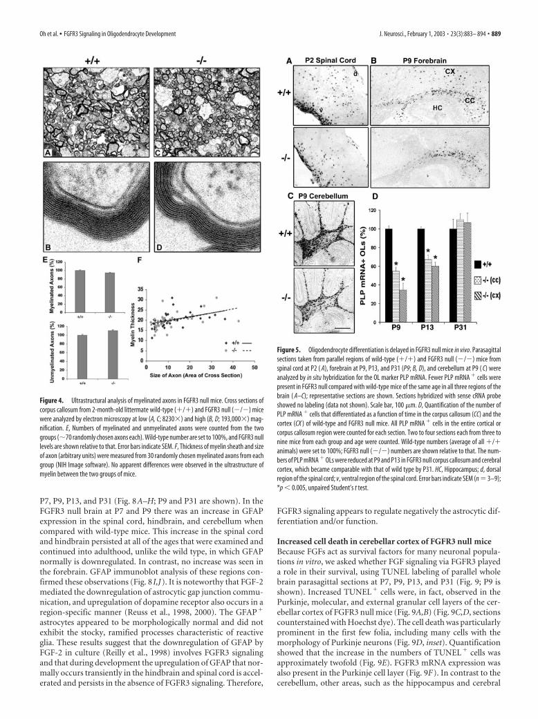
structurally. Therefore, we further analyzed adult FGFR3 null miceby electron microscopy (Fig. 4A–D). Cross sections from the corpuscallosum of 2-month-old wild-type and FGFR3 null littermatesshowed no significant differences in the number of myelinated andunmyelinated axons (Fig. 4E) or in the thickness of myelin sheaths(Fig. 4F).
These results show that myelination is delayed in FGFR3 nullmice but appears structurally normal. We conclude that FGFR3signaling is important for OLs to progress to the stage ofmyelination.
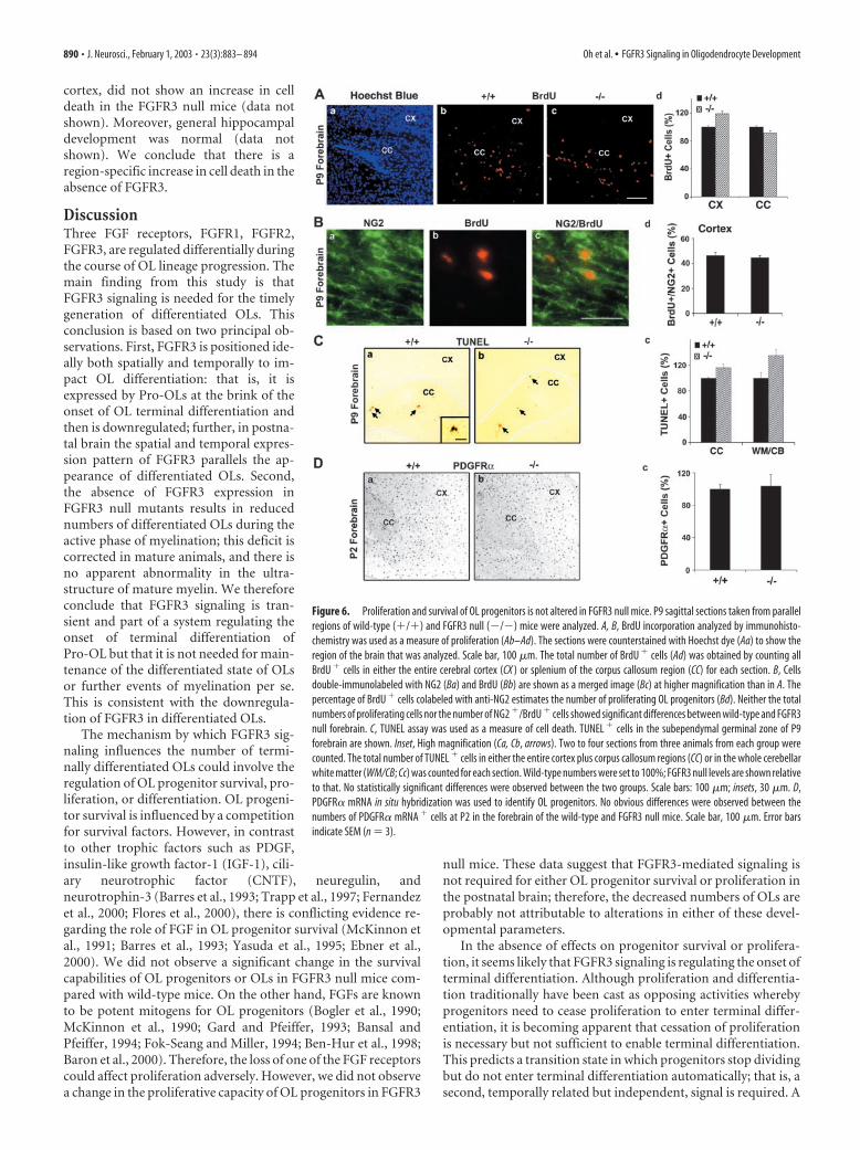
Reduced numbers of differentiated oligodendrocyte appear inFGFR3 null miceWe hypothesized that the reduced myelination at early postnatalages is attributable to a reduction in the number of terminallydifferentiated OLs. To examine this possibility, we determinedthe time course of OL differentiation in parallel parasagittal sec-tions from �/� and �/� littermates, using in situ hybridizationfor PLP mRNA to identify and count the number of OLs (Fig. 5;the estimation of OL cell numbers by immunohistochemistrywith myelin protein markers is difficult because of the back-ground of highly immunolabeled myelinated fibers). During theonset and active phase of myelination there were markedly fewerdifferentiated OLs in all regions of the brains from FGFR3 nullmice, including spinal cord at P2 (Fig. 5A; the most dorsal cervi-cal region is shown), and in the corpus callosum and cortex (Fig.5B) and cerebellum (Fig. 5C) at P9. Quantification of the data forthe corpus callosum and cortex (Fig. 5D) showed that there weretwofold to threefold fewer OLs expressing PLP mRNA in theFGFR3 null than in wild types at P9 and P13. The differencebetween wild type and mutant continued to become progres-sively smaller with increasing age, and the numbers of PLP� cellsreached control levels with subsequent development (P31) (Fig.5D), consistent with the normal level of myelination observed atthis age (Fig. 3Ae,Af). Similar differences in the number of OLsalso were observed in spinal cords from wild-type and mutantmice (data not shown). These results suggest that, during theperiod when myelination is initiated and is progressing actively,fewer differentiated OLs appear in FGFR3 null brain comparedwith wild-type mice.
The decrease in oligodendrocyte number in FGFR3 null miceis not attributable to either reduced proliferation or survivalof oligodendrocyte progenitorsThe reduced number of OLs expressing PLP mRNA observed inFGFR3 null mice during the active phase of myelination could becaused by a reduction in the number of OL progenitors availablefor differentiation to mature OLs, attributable in turn to reducedproliferation and/or increased cell death or, alternatively, to adecreased efficiency of terminal differentiation by OL progeni-tors. We investigated these mechanisms by analyzing whole brainparasagittal sections at P2, P7, P9, and P13 from wild-type (�/�)and FGFR3 null (�/�) mice.
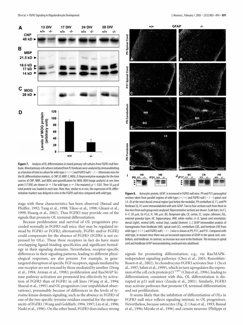
The proliferative capacity of OL progenitors was studied byinjecting BrdU intraperitoneally into these mice 2 hr before thebrains were harvested for double-immunofluorescence micros-copy with anti-BrdU and anti-NG2, a marker for OL progenitors(Fig. 6A,B). No differences in the total number of BrdU� cellswere observed between wild-type and FGFR3 null mice in eitherthe cortex or corpus callosum (Fig. 6Ab–Ad). Further, �40 –50%of the BrdU� cells in the cerebral cortex were also NG2� at P9(Fig. 6Bd), consistent with previous observations (Dawson et al.,2000; Mallon et al., 2002), in both wild-type and FGFR3 nullmice. These data suggest that the proliferation of OL progenitorsis not affected in the forebrain of FGFR3 null mice and cannotaccount for the reduced number of differentiated OLs.
OL cell death was studied in the white matter of FGFR3 null andwild-type mice via the TUNEL assay (a measure of apoptotic celldeath). In the normal mouse brain cell death occurs in the sub-ependymal germinal zone in the forebrain and cerebellum at an earlypostnatal period; then it is reduced progressively to barely detectablelevels in the adult brain (Levison et al., 2000). Although a fewTUNEL� cells were observed at P9 in the forebrain (Fig. 6Ca,Cb)and in the corpus callosum and white matter of the cerebellum(WM/CB) (Fig. 6Cc), the numbers did not differ between wild-typeand FGFR3 null mice. TUNEL� cells were not observed in the spinalcord or hindbrain (data not shown). We conclude that reduced sur-vival of OL progenitors or newly formed OLs is not the cause ofreduced numbers of differentiated OLs seen in FGFR3 null miceduring early postnatal differentiation.
Consistent with the absence of any changes in the proliferativeor survival capacities, the numbers of OL early progenitors, de-termined by in situ hybridization for the progenitor markersPDGFR� (Fig. 6D) or Olig-1 (data not shown), were similar inwild-type and FGFR3 null mice at P2. We conclude that changesin OL progenitor population size do not account for the reducednumber of terminally differentiated OLs in FGFR3 null mice.
In the absence of changes in OL progenitor survival, prolifera-tion, or cell number, we conclude that the decrease in the number ofterminally differentiated OLs in the FGFR3 null mice is attributableto delays in the timing of their differentiation process.
Oligodendrocyte differentiation also is inhibited in mixedprimary cultures from FGFR3 null forebrainOL development in vivo is orchestrated by complex interactionsamong at least three major cell types, OLs, astrocytes, and neu-rons. We studied the roles of these interactions on the regulationof OL number by examining the timing and extent of OL differ-entiation in mixed primary cultures grown in defined mediuminitiated from P1 forebrains of either wild-type (�/�) or FGFR3null (�/�) littermates (Fig. 7). These cultures are devoid of neu-rons, as indicated by the virtual absence of the neuron-specificmarker tubulin III (data not shown). FGF2, a ligand for FGFR3, ispresent in these cultures (immunoblot analyses; data not shown),
Figure 2. The expression of FGFR3 protein is regulated during the maturation of OL lineagecells. A, Purified cells from rat forebrain, at three stages of OL maturation and astrocytes, wereanalyzed by immunoblotting for FGFR3. Equal amounts of total protein (50 �g) were loaded ineach lane. FGFR3 protein is expressed by early progenitors (EP), is increased substantially as EPsmature to Pro-OLs, and is downregulated dramatically to undetectable levels with the terminaldifferentiation of progenitors into OLs (OL). Astrocytes (AST ) also express FGFR3 protein. Anal-yses of homogenates (2 �g) from transfected cell lines (3T3 and PC12) overexpressing FGFR1,FGFR2, or FGFR3 demonstrate the specificity of the antibody used for FGFR3. B, Immunoblots ofhomogenates (50 �g) from P9 wild types (�/�), heterozygotes (�/�), and FGFR3 null(�/�) hindbrains with anti-FGFR3 show the loss of FGFR3 protein to undetectable levels inFGFR3 null animals. Representative experiments of three are shown.
Oh et al. • FGFR3 Signaling in Oligodendrocyte Development J. Neurosci., February 1, 2003 • 23(3):883– 894 • 887
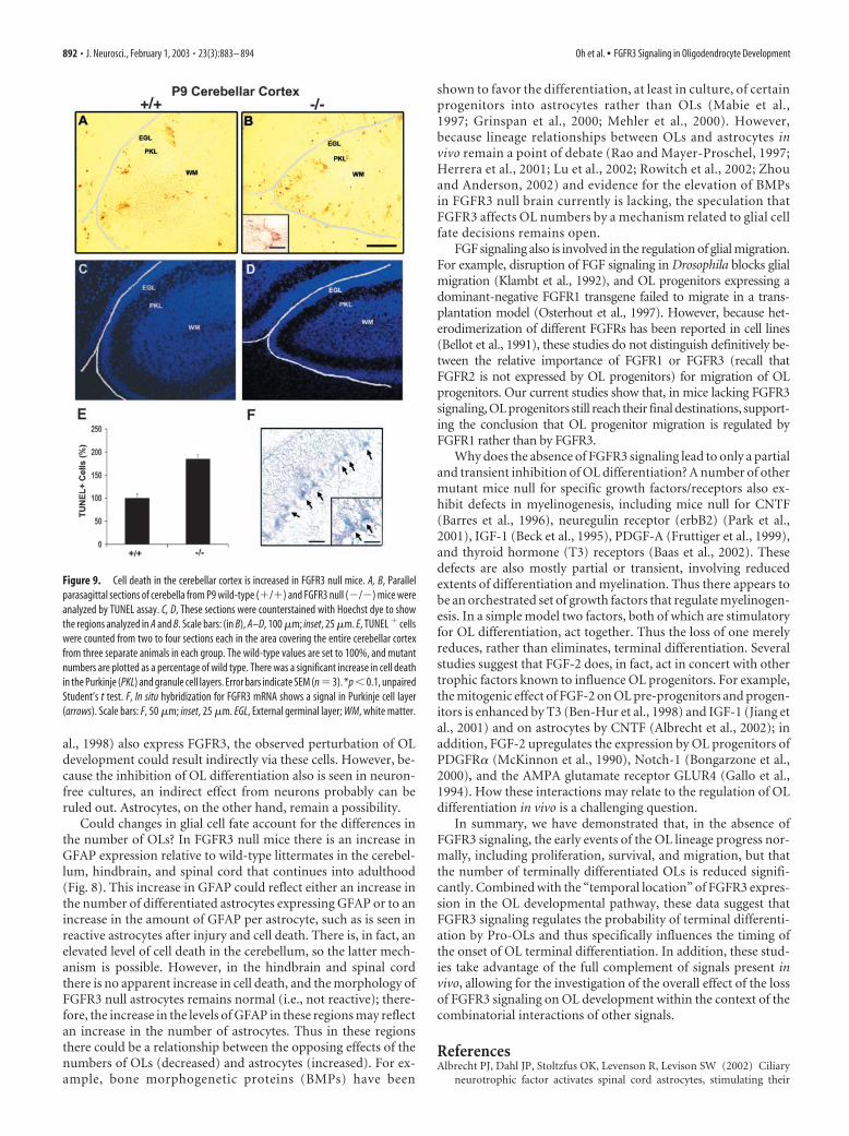
consistent with the reported expression ofFGF2 by astrocytes (Araujo and Cotman,1992). Immunoblot analyses of marker pro-teins for OL terminal differentiation, whichare expressed sequentially as CNP, MBP,and MOG, were performed as a function oftime in culture. In FGFR3 null cultures theexpression of all three markers lagged be-hind that seen in normal control cultures atP13, P17, and P24 d in vitro (DIV). Al-though the expression of CNP and MBPreached control levels by 28 DIV (Fig. 7A,B),the late marker MOG (Fig. 7C) still laggedbehind control levels at P28 (the latest pointthat was studied). As an example of the re-duced expression of OL markers, quantifi-cation of MOG protein levels is shown,demonstrating an approximate threefolddecrease at 17 DIV in FGFR3 null comparedwith control cells (Fig. 7C). This is consis-tent with a reduced number of MOG� cellsobserved by immunofluorescence micros-copy (data not shown). It is noteworthy thatthe expression of even the earliest marker ofterminal differentiation, CNP, was reduced.This indicates that the inhibition of differ-entiation occurs at or near the onset of ter-minal differentiation (a transient increase inthe number of cells at a stage just before theCNP� stage of the lineage would be predict-ed); however, this fleeting stage is difficult toobserve under the conditions of these anal-yses [i.e., “pre-GalC stage” (Bansal andPfeiffer, 1992)]. The levels of total proteinand the astrocytic marker GFAP were com-parable in the two groups (data not shown),suggesting that the decrease in OL markerswas not attributable to overall changes in theculture system. Note that these cultureswere initiated from forebrain, where nochange in GFAP expression was observed inFGFR3 null in vivo (see below). We con-clude that the lower levels of OL markerproteins are attributable to a reduction inthe rate of OL differentiation and that a sim-ilar delay of OL differentiation in the ab-sence of FGFR3 signaling observed in vivoalso occurs in culture in the absence of neu-rons, effectively ruling out the possibilitythat the effect on OL differentiation occursindirectly via neurons.
There is increased expression of theastrocytic marker GFAP in thehindbrain and cerebellum of FGFR3null mice, but not in the forebrainAstrocytes express FGFR3 (Fig. 2) (Bansalet al., 1996; Miyake et al., 1996) and proliferate in response toFGFs. During development the differentiation of astrocytes isaccompanied by the expression of GFAP, a marker for astrocyticmaturation (Goldman, 2001). In adults the astrocytes thendownregulate GFAP expression in a region-specific manner(DiProspero et al., 1997; Reilly et al., 1998). GFAP null mice
display abnormalities in myelination, suggesting a link betweenastrocytic function and myelination (Liedtke et al., 1996). Hy-pothesizing that the elimination of FGFR3 would have an impacton astrocytic function, we analyzed GFAP expression in astro-cytes by immunohistochemistry on whole brain parasagittal sec-tions (including the most rostral region of cervical spinal cord) at
Figure 3. Myelin formation is delayed in FGFR3 null mice brain. Parallel parasagittal sections from forebrain ( A) and cerebellum( B) of wild-type (�/�) and FGFR3 null (�/�) mice were analyzed by immunohistochemistry for MBP, a marker for myelinatedfibers. In the forebrain of FGFR3 null mice, when compared with wild-type mice, fewer myelinated fibers developed initially, asseen at both P9 (Aa, Ab; higher magnifications are shown in insets) and P13 (Ac, Ad); this difference became comparable with wildtype by P31 (Ae, Af ). B, In cerebellar white matter there were also fewer myelinated fibers in the FGFR3 null mice when comparedwith the wild type, as shown in a representative section taken at P7 (Ba, Bb). The boxed regions are shown at higher magnification(Bc, Bd). A, B, Two to four sections each from three to nine mice from each group and age were analyzed; similar results wereobtained. CC, Corpus callosum; CX, cortex; HC, hippocampus. Scale bars: Af, 100 �m; insets (for A, Bc, Bd), 50 �m. C, Immunoblotanalysis of forebrain homogenates from wild-type (�/�) and FGFR3 null (�/�) mice. Compared with wild-type mice, in FGFR3null mice the levels of CNP and MBP isoforms were reduced at P13; however, they reached wild-type levels by adulthood.
888 • J. Neurosci., February 1, 2003 • 23(3):883– 894 Oh et al. • FGFR3 Signaling in Oligodendrocyte Development
P7, P9, P13, and P31 (Fig. 8A–H; P9 and P31 are shown). In theFGFR3 null brain at P7 and P9 there was an increase in GFAPexpression in the spinal cord, hindbrain, and cerebellum whencompared with wild-type mice. This increase in the spinal cordand hindbrain persisted at all of the ages that were examined andcontinued into adulthood, unlike the wild type, in which GFAPnormally is downregulated. In contrast, no increase was seen inthe forebrain. GFAP immunoblot analysis of these regions con-firmed these observations (Fig. 8 I,J). It is noteworthy that FGF-2mediated the downregulation of astrocytic gap junction commu-nication, and upregulation of dopamine receptor also occurs in aregion-specific manner (Reuss et al., 1998, 2000). The GFAP�
astrocytes appeared to be morphologically normal and did notexhibit the stocky, ramified processes characteristic of reactiveglia. These results suggest that the downregulation of GFAP byFGF-2 in culture (Reilly et al., 1998) involves FGFR3 signalingand that during development the upregulation of GFAP that nor-mally occurs transiently in the hindbrain and spinal cord is accel-erated and persists in the absence of FGFR3 signaling. Therefore,
FGFR3 signaling appears to regulate negatively the astrocytic dif-ferentiation and/or function.
Increased cell death in cerebellar cortex of FGFR3 null miceBecause FGFs act as survival factors for many neuronal popula-tions in vitro, we asked whether FGF signaling via FGFR3 playeda role in their survival, using TUNEL labeling of parallel wholebrain parasagittal sections at P7, P9, P13, and P31 (Fig. 9; P9 isshown). Increased TUNEL� cells were, in fact, observed in thePurkinje, molecular, and external granular cell layers of the cer-ebellar cortex of FGFR3 null mice (Fig. 9A,B) (Fig. 9C,D, sectionscounterstained with Hoechst dye). The cell death was particularlyprominent in the first few folia, including many cells with themorphology of Purkinje neurons (Fig. 9D, inset). Quantificationshowed that the increase in the numbers of TUNEL� cells wasapproximately twofold (Fig. 9E). FGFR3 mRNA expression wasalso present in the Purkinje cell layer (Fig. 9F). In contrast to thecerebellum, other areas, such as the hippocampus and cerebral
Figure 4. Ultrastructural analysis of myelinated axons in FGFR3 null mice. Cross sections ofcorpus callosum from 2-month-old littermate wild-type (�/�) and FGFR3 null (�/�) micewere analyzed by electron microscopy at low (A, C; 8230�) and high (B, D; 193,000�) mag-nification. E, Numbers of myelinated and unmyelinated axons were counted from the twogroups (�70 randomly chosen axons each). Wild-type number are set to 100%, and FGFR3 nulllevels are shown relative to that. Error bars indicate SEM. F, Thickness of myelin sheath and sizeof axon (arbitrary units) were measured from 30 randomly chosen myelinated axons from eachgroup (NIH Image software). No apparent differences were observed in the ultrastructure ofmyelin between the two groups of mice.
Figure 5. Oligodendrocyte differentiation is delayed in FGFR3 null mice in vivo. Parasagittalsections taken from parallel regions of wild-type (�/�) and FGFR3 null (�/�) mice fromspinal cord at P2 ( A), forebrain at P9, P13, and P31 (P9; B, D), and cerebellum at P9 ( C) wereanalyzed by in situ hybridization for the OL marker PLP mRNA. Fewer PLP mRNA � cells werepresent in FGFR3 null compared with wild-type mice of the same age in all three regions of thebrain ( A–C); representative sections are shown. Sections hybridized with sense cRNA probeshowed no labeling (data not shown). Scale bar, 100 �m. D, Quantification of the number ofPLP mRNA � cells that differentiated as a function of time in the corpus callosum (CC) and thecortex (CX ) of wild-type and FGFR3 null mice. All PLP mRNA � cells in the entire cortical orcorpus callosum region were counted for each section. Two to four sections each from three tonine mice from each group and age were counted. Wild-type numbers (average of all �/�animals) were set to 100%; FGFR3 null (�/�) numbers are shown relative to that. The num-bers of PLP mRNA � OLs were reduced at P9 and P13 in FGFR3 null corpus callosum and cerebralcortex, which became comparable with that of wild type by P31. HC, Hippocampus; d, dorsalregion of the spinal cord; v, ventral region of the spinal cord. Error bars indicate SEM (n � 3–9);*p � 0.005, unpaired Student’s t test.
Oh et al. • FGFR3 Signaling in Oligodendrocyte Development J. Neurosci., February 1, 2003 • 23(3):883– 894 • 889
cortex, did not show an increase in celldeath in the FGFR3 null mice (data notshown). Moreover, general hippocampaldevelopment was normal (data notshown). We conclude that there is aregion-specific increase in cell death in theabsence of FGFR3.
DiscussionThree FGF receptors, FGFR1, FGFR2,FGFR3, are regulated differentially duringthe course of OL lineage progression. Themain finding from this study is thatFGFR3 signaling is needed for the timelygeneration of differentiated OLs. Thisconclusion is based on two principal ob-servations. First, FGFR3 is positioned ide-ally both spatially and temporally to im-pact OL differentiation: that is, it isexpressed by Pro-OLs at the brink of theonset of OL terminal differentiation andthen is downregulated; further, in postna-tal brain the spatial and temporal expres-sion pattern of FGFR3 parallels the ap-pearance of differentiated OLs. Second,the absence of FGFR3 expression inFGFR3 null mutants results in reducednumbers of differentiated OLs during theactive phase of myelination; this deficit iscorrected in mature animals, and there isno apparent abnormality in the ultra-structure of mature myelin. We thereforeconclude that FGFR3 signaling is tran-sient and part of a system regulating theonset of terminal differentiation ofPro-OL but that it is not needed for main-tenance of the differentiated state of OLsor further events of myelination per se.This is consistent with the downregula-tion of FGFR3 in differentiated OLs.
The mechanism by which FGFR3 sig-naling influences the number of termi-nally differentiated OLs could involve theregulation of OL progenitor survival, pro-liferation, or differentiation. OL progeni-tor survival is influenced by a competitionfor survival factors. However, in contrastto other trophic factors such as PDGF,insulin-like growth factor-1 (IGF-1), cili-ary neurotrophic factor (CNTF), neuregulin, andneurotrophin-3 (Barres et al., 1993; Trapp et al., 1997; Fernandezet al., 2000; Flores et al., 2000), there is conflicting evidence re-garding the role of FGF in OL progenitor survival (McKinnon etal., 1991; Barres et al., 1993; Yasuda et al., 1995; Ebner et al.,2000). We did not observe a significant change in the survivalcapabilities of OL progenitors or OLs in FGFR3 null mice com-pared with wild-type mice. On the other hand, FGFs are knownto be potent mitogens for OL progenitors (Bogler et al., 1990;McKinnon et al., 1990; Gard and Pfeiffer, 1993; Bansal andPfeiffer, 1994; Fok-Seang and Miller, 1994; Ben-Hur et al., 1998;Baron et al., 2000). Therefore, the loss of one of the FGF receptorscould affect proliferation adversely. However, we did not observea change in the proliferative capacity of OL progenitors in FGFR3
null mice. These data suggest that FGFR3-mediated signaling isnot required for either OL progenitor survival or proliferation inthe postnatal brain; therefore, the decreased numbers of OLs areprobably not attributable to alterations in either of these devel-opmental parameters.
In the absence of effects on progenitor survival or prolifera-tion, it seems likely that FGFR3 signaling is regulating the onset ofterminal differentiation. Although proliferation and differentia-tion traditionally have been cast as opposing activities wherebyprogenitors need to cease proliferation to enter terminal differ-entiation, it is becoming apparent that cessation of proliferationis necessary but not sufficient to enable terminal differentiation.This predicts a transition state in which progenitors stop dividingbut do not enter terminal differentiation automatically; that is, asecond, temporally related but independent, signal is required. A
Figure 6. Proliferation and survival of OL progenitors is not altered in FGFR3 null mice. P9 sagittal sections taken from parallelregions of wild-type (�/�) and FGFR3 null (�/�) mice were analyzed. A, B, BrdU incorporation analyzed by immunohisto-chemistry was used as a measure of proliferation (Ab–Ad). The sections were counterstained with Hoechst dye (Aa) to show theregion of the brain that was analyzed. Scale bar, 100 �m. The total number of BrdU � cells (Ad) was obtained by counting allBrdU � cells in either the entire cerebral cortex (CX ) or splenium of the corpus callosum region (CC) for each section. B, Cellsdouble-immunolabeled with NG2 (Ba) and BrdU (Bb) are shown as a merged image (Bc) at higher magnification than in A. Thepercentage of BrdU � cells colabeled with anti-NG2 estimates the number of proliferating OL progenitors (Bd). Neither the totalnumbers of proliferating cells nor the number of NG2 �/BrdU � cells showed significant differences between wild-type and FGFR3null forebrain. C, TUNEL assay was used as a measure of cell death. TUNEL � cells in the subependymal germinal zone of P9forebrain are shown. Inset, High magnification (Ca, Cb, arrows). Two to four sections from three animals from each group werecounted. The total number of TUNEL � cells in either the entire cortex plus corpus callosum regions (CC) or in the whole cerebellarwhite matter (WM/CB; Cc) was counted for each section. Wild-type numbers were set to 100%; FGFR3 null levels are shown relativeto that. No statistically significant differences were observed between the two groups. Scale bars: 100 �m; insets, 30 �m. D,PDGFR� mRNA in situ hybridization was used to identify OL progenitors. No obvious differences were observed between thenumbers of PDGFR� mRNA � cells at P2 in the forebrain of the wild-type and FGFR3 null mice. Scale bar, 100 �m. Error barsindicate SEM (n � 3).
890 • J. Neurosci., February 1, 2003 • 23(3):883– 894 Oh et al. • FGFR3 Signaling in Oligodendrocyte Development
stage with these characteristics has been observed (Bansal andPfeiffer, 1992; Tang et al., 1998; Tikoo et al., 1998; Ghiani et al.,1999; Huang et al., 2002). Thus FGFR3 may provide one of thesignals that promote OL terminal differentiation.
Because proliferation and survival of OL progenitors pro-ceeded normally in FGFR3 null mice, they may be regulated in-stead by FGFR1 or FGFR2; alternatively, FGFR1 and/or FGFR2could compensate for the absence of FGFR3 (FGFR4 is not ex-pressed by OLs). These three receptors in fact do have manyoverlapping ligand-binding specificities and significant homol-ogy in their signaling domains. Nevertheless, receptor-specificdifferences in their signaling patterns, leading to different physi-ological responses, are also present. For example, in gene-targeted disruption of specific FGF receptors, signals mediated byone receptor are not rescued by those mediated by another (Denget al., 1994; Arman et al., 1998); proliferation and Ras/MAP ki-nase pathway activation are promoted less effectively by activa-tion of FGFR3 than of FGFR1 in cell lines (Wang et al., 1994;Shaoul et al., 1995) and OL progenitors (our unpublished obser-vations), presumably because of differences in the levels of ty-rosine kinase domain signaling, such as the absence in FGFR3 ofone of the two specific tyrosine residues essential for the mitoge-nicity of FGFR1 (Wang and Goldfarb, 1994, 1997; Lin et al., 1996;Naski et al., 1996). On the other hand, FGFR3 does induce strong
signals for promoting differentiation, e.g., via Ras/MAPK-independent signaling pathways (Choi et al., 2001; Rozenblatt-Rosen et al., 2002). In chondrocytes FGFR3 activates Stat-1 (Su etal., 1997; Sahni et al., 1999), which in turn upregulates the expres-sion of the cell cycle protein p21 CIP1 (Chin et al., 1996), leading todifferentiation; consistent with this, OL differentiation is dis-rupted in p21 null mice (Zezula et al., 2001). Similarly, FGFR3may activate pathways that promote OL terminal differentiationand not proliferation.
It seems likely that the inhibition of differentiation of OLs inFGFR3 null mice reflects signaling intrinsic to OL progenitors.Nevertheless, because astrocytes (Fig. 2) (Asai et al., 1993; Bansalet al., 1996; Miyake et al., 1996) and certain neurons (Philippe et
Figure 7. Analysis of OL differentiation in mixed primary cell cultures from FGFR3 null fore-brain. Mixed primary cell cultures initiated from P2 forebrain were analyzed by immunoblottingas a function of time in culture for wild-type (�/�) and FGFR3 null (�/�) littermate mice forthe OL differentiation markers. A, CNP; B, MBP; C, MOG. D, Representative examples for the timecourses of CNP, MBP, and MOG and quantification for MOG (NIH Image analysis) at one timepoint (17 DIV) are shown (n � 3 for wild type; n � 5 for mutants); p � 0.05. Then 10 �g oftotal protein was loaded in each lane. Note that, similar to in vivo, the expression of OL differ-entiation markers was delayed in vitro in the FGFR3 null mice compared with wild type. Figure 8. Astrocytic protein, GFAP, is increased in FGFR3 null mice. P9 and P31 parasagittal
sections taken from parallel regions of wild-type (�/�) and FGFR3 null (�/�) spinal cord( A–D) at the most dorsal cervical region (just below the medulla), P9 cerebellum (E, F ), and P9forebrain (G, H ) were immunolabeled with anti-GFAP. Two to four sections each from three tofive mice from each group were analyzed. Representative sections are shown. Scale bars: (in F ),A–F, 50 �m; (in H ),G, H, 100 �m. BG, Bergmann glia; CX, cortex; CC, corpus callosum; EGL,external granular layer; HC, hippocampus; WM, white matter. A–D, Spinal cord orientation:dorsal (right), ventral (left), rostral (top), caudal (bottom). I, J, GFAP immunoblot analysis ofhomogenates from hindbrain (HB), spinal cord (SC), cerebellum (CB), and forebrain (FB) fromwild-type (�/�) and FGFR3 null (�/�) mice is shown at P9, P17, and P31. Compared withwild type, in mutant mice there was an increased expression of GFAP in the spinal cord, cere-bellum, and hindbrain. In contrast, no increase was seen in the forebrain. The increase in spinalcord and hindbrain GFAP immunolabeling continued into adulthood.
Oh et al. • FGFR3 Signaling in Oligodendrocyte Development J. Neurosci., February 1, 2003 • 23(3):883– 894 • 891
al., 1998) also express FGFR3, the observed perturbation of OLdevelopment could result indirectly via these cells. However, be-cause the inhibition of OL differentiation also is seen in neuron-free cultures, an indirect effect from neurons probably can beruled out. Astrocytes, on the other hand, remain a possibility.
Could changes in glial cell fate account for the differences inthe number of OLs? In FGFR3 null mice there is an increase inGFAP expression relative to wild-type littermates in the cerebel-lum, hindbrain, and spinal cord that continues into adulthood(Fig. 8). This increase in GFAP could reflect either an increase inthe number of differentiated astrocytes expressing GFAP or to anincrease in the amount of GFAP per astrocyte, such as is seen inreactive astrocytes after injury and cell death. There is, in fact, anelevated level of cell death in the cerebellum, so the latter mech-anism is possible. However, in the hindbrain and spinal cordthere is no apparent increase in cell death, and the morphology ofFGFR3 null astrocytes remains normal (i.e., not reactive); there-fore, the increase in the levels of GFAP in these regions may reflectan increase in the number of astrocytes. Thus in these regionsthere could be a relationship between the opposing effects of thenumbers of OLs (decreased) and astrocytes (increased). For ex-ample, bone morphogenetic proteins (BMPs) have been
shown to favor the differentiation, at least in culture, of certainprogenitors into astrocytes rather than OLs (Mabie et al.,1997; Grinspan et al., 2000; Mehler et al., 2000). However,because lineage relationships between OLs and astrocytes invivo remain a point of debate (Rao and Mayer-Proschel, 1997;Herrera et al., 2001; Lu et al., 2002; Rowitch et al., 2002; Zhouand Anderson, 2002) and evidence for the elevation of BMPsin FGFR3 null brain currently is lacking, the speculation thatFGFR3 affects OL numbers by a mechanism related to glial cellfate decisions remains open.
FGF signaling also is involved in the regulation of glial migration.For example, disruption of FGF signaling in Drosophila blocks glialmigration (Klambt et al., 1992), and OL progenitors expressing adominant-negative FGFR1 transgene failed to migrate in a trans-plantation model (Osterhout et al., 1997). However, because het-erodimerization of different FGFRs has been reported in cell lines(Bellot et al., 1991), these studies do not distinguish definitively be-tween the relative importance of FGFR1 or FGFR3 (recall thatFGFR2 is not expressed by OL progenitors) for migration of OLprogenitors. Our current studies show that, in mice lacking FGFR3signaling, OL progenitors still reach their final destinations, support-ing the conclusion that OL progenitor migration is regulated byFGFR1 rather than by FGFR3.
Why does the absence of FGFR3 signaling lead to only a partialand transient inhibition of OL differentiation? A number of othermutant mice null for specific growth factors/receptors also ex-hibit defects in myelinogenesis, including mice null for CNTF(Barres et al., 1996), neuregulin receptor (erbB2) (Park et al.,2001), IGF-1 (Beck et al., 1995), PDGF-A (Fruttiger et al., 1999),and thyroid hormone (T3) receptors (Baas et al., 2002). Thesedefects are also mostly partial or transient, involving reducedextents of differentiation and myelination. Thus there appears tobe an orchestrated set of growth factors that regulate myelinogen-esis. In a simple model two factors, both of which are stimulatoryfor OL differentiation, act together. Thus the loss of one merelyreduces, rather than eliminates, terminal differentiation. Severalstudies suggest that FGF-2 does, in fact, act in concert with othertrophic factors known to influence OL progenitors. For example,the mitogenic effect of FGF-2 on OL pre-progenitors and progen-itors is enhanced by T3 (Ben-Hur et al., 1998) and IGF-1 (Jiang etal., 2001) and on astrocytes by CNTF (Albrecht et al., 2002); inaddition, FGF-2 upregulates the expression by OL progenitors ofPDGFR� (McKinnon et al., 1990), Notch-1 (Bongarzone et al.,2000), and the AMPA glutamate receptor GLUR4 (Gallo et al.,1994). How these interactions may relate to the regulation of OLdifferentiation in vivo is a challenging question.
In summary, we have demonstrated that, in the absence ofFGFR3 signaling, the early events of the OL lineage progress nor-mally, including proliferation, survival, and migration, but thatthe number of terminally differentiated OLs is reduced signifi-cantly. Combined with the “temporal location” of FGFR3 expres-sion in the OL developmental pathway, these data suggest thatFGFR3 signaling regulates the probability of terminal differenti-ation by Pro-OLs and thus specifically influences the timing ofthe onset of OL terminal differentiation. In addition, these stud-ies take advantage of the full complement of signals present invivo, allowing for the investigation of the overall effect of the lossof FGFR3 signaling on OL development within the context of thecombinatorial interactions of other signals.
ReferencesAlbrecht PJ, Dahl JP, Stoltzfus OK, Levenson R, Levison SW (2002) Ciliary
neurotrophic factor activates spinal cord astrocytes, stimulating their
Figure 9. Cell death in the cerebellar cortex is increased in FGFR3 null mice. A, B, Parallelparasagittal sections of cerebella from P9 wild-type (�/�) and FGFR3 null (�/�) mice wereanalyzed by TUNEL assay. C, D, These sections were counterstained with Hoechst dye to showthe regions analyzed in A and B. Scale bars: (in B), A–D, 100 �m; inset, 25 �m. E, TUNEL � cellswere counted from two to four sections each in the area covering the entire cerebellar cortexfrom three separate animals in each group. The wild-type values are set to 100%, and mutantnumbers are plotted as a percentage of wild type. There was a significant increase in cell deathin the Purkinje (PKL) and granule cell layers. Error bars indicate SEM (n � 3). *p � 0.1, unpairedStudent’s t test. F, In situ hybridization for FGFR3 mRNA shows a signal in Purkinje cell layer(arrows). Scale bars: F, 50 �m; inset, 25 �m. EGL, External germinal layer; WM, white matter.
892 • J. Neurosci., February 1, 2003 • 23(3):883– 894 Oh et al. • FGFR3 Signaling in Oligodendrocyte Development
production and release of fibroblast growth factor-2, to increase motorneuron survival. Exp Neurol 173:46 – 62.
Araujo DM, Cotman CW (1992) Basic FGF in astroglial, microglial, andneuronal cultures: characterization of binding sites and modulation ofrelease by lymphokines and trophic factors. J Neurosci 12:1668 –1678.
Arman E, Haffner-Krausz R, Chen Y, Heath JK, Lonai P (1998) Targeteddisruption of fibroblast growth factor (FGF) receptor 2 suggests a role forFGF signaling in pregastrulation mammalian development. Proc NatlAcad Sci USA 95:5082–5087.
Asai T, Wanaka A, Kato H, Masana Y, Seo M, Tohyama M (1993) Differen-tial expression of two members of FGF receptor gene family, FGFR1 andFGFR2 mRNA, in the adult rat central nervous system. Brain Res MolBrain Res 17:174 –178.
Baas D, Legrand C, Samarut J, Flamant F (2002) Persistence of oligodendro-cyte precursor cells and altered myelination in optic nerve associated toretina degeneration in mice devoid of all thyroid hormone receptors. ProcNatl Acad Sci USA 99:2907–2911.
Bansal R (2002) Fibroblast growth factors and their receptors in oligoden-drocyte development: implications for demyelination and remyelination.Dev Neurosci 24:35– 46.
Bansal R, Pfeiffer SE (1992) Novel stage in the oligodendrocyte lineage de-fined by reactivity of progenitors with R-mAb prior to O-1 anti-galactocerebroside. J Neurosci Res 32:309 –316.
Bansal R, Pfeiffer SE (1994) Inhibition of protein and lipid sulfation in oli-godendrocytes blocks biological responses to FGF-2 and retards cytoar-chitectural maturation, but not developmental lineage progression. DevBiol 162:511–524.
Bansal R, Pfeiffer SE (1997) FGF-2 converts mature oligodendrocytes to anovel phenotype. J Neurosci Res 50:215–228.
Bansal R, Kumar M, Murray K, Morrison RS, Pfeiffer SE (1996) Regulationof FGF receptors in the oligodendrocyte lineage. Mol Cell Neurosci7:263–275.
Bansal R, Winkler S, Bheddah S (1999) Negative regulation of oligodendro-cyte differentiation by galactosphingolipids. J Neurosci 19:7913–7924.
Baron W, Metz B, Bansal R, Hoekstra D, de Vries H (2000) PDGF andFGF-2 signaling in oligodendrocyte progenitor cells: regulation of prolif-eration and differentiation by multiple intracellular signaling pathways.Mol Cell Neurosci 15:314 –329.
Barres BA, Schmid R, Sendnter M, Raff MC (1993) Multiple extracellularsignals are required for long-term oligodendrocyte survival. Develop-ment 118:283–295.
Barres BA, Burne JF, Holtmann B, Thoenen H, Sendtner M, Raff MC (1996)Ciliary neurotrophic factor enhances the rate of oligodendrocyte genera-tion. Mol Cell Neurosci 8:146 –156.
Beck KD, Powell-Braxton L, Widmer HR, Valverde J, Hefti F (1995) IGF1gene disruption results in reduced brain size, CNS hypomyelination, andloss of hippocampal granule and striatal parvalbumin-containing neu-rons. Neuron 14:717–730.
Bellot F, Crumley G, Kaplow JM, Schlessinger J, Jaye M, Dionne CA (1991)Ligand-induced transphosphorylation between different FGF receptors.EMBO J 10:2849 –2854.
Ben-Hur T, Rogister B, Murray K, Rougon G, Dubois-Dalcq M (1998)Growth and fate of PSA-NCAM � precursors of the postnatal brain.J Neurosci 18:5777–5788.
Bogler O, Wren D, Barnett SC, Land H, Noble M (1990) Cooperation be-tween two growth factors promotes extended self-renewal and inhibitsdifferentiation of oligodendrocyte-type-2 astrocyte (O-2A) progenitorcells. Proc Natl Acad Sci USA 87:6368 – 6372.
Bongarzone ER, Byravan S, Givogri MI, Schonmann V, Campagnoni AT(2000) Platelet-derived growth factor and basic fibroblast growth factorregulate cell proliferation and the expression of notch-1 receptor in a newoligodendrocyte cell line. J Neurosci Res 62:319 –328.
Chin YE, Kitagawa M, Su WC, You ZH, Iwamoto Y, Fu XY (1996) Cellgrowth arrest and induction of cyclin-dependent kinase inhibitor p21WAF1/CIP1 mediated by STAT1. Science 272:719 –722.
Choi DY, Toledo-Aral JJ, Lin HY, Ischenko I, Medina L, Safo P, Mandel G,Levinson SR, Halegoua S, Hayman MJ (2001) Fibroblast growth factorreceptor 3 induces gene expression primarily through Ras-independentsignal transduction pathways. J Biol Chem 276:5116 –5122.
Colvin JS, Bohne BA, Harding GW, McEwen DG, Ornitz DM (1996) Skel-etal overgrowth and deafness in mice lacking fibroblast growth factorreceptor 3. Nat Genet 12:390 –397.
Crossley PH, Martinez S, Martin GR (1996) Midbrain development in-duced by FGF8 in the chick embryo. Nature 380:66 – 68.
Dawson MR, Levine JM, Reynolds R (2000) NG2-expressing cells in thecentral nervous system: are they oligodendroglial progenitors? J NeurosciRes 61:471– 479.
Deng CX, Wynshaw-Boris A, Shen MM, Daugherty C, Ornitz DM, Leder P(1994) Murine FGFR1 is required for early postimplantation growth andaxial organization. Genes Dev 8:3045–3057.
Deng CX, Wynshaw-Boris A, Zhou F, Kuo A, Leder P (1996) Fibroblastgrowth factor receptor 3 is a negative regulator of bone growth. Cell84:911–921.
DiProspero NA, Meiners S, Geller HM (1997) Inflammatory cytokines in-teract to modulate extracellular matrix and astrocytic support of neuriteoutgrowth. Exp Neurol 148:628 – 639.
Ebner S, Dunbar M, McKinnon RD (2000) Distinct roles for PI3K in pro-liferation and survival of oligodendrocyte progenitor cells. J Neurosci Res62:336 –345.
Fernandez PA, Tang DG, Cheng L, Prochiantz A, Mudge AW, Raff MC(2000) Evidence that axon-derived neuregulin promotes oligodendro-cyte survival in the developing rat optic nerve. Neuron 28:81–90.
Flores AI, Mallon BS, Matsui T, Ogawa W, Rosenzweig A, Okamoto T, Mack-lin WB (2000) Akt-mediated survival of oligodendrocytes induced byneuregulins. J Neurosci 20:7622–7630.
Fok-Seang J, Miller RH (1994) Distribution and differentiation of A2B5 �
glial precursors in the developing rat spinal cord. J Neurosci Res37:219 –235.
Ford-Perriss M, Abud H, Murphy M (2001) Fibroblast growth factors in thedeveloping central nervous system. Clin Exp Pharmacol Physiol28:493–503.
Fruttiger M, Karlsson L, Hall AC, Abramsson A, Calver AR, Bostrom H,Willetts K, Bertold CH, Heath JK, Betsholtz C, Richardson WD (1999)Defective oligodendrocyte development and severe hypomyelination inPDGF-A knock-out mice. Development 126:457– 467.
Fukuchi-Shimogori T, Grove EA (2001) Neocortex patterning by the se-creted signaling molecule FGF8. Science 294:1071–1074.
Gallo V, Wright P, McKinnon RD (1994) Expression and regulation of aglutamate receptor subunit by bFGF in oligodendrocyte progenitors. Glia10:149 –153.
Gard AL, Pfeiffer SE (1993) Glial cell mitogens bFGF and PDGF differen-tially regulate development of O4 �GalC � oligodendrocyte progenitors.Dev Biol 159:618 – 630.
Ghiani CA, Eisen AM, Yuan X, DePinho RA, McBain CJ, Gallo V (1999)Neurotransmitter receptor activation triggers p27 Kip1 and p21 CIP1 accu-mulation and G1 cell cycle arrest in oligodendrocyte progenitors. Devel-opment 126:1077–1090.
Goldman JE (2001) Developmental origins of astrocytes. In: Glial cell devel-opment (Jessen KR, Richardson WD, eds), pp 55– 69. Oxford: Oxford UP.
Grinspan JB, Edell E, Carpio DF, Beesley JS, Lavy L, Pleasure D, Golden JA(2000) Stage-specific effects of bone morphogenetic proteins on the oli-godendrocyte lineage. J Neurobiol 43:1–17.
Herrera J, Yang H, Zhang SC, Proschel C, Tresco P, Duncan ID, Luskin M,Mayer-Proschel M (2001) Embryonic-derived glial-restricted precursorcells (GRP cells) can differentiate into astrocytes and oligodendrocytes invivo. Exp Neurol 171:11–21.
Huang Z, Tang XM, Cambi F (2002) Downregulation of the retinoblastomaprotein (rb) is associated with rat oligodendrocyte differentiation. MolCell Neurosci 19:250 –262.
Hudson LD, Friedrich Jr VL, Behar T, Dubois-Dalcq M, Lazzarini RA (1989)The initial events in myelin synthesis: orientation of proteolipid proteinin the plasma membrane of cultured oligodendrocytes. J Cell Biol109:717–727.
Jiang F, Frederick TJ, Wood TL (2001) IGF-I synergizes with FGF-2 to stim-ulate oligodendrocyte progenitor entry into the cell cycle. Dev Biol232:414 – 423.
Johnson DE, Williams LT (1993) Structural and functional diversity in theFGF receptor multigene family. Adv Cancer Res 60:1– 41.
Klambt C, Glazer L, Shilo BZ (1992) breathless, a Drosophila FGF receptorhomolog, is essential for migration of tracheal and specific midline glialcells. Genes Dev 6:1668 –1678.
Levison SW, Rothstein RP, Brazel CY, Young GM, Albrecht PJ (2000) Selec-tive apoptosis within the rat subependymal zone: a plausible mechanism
Oh et al. • FGFR3 Signaling in Oligodendrocyte Development J. Neurosci., February 1, 2003 • 23(3):883– 894 • 893

for determining which lineages develop from neural stem cells. Dev Neu-rosci 22:106 –115.
Liedtke W, Edelmann W, Bieri PL, Chiu FC, Cowan NJ, Kucherlapati R, RaineCS (1996) GFAP is necessary for the integrity of CNS white matter ar-chitecture and long-term maintenance of myelination. Neuron17:607– 615.
Lin YZ, Yao SY, Hawiger J (1996) Role of the nuclear localization sequencein fibroblast growth factor-1-stimulated mitogenic pathways. J Biol Chem271:5305–5308.
Lu QR, Sun T, Zhu Z, Ma N, Garcia M, Stiles CD, Rowitch DH (2002)Common developmental requirement for OLIG function indicates a mo-tor neuron/oligodendrocyte connection. Cell 109:75– 86.
Mabie PC, Mehler MF, Marmur R, Papavasiliou A, Song Q, Kessler JA (1997)Bone morphogenetic proteins induce astroglial differentiation of oligo-dendroglial–astroglial progenitor cells. J Neurosci 17:4112– 4120.
Mallon BS, Shick HE, Kidd GJ, Macklin WB (2002) Proteolipid promoteractivity distinguishes two populations of NG2-positive cells throughoutneonatal cortical development. J Neurosci 22:876 – 885.
McKinnon RD, Matsui T, Dubois-Dalcq M, Aaronson SA (1990) FGF mod-ulates the PDGF-driven pathway of oligodendrocyte development. Neu-ron 5:603– 614.
McKinnon RD, Matsui T, Aranda M, Dubois-Dalcq M (1991) A role forfibroblast growth factor in oligodendrocyte development. Ann NY AcadSci 638:378 –386.
McMorris FA, McKinnon RD (1996) Regulation of oligodendrocyte devel-opment and CNS myelination by growth factors: prospects for therapy ofdemyelinating disease. Brain Pathol 6:313–329.
Mehler MF, Mabie PC, Zhu G, Gokhan S, Kessler JA (2000) Developmentalchanges in progenitor cell responsiveness to bone morphogenetic pro-teins differentially modulate progressive CNS lineage fate. Dev Neurosci22:74 – 85.
Messersmith DJ, Murtie JC, Le TQ, Frost EE, Armstrong RC (2000) Fibro-blast growth factor 2 (FGF2) and FGF receptor expression in an experi-mental demyelinating disease with extensive remyelination. J NeurosciRes 62:241–256.
Miller RH (1996) Oligodendrocyte origins. Trends Neurosci 19:92–96.Miyake A, Hattori Y, Ohta M, Itoh N (1996) Rat oligodendrocytes and as-
trocytes preferentially express fibroblast growth factor receptor-2 and -3mRNAs. J Neurosci Res 45:534 –541.
Naski MC, Wang Q, Xu J, Ornitz DM (1996) Graded activation of fibroblastgrowth factor receptor 3 by mutations causing achondroplasia and thana-tophoric dysplasia. Nat Genet 13:233–237.
Oh LY, Yong VW (1996) Astrocytes promote process outgrowth by adulthuman oligodendrocytes in vitro through interaction between bFGF andastrocyte extracellular matrix. Glia 17:237–253.
Ornitz DM, Itoh N (2001) Fibroblast growth factors. Genome Biol 2:3005.Orr-Urtreger A, Givol D, Yayon A, Yarden Y, Lonai P (1991) Developmen-
tal expression of two murine fibroblast growth factor receptors, flg andbek. Development 113:1419 –1434.
Osterhout DJ, Ebner S, Xu J, Ornitz DM, Zazanis GA, McKinnon RD (1997)Transplanted oligodendrocyte progenitor cells expressing a dominant-negative FGF receptor transgene fail to migrate in vivo. J Neurosci17:9122–9132.
Park SK, Miller R, Krane I, Vartanian T (2001) The erbB2 gene is requiredfor the development of terminally differentiated spinal cord oligodendro-cytes. J Cell Biol 154:1245–1258.
Peters K, Werner S, Chen G, Williams LT (1992) Two FGF receptor genes aredifferentially expressed in epithelial and mesenchymal tissues during limbformation and organogenesis in the mouse. Development 114:233–243.
Peters K, Ornitz D, Werner S, Williams L (1993) Unique expression pattern ofthe FGF receptor 3 gene during mouse organogenesis. Dev Biol 155:423–430.
Pfeiffer SE, Warrington AE, Bansal R (1993) The oligodendrocyte and itsmany processes. Trends Cell Biol 3:191–197.
Philippe JM, Garces A, deLapeyiere O (1998) FGF-R3 is expressed in a sub-set of chicken spinal motor neurons. Mech Dev 78:119 –123.
Pringle NP, Mudhar HS, Collarini EJ, Richardson WD (1992) PDGF recep-tors in the rat CNS: during late neurogenesis, PDGF� receptor expressionappears to be restricted to glial cells of the oligodendrocyte lineage. De-velopment 115:535–551.
Qian X, Davis AA, Goderie SK, Temple S (1997) FGF-2 concentration reg-ulates the generation of neurons and glia from multipotent cortical stemcells. Neuron 18:81–93.
Rao MS, Mayer-Proschel M (1997) Glial-restricted precursors are derivedfrom multipotent neuroepithelial stem cells. Dev Biol 188:48 – 63.
Reilly JF, Maher PA, Kumari VG (1998) Regulation of astrocyte GFAP ex-pression by TGF-�1 and FGF-2. Glia 22:202–210.
Reuss B, Dermietzel R, Unsicker K (1998) Fibroblast growth factor-2(FGF-2) differentially regulates connexin (cx) 43 expression and functionin astroglial cells from distinct brain regions. Glia 22:19 –30.
Reuss B, Leung DS, Ohlemeyer C, Kettenmann H, Unsicker K (2000) Re-gionally distinct regulation of astroglial neurotransmitter receptors byfibroblast growth factor-2. Mol Cell Neurosci 16:42–58.
Rowitch DH, Lu QR, Kessaris N, Richardson WD (2002) An “oligarchy”rules neural development. Trends Neurosci 25:417– 422.
Rozenblatt-Rosen O, Mosonego-Ornan E, Sadot E, Madar-Shapiro L, Shei-nin Y, Ginsberg D, Yayon A (2002) Induction of chondrocyte growtharrest by FGF: transcriptional and cytoskeletal alterations. J Cell Sci115:553–562.
Sahni M, Ambrosetti DC, Mansukhani A, Gertner R, Levy D, Basilico C (1999)FGF signaling inhibits chondrocyte proliferation and regulates bone devel-opment through the STAT-1 pathway. Genes Dev 13:1361–1366.
Shaoul E, Reich-Slotky R, Berman B, Ron D (1995) Fibroblast growth factorreceptors display both common and distinct signaling pathways. Onco-gene 10:1553–1561.
Su WC, Kitagawa M, Xue N, Xie B, Garofalo S, Cho J, Deng C, Horton WA, FuXY (1997) Activation of STAT1 by mutant fibroblast growth-factor re-ceptor in thanatophoric dysplasia type II dwarfism. Nature 386:288 –292.
Tang XM, Strocchi P, Cambi F (1998) Changes in the activity of cdk2 andcdk5 accompany differentiation of rat primary oligodendrocytes. J CellBiochem 68:128 –137.
Tikoo R, Osterhout DJ, Casaccia-Bonnefil P, Seth P, Koff A, Chao MV (1998)Ectopic expression of p27 Kip1 in oligodendrocyte progenitor cells resultsin cell cycle growth arrest. J Neurobiol 36:431– 440.
Trapp BD, Nishiyama A, Cheng D, Macklin W (1997) Differentiation anddeath of premyelinating oligodendrocytes in developing rodent brain.J Cell Biol 137:459 – 468.
Vaccarino FM, Schwartz ML, Raballo R, Rhee J, Lyn-Cook R (1999) Fibro-blast growth factor signaling regulates growth and morphogenesis at mul-tiple steps during brain development. Curr Top Dev Biol 46:179 –200.
Wang JK, Goldfarb M (1997) Amino acid residues which distinguish themitogenic potentials of two FGF receptors. Oncogene 14:1767–1778.
Wang JK, Gao G, Goldfarb M (1994) Fibroblast growth factor receptors havedifferent signaling and mitogenic potentials. Mol Cell Biol 14:181–188.
Wang Q, Bardgett ME, Wong M, Wozniak DF, Lou J, McNeil BD, Chen C,Nardi A, Reid DC, Yamada K, Ornitz DM (2002) Ataxia and paroxysmaldyskinesia in mice lacking axonally transported FGF14. Neuron 35:25–38.
Warrington AE, Pfeiffer SE (1992) Proliferation and differentiation of O4 �
oligodendrocytes in postnatal rat cerebellum: analysis in unfixed tissueslices using anti-glycolipid antibodies. J Neurosci Res 33:338 –353.
Woodruff RH, Tekki-Kessaris N, Stiles CD, Rowitch DH, Richardson WD(2001) Oligodendrocyte development in the spinal cord and telenceph-alon: common themes and new perspectives. Int J Dev Neurosci19:379 –385.
Xu J, Liu Z, Ornitz DM (2000) Temporal and spatial gradients of FGF8 andFGF17 regulate proliferation and differentiation of midline cerebellarstructures. Development 127:1833–1843.
Yasuda T, Grinspan J, Stern J, Franceschini B, Bannerman P, Pleasure D(1995) Apoptosis occurs in the oligodendroglial lineage, and is preventedby basic fibroblast growth factor. J Neurosci Res 40:306 –317.
Yazaki N, Hosoi Y, Kawabata K, Miyake A, Minami M, Satoh M, Ohta M,Kawasaki T, Itoh N (1994) Differential expression patterns of mRNAsfor members of the fibroblast growth factor receptor family, FGFR1–FGFR4, in rat brain. J Neurosci Res 37:445– 452.
Ye W, Shimamura K, Rubenstein JL, Hynes MA, Rosenthal A (1998) FGFand Shh signals control dopaminergic and serotonergic cell fate in theanterior neural plate. Cell 93:755–766.
Zezula J, Casaccia-Bonnefil P, Ezhevsky SA, Osterhout DJ, Levine JM, DowdySF, Chao MV, Koff A (2001) p21 CIP1 is required for the differentiationof oligodendrocytes independently of cell cycle withdrawal. EMBO Rep2:27–34.
Zhou Q, Anderson DJ (2002) The bHLH transcription factors OLIG2 andOLIG1 couple neuronal and glial subtype specification. Cell 109:61–73.
894 • J. Neurosci., February 1, 2003 • 23(3):883– 894 Oh et al. • FGFR3 Signaling in Oligodendrocyte Development