Fibroblast Growth Factor-2 Protects Entorhinal Layer II ...
13
The Journal of Neuroscience, February 1, 1996, 76(3):886-898 Fibroblast Growth Factor-2 Protects Entorhinal Layer II Glutamatergic Neurons from Axotomy-Induced Death Daniel A. Peterson, Carrie A. Lucidi-Phillipi, Douglas P. Murphy, Jasodhara Ray, and Fred H. Gage Laboratory of Genetics, The Salk Institute, La Jolla, California 92037 The entorhinal cortex is a major relay between the hippocam- pus and other cortical and subcortical regions. Glutamatergic axons from layer II neurons form the entorhinal cortical projec- tion to the hippocampus via the perforant pathway. We have demonstrated previously that lesion of the perforant pathway causes the death of -30% of entorhinal layer II (ECL2) neurons. To elucidate mechanisms contributing to neuronal death and to investigate strategies preventing it, we identified the phenotype of the vulnerable neuronal population. Sections were immuno- labeled with antibodies to the neuronal markers NeuN, gluta- mate, and calbindin-D,,,, and to receptors for fibroblast growth factor-2 (FGFRl) and NMDA (NMDARl) and were examined using confocal microscopy. Calbindin immunoreactivity was strikingly lamina-specific to ECL2, where one-third of all ECL2 neurons were calbindin-positive. Localization of glutamate re- vealed that half of the glutamatergic ECU neurons coex- pressed calbindin. Quantification using unbiased stereology at 9 weeks after lesion of the per-forant pathway revealed that the only ECL2 neuronal population that experienced a significant (70%) loss (20% of the total) was the population of glutama- tergic ECL2 neurons that did not coexpress calbindin. All ECL2 neurons expressed FGFRI; therefore, we tested the role of FGF-2 in the survival of glutamatergic ECL2 neurons. We grafted fibroblasts genetically engineered to express nerve growth factor or FGFQ and found that only FGF-2 grafts pre- vented loss of the vulnerable glutamatergic/calbindin-negative neurons. We present a hypothesis for the selective vulnerability of these glutamatergic/calbindin-negative ECL2 neurons and address the role of FGF-2 in neuronal rescue. Key words: calbindin; glutamate; trophic factors; fibroblast growth factor; neuronal death; neuroprotection; oxidative stress; calcium; excitotoxicity; Alzheimer’s disease: ischemia; stereology The entorhinal cortex is a crucial relay providing a wide range of cortical input to the hippocampus and providing the hippocampus with an outlet for processed information to the cortical mantle. The principal entorhinal input to the hippocampus comes from glutamatergic projection neurons from entorhinal cortex layer II (ECL2) that, along with input from layer III neurons, travel via a major fiber tract, the perforant pathway (Fig. 1). Entorhinal- hippocampal circuitry has been widely studied with respect to both anatomical (Witter, 1993) and functional (Jones, 1993) con- nections. The entorhinal cortex has been hypothesized to play a functional role in organization and processing of learning and memory both at a systems level (Levisohn and Isacson, 1991; Wiig and Bilkey, 1994) and at the cellular level via long-term potenti- ation (LTP) (Bliss and Lomo, 1973; Abraham et al., 19Y3). How- ever, the entorhinal cortex appears to be particularly vulnerable to neurodegenerative and neuropathological insults, including epi- lepsy (Du and Schwartz, 1992; Heinemann et al., 1093) Alzhei- mer’s disease (Hyman et al., 1984, 1986; Armstrong et al., 1994) and Parkinson’s and Huntington’s diseases (Braak and Braak, 1092). The entorhinal cortex also demonstrates developmental Recervcd Aug. 21. lYY5: rcviscd Oct. 20. IYYS: acccptcd Oct. 23, IYYS. This work wit\ supported by National Institute of Neurological Diseases and Stroke Grant POI-NS2X121. WC thank Steve Forbes, Chung Ho. ;tnd John Leppert for technic;tl itssistancc. We &o thank the reviewers for their connncnts on this manuscrint. Corrc\oundence should be ;tddressed to Daniel A. Pctcrson. The Salk Institute (LOG), iOOl0 North Torrcy Pines Road, La Jolla, CA Y2037. Dr. Murphy‘s current address: University of Colomdo Health Sciences Center, Department of Psychiatry. 4200 East Yth Avenue, C-24’), Denver, CO XO2h2. Copyright 0 IYYO Society for Neuroscience 0270-h474/Y6/1h0XX0-13$05.00/0 abnormalities correlated with schizophrenia (Falkai et al., 1988; Arnold et al., 1991; Jakob and Beckmann, 1994). We have sought to model neuronal cell death in ECL2 neurons by transection of the perforant pathway, causing retrograde de- generation (Fig. 1). The perforant path lesion model makes it possible to investigate the specificity and mechanisms of the sequelae leading to neuronal cell death. With this knowledge, WC plan to develop strategies to interrupt or prevent the dcgcncrative process. In our first study (Peterson et al., 1994). we found that within 2 weeks after perforant pathway lesion, -30% of ECL2 neurons had died. However, these data could not reveal the phenotypic differences between those ECL2 neurons that survived and those that died because of the lesion. Using the pcrforant path lesion model in the prcscnt study, we have discriminated the vulnerable population of neurons in ECL2 and have determined that these neurons, like all ECL2 neurons, express receptors to fibroblast growth factor-2 (FGFRl). We have used a discrete delivery system of FGF-2 to the injured axons and compared the effect of FGF-2 to that of nerve growth factor (NGF). Our results lead to this hypothesis: only a select popula- tion of axotomized neurons is vulnerable to neuronal cell death, and FGF-2 can over-ride the axotomy-induced cascade of events that leads to death of the vulnerable population. MATERIALS AND METHODS Lesion arzd gaffing. Female Fischer 344 rats were anesthetized with a mixture of ketamine (75 mgikg). xylozine (4 mgikg). and accpromazine (5.6 mgikg) and placed in a Kopf stereotaxic frame, in which they received an aspirative right-unilateral perforant path Icsion, as described previ- ously (Peterson et al., 1994). The lesion cavity was tilled either with hydrated Gclfoam (Upjohn, Kalamazoo, MI) as a control graft or with
Transcript of Fibroblast Growth Factor-2 Protects Entorhinal Layer II ...

The Journal of Neuroscience, February 1, 1996, 76(3):886-898
Fibroblast Growth Factor-2 Protects Entorhinal Layer II Glutamatergic Neurons from Axotomy-Induced Death
Daniel A. Peterson, Carrie A. Lucidi-Phillipi, Douglas P. Murphy, Jasodhara Ray, and Fred H. Gage
Laboratory of Genetics, The Salk Institute, La Jolla, California 92037
The entorhinal cortex is a major relay between the hippocam- pus and other cortical and subcortical regions. Glutamatergic axons from layer II neurons form the entorhinal cortical projec- tion to the hippocampus via the perforant pathway. We have demonstrated previously that lesion of the perforant pathway causes the death of -30% of entorhinal layer II (ECL2) neurons. To elucidate mechanisms contributing to neuronal death and to investigate strategies preventing it, we identified the phenotype of the vulnerable neuronal population. Sections were immuno- labeled with antibodies to the neuronal markers NeuN, gluta- mate, and calbindin-D,,,, and to receptors for fibroblast growth factor-2 (FGFRl) and NMDA (NMDARl) and were examined using confocal microscopy. Calbindin immunoreactivity was strikingly lamina-specific to ECL2, where one-third of all ECL2 neurons were calbindin-positive. Localization of glutamate re- vealed that half of the glutamatergic ECU neurons coex- pressed calbindin. Quantification using unbiased stereology at
9 weeks after lesion of the per-forant pathway revealed that the only ECL2 neuronal population that experienced a significant (70%) loss (20% of the total) was the population of glutama- tergic ECL2 neurons that did not coexpress calbindin. All ECL2 neurons expressed FGFRI; therefore, we tested the role of FGF-2 in the survival of glutamatergic ECL2 neurons. We grafted fibroblasts genetically engineered to express nerve growth factor or FGFQ and found that only FGF-2 grafts pre- vented loss of the vulnerable glutamatergic/calbindin-negative neurons. We present a hypothesis for the selective vulnerability of these glutamatergic/calbindin-negative ECL2 neurons and address the role of FGF-2 in neuronal rescue.
Key words: calbindin; glutamate; trophic factors; fibroblast growth factor; neuronal death; neuroprotection; oxidative stress; calcium; excitotoxicity; Alzheimer’s disease: ischemia; stereology
The entorhinal cortex is a crucial relay providing a wide range of cortical input to the hippocampus and providing the hippocampus with an outlet for processed information to the cortical mantle. The principal entorhinal input to the hippocampus comes from glutamatergic projection neurons from entorhinal cortex layer II (ECL2) that, along with input from layer III neurons, travel via a major fiber tract, the perforant pathway (Fig. 1). Entorhinal- hippocampal circuitry has been widely studied with respect to both anatomical (Witter, 1993) and functional (Jones, 1993) con- nections. The entorhinal cortex has been hypothesized to play a functional role in organization and processing of learning and memory both at a systems level (Levisohn and Isacson, 1991; Wiig and Bilkey, 1994) and at the cellular level via long-term potenti- ation (LTP) (Bliss and Lomo, 1973; Abraham et al., 19Y3). How- ever, the entorhinal cortex appears to be particularly vulnerable to neurodegenerative and neuropathological insults, including epi- lepsy (Du and Schwartz, 1992; Heinemann et al., 1093) Alzhei- mer’s disease (Hyman et al., 1984, 1986; Armstrong et al., 1994) and Parkinson’s and Huntington’s diseases (Braak and Braak, 1092). The entorhinal cortex also demonstrates developmental
Recervcd Aug. 21. lYY5: rcviscd Oct. 20. IYYS: acccptcd Oct. 23, IYYS.
This work wit\ supported by National Institute of Neurological Diseases and Stroke Grant POI-NS2X121. WC thank Steve Forbes, Chung Ho. ;tnd John Leppert for technic;tl itssistancc. We &o thank the reviewers for their connncnts on this manuscrint.
Corrc\oundence should be ;tddressed to Daniel A. Pctcrson. The Salk Institute (LOG), iOOl0 North Torrcy Pines Road, La Jolla, CA Y2037.
Dr. Murphy‘s current address: University of Colomdo Health Sciences Center, Department of Psychiatry. 4200 East Yth Avenue, C-24’), Denver, CO XO2h2.
Copyright 0 IYYO Society for Neuroscience 0270-h474/Y6/1h0XX0-13$05.00/0
abnormalities correlated with schizophrenia (Falkai et al., 1988; Arnold et al., 1991; Jakob and Beckmann, 1994).
We have sought to model neuronal cell death in ECL2 neurons by transection of the perforant pathway, causing retrograde de- generation (Fig. 1). The perforant path lesion model makes it possible to investigate the specificity and mechanisms of the sequelae leading to neuronal cell death. With this knowledge, WC plan to develop strategies to interrupt or prevent the dcgcncrative process. In our first study (Peterson et al., 1994). we found that within 2 weeks after perforant pathway lesion, -30% of ECL2 neurons had died. However, these data could not reveal the phenotypic differences between those ECL2 neurons that survived and those that died because of the lesion.
Using the pcrforant path lesion model in the prcscnt study, we have discriminated the vulnerable population of neurons in ECL2 and have determined that these neurons, like all ECL2 neurons, express receptors to fibroblast growth factor-2 (FGFRl). We have used a discrete delivery system of FGF-2 to the injured axons and compared the effect of FGF-2 to that of nerve growth factor (NGF). Our results lead to this hypothesis: only a select popula- tion of axotomized neurons is vulnerable to neuronal cell death, and FGF-2 can over-ride the axotomy-induced cascade of events that leads to death of the vulnerable population.
MATERIALS AND METHODS Lesion arzd gaffing. Female Fischer 344 rats were anesthetized with a
mixture of ketamine (75 mgikg). xylozine (4 mgikg). and accpromazine (5.6 mgikg) and placed in a Kopf stereotaxic frame, in which they received an aspirative right-unilateral perforant path Icsion, as described previ- ously (Peterson et al., 1994). The lesion cavity was tilled either with hydrated Gclfoam (Upjohn, Kalamazoo, MI) as a control graft or with

Peterson et al. l FGF-2 Grafts Prevent Selective Glutamatergic Neuron Degeneration J. Neurosci., February 1, 1996, X(3):886-898 667
Parasaggital View Horizontal View
Normal Unlesioned
Petforant * Pathway Lesioned
Figure 1. Schematic of the normal, unlesioned rat brain showing the anatomical relationship of the hippocampus (HC) and the entorhinal cortex (ECX). The cell bodies of entorhinal cortex layer II (ECL2) project to the hippocampus, forming a wide ribbon of fibers known as the perforant pathway (PI’). These structures are indicated in a projection of parasagittal planes of section with a horizontal plane of section (HP) shown on the rig&. In the horizontal view, the entorhinal cortex (ECX) occupies the caudal pole of the cerebral cortex, whereas the ECL2 pro- jection fibers (PP) follow a course medial to the splenium of the corpus callosum, passing through the subiculum, to innervate (primarily) the outer molecular layer of the dentate gyrus (part of HC). When a perforant path lesion (L) is performed, the entorhinal-hippocampal fibers distal to the lesion cavity degenerate. The cell bodies of the projection neurons in ECL2 are damaged by the lesion, and a portion of them completely degenerated. The lesioned entorhinal-hippocampal anatomy viewed in the horizontal plane shows that only the right hemisphere receives an aspirative lesion (L), leaving the left hemisphere to serve as an intact control. The lesion completely transects the perforant pathway, causing the degeneration of a population of the ECL2 neurons on the lesioned side. The lesion cavity provides a ready site into which trophic factor- expressing grafts can be placed both to supply trophic support to the injured ECL2 neurons and to provide a substrate for any regenerative growth back into the hippocampus.
one of three types of genetically engineered fibroblasts suspended in reconstituted collagen, as described below.
Skin biopsies were obtained from inbred Fischer 344 rats as described previously (Fisher et al., 1991). Primary fibroblasts were transfected with a transgene to express NGF (Rosenberg et al., 1988), basic FGF-2 in its native, membrane-associated form (FGF-2b) (Ray et al., 199.5), or FGF-2 combined with a signal sequence that facilitated active secretion of the protein from the cell (FGF-2s) (Ray et al., 1995). Transfected fibroblasts were propagated in DMEM supplemented with 10% fetal bovine serum, 0.3 mg/ml (2 mM) glutamate, 2.5 &ml amphotericin, and 40 mg/ml gentamycin in a 10% CO, incubator. After reaching 90% confluence, fibroblasts were washed with PBS, removed from the vessel substrate by trypsinization, and resuspended in DMEM. Cells were counted and seeded at a final concentration of 600,000 cells/ml in 0.1% reconstituted type I collagen (Sigma, St. Louis, MO). Individual aliquots of the fibro- blast-collagen mixture (0.25 ml or 150,000 cells) were prepared sepa- rately in sterile conical Eppendorf tubes and placed in an incubator (10% CO,, 37°C) for 24 hr. After 24 hr in culture, timed to coincide with the day of surgery, the fibroblast-collagen mixture had constricted into a solid plug of an appropriate size and shape to fill the lesion cavity. Furthermore, each graft consisted of a standardized number of ceils occupying an equivalent volume.
Histology. At 9 weeks after lesion/grafting, animals were perfused transcardially under deep anesthesia with a fixative mixture of 4% p-formaldehyde and 0.1% glutaraldehyde in 100 mM phosphate buffer. The brains were removed and post-fixed overnight in fixative. One brain from each group was vibratome-sectioned (60 pm) in the horizontal plane, and the sections were stored in 100 mM phosphate buffer with 0.01% sodium azide at 4°C. After equilibration in 30% sucrose, the remaining brains were sectioned (50 pm) on a freezing-sliding microtome in the horizontal plane and the sections were stored in cryoprotectant (glycerol/ethylene glycoliphosphate buffer) at -20°C. A 1:6 series of
tissue sections was mounted on gelatin-coated slides and Nissl-stained using 0.5% aqueous thionin.
Immunocyrochemistry Sections selected for immunocytochemistry were washed twice in 100 mM Tris-buffered saline (TBS), blocked for at least 1 hr in TBS containing 5% donkey serum (Jackson Immunore- search, West Grove, PA) and 0.25% Triton X-100 (Sigma), and incubated with oooled orimarv antibodies diluted in TBS containine 0.25% Triton X-lob (TBS:). After incubation, the sections were washYed twice in the blocking solution and incubated with species-specific secondary antibod- ies conjugated to the desired fluorophores [donkey anti-species dichlo- rotriazinvl amino fluorescein (DTAF), Texas Red. or Cv3: Jackson Im- munoresearch] diluted in TBS+ for 2 hr in the dark, then washed and mounted. For biotin-streptavidin amplification, sections were incubated for 2 hr in biotinylated donkey anti-species antibody diluted in TBS+, washed twice in TBS+, incubated in streptavidin conjugated to the desired fluorophore (DTAF, Texas Red, or Cy3; Jackson Immunore- search) diluted in TBS+ for 2 hr in the dark, and then washed and mounted. After brief air-drying, mounted sections were coverslipped with a 10% solution of polyvinyl alcohol (Air Products, Allentown, PA) containing 2.5% 1,4-diazabicyclo-2,2,2-octane (Sigma). Primary antibod- ies used in this study were as follows: mouse a&-N&N (gift-of Dr. R. Mullen, Universitv of Utah. Salt Lake Citv. UT) used at 1:lO with amplification; rabbit anti-caibindin-D,,, used at i:iOOO (Swant, Bell- inzona, Switzerland); mouse anti-glutamate used at 1:lOO (Incstar, Still- water, MN); mouse anti-FGFRl used at 1:25 with amplification (gift of Dr. A. Baird, Scripps Institute, La Jolla, CA); mouse anti-p75 nerve growth factor receptor used at 1:5 with amplification (from Chandler et al., 1984); rabbit anti-t&A used at 1:1250 (gift of Dr. L. Reichert, University of California, San Francisco, CA); and mouse anti-NMDA receptor (anti-NMDARl) used at 1:lOOO (gift of Dr. S. Heinemann, The Salk Institute, La Jolla, CA).
Microscopy. Bright-field sections were imaged and photographed on an Olympus Vanox AH2 microscope (Lake Success, NY) using Kodak Ektachrome 1OOHC film (Rochester, NY). Selected frames were digitized using a Leaf Lumina digital camera (Leaf Systems, Southborough, MA). Fluorescent sections were imaged using a Bio-Rad MRC600 confocal microscope (Hemel Hempstead, UK) equipped with a krypton/argon laser and coupled to a Zeiss Axiovert 135M microscope (Thornwood, NY). Images were collected using the Kl/K2 block combination; each wavelength was collected using a separate and specific excitation filter and imaged on separate photomultiplier tubes using the COMOS oper- ating system from Bio-Rad. Bright-field and fluorescent digital images were processed using a Power Macintosh 8100 (Apple Computer, Cuper- tino, CA) running Photoshop 3.01 (Adobe Systems, Mountain View, CA) and Collage 2.01 (Specular International, Amherst, MA). Composited images were output on a Fujix Pictrography 3000 digital photographic printer (Fuji Photo Film, Elmsford, NY).
Stereology. Quantification of ECL2 number was accomplished using unbiased stereology as described in detail previously (Peterson et al., 1994). Briefly, ECL2 in thionin-stained sections was sampled for three- dimensional numerical density of neurons using the optical dissector procedure (Sterio, 1984; Peterson and Jones, 1993; Peterson et al., 1994) followed by an estimation of ECL2 volume using the Cavalieri procedure (Michel and Cruz-Orive, 1988; Peterson and Jones, 1993; Peterson et al., 1994). A combination of these two values-numerical density (per unit volume) and reference volume-yields an estimate of total or absolute number of neurons in ECLZ The boundaries used for quantification of ECL2 were those determined by tracer study as described previously (Peterson et al., 1994), which correspond essentially to the medial sub- division of the entorhinal cortex. The optical dissector procedure was accomplished using an Olympus Vanox AH2 microscope equipped with high-numerical-aperture objectives, a digital microcator (Heidenhain, Germany) for encodingr-axis travel, and a video camera (Sony) output to a high-resolution video monitor (Sony). An unbiased counting frame (Sterio, 1984) was printed on an acetate sheet and placed over the video screen. The same equipment was used for the Cavalieri procedure except for the use of a low-power objective and a point-counting grid (Michel and Cruz-Orive, 1988) printed on the acetate sheet. Linear calibration was accomplished using a stage micrometer.
Fluorescent labeling was used to examine the phenotype of ECL2 neurons axotomized by the lesion. The primary advantage of fluorescent immunolabels is that multiple labels can be resolved individually, and those individual images then can be recombined in registration to provide unambiguous identification of cell phenotype. The primary antibody NeuN was used to relate the quantitative results of the thionin-stained

666 J. Neurosci., February 1, 1996, 16(3):886-898 Peterson et al. l FGF-2 Grafts Prevent Selective Glutamatergic Neuron Degeneration
Unlesioned Entorhinal Cortex Lesioned Entorhinal Cortex
B B Control- Gelfoam Control- Gelfoam
NGF Graft
FGF-2s Graft
FGF-2b Graft
Figure 2. Appearance of the entorhinal cortex in thionin-stained horizontal sections. In unlesioned entorhinal cortex (A), layer II (ECLZ) forms a lamina of large, oval-shaped neurons six to eight cells thick. Above ECL2 is the relatively aneuronal molecular layer designated ECLZ. Below ECL2 is another lamina designated ECL3, which consists of smaller, more lightly staining oval neurons. Below this is ECU, also known as the lamina desicans, which contains few neurons and consists mostly of fiber tracts. At the bottom, there is another layer of neurons (ECL.5) below which is the deepest neuronal layer ECL6 (not shown). The appearance of ECL2 remained unchanged in the unlesioned hemisphere at 9 weeks after a perforant pathway lesion (C, E, G). In contrast, the ECL2 on the side receiving a perforant pathway lesion (B) showed considerable thinning of the neuronal population in this lamina (Andes). The same extent of loss was observed in animals receiving NGF grafts (D, arrows). Although less marked in qualitative appearance, animals receiving FGF-2s grafts also had thinning and loss of ECL2 neurons (F, arrows). However, animals receiving FGF-2b grafts (H) failed to demonstrate neuronal loss in ECL2. Scale bar (shown in A), 100 pm for all panels.

Peterson et al. l FGF-2 Grafts Prevent Selective Glutamatergic Neuron Degeneration J. Neurosci., February 1, 1996, 76(3):886-898 889
tissue to the data obtained using fluorcsccnt microscopy. This antibody
has been reported to bc specific for neuronal populations (Mullen et al., 19Y2), and immunopcroxidasc staining of tissue followed by a thionin
counterstain has demonstrated that this is the case for cntorhinal neurons (data not shown). We have assumed, therefore, that Huoresccntly labeled
NeuN-positive cells in ECL2 are the same population as the thionin- stained ECL2 neurons quantified as described above. This assumption was validated morphologically by examining NcuN immunofuorescencc counterstaincd with acridinc orange- or 4,6-diamidino-2-phcnylindole
(DAPI)-fluorescent counterstains to rcvcal the distribution of NeuN immunoreactivity relative to all other ccl1 nuclei (data not shown). Be- cause of the shallow pcnctration of the calbindin antibody, we were unable to quantify the relationship between NeuN-positive cells and calbindin-positive cells in three dimensions. Instead, a single focal plane
obtained in the confocal microscope was imaged for both NcuN signal and calbindin signal. The thin optical plant produced by the confocal microscope minimized sampling errors caused by overprojection. The number of cells in the field for each signal was quantified, and the results
were cxprcsscd as a pcrccntage of the NcuN-positive cells in the field that were calbindin-positive. The results were nveragcd over eight systcmati- tally sampled fields in four semiscrial sections, and the mean pcrccntage
was multiplied by the number of ECL2 neurons for that animal, as determined by stercological quantification of the thionin-stained sections. The mean of these values for each animal in a condition was taken as the number of calbindin-positive cells for that condition.
The estimates of the glutamatergic ncuronal populations were based on the ratio of glutamatcrgic cells to calhindin-positive cells imaged by double immtmofluorcscence. The number of cells in a held stained positive for glutamate, calbindin, or cocxprcssing both glutamate and
calbindin was quantified, and the results were expressed as a percentage of the calbindin-positive cells. The results were averaged over eight systematically aamplcd fields in four semiscrial sections, and the mean percentage of glutamatcrgic, glutamatcrgic/cnlbindin-positive, and
glutamatcrgic/calbindin-negative cells was multiplied by the number of calbindin-positive neurons for that animal as determined above. The number of nonphenotypcd neurons was calculated for each animal as the
number of neurons dctcrmined from the thionin-stained sections minus the number of calbindin-positive and glutamatergic/calhindin-negative neurons detcrmincd from the above procedures.
S/rrri,/ica/ UIIN~.~~.S. Quantitative data wcrc compiled and analyzed in StatView 4.01 for the Macintosh (Abacus Concepts, Bcrkcley, CA), and
a multiway ANOVA was performed with an u of 0.05. After this detcr- mination of bctwcen-group significance, ii Fisher’s post hoc test was applied with an u of 0.05. Error is exprcsscd as SEM.
RESULTS
Histological appearance
A semiserial (1:6) sequence of thionin-stained horizontal sections was examined for each animal to determine the precision of the lesion and position of the graft (Fig. 1). Animals in which the perforant pathway/angular bundle was transected and the lesion cavity contained a graft were included in the study for further analysis. The sample size for each condition was as follows: control-Gclfoam, n = 4; NGF graft, II = 6; FGF-2s graft, n = 6; FGF-2b graft, n = 6. Histological examination of ECL2 in the control-Gelfoam graft showed regions of neuronal loss similar to those reported previously (Peterson et al., 1994) (Fig. 2B). In contrast, the ECL2 in the hemisphere contralateral to the aspira- tive lesion showed no evidence of neuronal loss (Peterson et al., 1994) (Fig. 2A,C,E,G). Examination of the ECL2 ipsilateral to the aspirative lesion/graft of animals that received grafts of trophic factor-expressing fibroblasts revealed that ECL2 thinning and neuronal loss were still observed in the NGF graft condition (Fig. 20) and, to a lesser degree, in the FGF-2s graft condition (Fig. 25’) but not in animals that had received grafts of the FGF-2b- expressing fibroblasts (Fig. 2H). As in our previous study (Peter- son et al., 1994) entorhinal layer III showed no obvious cell loss after perforant pathway lesion.
Quantitative stereology
Analysis of the total number of ECL2 neurons in each condition provided by quantitative stereology demonstrated that, whereas the unlesioned, contralateral ECL2 showed no change in its ncuronal population (Peterson et al., 1994) (Table IA), the le- sioned ECL2 neuronal population was affected variably depend- ing on the graft type. When Gelfoam was placed into the lesion cavity as a control graft, the neuronal loss was signihcant @ < 0.0001) and of the same magnitude as reported previously (Peter- son et al., 1994) (Tables lA, 2). Grafts expressing NGF failed to alter the magnitude of ncuronal loss; therefore, thcrc was no significant difference compared with the control-Gclfoam grafts 0, = 0.5617), although the magnitude of loss was significant 0, < 0.0001) compared with the unlesioned ECL2 (Table IA). Grafts expressing FGF-2 in the secreted form (FGF-2s) also failed to alter the magnitude of neuronal loss compared with control- Gelfoam grafts 0, = 0.2326) although the loss of ECL2 neurons was significant (~1 < 0.0001) compared with the unlesioned ECL2 (Table 1A). The effects of NGF grafts and FGF-2s grafts on ECL2 neuronal survival also failed to differ from one another (,D = 0.4870).
Grafting of fibroblasts expressing FGF-2 in its native, membrane-bound form (FGF-2b) caused no loss 01 = 0.1422) in
the number of ECL2 neurons compared with the unlesioned ECL2 (Tables IA, 2). The number of ECL2 neurons in the
FGF-2b-grafted animals also was significantly grcatcr (JJ = 0.0014) than in the control-Gelfoam-grafted animals (Table IA). Furthermore, the number of surviving ECL2 neurons in FGF-2b- grafted animals was significantly greater than that of either the NGF-grafted 0, < 0.0001) or the FGF-2s-grafted @ = 0.0004) animals.
Calbindin immunoreactivity
Staining with antibodies to calbindin-Dz,, illustrated a striking
lamina-specific distribution of calbindin-positive neuronal somata and processes (Fig. 3A). Using double labeling with antibodies to NeuN, a neuron-specific antigen, to indicate the laminar distribu- tion of neurons within the entorhinal cortex, calbindin-positive neurons were found almost exclusively in layer II (Fig. 3A-C). Most of layer III was devoid of calbindin-positive cell bodies, although fine, calbindin-positive processes traversed layer III, suggesting axonal projections through this region. Occasionally, isolated calbindin-positive neurons were seen in layer III or layer IV (Fig. 3A-C). The calbindin cell body-immunonegative zone in layer III extended laterally to the anatomical transition between the entorhinal cortex and adjacent ncocortex (Fig. 3A). Thus, calbindin immunorcactivity is specific and characteristic in the rat cntorhinal cortex. Calbindin-positive processes were densely packed around their somata and extended into layer I, with a prominent tract of fibers running along layer IV, the lamina desicans (Fig. 3A-C). These calbindin-positive cells were deter- mined to be neuronal based on their absolute colocalization with antibodies to NeuN (Fig. 3C).
Calculation of the number of calbindin-positive ECL2 neurons obtained by double-labeling confocal microscopy revealed that one-third of ECL2 neurons in unlesioned animals expressed cal- bindin (Tables lB, 2; Fig. 3D-F). The population of calbindin- positive neurons was unaffected by the axotomizing lesion because their numbers remained relatively unchanged in lesioned ECL2, independent of the graft condition (Tables IB, 2). There was a trend of calbindin-positive cell loss observed in the absence of FGF-2 (Table lR), but this was not found to be significant in an

880 J. Neurosci., February 1, 1996, 76(3):886-898 Peterson et al. l FGF-2 Grafts Prevent Selective Glutamatergic Neuron Degeneration
Table 1. Number of surviving ECL2 neurons at 9 weeks after grafting according to pbenotypic identification
A. Thionin-stained ECL2 neurons (-tSEM) Y
Relative to unlesioned side
o/r Loss I’
Unlcsioned ECL2
Gclfoam 97690 NGF raft FGF- 9
97982 s graft 10020 I
FGF-2b graft 99712
Lesioned ECL2
Gelfoilm NGF raft FGF- 1 s graft FGF-2h graft
71911 74603 7749 I 93.544
3392 1219
2073 1704
4679 4668 3498 218b
<o.ooo I <o.ooo I 4l.0001
NS
B. Calbindin ’ ECL2 neurons (fSEM) P
Relative to unlesioned side
% Loss P
Unlesioncd ECL2
Lc5ioIled ECL2
c.
Unlesioned ECL2
Lesioned ECL2
D.
Gelfoam NGF raft
FGF- 9 s graft
FGF-2h graft
Gelfoam NGF raft FGF-$5 graft FGF-2b graft
Gelfoam NGF graft NGF raft
9 FGF- s graft FGF-2h graft
Gclfoam
31469 30341
%:
26954 27425 29484 31179
Glutamate ’ ECL2 neurons
50079 48902 48992
::stE
29972 32855 34877 49205
Glutamate’icalbindin’ ECL2 neurons
978 so0 x77
1247
1014 1159 1317 1430
(?SEM)
935 816 Xlb
19x9 1213
72X 1278 1895 1757
(?SEM)
14% 10% 12
g c ix
Relative to unlesioncd aide
% Loss I’
40% <0.0001 33% <o.ooo I
10.0001 “$F L NS
Relative to unlesioned side
% Loss I’
Unlesioned ECL2
Gelfoam NGF raft FGF-& graft FGF-2h graft
Lesioned ECLZ
Gelfoam NGF raft FGF-& graft FGF-2h graft
27693 2593.5 28732 2867 I
71.5 591 NA
III4 1050 zi
1019 1025 NA 1473 NA I4b2 NA
lb% 1% 4% 1%
E. Glutamate+/calhindin
ECL2 neurons (zSEM) I’
Relative to unleaioned side
o/C Loss I’
Unlesioned ECL2
Gelfoam NGT Jraft FGF-$ graft FGF-2b graft
Lrsioned ECL?
Gelfoam b59b NGF wift FGF- 1
7290 s graft 720 I
FGF-2h graft 20695
1358
t::i’ 1091
b81 520 903 x90
NA
K
7 1 ‘;i NS 6X% NS 70%> <0.0001 IOc/r’
4l.000 I -4.000 I <o.ooo I
NS
F. Nonphcnotyped ECL2 neurons (?SEM) I’
Relative to unlcsioncd side
9 Loss I’
Unlesioncd ECL2
Gelfoam 4239b NGF reft FGF- 4
4380 I s graft 44177
FGF-2b graft 44Sb4
Lesioncd ECL2
Gelfoam
I%X$aft FGF-2h graft
3836 I 4235b 408Ob 41670
2584 1210 NA
2457 1977 1::
The number of surviving ECL2 neurons 9 weeks after axotomizing lesion of the perforant pathway iis determined using unbiased stereology. For each letter designation h&w, 11 multiway ANOVA was conducted within groups (unlesioned or lesioned ECLZ) to determine whether a significant (a = 0.05) within-group diffcrcncc wits prehcnt. If there w;1s no significant within-group difference, thep-value was listed as NA (not applicable). If there was a significant within-group difference, i( post hoc Fisher test wtis pcrformcd and thepwluc wzls given or marked NS (not significant). Comparison was also made between unlesioned and lesioncd sides within treatment conditions (e.g.. Gelfoam, NGF graft, etc.). If a multiway ANOVA found a significant difference of the lcsioned side compared with the unlesioned side, thep-value of the subsequent Fisher post hoc test ws gwen; othewise, an NA was indicated. A, Quantitation of ECL2 neuronal number in thionin-stained sections. There was no difference between conditions for the unlcsioncd ECL2 ncuronal numbers. Between lesioned ECL2 conditions, grafts of NGF or FGF-2s did not differ from control Gelfoam grafts. However, the number of ECl.2 neurons in the FGF-2h condition was signiticantly greater than the other lesion/grafted conditions. When compared with the intact, unlesioned hemisphcrc, all lesion/grafted conditions showed significant neuronal loss, except for the FGF-2h-grafted ammals, which experienced no cell loss. B. Survival of calhindin-positive ECL2 neurons. There W:IS no signitic;mt loss of calbindin-positive ECL2 neurons in either the lesioned or unlesioned hemispheres. C, Survival of glutamatergic ECL2 neurons. There was no loss of glutam;ltcrgic neurons in the unlcsioned ECL2. Between lesioned ECL2 conditions, the extent of cell loss with grafts of NGF or FGF-2s did not diifer from control Gclfoam grafts. Howrvcr, the number of glutamatergic ECL2 neurons in the FGF-2h condition was significantly greater than the other lesion/grafted conditions. When compared with the intact.

Peterson et al. l FGF-2 Grafts Prevent Selective Glutamatergic Neuron Degeneration J. Neurosci., February 1, 1996, 76(3):6&X-698 891
Table 2. Percentage distribution by ECL2 neuronal phenotype relative to total number in the unlesioned control condition
Phenotype Unlesioned
Thionin NeuN Calbindin Glutamate Glut’/Calb’ Glut +/Calb~ Nonphenotyped
I00 I00 33
51 28
23
44
Lesioned Change
74 -26*
74 -26s
2x -5
31 -2o*
24 -4
7 -16*
39 -5
FGF-2b- grafted Change
96 -4
96 -4
32 -1
50 -I 29 I 21 -2
43 -1
The composition of the ECL2 hy neuronal phenotype as a percentage of the total number of ECL2 neurons in the unlesioned ECL2. Comparison is made between the lesioned Gclfoam-control graft and FGF-?b-grafted conditions. The NGF-grafted and FGF-Is-grafted conditions arc not represented because they did not differ from control-Gelfoam graft\. The percent change for each phenotype in the lesioned/ grafted conditions is indicated, with significant differences from the unlcsioned values marked by an asterisk. Percentage values are baaed on the means of the absolute numbers as presented in Table I.
ANOVA test of between-group means. Observation of the ECL2 integrity in NeuN-stained sections for the different conditions (Fig. 3E,H,K,N,Q) was consistent with the thionin-stained mate- rial (Fig. 2) in that all lesion/grafted conditions demonstrated cell loss and disorganization of ECL2, except for animals grafted with FGF-2b (Fig. 30). Study of the overlap of calbindin positivity and NeuN positivity in electronically merged images (Fig. 3F,I,L,O,R) revealed that the neuronal loss was greater in those neurons (NeuN-positive) that did not also express calbindin.
Glutamate immunoreactivity To investigate the relationship between calbindin-positive and glutamatergic neurons, sections double labeled with antibodies to calbindin and glutamate were examined by confocal microscopy. In unlesioned entorhinal cortices, glutamate-positive neurons were observed in layers II and III, in which half of the ECL2 neurons were glutamatergic (Tables lC, 2). Furthermore, half of the glutamate-positive neurons in layer II were colabeled with calbindin (Fig. U-C; Tables lC,D, 2). Preliminary studies of fluorogold retrogradely transported from dentate gyrus injections revealed that projecting ECL2 neurons are colabeled with anti- bodies to calbindin (data not shown). Heavily stained processes of glutamatergic cells also were found in layers I-III; the process labeling was so robust that it was difficult to distinguish the glutamatergic somata among the processes (Fig. 4B).
When lesionedigrafted entorhinal cortices were examined, there appeared to be no qualitative differences in glutamate staining among the control Gelfoam-grafted, NGF-grafted, or FGF-2s-grafted animals (Fig. 4D-L). In all of these conditions, glutamate staining was reduced (Fig. 4E,H,K), especially in neu- ronal processes, causing glutamatergic neuronal somata to be
t
more distinct. When quantified, the population of glutamatergic neurons declined from one-half to one-third of the total neuronal population (Table 2). Assessing the subpopulations of glutama- tergic neurons after lesioning revealed that the number of glutamatergicicalbindin-negative neurons was severely reduced, to 30% of that of the contralateral control population (Fig. 4FJ.L; Table 1E). In real terms, the number of glutamatergic/calbindin- negative neurons was reduced from 23 to 7% of the total neuronal population (Table 2). However, quantitation of the glutamatergici calbindin-positive neuronal population revealed that this pheno- type was unaffected by the lesion (Tables lD, 2). Furthermore, the loss of so many glutamatergic/calbindin-negative neurons meant that, of the surviving glutamatergic neurons, >75% coexpressed calbindin (Fig. 4F,Z,L; Tables lC,D, 2).
In contrast, animals with FGF-2b grafts showed levels of glu- tamate expression equivalent to the unlesioned side (Fig. 4N). Of importance, the number of glutamatergickalbindin-negative neu- rons also was unchanged from control levels (Fig. 40; Tables lE, 2). Thus, in the control Gelfoam-grafted, NGF-grafted, and FGF- 2s-grafted conditions, the glutamatergicicalbindin-negative neuro- nal population was vulnerable to cell death caused by the per- forant pathway lesion. In contrast, those animals that received FGF-2b grafts demonstrated no loss within this vulnerable popu- lation. These data are based on phenotypic recognition of char- acteristic cellular antigens; although reduced expression of gluta- mate immunoreactivity was observed, this could represent downregulation of this amino acid rather than the death of the neuron. However, the magnitude of loss documented with the glutamatergic/calbindin-negative phenotype (Tables IE, 2) is con- sistent with that observed after quantitation of the thionin-stained material (Tables IA, 2), which suggests that the observed effect is attributable to neuronal cell death. Regardless of whether the observations made represent cell death or expression downregu- lation, glutamatergicicalbindin-negative neurons lose their func- tional phenotype after perforant pathway transection unless they are supported by exogenous FGF-2.
Nonphenotyped neurons Double-immunofluorescence staining with antibodies to gluta- mate and calbindin did not identify all neuronal phenotypes present within the ECL2. It is important to note that calbindin- negative/glutamate-negative neurons exist in the entorhinal cortex (Fig. 4C, arrowheads). Calculating the number of nonphenotyped neurons in the ECL2 revealed that this population accounts for -40% of the neuronal population of the ECL2 (Tables lA,F, 2). Of importance, this population of unidentified neuronal pheno- type was nonresponsive to lesion of the perforant pathway and to the presence of trophic factor-expressing grafts (Tables lF, 2).
unlesioned hemisphere, all lesion/grafted conditions showed significant glutamatergic neuron loss except for the FGF-Zh-gratted antmals, whtch experienced no cell loss. Because the glutamatergic population consisted of two subtypes, those that coexpressed calbindin and those that did not, these subpopulations are represented separately below. D, Survival of glutamatergicicalbindin-positive ECL2 neurons. There was no significant loss of this subpopulation of glutamatergic/calbindin-positive ECL2 neurons in either the lesioned or the unlesioned hemispheres. E, Survival of glutamatergici calbindin-negative ECL2 neurons. There was no ECL2 neuronal loss in the unlesioned hemispheres. Between lesioned ECL2 conditions, the extent of cell loss with grafts of NGF or FGF-2s did not differ from control Gelfoam grafts. However, the number of glutamatergicicalbindin-negative ECL2 neurons in the FGF-2b condition was significantly greater than the other lesion/grafted conditions. When compared with the intact, unlcsioned hemisphere, all lesion/grafted conditions showed significant glutamatergickalbindin-negative neuron loss, except for the FGF-2b-grafted animals, which experienced no cell loss. This is the population that is most vulnerable to perforant pathway lesion and that demonstrates a trophic response to FGF-2. F, Survival of other, nonphenotyped ECL2 neurons. There was no significant loss of this subpopulation of ECL2 neurons of unidentified phenotype in either the lesioned or the unlesioned hemispheres.
ECLl
‘. : z ‘ (/I. . ’ . .
ECL3
ECL5 ;abindin+NeuN
Unlesioned ECL3
3 EC
ECL3
ECL4
Zalbindin ECLB
kontrol-Gelfoatn Graft
FG F-2s Graft
FGF-2b Graft
L

Peterson et al. . FGF-2 Grafts Prevent Selective Glutamatergic Neuron Degeneration J. Neurosci., February 1, 1996, 76(3)X386-898 693
Receptor staining Phenotypic identification of ECLP neurons To elucidate mechanisms of neuronal cell death and to provide Calbindin-D,,, is one of a number of calcium-binding proteins information on which to base strategies for intervention, ECL2 including parvalbumin and calretinin expressed in the central neurons were characterized for their expression of relevant recep- nervous system. Calcium-binding proteins are thought to enable tars. Double-immunofluorescence staining of sections with anti- these neurons to survive overloading of calcium homeostasis bodies to calbindin and NMDARl revealed that ECL2 neurons caused by excitotoxic insult (Mattson et al., 1991; Miller, 1991). In express NMDARl at high concentration in the cell bodies, to a both an experimental model of Parkinson’s disease (Iacopino et lesser extent on neuronal processes, and at low density in the al., 1992) and in postmortem Parkinson brains (Yamada et al., nuclear region (Fig. %I). Both calbindin-positive and calbindin- 1990), surviving nigral dopaminergic neurons coexpressed calbin- negative ECL2 neurons express NMDARl (Fig. 5&C). Although din. However, calbindin levels were reduced in Alzheimer’s dis- quantitation of NMDARl staining was not attempted, it appeared ease (Ichimiya et al., 1988; Iacopino and Christakos, 1990; Hof that most, if not all, ECL2 neurons expressed NMDARI. and Morrison, 1991; Ferrer et al., 1993), and in Parkinson’s and
Adjacent sections also were stained for antibodies against the Huntington’s diseases (Iacopino and Christakos, 1990) which ~75 low-affinity NGF receptor, the high-affinity NGF receptor suggests that calbindin-expressing neurons fail to survive the neu- trkA, and the FGF-2 frg receptor FGFRl. Although both ~75 rodegenerative progression of these diseases. low-affinity NGF receptor-positive neurons and trkA high-affinity Although the distribution of calbindin immunoreactivity has NGF receptor-positive neurons could be detected in areas where been described for human entorhinal cortex (Beall and Lewis, they are known to occur, no positive cells were observed in ECL2 1992; Tunon et al., 1992), calbindin-immunoreactive cytoarchitec- (data not shown). However, numerous ECL2 cells, both neuronal ture in the rat entorhinal cortex has not been described in detail and non-neuronal, stained positive for the FGFRl antibody (Fig. (Celio, 1990). We report that calbindin-positive neurons are 5D). Astrocytes are known to express FGFRl (Balaci et al., 1994). found almost exclusively in layer II, in which they are colocalized Staining for FGFRl was localized in cell bodies and cell processes with the glutamatergic projection neurons known to make the but was absent in the nucleolar region (Fig. 5D). Colocalization major contribution to the perforant pathway (Witter, 1993). with antibodies against calbindin demonstrated that both The stability of the calbindin-positive neuronal population sug- calbindin-positive and calbindin-negative ECL2 neurons ex- gests that calcium-binding protein expression facilitates survival of pressed FGFRl (Fig. 5&F). These data provided the rationale for these cells independent of trophic support. Although most the choice of grafting trophic factor-expressing cells. This same glutamatergicicalbindin-negative neurons were vulnerable to the pattern of FGFRl immunoreactivity was observed in the lesioned lesion, a small population survived even in the absence of FGF-2. ECL2 of all graft conditions (data not shown). Although quanti- These particular glutamatergic neurons may not contribute to the tation of FGFRl staining was not attempted, it appeared that perforant pathway, but project elsewhere or have multiple pro- most, if not all, ECL2 neurons expressed FGFRl. jections (Witter, 1993), or they may express one of the other
DISCUSSION calcium-binding proteins (Demeulemeester et al., 1991; Gulyas et al., 1991; Morino-Wannier et al., 1992; Rogers, 1992; Toth and
We present three major findings in this report. First, we describe the specific patterns of calbindin-positive cells in the rat entorhi- nal cortex, demonstrate the extensive colocalization of calbindin with glutamate in ECL2, and provide quantitative estimates for these populations of ECL2 neurons. Second, we demonstrate that the previously described ECL2 neuronal death caused by per- forant pathway lesion (Peterson et al., 1994) can be attributed to a specific vulnerable subpopulation of ECL2 neurons, namely, the glutamatergic/calbindin-negative neurons. Third, we show that this vulnerable population of neurons can be rescued by the appropriate trophic factor support from FGF-2b-expressing fibro- blast grafts. In conclusion, we offer a hypothesis of the sequence of neuronal vulnerability and the mechanism of trophic factor rescue suggested by these data.
Freund, 1992). Alternatively, these surviving ECL2 glutamatergic neurons may die eventually if examined during a longer postlesion time interval (Peterson et al., 1994).
FGF-Pb rescue of glutamatergickalbindin-negative ECLP neurons FGF-2 enhances neuron survival and promotes neurite outgrowth when added to cultures from a variety of brain regions, including entorhinal cortex (Walicke, 1988) and hippocampus (Walicke et al., 1986; Walicke, 1988; Ray et al., 1993). Furthermore, addition of FGF-2 protects cultured neurons from a variety of cytotoxic insults, including excitotoxins (Freese et al., 1992; Louis et al., 1993; Mattson et al., 1993b; Skaper et al., 1993), ischemia (Cheng and Mattson, 1991, 1992; Cheng et al., 1993; Louis et al., 1993),
C
&tre 3. Calbindin (red, Cy3) and NeuN (green, DTAF) -
nnmunofluorescence in the entorhinal cortex imaged using confocal microscopy. Calbindin- positive cells are seen in the hippocampal formation in the dentate gyrus, the mossy fiber projections of the dentate gyrus, and in area CA1 (A). Within the entorhinal cortex, calbindin immunoreactivity revealed prominent cell body staining in ECL2 with substantial process arborization into ECLI (A-C). Fine processes, probably axons, descend from calbindin-positive neurons in ECL2 and coalesce into a prominent fiber tract in ECU (B). There is a striking absence of calbindin-positive cell bodies in ECL3 that persists laterally to the border of entorhinal cortex (A). Confirmation of the neuronal identity of calbindin-positive neurons (red) is provided by double immunofluorescence with antibodies to NeuN (green), a specific neuronal marker (C). Within the experimental conditions, immunostaining with calbindin revealed that calbindin-positive ECL2 neuronal number and organization remained constant irrespective of lesion or grafting conditi%n (D, G, J, M, P). However, overall ECL2 neuronal number declined aid there was ECL2 disoreanization after uerforant oathwav lesion when animals received Gelfoam, NGF, or FGF-2s grafts (H, K, IV). Each immunofluorescent wavelength was &ected in re&;ration, so-it was ‘possible to merge the two signals electronically to examine-the extent of colocalization. In the unlesioned ECL2 (F) and in FGF-26 grafts (R), one-third of NeuN-positive neurons also coexprcss calbindin (Table 2). Despite the apparent percentage increase of calbindin-positive neurons in Gelfoam (I), NGF (L), and FGF-2s (0) grafts, the total number of calbindin-positive cells remained unchanged (Table 1B). The apparent percentage increase is attributable to loss of NeuN-positive cells (Table IA) in these conditions (H, K, N) that did not occur with FGF-2b grafts (Q). Scale bar: 1.0 mm in A. Scale bar (shown in B): 100 pm for B and C. Scale bar (shown in D): 100 pm for D through R.
894 J. Neurosci., Februaty 1, 1996, 76(3):886-898
Calbindin
Control-Gelfoam Graft
NGF Graft
” L U$&!, ;
0 4 4
FGF-2s Graft ’ I
FGF-2b Graft
Control-Genoam bran ‘GF-2b Graft ’

Peterson et al. l FGF-2 Grafts Prevent Selective Glutamaterglc Neuron Degeneration
FGFRI Calbindin
J. Neurosci.. February 1, 1996, 16(3):886-898 895
erged Image
Figure 5. Localization of receptors for NMDA and FGF-2 in ECL2 neurons imaged using confocal microscopy. A. NMDARl (red, Cy3) is localized in punctate staining at high density in ECL2 neuronal somata, with the exception of the nucleus, in which staining is at a lower density. Some staining of proximal fibers is also present. B, Calbindin-positive ECL2 neurons (green, DTAF) imaged in the same field of view as A. C, Electronic merging of the registered images in A and B shows that most ECL2 neurons, calbindin-positive and -negative, express the NMDARl. 0, The fig receptor for FGF-2, FGFRl (red, Cy3) is found on both neuronal and non-neuronal ECL2 cells localized in punctate staining at high density in cell bodies and processes. Only the nucleolar region is devoid of FGFRl staining. E, Calbindin-positive ECL2 neurons (green, DTAF) imaged in the same field of view as D. F, Electronic merging of the registered images in D and E shows that most ECL2 neurons, calbindin-positive and -negative, express FGFRl. Scale bar, 25 pm for all panels.
and oxidative stress (Enokido et al., 1992). A neuroprotective role for FGF-2 also has been reported in viva in experimental models administering FGF-2 in conjunction with lesions of the medial
septum (Anderson et al., 1988; Otto et al., 1989), striatum (Otto
and Unsicker, 1990; Nozaki et al., 1993), thalamus (Yamada et al.,
1991), entorhinal cortex (Cummings et al., 1992), and spinal cord
(Blottner and Baumgarten, 1992a,b). Unlike other neurotrophins, FGF-2 is not secreted from cells;
rather, it is expressed in a membrane-associated form in which its sequestration by the extracellular matrix protects it from nonspe- cific proteolytic or thermal degradation (Klagsbrun, 1989). The response of neurons is mediated by the high-affinity FGFRl (Asai et al., 1993), which internalizes FGF-2 where it is cleaved into two metabolic fragments and transported to the neuronal nucleus (Walicke and Baird, 1991). We report here that ECL2 neurons
t
express FGFRl. Consistent with our previous studies (Ray et al.,
1995; Takayama et al., 199.5), the results presented here suggest that FGF-2b elicits a greater neurotrophic response than does FGF-2s.
Nevertheless, the absence of a neuroprotective effect with FGF-2s grafts was unexpected in view of previous reports of neuroprotection in vivo after FGF infusions. This may be attrib- utable to FGF-2 concentration differences between chronic infu- sions and the production of the FGF-2s fibroblasts. In fact, extra- cellular matrix-sequestered FGF-2b should achieve a higher local concentration than the FGF-2s. Furthermore, secreted FGF-2 would have been vulnerable to proteolytic degradation. Finally, Western blot analysis of FGF-2 released in the media of FGF-2s cultures revealed that a majority of FGF-2 protein was the 35 kDa form reported to be inactive, whereas the active 18 kDa form was
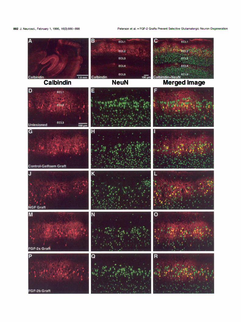
Figure 4. Calbindin (red, Cy3) and glutamate (green, DTAF) immunofluorescence in the entorhinal cortex imaged using confocal microscopy. Although calbindin-positive cell staining remained unaffected by lesion or grafting (A, D, G, J, M), as observed previously (Fig. 3), entorhinal glutamate staining was changed by perforant pathway lesion. Glutamate immunofluorescence of the intact entorhinal cortex (B) revealed strong labeling of neuronal somata and fibers; fiber staining was so robust that individual aomata are difficult to discern. However, lesion of the perforant pathway followed by Gelfoam (E), NGF (H), or FGF-2s (K) grafts showed a marked attenuation of glutamate staining, which also helped to make the presence of glutamatergic neuronal somata more distinct. When colocalization of glutamate with calbindin was examined, the unlesioned ECL2 (C) revealed that, although most calbindin-positive cells expressed glutamate, some did not. Of the more numerous glutamatergic cells in ECL2, -50% expressed calbindin (Table 2). Fluorescent-negative regions suggest that not all ECL2 neurons express glutamate or calbindin (C, arrow/zeads). When coexprcssion in lesion/grafting conditions was examined, it was observed that most of the surviving ECL2 glutamatergic neurons in animals receiving Gelfoam (F, see also P), NGF (I), or FGl+-2s (L) grafts also expressed calbindin. Only a minority ofglutamatergic neurons not expressing calbindin were observed in these conditions (Table 2). These results illustrate that the population of glutamatergicicaibindin-negative ECL2 neurons is vulnerable to perforant pathway lesion. Animals that received FGF-2b grafts demonstrate equivalent glutamate staining (N) and the same ratio of calbindin coexpression of glutamatergic ECL2 neurons (0, see also Q) as the unlesioned animals (C). To illustrate this distinction further in the FGF-2b-grafted animals, a higher-power view (Q) is contrasted with the control-Gelfoam condition (P). These results illustrate that FGF-2b prevents the death of the vulnerable glutamatergicicalbindin-negative ECL2 neurons. Scale bar (shown in A): 50 km for A through 0. Scale bar (shown in P): 50 Km for P and Q.

896 J. Neurosci., February 1. 1996, 76(3):886-898 Peterson et al. l FGF-2 Grafts Prevent Selective Glutamatergic Neuron Degeneration
present at low levels (Ray et al., 199.5), suggesting that the FGF-2s grafts were of limited utility in presenting active FGF-2 to the ECL2 neurons.
Hypothesis of cell death/rescue The postlesion survival of glutamatergicicalbindin-positive neu- rons in the absence of FGF-2 may reflect a sequence of excito- toxicity and oxidative stress followed by calbindin-mediated intra- cellular calcium buffering. Axotomy of glutamatergic ECL2 neurons may lead to a transitory increase in local extracellular glutamate concentrations, as was reported after ischemia (Gra- ham et al., 1990) or concussive injury (Katayama et al., 1990). Glutamate is the major effector of oxidative stress in the brain, acting primarily via ionotropic receptors opening ion channels. The resultant calcium influx produces oxygen radicals, which has serious cytotoxic consequences (Coyle and Puttfarcken, 1993). We have demonstrated that most ECL2 neurons express NMDARl and, therefore, would be susceptible to extracellular glutamate concentrations. Of interest, injection of excitotoxins into the en- torhinal cortex can cause loss of layer II (Levisohn and Isacson, 1991; Yee et al., 1993) or layer III neurons (Du and Schwartz, 1992; Eid et al., 1994).
The neuroprotective function of FGF-2 has been investigated using experimental manipulations modeling excitotoxicity (Freese et al., 1992; Liu et al., 1993; Nozaki et al., 1993; Skaper et al., 1993), ischemia (Nakata et al., 1993; Nozaki et al., 1993; Regli et al., 1994), and oxidative stress (Enokido et al., 1992). One com- mon component of these investigations has been the modulation of intracellular calcium levels. Studies conducted by Mattson and coworkers modeling excitotoxicity (Mattson et al., 1993b), isch- emia (Cheng and Mattson, 1991, 1992; Cheng et al., 1993; Matt- son et al., 1993c), and oxidative stress (Zhang et al., 1993), in addition to reports by others (Ferhat et al., 1993; Louis et al., 1993), have confirmed that changes in intracellular calcium are specifically modulated by FGF-2. Modulation of intracellular cal- cium also has been described for a number of other neurotrophic factors, including NGF (Mattson and Cheng, 1993; Mattson et al., 1993a).
However, NGF failed to protect the vulnerable glutamatergici calbindin-negative neurons. Furthermore, staining with antibodies to both the high-affinity trkA and low-affinity ~75 NGF receptors reveals that layer II neurons do not possess receptors for NGF (data not shown). The difference in efficacy also may be explained by a mechanism unique to FGF-2 by which intracellular calcium may be stabilized. For example, Mattson et al. (1993d) reported that the expression of the glutamate-binding subunit of the NMDA receptor (NMDARP-71) is selectively suppressed by FGF-2. The disabling of this receptor may prevent neuronal response to glutamate, thereby obviating challenge to intracellular calcium homeostasis. Via this mechanism, FGF-2, but not NGF or EGF (Mattson et al., 1993d), may regulate the calcium pathways of the neuron.
Based on these data, we propose a two-part hypothesis describ- ing the cascade of events leading to the axotomy-induced death of the vulnerable glutamatergicicalbindin-negative neurons and their rescue by FGF-2. We propose that local extracellular glutamate levels are increased in layer II because of cellular damage to glutamatergic ECL2 neurons after the axotomizing lesion. This increased extracellular glutamate may activate the NMDARl present on these ECL2 neurons, causing a calcium influx. Those glutamatergic cells that coexpress calbindin would be able to buffer this calcium, whereas the vulnerable calbindin-negative
population would undergo calcium-mediated oxidative stress cul- minating in their death. As a second part to the hypothesis, we propose that FGF-2 rescues the vulnerable glutamatergici calbindin-negative by obstructing the initiation of calcium- mediated oxidative stress. The mechanism for this may be that described above by which the NMDARl capacity is compromised in the presence of FGF-2 (Mattson et al., 1993d). Future exper- iments will be directed toward testing these hypotheses.
The data presented here implicate the loss of calcium ho- meostasis in the death of the axotomized glutamatergic/calbindin- negative neurons. The rescue of this population of neurons by the availability of FGF-2 suggests that therapeutic strategies can be developed to prevent neuronal death caused by calcium-mediated cytotoxic stress in cases of excitotoxicity, ischemia, physical trauma and, possibly, in neurodegenerative diseases. The demon- stration of neuroprotection in the entorhinal cortex, a region of specific neurodegeneration in Alzheimer’s disease, suggests that further studies should be designed to test hypotheses of mecha- nisms by which neurodegeneration may be prevented.
REFERENCES Abraham WC, Mason SE, Demmer J, Williams JM, Richardson CL, Tate
WP, Lawlor PA, Dragunow M (1993) Correlations between immcdiatc early gene induction and the persistence of long-term potentiation. Neuroscience 56:717-727.
Anderson KJ, Dam D, Lee S, Cotman CW (1988) Basic fibroblast growth factor prevents death of lcsioned cholinergic neurons in viva. Nature 332:360-361.
Armstrong DM, Ikonomovic MD, Sheffield R, Wcnthold RJ (1994) AMPA-selective glutamate receptor subtype immunorcactivity in the entorhinal cortex of non-demented elderly and patients with Alzhei- mer’s disease. Brain Res 639:207-2 16.
Arnold SE, Hyman BT, Vanhoesen GW, Damasio AR (1991) Some cytoarchitcctural abnormalities of the entorhinal cortex in schizophrc- nia. Arch Gen Psychiatry 48:625-632.
Asai T, Wanaka A, Kato H, Masana Y, Sco M, Tohyama M (1993) Differential expression of two members of FGF receptor gcnc iamilyl FGFR-1 and FGFR-2 mRNA. in the adult rat central nervous svstem. Mol Brain Res 17:174-17X.
>
Balaci L, Presta M, Ennas MC, Dell’Era P, Sogos V, Lauro G, Gremo F (1994) Differential expression of fibroblast growth factor receptors by human neurones, astrocytes and microglia. NeuroReport 6: 197-200.
Beall MJ, Lewis DA (1992) Heterogeneity of layer-11 neurons in human entorhinal cortex. J Comp Neurol 321:241-266.
Bliss TVP, time T (1973) Long-lasting potentiation of synaptic trans- mission in the dentate area of the anaesthetized rabbit following stim- ulation of the perforant path. J Physiol (Lond) 232:331-356.
Blottner D, Baumgarten HG (1992a) Basic fibroblast growth factor pre- vents neuronal death and atrophy of retrogradely labeled prcganglionic neurons in vivo. Exp Neural I1 8:35-46.
Blottner D, Baumgarien HG (1992b) Insulin-like growth factor-1 coun- teracts bFGF-induced survival of nitric oxide svnthasc fNOS)-oositivc I 1 spinal cord neurons after target-lesion in bivo. J Neurosci Res 32:471-480.
Braak H, Braak E (1992) The human entorhinal cortex: normal morphol- ogy and lamina-specific pathology in various discascs. Neurosci Rcs l5:6-31.
Celio MR (1990) Calbindin D-28k and parvalbumin in the rat nervous system. Neuroscience 35:375-475.
Chandler CE, Parson LM, Hosang M, Shooter EM (1984) A monoclonal antibody modulates the interaction of nerve growth factor with PC12 cells. J Biol Chem 259:6882-6889.
Cheng B, Mattson MP (1991) NGF and bFGF protect rat hippocdmpal and human cortical neurons against hypoglycemic damage by stabilizing calcium homeostasis. Neuron 7:1031-1041.
Cheng B, Mattson MP (1992) Glucose deprivation elicits neurolibrillary tangle-like antigenic changes in hippocampal neurons: prevention by NGF and bFGF. Exp Neurol 117:114-123.
Cheng B, McMahon DG, Mattson MP (1993) Modulation of calcium current, intracellular calcium levels and cell survival by glucose dcpri-

Peterson et al. . FGF-2 Grafts Prevent Selective Glutamatergic Neuron Degeneration J. Neurosci.. February 1, 1996, 76(3):886-898 897
vation and growth factors in hippocampal neurons. Brain Res 607:275-2X5.
Coyle JT, Puttfarckcn P (1993) Oxidative stress, glutamate, and neuro- degenerative disorders. Science 262:689-695.
Cummings BJ, Yee GJ, Cotman CW (1992) bFGF promotes the survival of entorhinal layer-11 neurons after perforant path axotomy. Brain Res 591:271-276.
Demeulemeester H, Arckens L, Vandesande F, Orban GA, Heizmann CW, Pochet R (1991) Calcium binding proteins and neuropeptides as molecular markers of GABAergic interneurons in the cat visual cortex. Exp Brain Res 84:538-544.
Du F, Schwartz R (1902) Aminooxyacetic acid causes selective neuronal loss in layer-111 of the rat medial entorhinal cortex. Neurosci Lett 147:185-188.
Eid T, Du F, Schwartz R (1994) Differential neuronal vulnerability to aminooxyacetic acid and quinolinic acid in the rat entorhinal cortex. Sot Neurosci Abstr 24:270.
Enokido Y, Akaneya Y, Niinobe M, Mikoshiba K, Hatanaka H (1992) Basic fibroblast growth factor rescues CNS neurons from cell death caused by high oxygen atmosphere in culture. Brain Res 599:261-271.
Falkai P, Bogerts B, Rozumek M (1988) Limbic pathology in schizophre- nia: the entorhinal region-a morphometric study. Biol Psycho1 24:515-521.
Ferhat L, Khrestchatisky M, Roisin MP, Barbin G (1993) Basic fibroblast growth factor-induced increase in zifi268 and c-fos mRNA levels is Ca7+ dependent in primary cultures of hippocampal neurons. J Neurochem 61:1105-1112.
Ferrer I. Tunon T, Soriano E, del Rio A, Iraizoz I, Fonseca M, Guionnet N (1993) Calbindin D-28k immunoreactivity in the temporal neocortex in patients with Alzheimer’s disease. Clin Neuropathol 12:53-58.
Fisher LJ, Jinnah HA, Kale LC, Higgins GA, Gage FH (1991) Survival and function of intrastriatally grafted primary fibroblasts genetically modified to product L-dopa. Neuron 6:371-380.
Freese A, Finklestein SP, DiFiglia M (1992) Basic libroblast growth factor protects striatal neurons in vitro from NMDA receptor-mediated cxcitotoxicity. Brain Res 575:351-355.
Graham SH. Shiraishi K, Panter SS, Simon RP, Faden AI (1990) Changes in extracellular amino acid neurotransmitters produced by focal cerebral ischemia. Neurosci Lett I10:124-130.
Gulyas AI, Toth K, Danos P, Freund TF (1991) Subpopulations of GABAergic neurons containing parvalbumin, calbindin D28k, and cho- lecystokinin in the rat hippocampus. J Comp Neural 312:371-378.
Heinemann U, Zhang CL, Eder C (1993) Entorhinal cortex- hippocampal interactions in normal and epileptic temporal lobe. Hip- pocampus 3:89-97.
Hof PR, Morrison JH (1991) Neocortical neuronal subpopulations la- beled by a monoclonal antibody to calbindin exhibit differential vulner- ability in Alzheimer’s disease. Exp Neurol I I1:293-301.
Hyman BT, Damasio AR, Van Hoesen GW, Barnes CL (1984) Alzhei- mer’s disease: cell specific pathology isolates the hippocampal forma- tion. Science 225:116X-1170.
Hyman BT, Van Hoesen GW, Kromer LJ, Damasio AR (1986) Per- forant pathway changes and the memory impairment of Alzheimer’s disease. Ann Neurol 20:472-48 1.
Iacopino AM, Christakos S (1990) Specific reduction of calcium-binding protein (28-kilodalton calbindin-D) gene expression in aging and neu- rodegenerative diseases. Proc Nat1 Acad Sci USA 87:4078-4082.
Iacopino AM, Christakos S, German D, Sonsalla PK, Altar CA (1992) Calbindin-D28K-containing neurons in animal models of neurodegen- eration: possible protection from excitotoxicity. Mol Brain Res l3:251-261.
Ichimiya Y, Emson PC, Mountjoy CQ, Lawson DE, Heizmann CW (1988) Loss of calbindin-28K immunoreactive neurones from the cor- tex in Alzheimer-type dementia. Brain Res 475:156-159.
Jakob H, Beckmann H (1994) Circumscribed malformation and nerve cell alterations in the entorhinal cortex of schizophrenics: pathogenetic and clinical aspects. J Neural Trans 98:83-106.
Jones RSG (1903) Entorhinal hippocampal connections: a speculative view of their function. Trends Neurosci 16:58-64.
Katayama Y, Becker DP, Tamura T, Hovda DA (1990) Massive in- creases in extracellular potassium and the indiscriminate release of glutamate following concussive brain injury. J Neurosurg 73:889-900.
Klagsbrun M (1989) The fibroblast growth factor family: structural and biological properties. Prog Growth Factor Res 1:207-235.
Levisohn LF, Isacson 0 (1991) Excitotoxic lesions of the rat entorhinal cortex: effects of selective neuronal damage on acquisition and retention of a non-spatial reference memory task. Brain Res 564:230-244.
Liu Z, D’Amore PA, Mikati M, Gatt A, Holmes GL (1993) Neuropro- tective effect of chronic infusion of basic fibroblast growth factor on seizure-associated hippocampal damage. Brain Res 626:335-338.
Louis JC, Magal E, Gerdes W, Seifert W (1993) Survival-promoting and protein kinase C-regulating roles of basic FGF for hippocampal neurons exposed to phorbol ester, glutamate and ischaemia-like conditions. Eur J Neurosci 5:1610-1621.
Mattson MP, Cheng B (1993) Growth factors protect neurons against excitotoxiciischemic damage by stabilizing calcium homeostasis. Stroke 24:1136-1140.
Mattson MP, Rychlik B, Chu C, Christakos S (1991) Evidence for calcium-reducing and excite-protective roles for the calcium-binding protein calbindin-D28k in cultured hippocampal neurons. Neuron 6:41-51.
Mattson MP, Rydel RE, Lieberburg I, Smith-Swintosky VL (lY93a) Al- tered calcium signaling and ncuronal injury: stroke and Alzheimer’s disease as examples. Ann NY Acad Sci 679:1-21.
Mattson MP, Tomaselli KJ, Rydel RE (1993b) Calcium-destabilizing and neurodegenerative effects of aggregated /3-amyloid peptidc arc attenu- ated by basic FGF. Brain Rcs 621:35-49.
Mattson MP, Zhang Y, Bose S (1993~) Growth factors prevent mito- chondrial dysfunction, loss of calcium homeostasis. and cell injury, but not ATP depletion in hippocampal neurons deprived of glucose. Exp Neural 121:1-13.
Mattson MP, Kumar KN, Wang H, Cheng B, Michaelis EK (1993d) Basic FGF regulates the expression of a functional 71 kDa NMDA receptor protein that mediates calcium influx and neurotoxicity in hippocampal neurons. J Neurosci 13:4575-4588.
Michel RP, Cruz-Orive LM (1988) Application of the Cavalieri principle and vertical sections method to lung: estimation of volume and pleural surface area. J Microsc 150:117-136.
Miller RJ (1991) The control of ncuronal Ca” homeostasis. Prog Neu- robiol 37:255-285.
Morino-Wannier P, Fujita SC, Jones EG (1992) GABAergic neuronal populations in monkey primary auditory cortex defined by co-localized calcium binding proteins and surface antigens. Exp Brain Res 88:422-432.
Mullen RJ, Buck CR, Smith AM (1992) NcuN, a neuronal specific nu- clear protein in vertebrates. Development 116:201-211.
Nakata N, Kato H, Kogure K (1993) Protective effects of basic fibroblast growth factor against hippocampal neuronal damage following ccrcbral ischemia in the gerbil. Brain Res 605:354-356.
Nozaki K, Finklestein SP, Beal MF (1993) Basic fibroblast growth factor protects against hypoxia-ischemia and NMDA neurotoxicity in neonatal rats. J Cereb Blood Flow Metab 13:221-22X.
Otto D, Unsicker K (1990) Basic FGF reverses chemical and morpho- logical deficits in the nigrostriatal system of MPTP-treated mice. J Neurosci 10:1912-1921.
Otto D, Frotscher M, Unsicker K (1989) Basic fibroblast growth factor and nerve growth factor administered in gel foam rescue medial septal neurons after fimbria fornix transection. J Neurosci Res 22:83-Y].
Peterson DA, Jones DG (1993) Determination of neuronal number and process surface area: a stereological approach to in vim quantitation. J Neurosci Methods 46:107-120.
Peterson DA, Lucidi-Phillipi CA, Eagle KA, Gage FH (1994) Pcrforant path damage results in progressive neuronal death and somal atrophy in layer II of entorhinal cortex and functional impairment with increasing postdamage age. J Neurosci 14:6872-6885.
Ray J, Hogg J, Beutler AS, Takayama H, Baird A, Gage FH (lY95) Expression of biologically active basic fibroblast growth factor by genetically modified rat primary skin fibroblasts. J Neurochcm 64:503-513.
Ray J, Peterson DA, Schinstine M, Gage FH (1993) Proliferation, dif- ferentiation and long-term culture of primary hippocampal neurons. Proc Nat1 Acad Sci USA 90:3602-3606.
Regli L, Anderson RE, Meyer FB (1994) Basic fibroblast growth factor increases cortical blood flow in viva. Brain Rcs 665: 155-157.
Rogers JH (1992) Immunohistochemical markers in rat cortex: co- localization of calretinin and calbindin-D28k with neuropeptidcs and GABA. Brain Res 587:147-157.
Rosenberg MB, Friedmann T, Robertson RC, Tuszynski M, Wolff JA, Breakefield X0, Gage FH (1988) Grafting of gcnctically modified

898 J. Neurosci., February 1, 1996, 76(3):886-898 Peterson et al. . FGF-2 Grafts Prevent Selective Glutamatergic Neuron Degeneration
cells to the damaged brain: restorative effects of NGF gene expression. Science 242:1575-1578.
Skaper SD, Leon A, Facci L (1993) Basic fibroblast growth factor modulates sensitivity of cultured hippocampal pyramidal neurons to glutamate cyto- toxicity: interaction with ganglioside GMl. Dev Brain Res 71:1-B.
Sterio DC (1984) The unbiased estimation of number and sizes of arbi- trary particles using the dissector. J Microsc 134:127-136.
Takayama H, Ray J, Raymon HK Baird A, Hogg J, Fisher LI, Gage FH (1995) Basic fibroblast growth factor increases dopaminergic graft survival and function in a rat model of Parkinson’s disease. Nature Med 1:53-58.
Toth K, Freund TF (1992) Calbindin D28kcontaining nonpyramidal cells in the rat hippocampus: their immunoreactivity for GABA and projection to the medial septum. Neuroscience 49:793-805.
Tunon T, Insausti R, Ferrer I, Sobreviela T, Soriano E (1992) Parval- bumin, and calbindin D-28K in the human entorhinal cortex: an immu- nohistochemical study. Brain Res 589:24-32.
Walicke PA (1988) Basic and acidic fibroblast growth factors have tro- phic effects on neurons from multiple CNS regions. J Neurosci 8:2618-2627.
Walicke PA, Baird A (1991) Internalization and processing of basic fi- broblast growth factor by neurons and astrocytes. J Neurosci 11:2249-2258.
Walicke PA, Cowan WM, Ueno N, Baird A, Guillemin R (1986) Fibro- blast growth factor promotes survival of dissociated hippocampal neu- rons and enhances neurite extension. Proc Nat1 Acad Sci USA 83:3012-3016.
Wiig KA, Bilkey DK (1994) The effects of perirhinal cortical lesions on spatial reference memory in the rat. Behav Brain Res 63:101-109.
Witter MP (1993) Organization of the entorhinal-hippocampal system: a review of current anatomical data. Hippocampus 3:33-44.
Yamada K, Kinoshita A, Kohmura E, Sakaguchi T, Taguchi J, Kataoka K, Hayakawa T (1991) Basic fibroblast growth factor prevents thalamic degeneration after cortical infarction. J Cereb Blood Flow Metab 11~472-478.
Yamada T, Mcgeer PL, Baimbridge KG, Mcgeer EG (1990) Relative sparing in Parkinson’s disease of substantia nigra dopamine neurons containing calbindin-D28K. Brain Res 526:303-307.
Yee WM, Frim DM, Isacson 0 (1993) Relationships between stress protein induction and NMDA-mediated neuronal death in the entorhi- nal cortex. Exp Brain Res 94:193-202.
Zhang Y, Tatsuno T, Carney JM, Mattson MP (1993) Basic FGF, NGF, and IGFs protect hippocampai and cortical neurons against iron- induced degeneration. J Cereb Blood Flow Metab 13:378-388.