
FEVER and RASH Lan-fang Tang Dept. Pediatric Pulmonology Email: [email protected] Pediatrics.

Fever and Rash: Common clinical syndromes
Christina Hermos, MD Primary Care Days
April 10th, 2013 Westborough, MA
Approach to Patient with Fever and Rash
1. Description of Rash 2. Associated Signs and Symptoms 3. Exposures

Describe the Rash
• Timing • Distribution
– Where did it start? – Where has it spread? – Does it move (evanescent) or not (fixed)?
• Symptoms – Itching – Pain – Swelling

Describe the Rash Characteristics and common terminology
• Type of lesion – Macule (flat) – Papule – Nodule – Vesicle – Pustule – Abscess – Plaque – Wheal
• Color – Erythematous (red) – Violacious (purple)
• Vascularity – Blanching – Petechiae – Purpura – Ecchymosis
• Arrangement/shape – Scattered – Grouped – Well demarcated – Morbilliform – Coalescent – Linear – Annular – Serpiginous – Targetoid – Lacey
• Consistency – Desquamation – Sandpaper – Crust (scab)

Signs/Symptoms Associated with Rash
• Fever duration and characteristics • Signs of shock
– Hypotension, poor perfusion, decreased consciousness
• Irritability • Headache • Respiratory symptoms • Eye changes • Mucous membrane lesions or pain • Joint pain or swelling

Exposures • Sick contacts • Medications • Vaccines
– Recent vaccines? – Incomplete suggesting susceptible host?
• Daycare • Travel • Season • Outdoor exposures
– Ticks, other vectors • Menses/Tampon use

Case 1
•Diffuse •Erythematous •Blanching •“Erythroderma”
•Sunburn

Case 1 Associated signs/sxs Exposures
• Fever: 40ºC, 1 day • Signs of shock: Yes • Headache • Injected bulbar
conjunctiva • Hyperemic mucous
membranes: • Dizzyness • Myalgias • Vomiting and diarrhea
• Menses/Tampon use

Toxic Shock Syndrome
• Staphylococcus aureus – Menstrual and non-menstrual cases – Toxic shock syndrome toxin (TSST) and
others – Bacteremia uncommon
• Streptococcus pyogenes – TSS Complicates 1/3 of invasive GAS
infections, most commonly necrotizing fasciitis – Bacteremia common

Toxic Shock Syndrome in the United States: Surveillance Update, 1979–1996.
• Hajjeh RA, Reingold A, Weil A, Shutt K, Schuchat A, Perkins BA. Emerg Infect Dis [serial on the Internet]. 1999, Dec

Staphylococcus aureus Toxic Shock Syndrome: Clinical Case Definition
Clinical Findings • Fever: temperature 38.9°C (102.0°F) or greater • Rash: diffuse macular erythroderma • Hypotension: SBP <= 90 for adults; < 5% for children < 16 years of age; orthostatic
drop in DBP >=15 from lying to sitting; orthostatic syncope/dizziness • Multisystem organ involvement: 3 or more of the following: • Gastrointestinal: vomiting or diarrhea at onset of illness • Muscular: severe myalgia or CPK > twice the upper limit of normal • Mucous membrane: vaginal, oropharyngeal, or conjunctival hyperemia • Renal: BUN or Cr > twice the upper limit of normal or urinary sediment with >=5
WBC • Hepatic: total bilirubin, AST, or ALT > twice the upper limit of normal • Hematologic: platelet count <=100 • Central nervous system: disorientation or alterations in consciousness without focal
neurologic signs when fever and hypotension are absent Laboratory Criteria • Negative results on the following tests, if obtained: • Blood, throat, or CSF; blood culture may be positive for S aureus • Serologic tests for Rocky Mountain spotted fever, leptospirosis, or measles
© 2009 by the American Academy of Pediatrics. All rights reserved.

Streptococcal Toxic Shock Syndrome: Clinical Case Definition
I. Isolation of group A streptococcus (Streptococcus pyogenes) A. From a normally sterile site (Definite case)
B. From a non-sterile site (Probable case)
II. Clinical signs of severity A. Hypotension: systolic pressure <=90 adults or <5% for
age in children AND
B. Two or more of the following signs: • Renalt: Cr >= 2 x upper limit of normal for age
• Coagulopathy: platelet count <=100 or less or DIC • Hepatic: total bili, AST, or ALT > 2x the ULN • ARDS • Generalized erythematous macular rash • Soft tissue necrosis, including necrotizing fasciitis or
myositis, or gangrene

Table 3.71. Management of Streptococcal Toxic Shock Syndrome Without Necrotizing Fasciitis
• Fluid management to maintain adequate venous return and cardiac filling pressures to prevent end-organ damage
• Anticipatory management of multisystem organ failure
• Parenteral antimicrobial therapy at maximum doses
• Kill organism with bactericidal cell wall inhibitor (eg, beta-lactamase-resistant antistaphylococcal antimicrobial agent)
• Stop enzyme, toxin, or cytokine production with protein synthesis inhibitor (eg, clindamycin)
• Immune Globulin Intravenous may be considered for infection refractory to several hours of aggressive therapy or in the presence of an undrainable focus or persistent oliguria with pulmonary edema
Table 3.68. Management of Staphylococcal Toxic Shock Syndrome
• Fluid management to maintain adequate venous return and cardiac filling pressures to prevent end-organ damage
• Anticipatory management of multisystem organ failure
• Parenteral antimicrobial therapy at maximum doses
• Kill organism with bactericidal cell wall inhibitor (eg, beta-lactamase-resistant antistaphylococcal antimicrobial agent)
• Stop enzyme, toxin, or cytokine production with protein synthesis inhibitor (eg, clindamycin)
• Immune Globulin Intravenous may be considered for infection refractory to several hours of aggressive therapy, or in the presence of an undrainable focus, or persistent oliguria with pulmonary edema


Case 2
• Macular/papular – morbilliform
• Begins on face and
spreads caudally • Usually spares palms and
soles • Over time:
– Confluence – Deepening color – Desquamation

• Fever: 4th day • Cough and rhinorrhea • Conjunctivitis • Vomiting, loose stools
• No live vaccines • France 1 week ago
Case 2 Associated signs/sxs Exposures

Measles
• Rubeola virus • Transmission: Droplet and Airborne • Highly contagious

Head to toe

Koplik Spots: Red with blue/white center

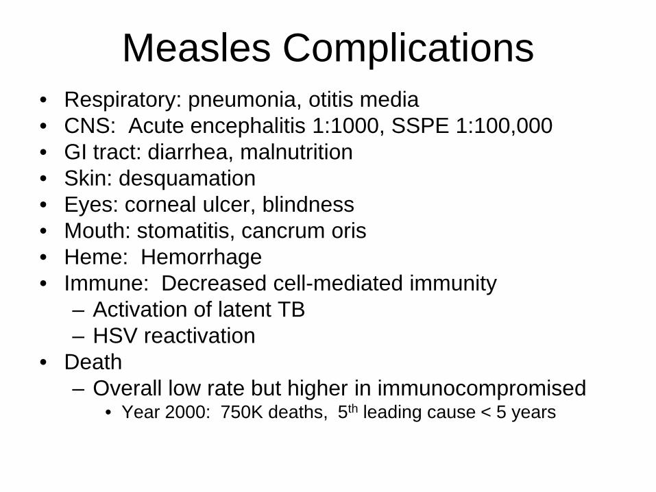
Measles Complications • Respiratory: pneumonia, otitis media • CNS: Acute encephalitis 1:1000, SSPE 1:100,000 • GI tract: diarrhea, malnutrition • Skin: desquamation • Eyes: corneal ulcer, blindness • Mouth: stomatitis, cancrum oris • Heme: Hemorrhage • Immune: Decreased cell-mediated immunity
– Activation of latent TB – HSV reactivation
• Death – Overall low rate but higher in immunocompromised
• Year 2000: 750K deaths, 5th leading cause < 5 years


MMR recommendations for travelers
• All persons aged ≥6 months who will be traveling outside the US and are eligible to receive MMR vaccine should be vaccinated before travel. Children aged ≥12 months should receive 2 doses of MMR vaccine separated by at least 28 days, before travel

Case 3 •Morbilliform •Blanching and Purpuric •Targetoid •Involves palms and soles •Mucous membrane ulceration

• Fever: 2 days low grade • Painful conjunctivitis • Ulcerative Mucous
membranes • Dysuria • Myalgias
• Bactrim
Case 3 Associated signs/sxs Exposures

Stevens-Johnson Syndrome • Idiosyncratic reaction to
– Medications: sulfa drugs, penicillin, others – Infections: HSV, Mycoplasma
• Fever • Rash
– Erythematous macules, purpuric, targetoid, burning/painful – Bulla formation and necrosis, sloughing of the skin, Nikolsky’s sign – <10% Body Surface area (BSA) – >=2 mucous membranes involved
• Oral, conjunctival, genital
Toxic Epidermal Necrolysis
>30% BSA
Erythema multiforme No sloughing
0-1 mucous membrane involved
Infectious triggers more likely (HSV, Mycoplasma)
Drugs, CA, autoimmune
SJS SJS/TEN

EM/SJS

TEN

Case 4
• Petechial • Purpuric • Extremities

• Fever: 1 day • Signs of shock: • Headache: • Sore throat • Myalgias, leg pain
• No MCV4
Case 4 Associated signs/sxs Exposures

Meningococcemia
• 30% of cases without associated meningitis • Prodrome with fever, myalgias often with non-
specific non-petechial rash – Associated symptoms can include nausea, vomiting,
pharyngitis (non-exudative) • Petechiae may first appear at pressure points • Mortality: 10% • Skin grafting or amputation: 1-5%
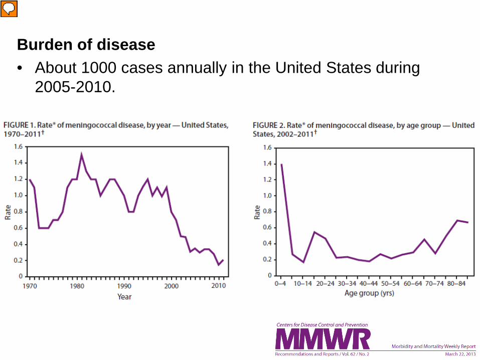
Burden of disease • About 1000 cases annually in the United States during
2005-2010.

ACIP Recommendations for Infants at Increased Risk for Meningococcal Disease
• Infants at increased risk for meningococcal disease should be
vaccinated with a 4-dose series of Hib-MenCY-TT. • These include infants with recognized persistent complement
pathway deficiencies and infants who have anatomic or functional asplenia including sickle cell disease.
• Infant Meningococcal Vaccination: Advisory Committee on Immunization Practices (ACIP) Recommendations and Rationale
– Morbidity and Mortality Weekly Report 52 MMWR / January 25, 2013 / Vol. 62 / No. 3

Case 5
•Petechial •Involves palms (and soles)

• Fever: 3 days • Signs of shock • Headache • Myalgias
• Summer • Recent camping trip
to Webster • Removed a large tick
Case 5 Associated signs/sxs Exposures

Rocky Mountain Spotted Fever
• Rickettsia rickettsii • Tick borne (American dog, RM wood ticks) • Rash spreads from wrists/ankles to palms/soles
and trunk • Endemic to East Coast (south and central)

RMSF: Incidence and case fatality from 1920-2010

http://www.cdc.gov/rmsf/stats/index.html#geography
RMSF

• CDC-National Notifiable Diseases Surveillance System
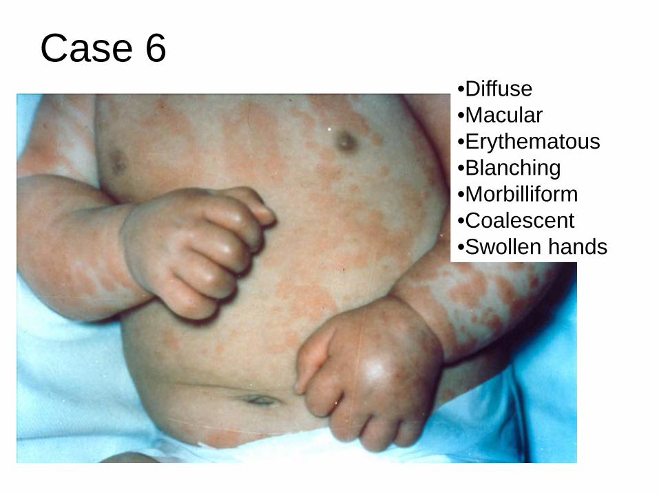
Case 6 •Diffuse •Macular •Erythematous •Blanching •Morbilliform •Coalescent •Swollen hands

• Fever: 6th day • Irritability: • Bulbar conjunctivitis
without exudate • Cracked lips, beefy
red (strawberry) tongue
• None
Case 6 Associated signs/sxs Exposures

Kawasaki Disease
• Etiology: • Unknown. • An infectious cause has been suspected but none has been identified
• Epidemiology:
• Most cases occur in young children (80% before age 4 years) • Males outnumber females 1.6:1. • Most common in the winter and spring in the U.S. • Any ethnic group, those of Asian ancestry are at highest risk.
• Clinical
• Small/medium-sized vessel vasculitis • Acute phase: high fever, rash, conjunctival hyperemia, cervical
lymphadenopathy, redness of the oral and pharyngeal mucosa, “strawberry tongue”, and redness and swelling of the palms and soles.
• Subacute phase >10 days: lower fevers, desquamation of the fingertips, thrombocytosis, arthralgia, and carditis.
• Cardiac complications: coronary artery vasculitis may lead to aneurysms in 25% of patients, with risk of myocardial infarction and death.

• Diagnosis:
• Clinical diagnostic criteria: a. Fever of 5 or more days’ duration b. Four of the following five conditions
1. Conjunctivitis – bilateral, without exudate 2. Rash 3. Cervical Adenopathy >1.5cm 4. Mucous membrane changes of the upper respiratory tract,
such as injected pharynx; injected lips; dry, fissured lips; Strawberry tongue
5. Changes in the Hands and feet, such as edema and erythema, with desquamation in the healing phase
c. Illness not explained by another disease (e.g. measles, TSS) • Incomplete Kawasaki • 2-3 clinical findings plus >=3 lab abnormalities
• WBC >15, anemia, platelets >450, ALT, low albumin, sterile pyuria • Therapy:
• Intravenous immune globulin (IVIG) prevents coronary aneurysms. • Aspirin is also given for its anti-inflammatory and anti-platelet effect.
Kawasaki Disease (continued)


• Evaluation of suspected incomplete Kawasaki disease (KD)
• (2) Infants <=6 months old on day >=7 of fever without other explanation should undergo laboratory testing and, if evidence of systemic inflammation is found, an echocardiogram, even if the infants have no clinical criteria.
• (4) Supplemental laboratory criteria include
– albumin <=3.0 – anemia for age, – elevation of ALT – platelets after 7d >450 – WBC >15 000/mm3 – urine WBC >10
• Reprinted from Newburger JW, et al. Pediatrics. 2004;114(6):17058-1733.

Table 1.9. Suggested Intervals Between Immune Globulin Administration and Measles Immunization (MMR or MMRV)
Dose
Indications or Product Route U or mL mg IgG/kg Interval, moa
Tetanus prophylaxis (as TIG) IM 250 U 10 3 Hepatitis A prophylaxis (as IG)
Contact prophylaxis IM 0.02 mL/kg 3.3 3 International travel IM 0.06 mL/kg 10 3
Hepatitis B prophylaxis (as HBIG) IM 0.06 mL/kg 10 3 Rabies prophylaxis (as RIG) IM 20 IU/kg 22 4
Varicella prophylaxis (as VariZIG) IM 125 U/10 kg (maximum 625 U) 20-40 5
Measles prophylaxis (as IG) Standard IM 0.25 mL/kg 40 5 Immunocompromised host IM 0.50 mL/kg 80 6
RSV prophylaxis (palivizumab monoclonal antibody)b IM ... 15 mg/kg (monoclonal) None
Cytomegalovirus Immune Globulin IV 3 mL/kg 150 6 Blood transfusion
Washed RBCs IV 10 mL/kg Negligible 0 RBCs, adenine-saline added IV 10 mL/kg 10 3 Packed RBCs IV 10 mL/kg 20-60 5 Whole blood IV 10 mL/kg 80-100 6 Plasma or platelet products IV 10 mL/kg 160 7
Replacement (or therapy) of immune deficiencies (as IGIV) IV ... 300-400 8
Therapy for ITP (as IGIV) IV ... 400 8 Therapy for ITP IV ... 1000 10 Therapy for ITP or Kawasaki disease (as IGIV) IV ... 1600-2000 11

Case 7 •Sandpapery •Itchy (pruritic) •Coalesced in the axilla

• Sore throat • Headache • Abdominal pain
• Winter
Case 7 Associated signs/sxs Exposures

Scarlet Fever
• Streptococcus pyogenes (Group A Strep) – Delayed type reaction to erythrogenic toxin
• Face to trunk spread of rash – Circumoral pallor – Strawberry tongue – Desquamation


Case 8
•Macular •Cheeks •Blanching •Lacey (“reticular”)

• Fever: 3 days low grade
• Rhinorrhea • Vomiting and diarrhea
• Daycare • Spring
Case 8 Associated signs/sxs Exposures

Parvovirus B19 • Erythema infectiosum
– 5th’s Disease – “Slapped cheeks” – Prodrome
• Fever, rhinorrhea, nausea, vomiting – Rash
• Slapped cheek with circumoral pallor • Lacey, reticular rash of trunk and extremities • Child improving when it appears • Immune mediated
• Aplastic Crisis in Sickle Cell • Hydrops fetalis (non-immune) with intrauterine infection

• 9-month-old male was referred to the dermatology clinic for a rash involving his hands and feet. His skin eruption was associated with pruritus, and he had had low grade fevers and upper respiratory symptoms for 2 days. Physical examination revealed petechial, erythematous patches of the palms and soles (Figure 2). The remainder of his physical examination was unremarkable, including a normal oral mucosal examination and the absence of lymphadenopathy
• http://www2.luriechildrens.org/ce/online/article.aspx?articleID=89

gloves and socks syndrome • Papular-purpuric gloves and socks syndrome
(PPGSS) • Purpuric erythema involving hands and feet
(especially palms and soles) in a “glove and stocking” distribution
• Pruritus, tingling, and pain • Sometimes oral ulcerative lesions • Most show IgM antibodies to parvovirus B19
– HHV6, HHV7, measles and CMV • Low grade fever common, often developing 2 to
4 days following the onset of the rash

Case 9: Fever… then rash!
• Roseola infantum (exanthem subitum) • HHV-6 (or HHV-7) • Rash presents after 3-5 days of high fever
– Other symptoms include URI, otitis, irritability, GI sxs, and febrile seizures
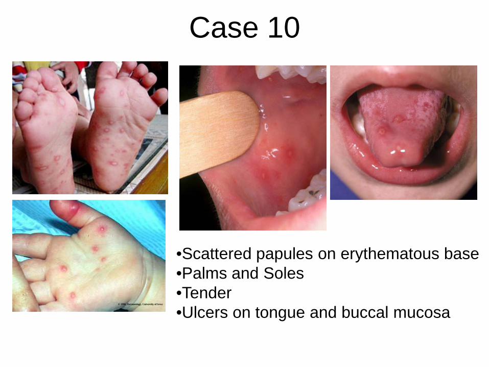
Case 10
•Scattered papules on erythematous base •Palms and Soles •Tender •Ulcers on tongue and buccal mucosa

• Poor feeding
• Summer
Case 10 Associated signs/sxs Exposures

Hand Foot and Mouth
• Coxsackie A – Picornavirus
• Enterovirus – Polio – Non-polio: Coxsackie A and B, Enteroviruses, Echovirus,
• Young children – Transmitted via oral/nasal secretions
• Self limited

Severe Hand, Foot, and Mouth Disease Associated with Coxsackievirus A6 — Alabama, Connecticut, California, and Nevada, November 2011–February 2012 MMWR / March 30, 2012 / Vol. 61 / No. 12

•Scattered and grouped vesicles •Diffuse •Erythematous base •Crust
Case 11

• Itchy/pruritic rash
• Unvaccinated
Case 11 Associated signs/sxs Exposures

Varicella-zoster virus • Primary infection: Chicken Pox • Reactivated infection: Zoster • Other manifestations
– Meningitis and encephalitis – Disseminated disease in neonates/immunocompromised host
• Hepatitis, pneumonitis – Bacterial (GAS) superinfection: Necrotizing Fasciitis and TSS – Reye syndrome: ASA use
• Droplet and airborne transmission • Vaccine: Live attenuated
– Breakthrough disease is milder with fewer complications • Treatment: Acyclovir • Prophylaxis: Acyclovir, VariZIG

Pre-vaccine (1995) Vs Now
• EACH YEAR, >3.5 MILLION CASES, 9,000 HOSPITALIZATIONS, AND 100 DEATHS ARE PREVENTED BY VARICELLA VACCINATION IN THE UNITED STATES.
• VARICELLA INCIDENCE IN 26 STATES, WHICH HAD ADEQUATE AND CONSISTENT REPORTING TO THE NNDSS DECLINED 82% FROM 2000 TO 2010.
• IN PEOPLE <20 YEARS, HOSPITALIZATION RATES DECLINED BY APPROXIMATELY 95%.
• VARICELLA DEATHS DECLINED BY 98.5% IN CHILDREN AND ADOLESCENTS <20 YEARS .
• VARICELLA AMONG HIV-INFECTED CHILDREN DECLINED 63%. • VARICELLA INCIDENCE AMONG INFANTS, A GROUP NOT ELIGIBLE
FOR VARICELLA VACCINATION, DECLINED BY 90%.
• http://www.cdc.gov/chickenpox/surveillance.html

Varicella vs. Smallpox
• Vesicles different stages – Successive crops appear over
4 days – Crust by day 6

Gianotti-Crosti syndrome

Summary • Fever and rash syndromes can often be diagnosed by
– History of Present Illness – Past Medical History – Exposure History
• Keep a high index of suspicion for life-threatening illnesses which may present early with non-specific findings – Meningococcemia – Toxic Shock Syndrome – Rocky Mountain Spotted Fever
• Remember a vaccine history – Meningococcus – Measles – Varicella
• Remember non-infectious syndromes – Kawasaki Disease – Stevens-Johnson Syndrome

• An 18 yo girl presents to the emergency department with high fever, vomiting, diarrhea, muscle aches, dizziness and a rash covering her stomach and back. Vital signs indicate hypotension. The patient's mother reports that her daughter had a "major allergic reaction" to an antibiotic given around age 2 years for an ear infection. Vigorous administration of intravenous fluids is initiated. What empiric antibiotic(s) are most appropriate for this patient?
• A: Vancomycin • B: Clindamycin • C: Doxycycline • D: Azithromycin • E: Penicillin

• A 4 yo is brought to your clinic with primary complaint of high fever, runny nose, and general malaise. He also has diffuse rash. His parents report that he has no major medical history. The family emigrated from Vietnam about 5 months ago. The child has not been seen by a health care provider prior to this time. On examination you also note bluish-gray lesions in side the mouth. What is the most common complication associated with this presentation?
• A: Pneumonia • B: Death • C: Encephalitis • D: Otitis media • E: Diarrhea

• A teenager presents to the emergency department complaining of fever, headache, body aches and vomiting. Empiric antibiotics are initiated after blood cultures are drawn. In the lab, the following is noted on a blood smear which has been gram stained.
• • What is the most common neurologic sequelae of
this infection? • A: Cerebral thrombosis • B: Ataxia • C: Blindness • D: Deafness • E: Quadraparesis

• A 46 year old male presents to your preceptor’s office with complaint of fever and rash. He tells you he was in North Carolina about 10 days ago. After returning from a hike with friends, he noted a tick on his left arm. This was properly removed without any further local symptoms. Over the past 3 days he has had fever, headache and general malaise. Yesterday he woke with a red flat rash across his wrists and ankles. He notes the rash today on his hands and bottoms of his feet. Some of the “early spots” now seem much darker. What is the target cell of the causative organism in this case?
• A: Vascular endothelium • B: Dermal lymphocytes • C: Blood monocytes • D: Tissue macrophages • E: Red blood cells

• A 6 yo female presents to her pediatrician’s office with chief complaint of fever. Mom reports that the fever has been ongoing for the past 10 days and recurs as soon as “fever medications” wear off. Mom has also noticed that the child has very red eyes. On examination you note the following:
• What is a critical test to obtain in the evaluation/management of this patient?
• A: Chest xray • B: Blood cultures • C: Lumbar pucture • D: Throat culture • E: Echocardiogram

• A 32 yo woman presented to her primary care doctor reporting pain and swelling in her hands and feet. This has been ongoing for the past 2 weeks. She does note that there is some decreased ability to move the affected joints. On exam, the swelling is symmetrical in distribution and they are slightly red and swollen. She has not had any other symptoms, except a fever 10 days or so ago, which she attributed to the illness her young children had at the time. What is the most likely symptom her children exhibited at that time?
• A: Vomiting • B: Wheezing • C: Red cheeks • D: Sore throat • E: Cough

• An 8 month old baby girl is brought to her pediatrician’s office with mom reporting high fever for the past 3 days. She become alarmed when the baby broke out in a diffuse skin rash today. The child has never had rash like this before.
What is the most likely causative organism responsible
for this child’s illness? • A: Adenovirus • B: HHV6 • C: Parainfluenza • D: Rotavirus • E: RSV

• This child is brought to the pediatrician’s office with fever and rash. The fever started 2 days ago, then the rash began yesterday and seems to be spreading. The child is quite itchy and fussy, but is eating and drinking well. She does not have many mucosal lesions. No cough or respiratory symptoms otherwise. The child has no medical history otherwise. What treatment is recommended in this situation?
• A: Acyclovir • B: Amoxicillin • C: Oseltamivir • D: Bactrim • E: Supportive care
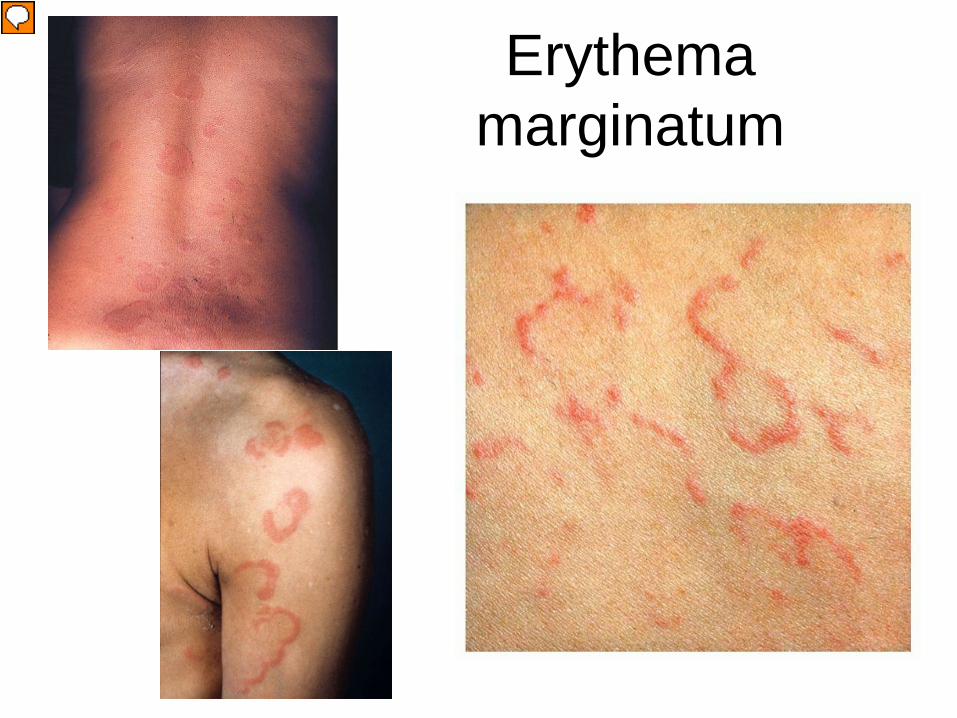
Erythema marginatum

• Toxigenic – Scarlet fever and TSS
• Immunologic – Rheumatic Fever
• Mediated by antibody to M-protein • J-Joints-Polyarthritis • O- -Carditis • N-Subcutaneous Nodules • E-Erythema marginatum • S-Sydenham’s chorea
– Glomerulonephritis – Anti-streptolysin O (ASO) titers to help diagnose
Other non-suppurative complication of GAS

Neisseria
• Gram negative diplococci
• N. meningitidis • Ferments maltose and glucose • IgA protease • Polysaccharide capsule • Vaccine • Respiratory/oral secretions • Meningitis, meningococcemia,
Waterson-Friderichsen syndrome
• N. gonnorhoeae • Ferments glucose • IgA protease • No polysaccharide capsule • No vaccine • Genital secretions/STI • Gonorrhea, pelvic
inflammatory disease, septic arthritis, neonatal conjunctivitis

Other Rickettsial Diseases • R. typhi
– Endemic typhus – Fleas
• R. prowazekii – Epidemic typhus – Human body louse
• Ehrlichia – Ehrilichiosis – Tick
• Coxiella burnetii – Q-fever – Inhaled aeresols from infected animal
• Diagnosis: Serology – Weil Felix Reaction: antiricketsial antibodies cross react with
Proteus antigen. Negative for Q-fever • Treatment for all: Tetracyclines (Doxycycline)

Case 12
• Localized • Red • Swollen • Pustule • Abscess • Fluctuant • Tender • Warm

• Cluster of pustules

Case 12: Associated signs/symptoms
• Fever: Mild • Signs of shock: No • Irritability: No • Headache: No • Respiratory symptoms: No • Eye changes: No • mucous membranes: No • Joint pain or swelling: No • Other: Pain at site

Case 12: Exposures
• Sick contacts • Medications • Vaccines • Daycare • Travel • Season • Outdoor/vectors • Menses/Tampon use
• Family member prior abscess • No • UTD • Yes • No • Fall • No • No
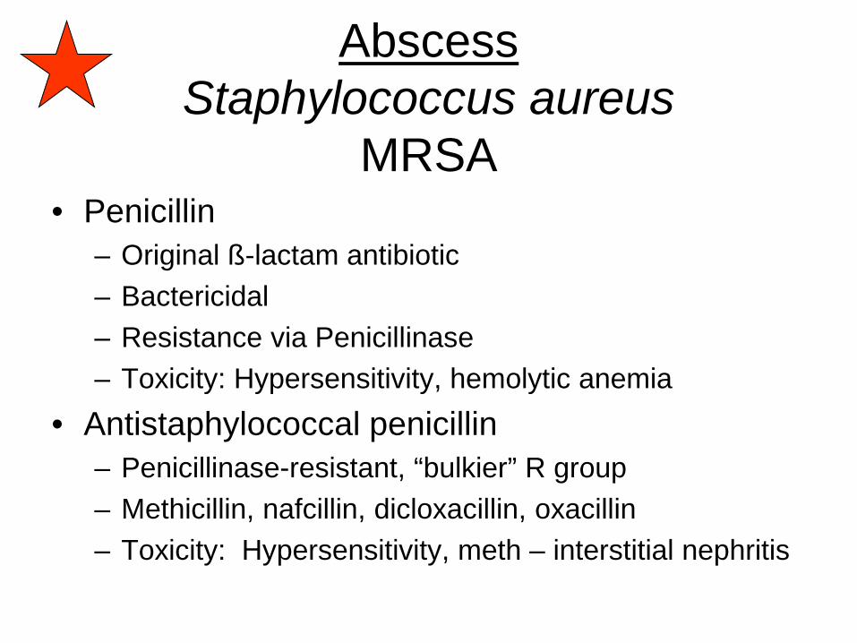
Abscess Staphylococcus aureus
MRSA • Penicillin
– Original ß-lactam antibiotic – Bactericidal – Resistance via Penicillinase – Toxicity: Hypersensitivity, hemolytic anemia
• Antistaphylococcal penicillin – Penicillinase-resistant, “bulkier” R group – Methicillin, nafcillin, dicloxacillin, oxacillin – Toxicity: Hypersensitivity, meth – interstitial nephritis

Penicillin binding protein (Transpeptidase)
Cross-links cell wall
β-Lactams block cell wall synthesis by blocking penicillin binding protein active site

active site
blocked active site
MRSA encodes altered PBP (PBP2a) conferring β-lactam resistance
working active site
PBP2a
Wild type PBP
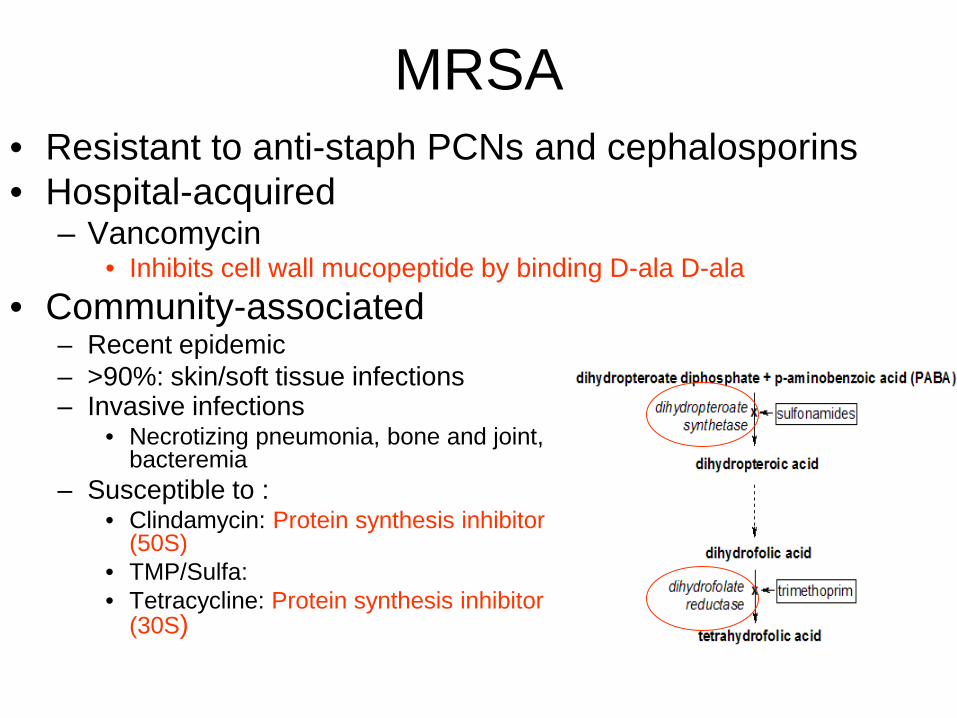
MRSA • Resistant to anti-staph PCNs and cephalosporins • Hospital-acquired
– Vancomycin • Inhibits cell wall mucopeptide by binding D-ala D-ala
• Community-associated – Recent epidemic – >90%: skin/soft tissue infections
– Invasive infections
• Necrotizing pneumonia, bone and joint, bacteremia
– Susceptible to : • Clindamycin: Protein synthesis inhibitor
(50S) • TMP/Sulfa: • Tetracycline: Protein synthesis inhibitor
(30S)


