Fetal Cells in Maternal Blood - cme-utilities.comcme-utilities.com/mailshotcme/Material for...
69
Fetal Cells in Maternal Blood Game Changer in Non-Invasive Prenatal Diagnosis RIPUDAMAN SINGH. PhD, MBA Chief Technology Officer ARCEDI Biotech ApS, Denmark 1
Transcript of Fetal Cells in Maternal Blood - cme-utilities.comcme-utilities.com/mailshotcme/Material for...

Fetal Cells in Maternal BloodGame Changer in Non-Invasive Prenatal Diagnosis
RIPUDAMAN SINGH. PhD, MBA
Chief Technology Officer
ARCEDI Biotech ApS, Denmark
1
Cell-based Non-Invasive Prenatal Diagnosis (cbNIPD)
2
“If you can do it, it changes the game. There’s no question about it.”

Disclosures
3
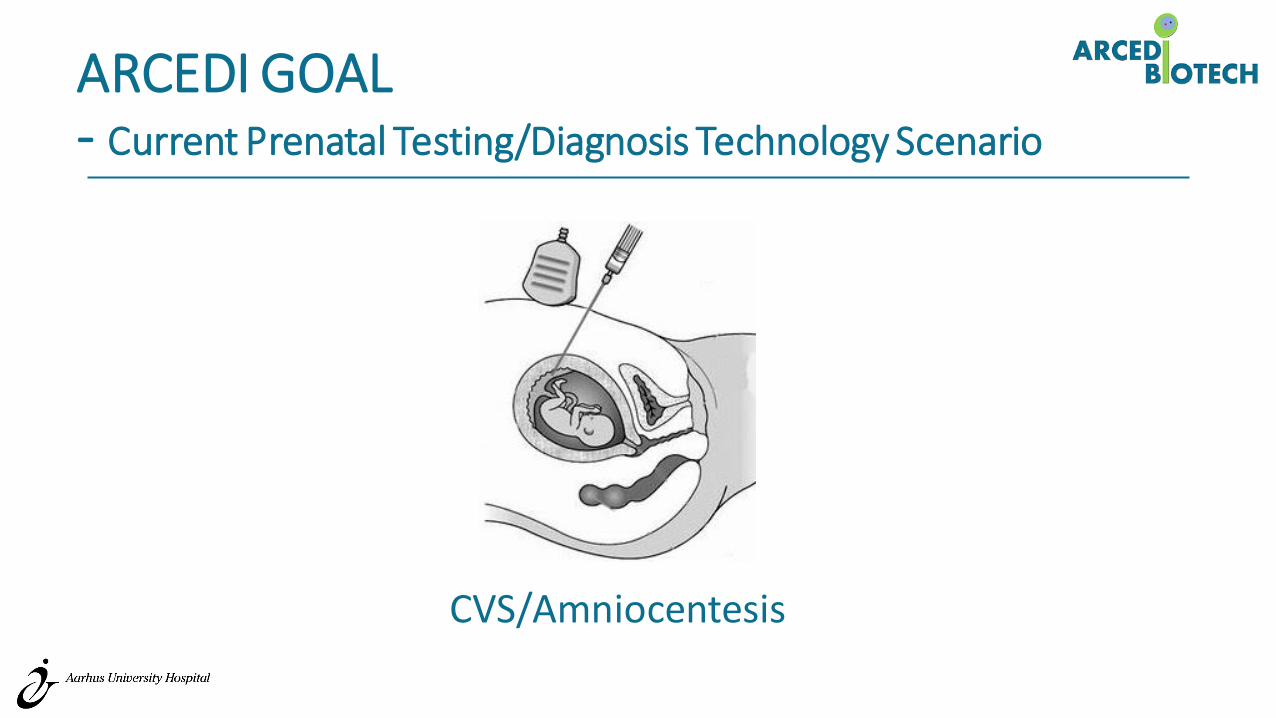
CVS/Amniocentesis
ARCEDI GOAL- Current Prenatal Testing/Diagnosis Technology Scenario

ARCEDI GOAL- Future Prenatal Testing/Diagnosis Technology Scenario
Cell-based
5

RATIONALE- High coverage with low risk
6
cbNIPD CVS/Amnio
cfNIPT
Low
High
Low High
Fetal genome
purity /
Coverage
Risk / Stress

ARCEDI TECHNOLOGY OVERVIEW- Isolation, Extraction and Analyses of Circulating Fetal Cells
Scanning and Identification
of Fetal Cells Selection and
Staining using
ARCEDI markers
Pregnant
women
GA: 10 to 13
weeks
Blood
Processing
(30mL of whole
blood)
Positively Identified Fetal
Cells
‘Picking’ Fetal CellsCbNIPD using
CMA/NGS
Whole Genome
Amplification (WGA)

Fetal Cells in Maternal Blood – A History

FETAL CELLS IN MATERNAL BLOOD
9
• It’s been known for years that fetal cells do circulate in pregnant women’s blood
• Alternative to invasive prenatal diagnosis was envisaged – Focus on fetal cells. Reasons:• Mitigate the risk of intervention associated with invasive methods
• Make prenatal diagnostics simple and cost-effective
• Earlier attempts to isolate fetal cells from maternal circulation consistently and in good numbers were not very successful
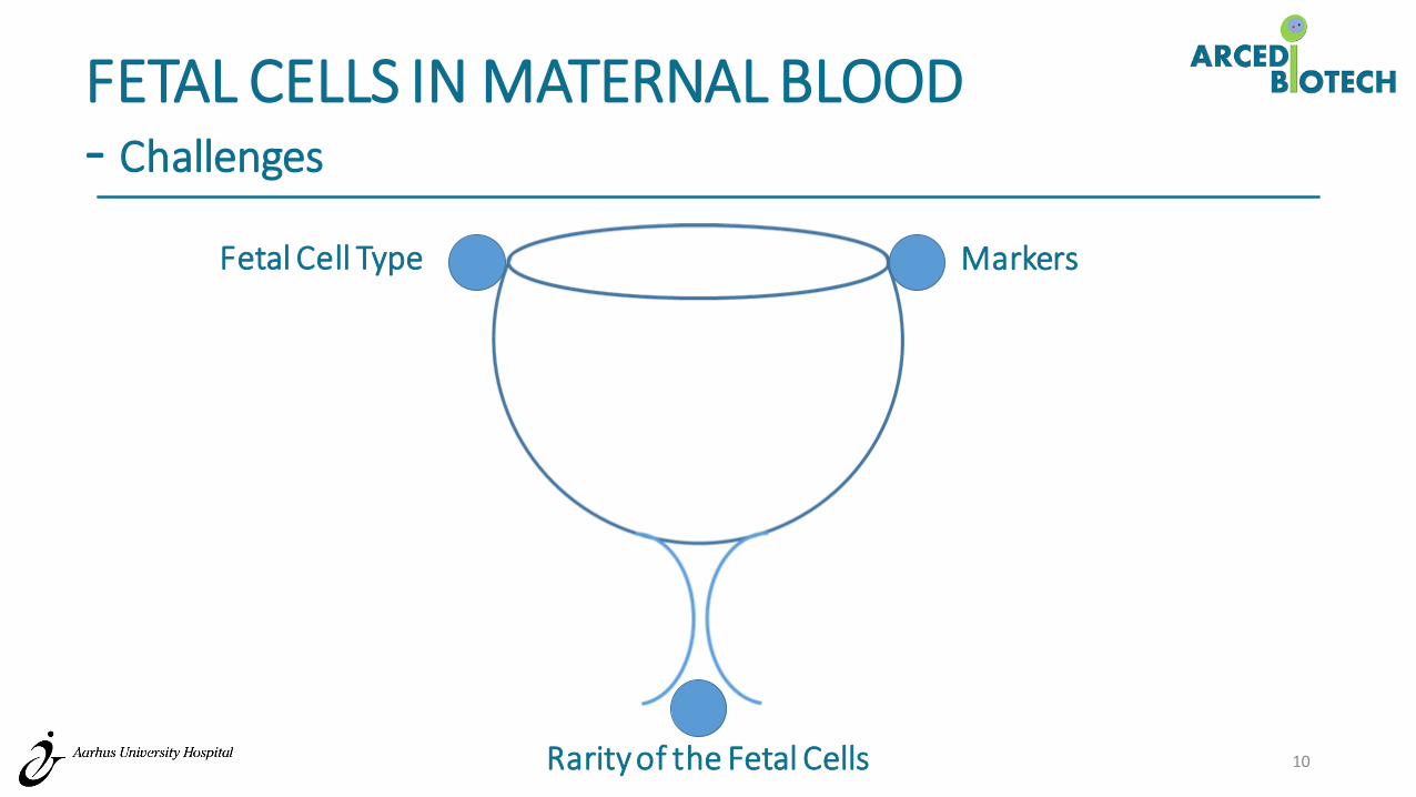
FETAL CELLS IN MATERNAL BLOOD- Challenges
10
Fetal Cell Type Markers
Rarity of the Fetal Cells

FETAL CELLS IN MATERNAL BLOOD- A Love story rekindled
11
Singh et al. 2017

FETAL CELLS IN MATERNAL BLOOD- A Love story rekindled
12
Beginning
Singh et al. 2017

FETAL CELLS IN MATERNAL BLOOD- A Love story rekindled
13
Beginning Cb-NIPT
Singh et al. 2017

FETAL CELLS IN MATERNAL BLOOD- A Love story rekindled
14
Beginning Cb-NIPT Enrichment from MB
Singh et al. 2017

FETAL CELLS IN MATERNAL BLOOD- A Love story rekindled
15
Beginning Cb-NIPT Enrichment from MB
Skepticism
Singh et al. 2017

FETAL CELLS IN MATERNAL BLOOD- A Love story rekindled
16
Beginning Cb-NIPT Enrichment from MB
Skepticism
ARCEDI
Singh et al. 2017

FETAL CELLS IN MATERNAL BLOOD- A Love story rekindled
17
ARCEDI
Singh et al. 2017

Addressing the Challenges – The ARCEDI Way
CHECKLIST- Technology to be approved as a cbNIPD should fulfil the following criteria
19
• The technology should target specific cell type(s).
• There should be antibodies that are both sensitive and specific for those cell types.
• Technology should be platform and parameter independent.
• After identifying the true fetal cells it should be possible to get access to the cells for downstream applications.
• The fidelity of the DNA from enriched fetal cells should be high so that genetic analyses using chromosomal microarray (CMA) or next-generation sequencing (NGS) can be performed.
Singh et al. 2017

CHECKLIST- Technology to be approved as a cbNIPD should fulfil the following criteria
20
• The technology should target specific cell type(s).
• There should be antibodies that are both sensitive and specific for those cell types.
• Technology should be platform and parameter independent.
• After identifying the true fetal cells it should be possible to get access to the cells for downstream applications.
• The fidelity of the DNA from enriched fetal cells should be high so that genetic analyses using chromosomal microarray (CMA) or next-generation sequencing (NGS) can be performed.
Singh et al. 2017

CHECKLIST- Technology to be approved as a cbNIPD should fulfil the following criteria
21
• The technology should target specific cell type(s).
• There should be antibodies that are both sensitive and specific for those cell types.
• Technology should be platform and parameter independent.
• After identifying the true fetal cells it should be possible to get access to the cells for downstream applications.
• The fidelity of the DNA from enriched fetal cells should be high so that genetic analyses using chromosomal microarray (CMA) or next-generation sequencing (NGS) can be performed.
Singh et al. 2017

CHECKLIST- Technology to be approved as a cbNIPD should fulfil the following criteria
22
• The technology should target specific cell type(s).
• There should be antibodies that are both sensitive and specific for those cell types.
• Technology should be platform and parameter independent.
• After identifying the true fetal cells it should be possible to get access to the cells for downstream applications.
• The fidelity of the DNA from enriched fetal cells should be high so that genetic analyses using chromosomal microarray (CMA) or next-generation sequencing (NGS) can be performed.
Singh et al. 2017

CHECKLIST- Technology to be approved as a cbNIPD should fulfil the following criteria
23
• The technology should target specific cell type(s).
• There should be antibodies that are both sensitive and specific for those cell types.
• Technology should be platform and parameter independent.
• After identifying the true fetal cells it should be possible to get access to the cells for downstream applications.
• The fidelity of the DNA from enriched fetal cells should be high so that genetic analyses using chromosomal microarray (CMA) or next-generation sequencing (NGS) can be performed.
Singh et al. 2017

CHECKLIST- Technology to be approved as a cbNIPD should fulfil the following criteria
24
• The technology should target specific cell type(s).
• There should be antibodies that are both sensitive and specific for those cell types.
• Technology should be platform and parameter independent.
• After identifying the true fetal cells it should be possible to get access to the cells for downstream applications.
• The fidelity of the DNA from enriched fetal cells should be high so that genetic analyses using chromosomal microarray (CMA) or next-generation sequencing (NGS) can be performed.
Singh et al. 2017
FETAL CELL TYPE

FETAL CELL TYPE- Establishing the Fetal Cell gene expression profile
26

FETAL CELL TYPE- Trophoblast mediated Uterine Vessel Remodelling
27
Moser et al. 2016(Histochem Cell Biol)

FETAL CELL TYPE- Trophoblast mediated Uterine Vessel Remodelling
28
Moser et al. 2016(Histochem Cell Biol)

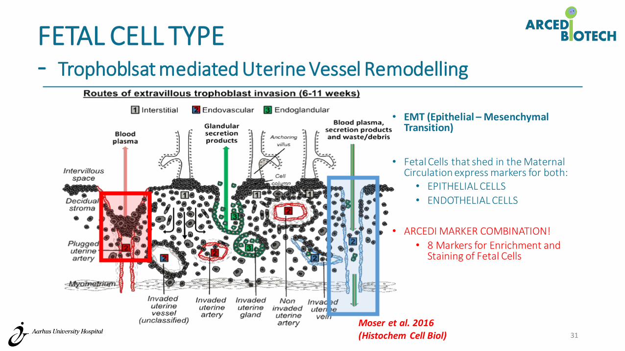
FETAL CELL TYPE- Trophoblsat mediated Uterine Vessel Remodelling
29
Moser et al. 2016(Histochem Cell Biol)

FETAL CELL TYPE- Trophoblsat mediated Uterine Vessel Remodelling
30
Moser et al. 2016(Histochem Cell Biol)
FETAL CELL TYPE- Trophoblsat mediated Uterine Vessel Remodelling
31
• EMT (Epithelial – Mesenchymal Transition)
• Fetal Cells that shed in the Maternal Circulation express markers for both:• EPITHELIAL CELLS
• ENDOTHELIAL CELLS
• ARCEDI MARKER COMBINATION!
• 8 Markers for Enrichment and Staining of Fetal Cells
Moser et al. 2016(Histochem Cell Biol)

FETAL CELL MARKERS- IP on the Fetal Cell Selection and Staining Markers
32

FETAL CELL MARKERS

ARCEDI METHOD – MARKER SENSITIVITY- Performance - Retrieval of Fetal Cells
• 190 PREGNANT WOMEN at NT SCAN- 30ml Blood.• 99 SAMPLES FROM ‘LOW RISK’ GROUP
• 91 SAMPLES FROM ‘HIGH RISK’ GROUP (offered CVS)
34

ARCEDI METHOD – MARKER SENSITIVITY- Performance - Retrieval of Fetal Cells
• 190 PREGNANT WOMEN at NT SCAN- 30ml Blood.• 99 SAMPLES FROM ‘LOW RISK’ GROUP
• 91 SAMPLES FROM ‘HIGH RISK’ GROUP (offered CVS)
PARAMETER VALUE
No of Fetal Cells 2440
Mean (per sample) 12.8/30ml blood
Median 10
Range 1-46
35

ARCEDI METHOD – MARKER SENSITIVITY- Frequency Distribution (‘High Risk’ vs ‘Low Risk’)
36

ARCEDI METHOD – MARKER SENSITIVITY- Frequency Distribution (‘High Risk’ vs ‘Low Risk’)
37
Fetal Cell from Every Sample!
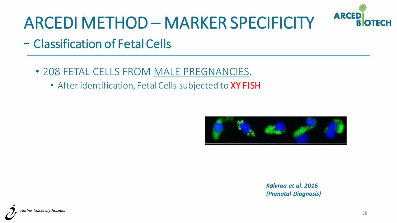
ARCEDI METHOD – MARKER SPECIFICITY- Classification of Fetal Cells
• 208 FETAL CELLS FROM MALE PREGNANCIES.• After identification, Fetal Cells subjected to XY FISH
38
Kølvraa et al. 2016(Prenatal Diagnosis)

ARCEDI METHOD – MARKER SPECIFICITY- Classification of Fetal Cells
• 208 FETAL CELLS FROM MALE PREGNANCIES.• After identification, Fetal Cells subjected to XY FISH
XY 164
XX 0
No. FISH Signal 32
Lost during FISH 12
39
Kølvraa et al. 2016(Prenatal Diagnosis)

FETAL CELL MORPHOLOGY

FETAL CELLS- Gallery
41

FETAL CELLS- Gallery
42
Fetal Cell Identification Criteria
• Cytoplasmic staining pattern (green)• Nuclear morphology/staining pattern (blue)• Counterstain• Size of the cell

METHOD ROBUSTNESS

ARCEDI METHOD – ROBUSTNESS- Turnaround time/sample
0 1 5
ARCEDI method: Processing Time per sample in Hours (continuous)
Sampling BloodProcessing
FetalCellEnrichment and
staining
Mounting andscanning
Visual inspection Picking
14
WGA
44
8
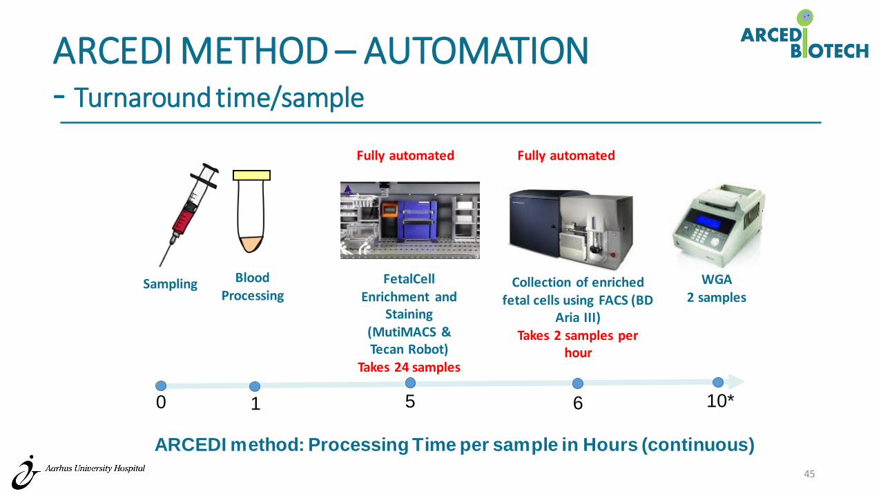
ARCEDI METHOD – AUTOMATION- Turnaround time/sample
ARCEDI method: Processing Time per sample in Hours (continuous)
45
0 1 5
Sampling BloodProcessing
FetalCellEnrichment and
Staining (MutiMACS & Tecan Robot)
Takes 24 samples
Collection of enriched fetal cells using FACS (BD
Aria III) Takes 2 samples per
hour
10*
WGA2 samples
6
Fully automatedFully automated

ARCEDI METHOD – ROBUSTNESS- Sample Stability (Parameter/Platform Independant)
Fetal Cell Number and Distribution unaffected by:
• Blood collection tubes (BD vs Streck Tubes)
• Time before blood processed – 72 hrs
• Transportation – air and road and processed after 72 hrs
• Fetal cells from every sample!
46
GENETIC ANALYSES ON FETAL CELLS

DNA FIDELITY- Decifering Genetic Information from Fetal Cells
48
WGAPicoplex
CMA (180K)

DNA FIDELITY- One ‘High Risk’ case
49

• ’High Risk’ Pregnancy offered Amniocentesis
• Maternal Blood collected before Amniocentesis
• 2 Fetal Cells enriched and identified using ARCEDI Method
• WGA and Array CGH (Agilent 180k microarray) performed at Baylor College of Medicine, Houston TX.
50
DNA FIDELITY- One ‘High Risk’ case

DNA FIDELITY- NIPT413: Mosaicism [45,X/46,X,r(X)] – array CGH
377 450
51
Kølvraa et al. 2016(Prenatal Diagnosis)
377
Cell 1

377 450
450
52
Kølvraa et al. 2016(Prenatal Diagnosis)
377
450
Cell 1
Cell 2
DNA FIDELITY- NIPT413: Mosaicism [45,X/46,X,r(X)] – array CGH

377 450
450
53
377
450
Cell 1
Cell 2
Amnio
DNA FIDELITY- NIPT413: Mosaicism [45,X/46,X,r(X)] – array CGH
Kølvraa et al. 2016(Prenatal Diagnosis)

45,X: cell 377
46,X,r(X): cell 450
54
DNA FIDELITY- NIPT413: Mosaicism [45,X/46,X,r(X)] – NGS
Kølvraa et al. 2016(Prenatal Diagnosis)

GOING BEYOND ANEUPLOIDIES - Five ‘High Risk’ cases
55

GOING BEYOND COMMON ANEUPLOIDIES - Five ‘High Risk’ cases
56(Vestergaard et al. 2017; In Press)
T21
T13
T2
12.4Mb dup. Chr 21
31Mb del Chr 4 and 30Mb dup Chr 8

GOING BEYOND COMMON ANEUPLOIDIES - Five ‘High Risk’ cases
57(Vestergaard et al. 2017; In Press)
T13
T2
12.4Mb dup. Chr 21
31Mb del Chr 4 and 30Mb dup Chr 8
T21

GOING BEYOND COMMON ANEUPLOIDIES - Five ‘High Risk’ cases
58
cbNIPD
(Vestergaard et al. 2017; In Press)

GOING BEYOND COMMON ANEUPLOIDIES - Five ‘High Risk’ cases
59
(Vestergaard et al. 2017; In Press)
CVS
cbNIPD

GOING BEYOND COMMON ANEUPLOIDIES - 4.9 Mb Deletion on Chr 3 (Male Fetus)
60
cbNIPD
3 fetal cells

GOING BEYOND COMMON ANEUPLOIDIES - 4.9 Mb Deletion on Chr 3 (Male Fetus)
61
CVS
cbNIPD
3 fetal cells

CLINICAL VALIDATION - ONGOING

CLINICAL VALIDATION - (CVS vs cbNIPD vs cfNIPT)
63
Pregnancies
(6 Hospitals in DK)
CVS cfNIPT cbNIPD
• Recruit ‘High Risk’ pregnancies (who are undergoing CVS) from 6 different hospitals in Denmark
• Inclusion criteria: Singleton pregnancies between the GA of 10-13 weeks, opting for CVS.
• Enrich Fetal Cells from the blood and perform cell based fetal DNA analysis on the the cells
• Perform cell free NIPT
• Check whether the results from three tests correlate

Sample ID cbNIPD cfNIPT CVS FF (%)
1 Normal Normal Normal 12
2 Normal Normal Normal 13
3 Normal Normal Normal 16
4 Normal Normal Normal 7
5 T21 No-call T21 (Mosaic 80-85%) 0
6 Normal Normal Normal 7
7 Normal Normal Normal 3
8 Normal Normal Normal 13
9 T13 T13 T13 (Mosaic 90%) 8
10* T16 ? na ?
11 Normal Normal Normal 3
CLINICAL VALIDATION (CVS vs cbNIPD vs cfNIPT)

cbNIPD TEST LAUNCH IN DENMARK

TEST LAUNCH
66
HIGH RISK PREGNANCIES
CVS
NON-INVASIVE
CELL BASED
CELL FREE
2/3
1/3
If Abnormal

IN BRIEF
• Identified the Circulating Fetal Cell Type
• Tested Markers which are highly sensitive and specific
• Robust method of enriching fetal cells
• Picked the cells and perform downstream analyses (WGA/array CGH/NGS)
• Detected Aneuploidies as well as CNVs using Fetal Cells
67

ACKNOWLEDGEMENTS
ARCEDI Biotech• PALLE SCHELDE• LOTTE HATT• KATARINA RAVN• PETER SCHELDE HØY• SIMON TABI ARREY• MATHIAS KØLVRAA• SOFIE KRUKOW• ANNE SCHELDE HØY• MICHEL RAVN• MAIKEN B. KRISTENSEN• FILIZ KEZGIN• HAI QING• RIE BRUUN• KARIN FREDBORG
68
AUH (Dept. Gyn/Obs)• NIELS ULDBJERG• OLAV B. PETERSEN• MARIANNE RAVNDAL • LOTTE MATHIASEN• ALICE SØRENSEN
AUH (Dept. Clinical Genetics)• IDA VOGEL• ELSE MARIE VESTERGAARD• RIKKE CHRISTENSEN