Fetal cell microchimerism in the maternal mouse … · Fetal cell microchimerism in the maternal...
9
Neurosci Bull February 1, 2014, 30(1): 81–89. http://www.neurosci.cn DOI: 10.1007/s12264-013-1392-1 81 ·Original Article· Fetal cell microchimerism in the maternal mouse spinal cord Guohui Zhang 1,3 , Yunan Zhao 3 , Xin-Min Li 2 , Jiming Kong 1,3 1 Department of Forensic Medicine, Hebei North University, Zhangjiakou 075000, China 2 Department of Psychiatry, University of Manitoba, PZ432-771 Bannatyne Avenue, Winnipeg, Manitoba R3E 3N4, Canada 3 Department of Human Anatomy and Cell Science, Faculty of Medicine, University of Manitoba, 745 Bannatyne Avenue, Winnipeg, Manitoba R3E 0J9, Canada Corresponding author: Jiming Kong. E-mail: [email protected] © Shanghai Institutes for Biological Sciences, CAS and Springer-Verlag Berlin Heidelberg 2014 ABSTRACT Fetal cell microchimerism refers to the persistence of fetal cells in the maternal tissues following pregnancy. It has been detected in peripheral organs and the brain, but its existence in the spinal cord has not been reported. Our aim was to detect fetal cell microchimerism in the spinal cord of maternal mice. C57BL/6 female mice were crossed with GFP transgenic male mice and sacrificed after their first or third delivery. GFP-positive cells, which were presumably from fetuses whose fathers were GFP transgenic, were detected in the spinal cord by fluorescence microscopy and immunohistochemistry. PCR was also performed to detect GFP DNA, which must come from GFP hemizygous fetuses. We found GFP-positive cells and detectable GFP DNA in most of the maternal spinal cords. Twenty percent (1/5) of the mice that were only pregnant once had detectable fetal cells, while 80% (4/5) of those that were pregnant three times had detectable fetal cells. Some fetal cells, which not only emitted green fluorescence but also expressed NeuN, were detected in the spinal cords from maternal mice. These results indicate that fetal cells migrate into the spinal cord of a maternal mouse during and/or after the gestational period, and the fetal cells may differentiate into neurons in the spinal cord. Keywords: fetal cell microchimerism; green fluorescent protein; spinal cord; mouse INTRODUCTION Fetal cells have been observed in the maternal circulation after human pregnancy [1] and persist there for decades [2] . In addition to the peripheral circulation, fetal cells have also been found to engraft in almost all peripheral tissues in both humans and animal models [3-10] . These engrafted cells appear to have the ability of multi-directional differentiation and express differentiation markers. For example, fetal microchimeric cells are capable of engraftment and differentiation along the hematopoietic pathway [11-13] . Studies from Dr. Nelson’s lab [14] showed that fetal cells found in the liver of women who had given birth to sons included cells that expressed hepatocyte antigens (cytokeratins); studies by Wang et al. [15] showed that, after tissue injury in the liver and kidneys, engrafted fetal cells transformed into hepatocytes and tubular cells. In view of these findings, it is believed that persistent fetal cells provide a rejuvenating source of fetal progenitor cells that may have the capacity to participate in maternal tissue repair [16] . Furthermore, Tan et al. [17] demonstrated that fetal cells can even pass through the blood-brain barrier, respond to brain tissue injury, and adopt the location, morphology, and expression of immunocytochemical markers indicative of perivascular macrophage-, neuron-, astrocyte-, and oligodendrocyte-like cell types. This finding has raised the hope that exogenously-engrafted fetal cells contribute to adult neurogenesis. Although it is uncertain whether fetal stem- cells in the adult brain have a competitive advantage over endogenous neuronal stem-cells, it seems that fetal stem-cells potentially possess therapeutic value for spinal cord injury [18] ,
-
Upload
nguyenkien -
Category
Documents
-
view
218 -
download
0
Transcript of Fetal cell microchimerism in the maternal mouse … · Fetal cell microchimerism in the maternal...
Neurosci Bull February 1, 2014, 30(1): 81–89. http://www.neurosci.cnDOI: 10.1007/s12264-013-1392-1 81
·Original Article·
Fetal cell microchimerism in the maternal mouse spinal cordGuohui Zhang1,3, Yunan Zhao3, Xin-Min Li2, Jiming Kong1,3
1Department of Forensic Medicine, Hebei North University, Zhangjiakou 075000, China2Department of Psychiatry, University of Manitoba, PZ432-771 Bannatyne Avenue, Winnipeg, Manitoba R3E 3N4, Canada 3Department of Human Anatomy and Cell Science, Faculty of Medicine, University of Manitoba, 745 Bannatyne Avenue,
Winnipeg, Manitoba R3E 0J9, Canada
Corresponding author: Jiming Kong. E-mail: [email protected]
© Shanghai Institutes for Biological Sciences, CAS and Springer-Verlag Berlin Heidelberg 2014
ABSTRACT
Fetal cell microchimerism refers to the persistence of fetal cells in the maternal tissues following pregnancy. It has been detected in peripheral organs and the brain, but its existence in the spinal cord has not been reported. Our aim was to detect fetal cell microchimerism in the spinal cord of maternal mice. C57BL/6 female mice were crossed with GFP transgenic male mice and sacrificed after their first or third delivery. GFP-positive cells, which were presumably from fetuses whose fathers were GFP transgenic, were detected in the spinal cord by fl uorescence microscopy and immunohistochemistry. PCR was also performed to detect GFP DNA, which must come from GFP hemizygous fetuses. We found GFP-positive cells and detectable GFP DNA in most of the maternal spinal cords. Twenty percent (1/5) of the mice that were only pregnant once had detectable fetal cells, while 80% (4/5) of those that were pregnant three times had detectable fetal cells. Some fetal cells, which not only emitted green fluorescence but also expressed NeuN, were detected in the spinal cords from maternal mice. These results indicate that fetal cells migrate into the spinal cord of a maternal mouse during and/or after the gestational period, and the fetal cells may differentiate into neurons in the spinal cord.
Keywords: fetal cel l microchimerism; green
fl uorescent protein; spinal cord; mouse
INTRODUCTION
Fetal cells have been observed in the maternal circulation after human pregnancy[1] and persist there for decades[2]. In addition to the peripheral circulation, fetal cells have also been found to engraft in almost all peripheral tissues in both humans and animal models[3-10]. These engrafted cells appear to have the ability of multi-directional differentiation and express differentiation markers. For example, fetal microchimeric cells are capable of engraftment and differentiation along the hematopoietic pathway[11-13]. Studies from Dr. Nelson’s lab[14] showed that fetal cells found in the liver of women who had given birth to sons included cells that expressed hepatocyte antigens (cytokeratins); studies by Wang et al.[15] showed that, after tissue injury in the liver and kidneys, engrafted fetal cells transformed into hepatocytes and tubular cells. In view of these fi ndings, it is believed that persistent fetal cells provide a rejuvenating source of fetal progenitor cells that may have the capacity to participate in maternal tissue repair[16].
Furthermore, Tan et al.[17] demonstrated that fetal cells can even pass through the blood-brain barrier, respond to brain tissue injury, and adopt the location, morphology, and expression of immunocytochemical markers indicative of perivascular macrophage-, neuron-, astrocyte-, and oligodendrocyte-like cell types. This finding has raised the hope that exogenously-engrafted fetal cells contribute to adult neurogenesis. Although it is uncertain whether fetal stem-cells in the adult brain have a competitive advantage over endogenous neuronal stem-cells, it seems that fetal stem-cells potentially possess therapeutic value for spinal cord injury[18],
Neurosci Bull February 1, 2014, 30(1): 81–8982
and diseases such as amyotrophic lateral sclerosis[19], multiple sclerosis[20], and cervical spondylotic myelopathy[21]. Here, we report fetal microchimerism in the maternal mouse spinal cord using GFP-based detection, together with morphological and immunocytochemical evidence.
MATERIALS AND METHODS
Mice All animal protocols were approved by the University of Manitoba Animal Care Ethics Committee. Twelve- to 16-week-old male homozygous C57BL/6 Cr Slc TgN (act-EGFP) OsbC14-Y01-FM131 mice (green mice) were crossed with young adult female wild-type C57BL/6 mice (6–8 weeks old). All the offspring were hemizygous green mice. Control wild-type young adult female C57BL/6 mice remained virgins. Young adult green mouse pups provided positive controls. Ex-breeder wild-type female C57BL/6 mice and male green mice were purchased from the Central Animal Care Breeding Facility, University of Manitoba, Winnipeg, Canada. These mice had been held as breeding stock since the age of 6–8 weeks. Three weeks after the first delivery, the mothers underwent a second or third breeding. One week or four weeks after delivering their last litter, the mothers were euthanized with an overdose of isofl urane and perfused with 0.9% ice-cold saline followed by 4% paraformaldehyde in phosphate buffer (4°C, pH 7.4). Thirty minutes later, the spinal cords were removed and fixed again in cold 4% paraformaldehyde for 12 h. After fixation, the cords were immersed in cold 30% sucrose solution. One day later, the cords were removed and stored at –80°C until analyzed.
Visualization of Fetal Green Mouse CellsThe spinal cords were serially sectioned (20 μm) on a Shandon SME Cryotome Cryostat (Ramsey, MN). Selected sections were first washed three times (10 min each) in phosphate-buffered saline (PBS). Then, they were dipped briefl y in distilled H2O, treated with 1 mmol/L CuSO4 (Fisher Scientific, Ottawa, CA) in ammonium acetate buffer (50 mmol/L CH3COONH4, pH 5.0) for 20 min, rinsed briefly in distilled H2O, and returned to PBS[22]. Then they were mounted with FluoromountTM aqueous mounting medium (Sigma) and viewed under a Nikon TE2000-E microscope equipped with a RETIGA camera (QImaging).
ImmunohistochemistryThe spinal cords were serially sectioned (20 μm) on a cryostat and selected sections were immunostained. The primary antibodies were mouse monoclonal anti-GFP (1:250, Abcam, Cambridge, MA), rabbit polyclonal anti-GFP (1:250, Abcam), and mouse monoclonal anti-NeuN (1:200, Chemicon, Temecula, CA). The secondary antibodies were Alexa Fluor 594-labelled goat anti-mouse IgG (Invitrogen, Burlington, CA), and Alexa Fluor 488-labelled goat anti-rabbit IgG (Invitrogen). Both of the secondary antibodies were used at 1:200 dilution.
Briefly, sections were washed three times in PBS (10 min each), transferred to PBS containing 0.3% Triton X-100 (PBST) for 30 min, and blocked with 5% goat serum in PBST for 1 h at room temperature. Then they were incubated with appropriate primary antibody overnight at 4°C. After washing three times in PBS, the sections were incubated with the appropriate secondary antibody for 1 h at room temperature in the dark, followed by three washes (10 min each) in 0.1% PBS-Tween20. After staining with Hoechst 33342 (Calbiocam, San Diego, CA) for 3 min, the sections were treated with 1 mmol/L CuSO4 in ammonium acetate buffer for 20 min, dipped briefly in distilled H2O, and returned to PBS. The sections were finally mounted with FluoromountTM aqueous mounting medium and viewed under the TE2000-E microscope.
Detection of GFP-specifi c DNA by PCR The spinal cord sections prepared above were washed three times (20 min each) in PBS, then they were incubated in lysis buffer (10 mmol/L Tris-HCl, 1 mmol/L EDTA, 1% SDS, 1 mg/mL proteinase K, pH ~6.0) at 56°C overnight. After centrifugation at 12 000 g for 10 min, DNA was extracted from the supernatant with the GenEluteTM Mammalian Genomic DNA Miniprep kit (Sigma) and resuspended in distilled water. The amount of sample DNA was quantified by fluorometry, and the concentration was adjusted to 500 ng/μL.
The following GFP-specific PCR primers were used: forward: 5’-GTAAACGGCCACAAGTTCAGC-3’; reverse: 5’-CATGCCGAGAGTGATCCCG-3’. PCR amplification was carried out by adding 1 ng DNA to 50 μL amplifi cation mixture [0.2 mmol/L dNTP, 2 mmol/L MgCl2, 0.2 μmol/L of each primer, 50 mmol/L Tris-HCl, 10 mmol/L KCl and 1 U of Taq Platinium DNA polymerase (Gibco)]. The thermal
Guohui Zhang, et al. Fetal cell microchimerism in the maternal mouse spinal cord 83
profile was 94°C for 3 min, followed by 35 cycles at 94°C for 45 s, 58°C for 30 s, 72°C for 1 min, and a final extension at 72°C for 10 min. To check for variability in DNA extraction and the absence of inhibition during the PCR, the housekeeping gene β-actin was analyzed in parallel for each sample using the following primers: forward: 5’-GCCTGTGGTACGACCAGAGGCATACAG-3’; reverse: 5’-GATGACGATATCGCTGCGCTGGTCG-3’. The thermal profi le was 94°C for 3 min, followed by 25 cycles at 94°C for 45 s, 60°C for 30 s, 72°C for 1 min, and a fi nal extension at 72°C for 10 min.
Serial dilutions of DNA were prepared with GFP cells diluted in wild-type cells (ranging from 0.001% to 1%) and the samples were amplifi ed in order to compare their DNA concentrations. PCR products were separated on 1.5% agarose gel, visualized by ethidium bromide (Sigma) staining, and analyzed using a FluorChem 8900 imager (Alpha Innotech, San Leandro, CA). The optical density multiplied by the area of the band was used to semi-quantify the PCR product. The value of GFP PCR product was normalized against the amount of PCR product for actin obtained from the same sample. A standard curve was prepared between the number of GFP cells and the ratio of PCR product optical density, from which unknown samples were interpolated.
Statistical AnalysisStatistical differences between two groups of pregnant mice were assessed by the χ2 test. Statistical signifi cance was assumed when P <0.05.
RESULTS
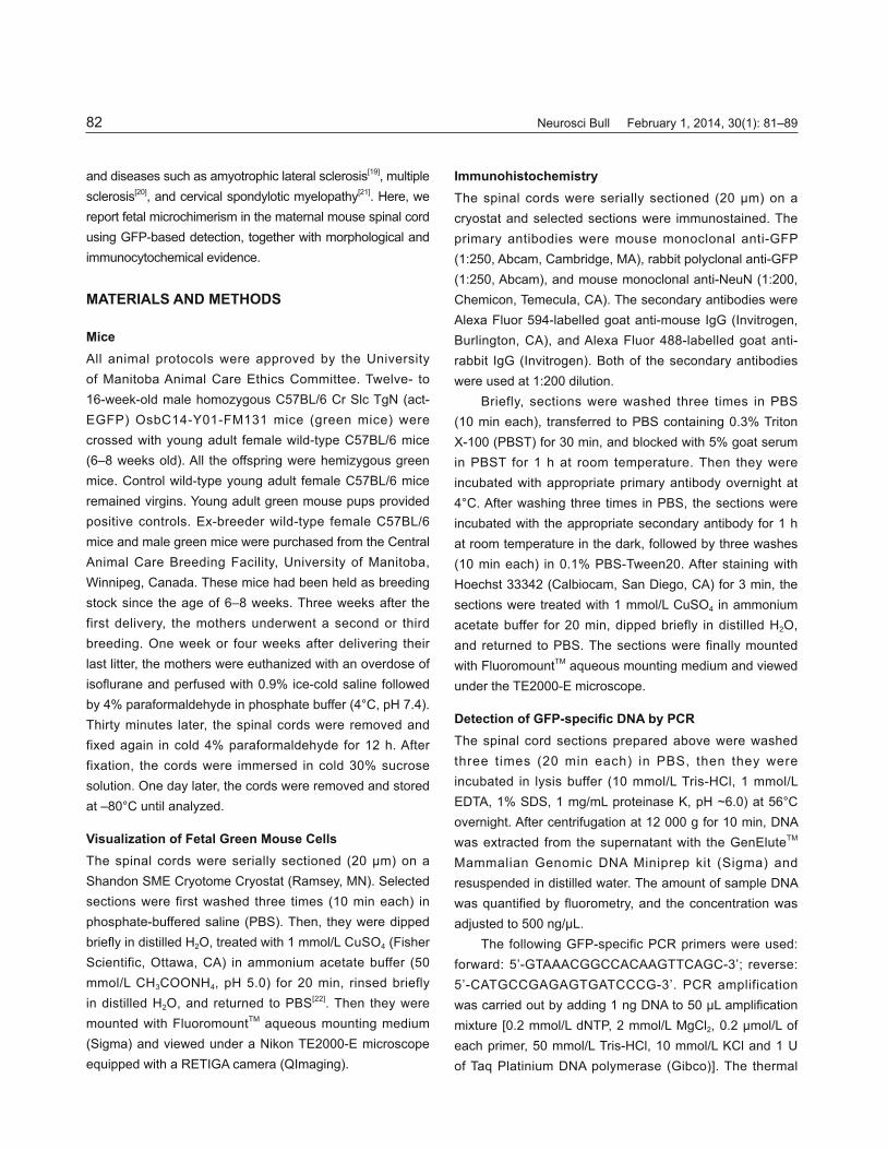
Cupric Sulfate Reduces Natural Fluorescence without Effects on GFP Fluorescence and Immunofl uorescent LabelingNaturally-fl uorescent pigments (i.e., lipofuscin, fl avins, and porphyrins) accumulate in the cytoplasm of many cell types and can often complicate fl uorescence microscopy because of their broad excitation and emission spectra[23, 24]. These pigments often emit green (Fig. 1A) and red light (Fig. 1C), making it difficult to distinguish specific from nonspecific autofl uorescence[25]. Based on the literature[22, 26], we tested the effect of CuSO4 on reducing the natural fluorescence and found that 1 mmol/L CuSO4 almost eliminated the
background autofluorescence (Fig. 1B, D) while retaining the GFP fl uorescence (Fig. 1E, F).
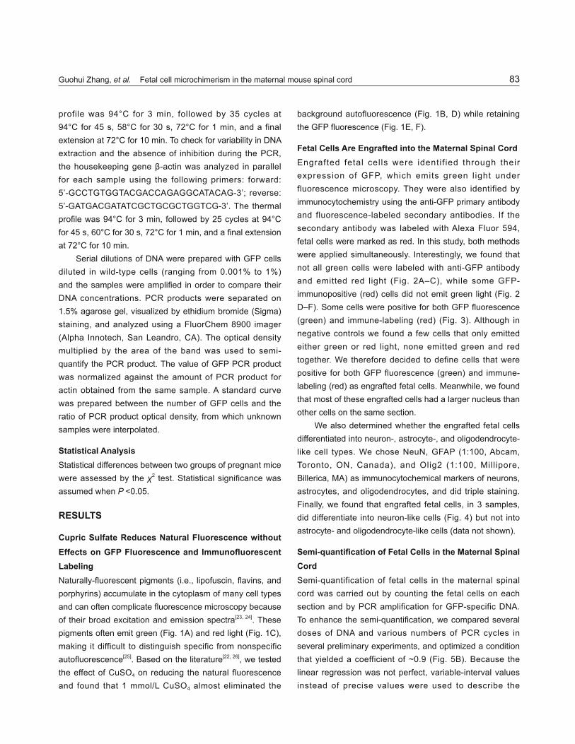
Fetal Cells Are Engrafted into the Maternal Spinal CordEngrafted fetal cel ls were identif ied through their expression of GFP, which emits green l ight under fluorescence microscopy. They were also identified by immunocytochemistry using the anti-GFP primary antibody and fluorescence-labeled secondary antibodies. If the secondary antibody was labeled with Alexa Fluor 594, fetal cells were marked as red. In this study, both methods were applied simultaneously. Interestingly, we found that not all green cells were labeled with anti-GFP antibody and emitted red light (Fig. 2A–C), while some GFP-immunopositive (red) cells did not emit green light (Fig. 2 D–F). Some cells were positive for both GFP fl uorescence (green) and immune-labeling (red) (Fig. 3). Although in negative controls we found a few cells that only emitted either green or red light, none emitted green and red together. We therefore decided to define cells that were positive for both GFP fluorescence (green) and immune-labeling (red) as engrafted fetal cells. Meanwhile, we found that most of these engrafted cells had a larger nucleus than other cells on the same section.
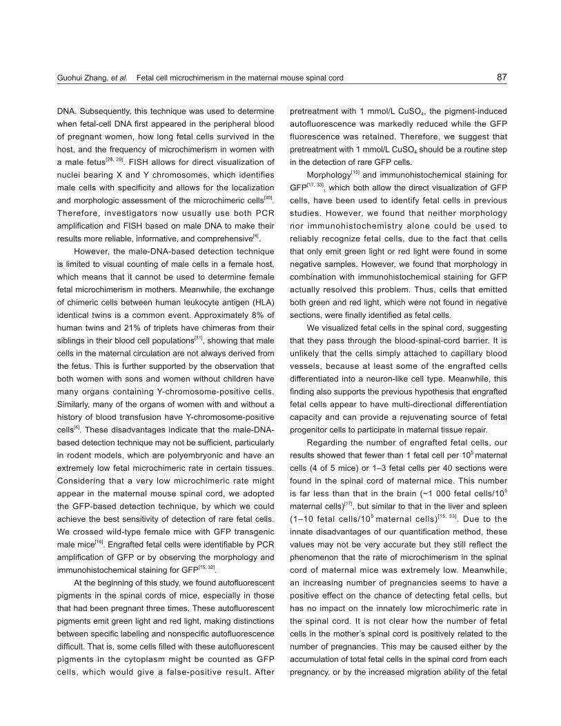
We also determined whether the engrafted fetal cells differentiated into neuron-, astrocyte-, and oligodendrocyte-like cell types. We chose NeuN, GFAP (1:100, Abcam, Toronto, ON, Canada), and Olig2 (1:100, Millipore, Billerica, MA) as immunocytochemical markers of neurons, astrocytes, and oligodendrocytes, and did triple staining. Finally, we found that engrafted fetal cells, in 3 samples, did differentiate into neuron-like cells (Fig. 4) but not into astrocyte- and oligodendrocyte-like cells (data not shown).
Semi-quantifi cation of Fetal Cells in the Maternal Spinal CordSemi-quantification of fetal cells in the maternal spinal cord was carried out by counting the fetal cells on each section and by PCR amplification for GFP-specific DNA. To enhance the semi-quantifi cation, we compared several doses of DNA and various numbers of PCR cycles in several preliminary experiments, and optimized a condition that yielded a coefficient of ~0.9 (Fig. 5B). Because the linear regression was not perfect, variable-interval values instead of precise values were used to describe the

Neurosci Bull February 1, 2014, 30(1): 81–8984
Fig. 2. Fetal cells can be identifi ed through the combination of GFP fl uorescence (green) and GFP immunofl uorescence (red). Not all green cells (A, C), however, were immune-labeled with the anti-GFP antibody (B, C), and some GFP-positive (red) cells (D, F) did not emit green light (E, F, arrows). C is the overlay of A and B, and F is of D and E.
Fig. 1. Cupric sulfate reduces natural fl uorescence while retaining GFP fl uorescence (×10 magnifi cation). Natural fl uorescent pigments often emit green (A) and red light (C). CuSO4 (1 mmol/L) evidently eliminated the fl uorescent pigment-induced autofl uorescence (B, D). Regarding GFP fl uorescence from GFP transgenic mice (E), 1 mmol/L CuSO4 did not have any effects (F).
amounts of fetal cells (Table 1). Our results showed that fetal cells were undetectable in the negative control group. Some mice that had either one or three gestations had detectable fetal cells in their spinal cords three weeks after
their last delivery. However, the frequency of fetal cells was generally low. The morphologic method revealed that only 1–3 cells per 40 sections were GFP-positive green mouse fetal cells in the spinal cord. Meanwhile, the semi-

Guohui Zhang, et al. Fetal cell microchimerism in the maternal mouse spinal cord 85
Fig. 3. Fetal cells that had GFP were identified through fluorescence microscopy directly (green, B, E), and by immunofluorescence staining (red, A, D) using monoclonal anti-GFP as primary antibody and Alexa Fluor 594 as secondary antibody. C and F are merged images of A and B, and D and E, respectively.
Fig. 4. Fetal cells can differentiate into a neuron-like cell type. Fetal cells with a combination of GFP fl uorescence (A, green) and GFP immunofl uorescence (B, blue) were identifi ed. The section was immunostained with an antibody to NeuN, a neuronal marker (C, red). The merged picture indicates that a fetal cell (arrow) expressed NeuN in the nucleus (D).
quantifi ed PCR of genomic DNA showed that fewer than 1 fetal cell per 105 maternal cells were GFP-positive green mouse fetal cells (Table 1). Interestingly, 20% of maternal mice with one pregnancy had detectable fetal cells, while
80% of those with three pregnancies had them, indicating that the number of pregnancies has a positive effect on the engrafting of fetal cells (χ2 = 3.6, χ2
0.05 (1) = 3.84, 0.05 < P < 0.1) (Table 1).

Neurosci Bull February 1, 2014, 30(1): 81–8986
DISCUSSIONThe first molecular biological techniques used to detect fetal cells in the maternal body were the PCR amplifi cation and fluorescence in situ hybridization (FISH) of a
Y-chromosome-specifi c sequence. In 1989, Lo et al.[27] were
the fi rst to show that the Y-chromosome-specifi c sequence
from a male fetus could be amplifi ed from blood samples
of pregnant women by PCR amplification based on male
Fig. 5. Semi-quantifi cation of fetal cells in maternal spinal cord through PCR amplifi cation for GFP-specifi c DNA. DNA dilution series prepared from GFP cells diluted in wild-type cells (ranging from 0.001% to 1%) were amplifi ed (A). PCR products of GFP and β-actin were separated on 1.5% agarose gels, visualized by ethidium bromide staining, and analyzed using a FluorChem 8900 imager. The values of the GFP PCR product were normalized against the amount of PCR product for actin obtained from the same sample. The standard curve was prepared between the number of GFP cells and the ratio of PCR product optical density, from which samples were interpolated (B).
Table 1 Semi-quantifi cation analysis of fetal cells in maternal spinal cord
Number of pregnancies Microscopy (fetal cells/40 sections) PCR amplifi cation (fetal cells/wild-type cells)
0 - -0 - -0 - -0 - -1 - -1 - -1 2 0–1/105
1 - -1 - -3 - -3 1 0–1/105
3 2 0–1/105
3 3 1/105–10/105
3 1 0–1/105
Mice were assigned to three groups: controls, mice with one gestation, and mice with three gestations. Semi-quantifi cation of fetal cells in the
maternal spinal cord was carried out by counting the fetal cells in each section, in combination with PCR amplifi cation for GFP-specifi c DNA. The
sign ‘-’ indicates that fetal cells were not found in the sections or there was no amplifi cation band on 1.5% agarose gels. We obtained the same
positive results with two types of semi-quantifi cation. Five of 10 pregnant mice were found to have engrafted fetal cells in the cord. The χ2 test
showed that an increased number of pregnancies had a positive effect on the chance of detecting fetal cells (χ2 = 3.6, χ2 0.05 (1) = 3.84, 0.05 < P < 0.1).
Guohui Zhang, et al. Fetal cell microchimerism in the maternal mouse spinal cord 87
DNA. Subsequently, this technique was used to determine when fetal-cell DNA fi rst appeared in the peripheral blood of pregnant women, how long fetal cells survived in the host, and the frequency of microchimerism in women with a male fetus[28, 29]. FISH allows for direct visualization of nuclei bearing X and Y chromosomes, which identifies male cells with specificity and allows for the localization and morphologic assessment of the microchimeric cells[30]. Therefore, investigators now usually use both PCR amplifi cation and FISH based on male DNA to make their results more reliable, informative, and comprehensive[4].
However, the male-DNA-based detection technique is limited to visual counting of male cells in a female host, which means that it cannot be used to determine female fetal microchimerism in mothers. Meanwhile, the exchange of chimeric cells between human leukocyte antigen (HLA) identical twins is a common event. Approximately 8% of human twins and 21% of triplets have chimeras from their siblings in their blood cell populations[31], showing that male cells in the maternal circulation are not always derived from the fetus. This is further supported by the observation that both women with sons and women without children have many organs containing Y-chromosome-positive cells. Similarly, many of the organs of women with and without a history of blood transfusion have Y-chromosome-positive cells[4]. These disadvantages indicate that the male-DNA-based detection technique may not be suffi cient, particularly in rodent models, which are polyembryonic and have an extremely low fetal microchimeric rate in certain tissues. Considering that a very low microchimeric rate might appear in the maternal mouse spinal cord, we adopted the GFP-based detection technique, by which we could achieve the best sensitivity of detection of rare fetal cells. We crossed wild-type female mice with GFP transgenic male mice[16]. Engrafted fetal cells were identifi able by PCR amplifi cation of GFP or by observing the morphology and immunohistochemical staining for GFP[15, 32].
At the beginning of this study, we found autofl uorescent pigments in the spinal cords of mice, especially in those that had been pregnant three times. These autofl uorescent pigments emit green light and red light, making distinctions between specifi c labeling and nonspecifi c autofl uorescence diffi cult. That is, some cells fi lled with these autofl uorescent pigments in the cytoplasm might be counted as GFP cells, which would give a false-positive result. After
pretreatment with 1 mmol/L CuSO4, the pigment-induced autofluorescence was markedly reduced while the GFP fluorescence was retained. Therefore, we suggest that pretreatment with 1 mmol/L CuSO4 should be a routine step in the detection of rare GFP cells.
Morphology[15] and immunohistochemical staining for GFP[17, 33], which both allow the direct visualization of GFP cells, have been used to identify fetal cells in previous studies. However, we found that neither morphology nor immunohistochemistry alone could be used to reliably recognize fetal cells, due to the fact that cells that only emit green light or red light were found in some negative samples. However, we found that morphology in combination with immunohistochemical staining for GFP actually resolved this problem. Thus, cells that emitted both green and red light, which were not found in negative sections, were fi nally identifi ed as fetal cells.
We visualized fetal cells in the spinal cord, suggesting that they pass through the blood-spinal-cord barrier. It is unlikely that the cells simply attached to capillary blood vessels, because at least some of the engrafted cells differentiated into a neuron-like cell type. Meanwhile, this fi nding also supports the previous hypothesis that engrafted fetal cells appear to have multi-directional differentiation capacity and can provide a rejuvenating source of fetal progenitor cells to participate in maternal tissue repair.
Regarding the number of engrafted fetal cells, our results showed that fewer than 1 fetal cell per 105 maternal cells (4 of 5 mice) or 1–3 fetal cells per 40 sections were found in the spinal cord of maternal mice. This number is far less than that in the brain (~1 000 fetal cells/105
maternal cells)[17], but similar to that in the liver and spleen (1–10 fetal cells/105 maternal cells)[15, 33]. Due to the innate disadvantages of our quantification method, these values may not be very accurate but they still reflect the phenomenon that the rate of microchimerism in the spinal cord of maternal mice was extremely low. Meanwhile, an increasing number of pregnancies seems to have a positive effect on the chance of detecting fetal cells, but has no impact on the innately low microchimeric rate in the spinal cord. It is not clear how the number of fetal cells in the mother’s spinal cord is positively related to the number of pregnancies. This may be caused either by the accumulation of total fetal cells in the spinal cord from each pregnancy, or by the increased migration ability of the fetal

Neurosci Bull February 1, 2014, 30(1): 81–8988
cell with repetitive pregnancies.The results of this study demonstrate that fetal
microchimeric cells can pass through the blood-spinal-cord barrier and enter maternal spinal cord tissue. Although the microchimeric rate is very low, engrafted fetal cells have a multi-lineage capacity and can differentiate into a neuron-like cell type. Further studies are needed to address what factors or methods can promote the engraftment of fetal cells in the maternal spinal cord and increase the microchimeric rate, which will lay a foundation for the hypothesis that engrafted fetal cells can functionally improve a variety of degenerative disorders of the spinal cord.
ACKNOWLEDGEMENTS
This work was supported by the Manitoba Health Research Council (MHRC) and the Canadian Institutes for Health Research (CIHR). We thank Jacqueline Hogue for her assistance in preparing the manuscript.
Received date: 2013-01-07; Accepted date: 2013-03-11
REFERENCES
[1] Ariga H, Ohto H, Busch MP, Imamura S, Watson R, Reed W, et al. Kinetics of fetal cellular and cell-free DNA in the maternal circulation during and after pregnancy: implications for noninvasive prenatal diagnosis. Transfusion 2001, 41: 1524–1530.
[2] Bianchi DW, Zickwolf GK, Weil GJ, Sylvester S, DeMaria MA. Male fetal progenitor cells persist in maternal blood for as long as 27 years postpartum. Proc Natl Acad Sci U S A 1996, 93: 705–708.
[3] Gadi VK, Nelson JL. Fetal microchimerism in women with breast cancer. Cancer Res 2007, 67: 9035–9038.
[4] Koopmans M, Kremer Hovinga IC, Baelde HJ, Fernandes RJ, de Heer E, Bruijn JA, et al. Chimerism in kidneys, livers and hearts of normal women: implications for transplantation studies. Am J Transplant 2005, 5: 1495–1502.
[5] Lu JQ, Joseph JT, Nash RA, Storek J, Stevens AM, Metz LM, et al. Neuroinflammation and demyelination in multiple sclerosis after allogeneic hematopoietic stem cell transplantation. Arch Neurol 2010, 67: 716–722.
[6] Lu JQ, Storek J, Metz L, Yong VW, Stevens AM, Nash RA, et al. Continued disease activity in a patient with multiple sclerosis after allogeneic hematopoietic cell transplantation. Arch Neurol 2009, 66: 116–120.
[7] Matsubara K, Uchida N, Matsubara Y, Hyodo S, Ito M.
Detection of fetal cells in the maternal kidney during gestation in the mouse. Tohoku J Exp Med 2009, 218: 107–113.
[8] O’Donoghue K, Chan J, de la Fuente J, Kennea N, Sandison A, Anderson JR, et al. Microchimerism in female bone marrow and bone decades after fetal mesenchymal stem-cell traffi cking in pregnancy. Lancet 2004, 364: 179–182.
[9] Sawaya HH, Jimenez SA, Artlett CM. Quantifi cation of fetal microchimeric cells in clinically affected and unaffected skin of patients with systemic sclerosis. Rheumatology (Oxford) 2004, 43: 965–968.
[10] Khosrotehrani K, Johnson KL, Guegan S, Stroh H, Bianchi DW. Natural history of fetal cell microchimerism during and following murine pregnancy. J Reprod Immunol 2005, 66: 1–12.
[11] Artlett CM, Cox LA, Ramos RC, Dennis TN, Fortunato RA, Hummers LK, et al. Increased microchimeric CD4+ T lymphocytes in peripheral blood from women with systemic sclerosis. Clin Immunol 2002, 103: 303–308.
[12] Evans PC, Lambert N, Maloney S, Furst DE, Moore JM, Nelson JL. Long-term fetal microchimerism in peripheral blood mononuclear cell subsets in healthy women and women with scleroderma. Blood 1999, 93: 2033–2037.
[13] Guetta E, Gordon D, Simchen MJ, Goldman B, Barkai G. Hematopoietic progenitor cells as targets for non-invasive prenatal diagnosis: detection of fetal CD34+ cells and assessment of post-delivery persistence in the maternal circulation. Blood Cells Mol Dis 2003, 30: 13–21.
[14] Stevens AM, McDonnell WM, Mullarkey ME, Pang JM, Leisenring W, Nelson JL. Liver biopsies from human females contain male hepatocytes in the absence of transplantation. Lab Invest 2004, 84: 1603–1609.
[15] Wang Y, Iwatani H, Ito T, Horimoto N, Yamato M, Matsui I, et al. Fetal cells in mother rats contribute to the remodeling of liver and kidney after injury. Biochem Biophys Res Commun 2004, 325: 961–967.
[16] Bianchi DW. Robert E. Gross Lecture. Fetomaternal cell trafficking: a story that begins with prenatal diagnosis and may end with stem cell therapy. J Pediatr Surg 2007, 42: 12–18.
[17] Tan XW, Liao H, Sun L, Okabe M, Xiao ZC, Dawe GS. Fetal microchimerism in the maternal mouse brain: a novel population of fetal progenitor or stem cells able to cross the blood-brain barrier? Stem Cells 2005, 23: 1443–1452.
[18] Hedlund E, Hefferan MP, Marsala M, Isacson O. Cell therapy and stem cells in animal models of motor neuron disorders. Eur J Neurosci 2007, 26: 1721–1737.
[19] Ferguson TA, Elman LB. Clinical presentation and diagnosis of amyotrophic lateral sclerosis. NeuroRehabilitation 2007, 22: 409–416.
[20] Kenner M, Menon U, Elliott DG. Multiple sclerosis as a
Guohui Zhang, et al. Fetal cell microchimerism in the maternal mouse spinal cord 89
painful disease. Int Rev Neurobiol 2007, 79: 303–321.[21] Baron EM, Young WF. Cervical spondylotic myelopathy:
a brief review of its pathophysiology, clinical course, and diagnosis. Neurosurgery 2007, 60: S35–41.
[22] Schnell SA, Staines WA, Wessendorf MW. Reduction of lipofuscin-like autofluorescence in fluorescently labeled tissue. J Histochem Cytochem 1999, 47: 719–730.
[23] Barden H. In ter ference f i l te r micro f luorometry o f neuromelanin and lipofuscin in human brain. J Neuropathol Exp Neurol 1980, 39: 598–605.
[24] Brizzee KR, Ordy JM, Kaack B. Early appearance and regional differences in intraneuronal and extraneuronal l ipofuscin accumulat ion with age in the brain of a nonhuman primate (Macaca mulatta). J Gerontol 1974, 29: 366–381.
[25] Correa FM, Innis RB, Rouot B, Pasternak GW, Snyder SH. Fluorescent probes of alpha- and beta-adrenergic and opiate receptors: biochemical and histochemical evaluation. Neurosci Lett 1980, 16: 47–53.
[26] Billinton N, Knight AW. Seeing the wood through the trees: a review of techniques for distinguishing green fluorescent protein from endogenous autofluorescence. Anal Biochem 2001, 291: 175–197.
[27] Lo YM, Patel P, Wainscoat JS, Sampietro M, Gillmer MD,
Fleming KA. Prenatal sex determination by DNA amplifi cation from maternal peripheral blood. Lancet 1989, 2: 1363–1365.
[28] Herzenberg LA, Bianchi DW, Schroder J, Cann HM, Iverson GM. Fetal cells in the blood of pregnant women: detection and enrichment by fl uorescence-activated cell sorting. Proc Natl Acad Sci U S A 1979, 76: 1453–1455.
[29] Thomas MR, Williamson R, Craft I, Yazdani N, Rodeck CH. Y chromosome sequence DNA amplifi ed from peripheral blood of women in early pregnancy. Lancet 1994, 343: 413–414.
[30] Johnson KL, Zhen DK, Bianchi DW. The use of fl uorescence in situ hybridization (FISH) on paraffin-embedded tissue sections for the study of microchimerism. Biotechniques 2000, 29: 1220–1224.
[31] van Dijk BA, Boomsma DI, de Man AJ. Blood group chimerism in human multiple births is not rare. Am J Med Genet 1996, 61: 264–268.
[32] Khosrotehrani K, Wataganara T, Bianchi DW, Johnson KL. Fetal cell-free DNA circulates in the plasma of pregnant mice: relevance for animal models of fetomaternal traffi cking. Hum Reprod 2004, 19: 2460–2464.
[33] Khosrotehrani K, Reyes RR, Johnson KL, Freeman RB, Salomon RN, Peter I, et al. Fetal cells participate over time in the response to specifi c types of murine maternal hepatic injury. Hum Reprod 2007, 22: 654–661.