Fetal Abnormalities and Anomalies. Fetal Abnormalities Detectable by Ultrasound Brain –Anencephaly...
36
Fetal Abnormalities and Anomalies
-
Upload
giana-frisbee -
Category
Documents
-
view
248 -
download
4
Transcript of Fetal Abnormalities and Anomalies. Fetal Abnormalities Detectable by Ultrasound Brain –Anencephaly...
Fetal Abnormalities and Anomalies
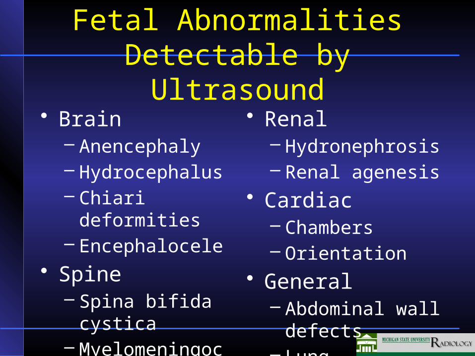
Fetal Abnormalities Detectable by Ultrasound
• Brain– Anencephaly– Hydrocephalus– Chiari deformities– Encephalocele
• Spine– Spina bifida
cystica– Myelomeningocel
e
• Renal– Hydronephrosis– Renal agenesis
• Cardiac– Chambers– Orientation
• General– Abdominal wall
defects– Lung abnormalities
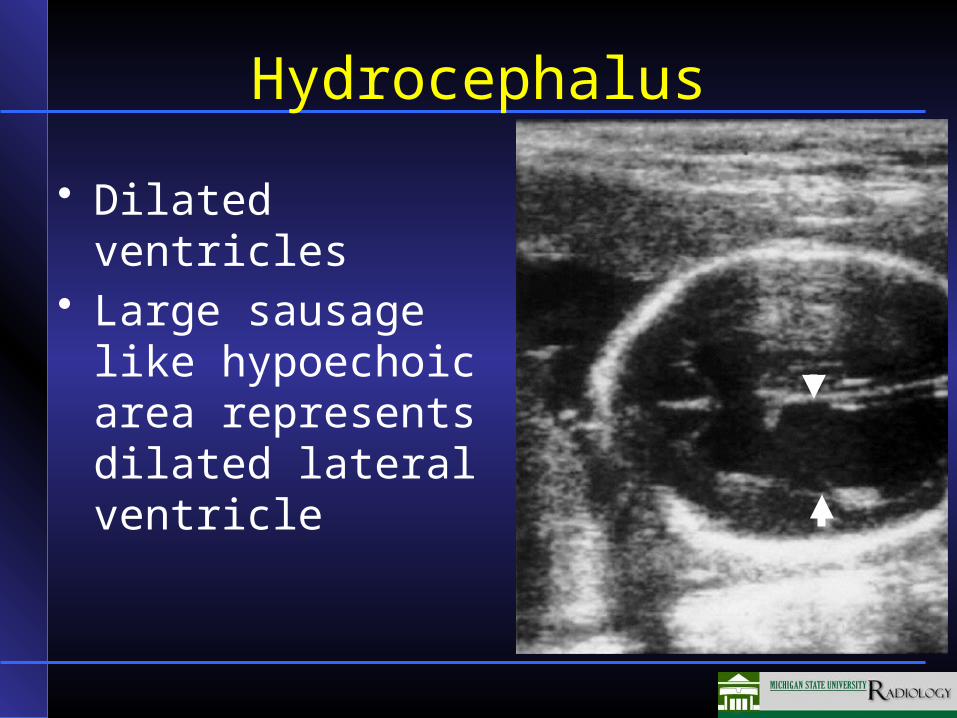
Hydrocephalus
• Dilated ventricles• Large sausage like
hypoechoic area represents dilated lateral ventricle
Intestinal Tract AbnormalitiesDetectable by Ultrasound
• Omphalocele• Abdominal wall defects and
gastroschisis• Midgut malrotation• Focal intestinal atresia
Normal Development of Intestinal Tract
• At 9 weeks there is physiologic herniation of the small bowel into the umbilical cord
• The small bowel rotates 90 degrees counterclockwise around the superior mesenteric artery
• At 12 weeks the small bowel returns into the abdominal cavity while rotating an additional 180 degrees counterclockwise around the superior mesenteric artery
• Total rotation of 270 degrees
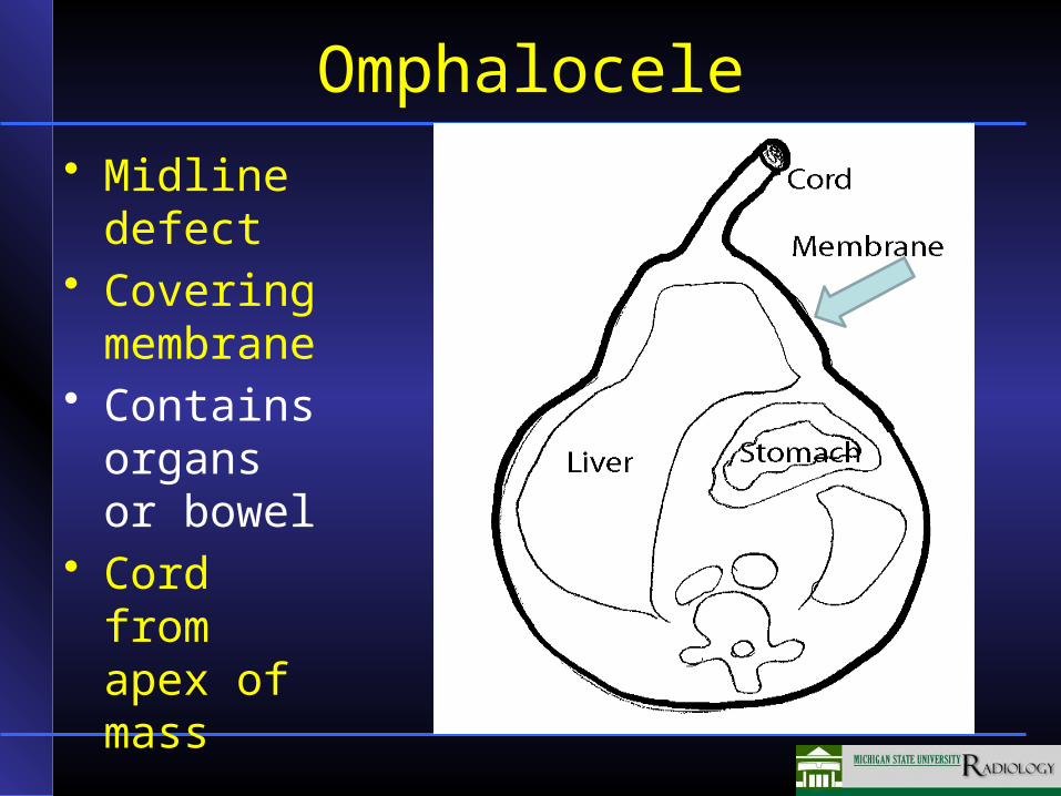
Omphalocele
• Midline defect
• Covering membrane
• Contains organs or bowel
• Cord from apex of mass
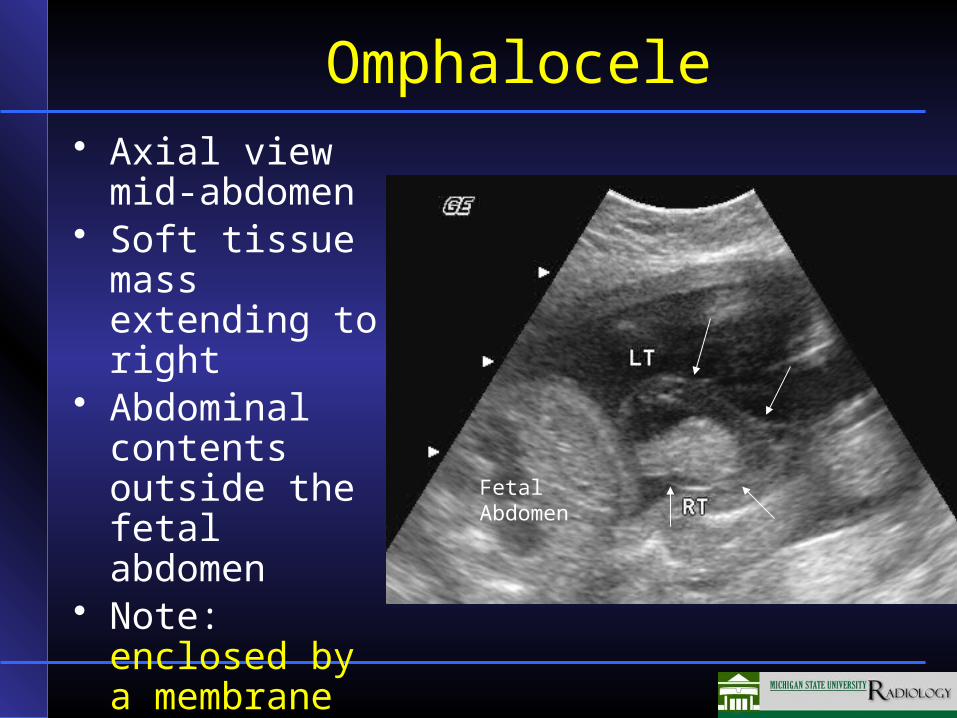
Omphalocele• Axial view mid-
abdomen• Soft tissue
mass extending to right
• Abdominal contents outside the fetal abdomen
• Note: enclosed by a membrane (arrows)
FetalAbdomen
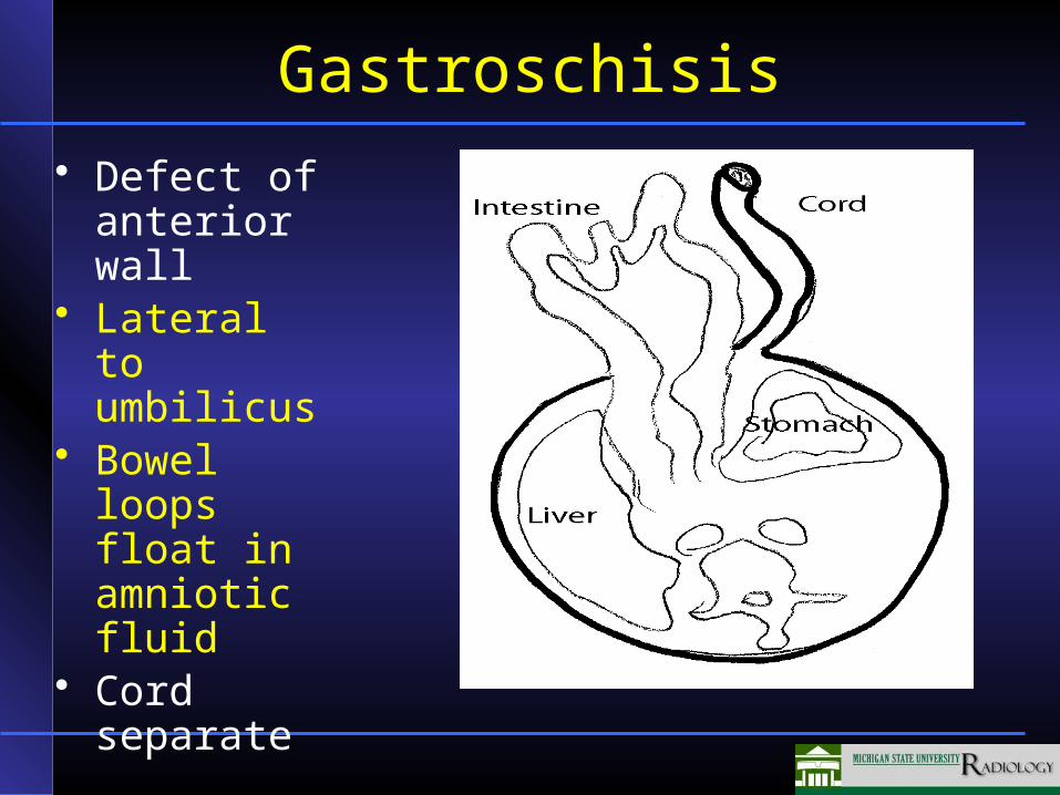
Gastroschisis
• Defect of anterior wall
• Lateral to umbilicus
• Bowel loops float in amniotic fluid
• Cord separate
Gastroschisis
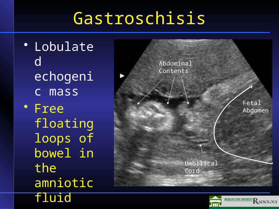
• Lobulated echogenic mass
• Free floating loops of bowel in the amniotic fluid
FetalAbdomen
UmbilicalCord
AbdominalContents
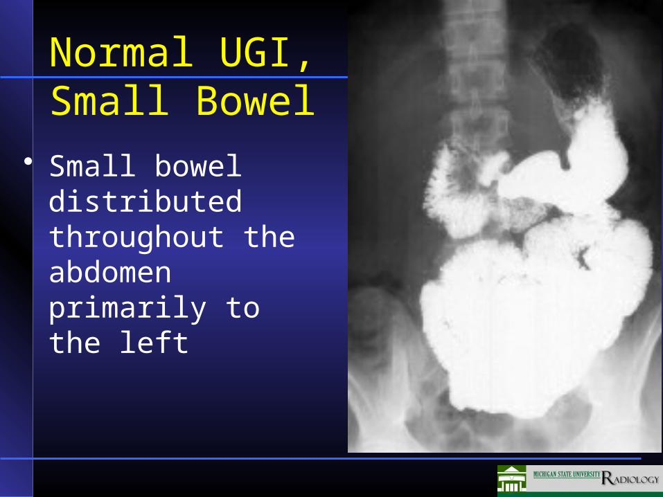
Normal UGI, Small Bowel
• Small bowel distributed throughout the abdomen primarily to the left
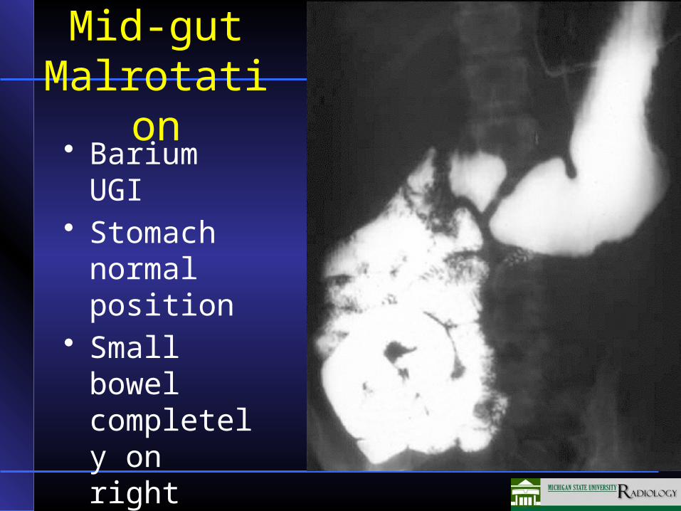
Mid-gut Malrotation• Barium UGI• Stomach
normal position
• Small bowel completely on right side of abdomen
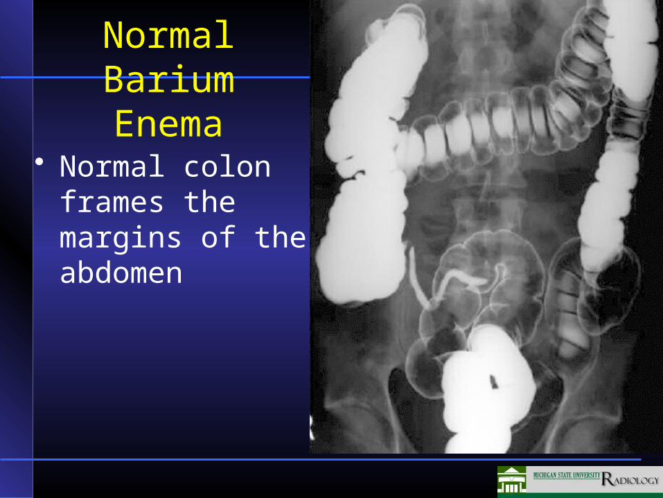
Normal Barium Enema
• Normal colon frames the margins of the abdomen
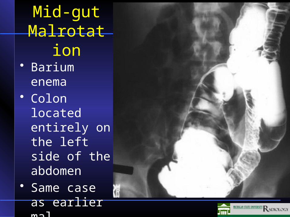
Mid-gut Malrotation
• Barium enema• Colon located
entirely on the left side of the abdomen
• Same case as earlier mal-rotation case
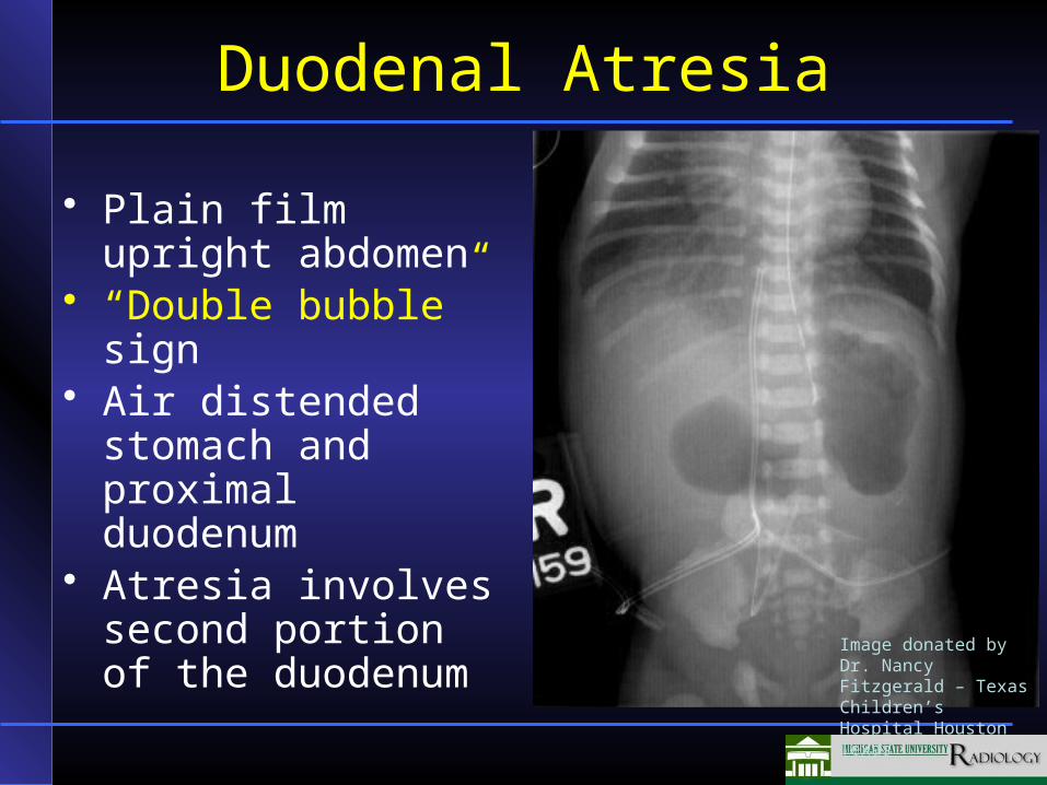
Duodenal Atresia
• Plain film upright abdomen
• “Double bubble” sign• Air distended
stomach and proximal duodenum
• Atresia involves second portion of the duodenum
Image donated by Dr. Nancy Fitzgerald – Texas Children’s Hospital Houston Texas
Skeletal Development Long Bones
• Diaphysis ossified at birth (shaft of long bone)
• Epiphysis radiolucent (cartilage) at birth except for distal femoral epiphysis– Develop Epiphyseal Ossification Centers
(EOC) later in life
Skeletal Development Long Bones
• Physis– Cartilaginous plate between EOC and
metaphysis– Responsible for growth in length– When ossifies (closes) – longitudinal growth
stops– Weak point in the bone
• Metaphysis– Active bone formation via formation and
calcification of osteoid
Bone Growth Abnormalities
• Cartilage growth deficiency– Example: Achondroplasia
• Ossification growth deficiency– Example: Osteogenic imperfecta
• Metabolic defect– Example: Hypophosphatasia
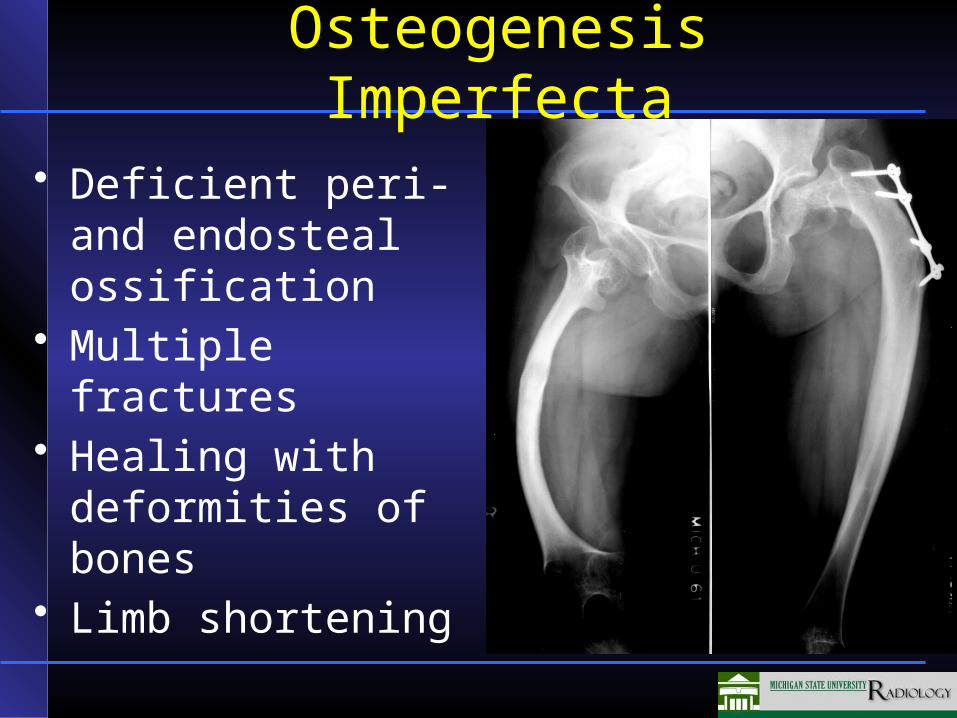
Osteogenesis Imperfecta
• Deficient peri- and endosteal ossification
• Multiple fractures• Healing with
deformities of bones• Limb shortening
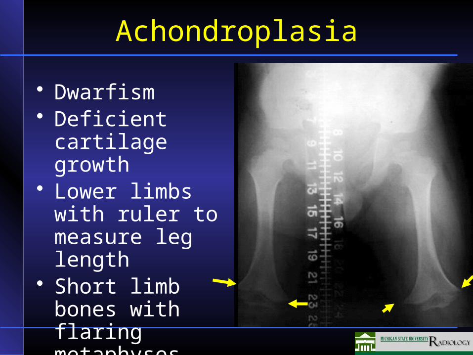
Achondroplasia
• Dwarfism• Deficient cartilage
growth• Lower limbs with
ruler to measure leg length
• Short limb bones with flaring metaphyses
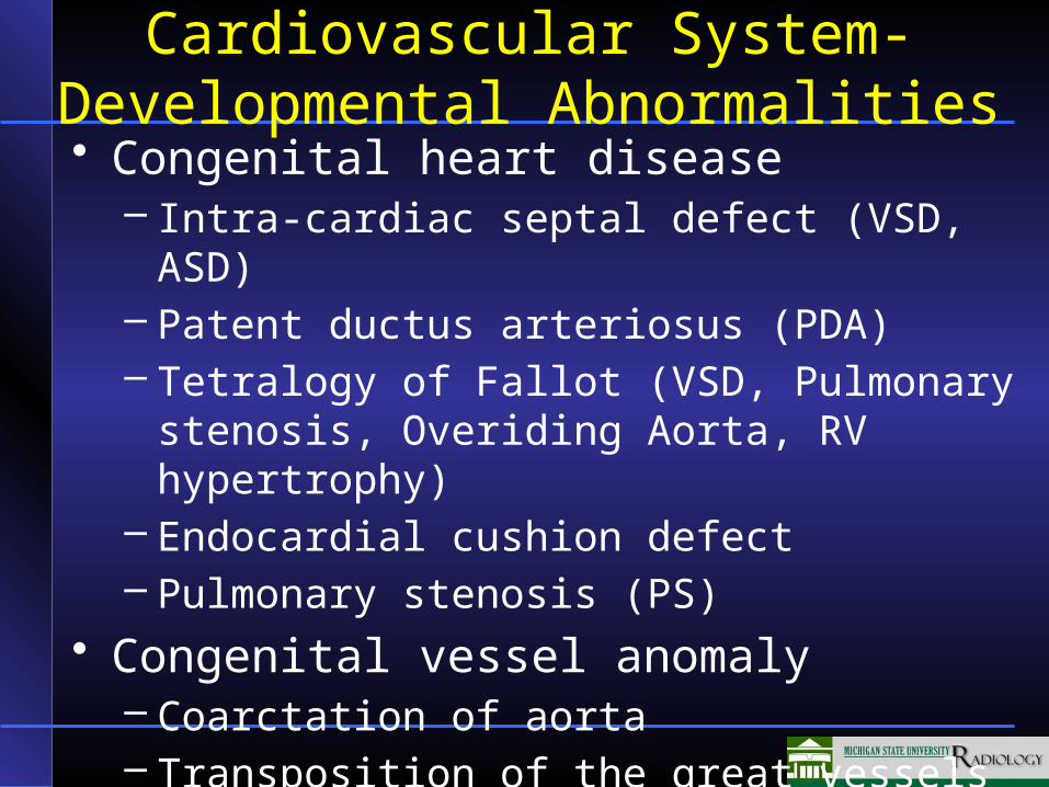
Cardiovascular System- Developmental Abnormalities
• Congenital heart disease– Intra-cardiac septal defect (VSD, ASD)– Patent ductus arteriosus (PDA)– Tetralogy of Fallot (VSD, Pulmonary stenosis,
Overiding Aorta, RV hypertrophy)– Endocardial cushion defect– Pulmonary stenosis (PS)
• Congenital vessel anomaly– Coarctation of aorta– Transposition of the great vessels
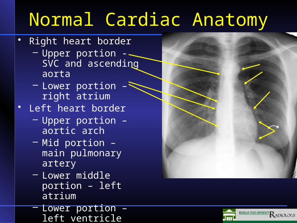
Normal Cardiac Anatomy• Right heart border
– Upper portion - SVC and ascending aorta
– Lower portion – right atrium
• Left heart border– Upper portion – aortic
arch– Mid portion – main
pulmonary artery– Lower middle portion –
left atrium– Lower portion – left
ventricle
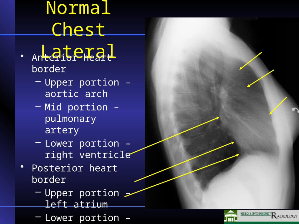
Normal Chest Lateral
• Anterior heart border– Upper portion –
aortic arch– Mid portion –
pulmonary artery– Lower portion – right
ventricle• Posterior heart border
– Upper portion – left atrium
– Lower portion – left ventricle and IVC
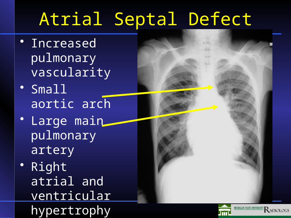
Atrial Septal Defect• Increased
pulmonary vascularity
• Small aortic arch
• Large main pulmonary artery
• Right atrial and ventricular hypertrophy
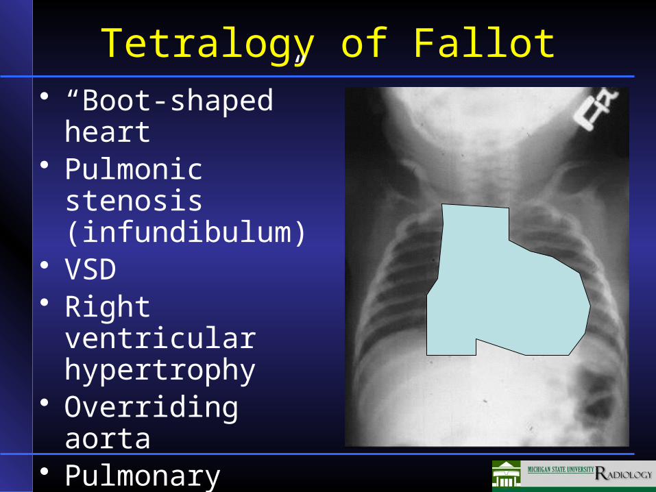
Tetralogy of Fallot• “Boot-shaped” heart• Pulmonic stenosis
(infundibulum)• VSD• Right ventricular
hypertrophy• Overriding aorta• Pulmonary
circulation decreased
Renal Abnormalities
• Anomalies in size and form– Horseshoe kidney
• Anomalies in position– Malrotation– Ectopia
• Anomalies in structure– Polycystic kidney
• Anomalies of drainage system– Duplicated kidney, ureter
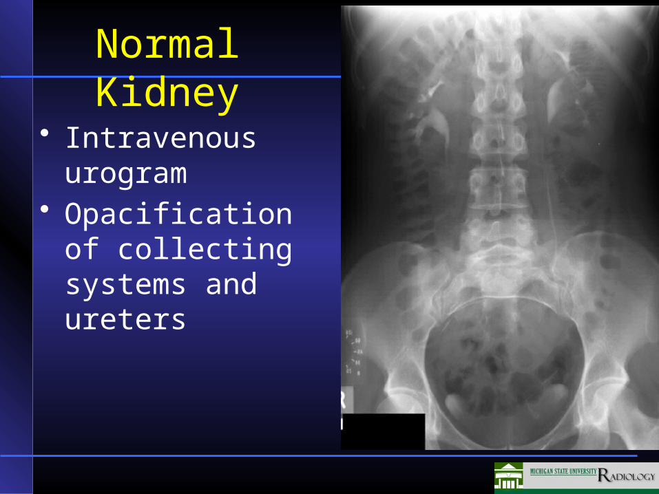
Normal Kidney
• Intravenous urogram
• Opacification of collecting systems and ureters
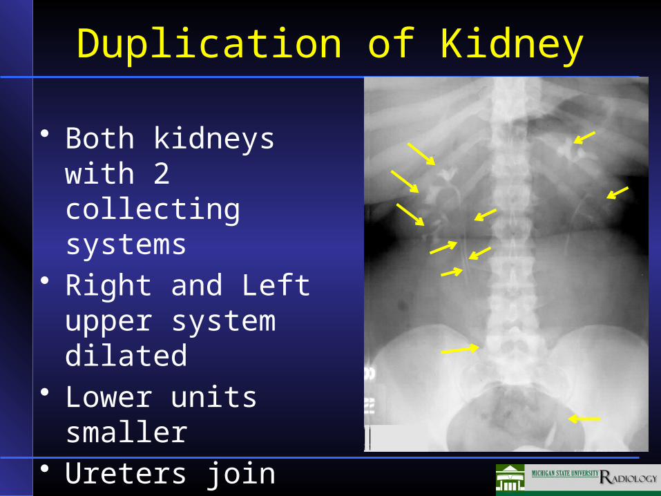
Duplication of Kidney
• Both kidneys with 2 collecting systems
• Right and Left upper system dilated
• Lower units smaller• Ureters join before
bladder
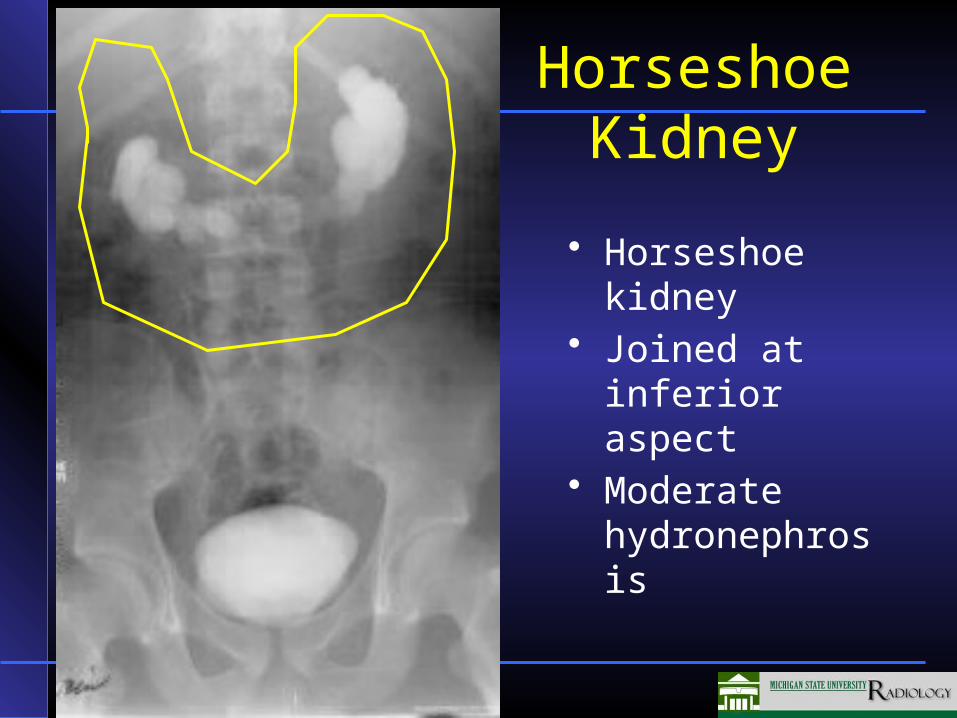
• Horseshoe kidney
• Joined at inferior aspect
• Moderate hydronephrosis
Horseshoe Kidney
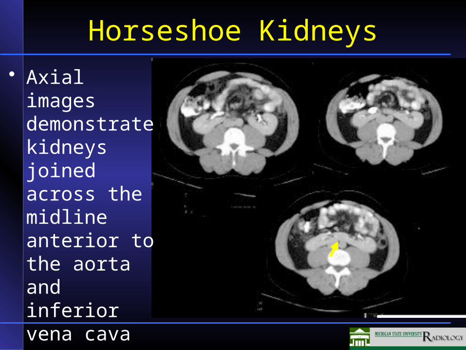
Horseshoe Kidneys
• Axial images demonstrate kidneys joined across the midline anterior to the aorta and inferior vena cava
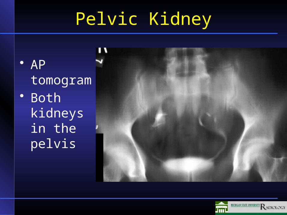
Pelvic Kidney
• AP tomogram
• Both kidneys in the pelvis
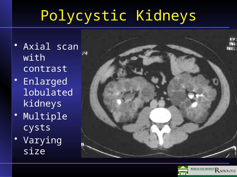
Polycystic Kidneys
• Axial scan with contrast
• Enlarged lobulated kidneys
• Multiple cysts• Varying size
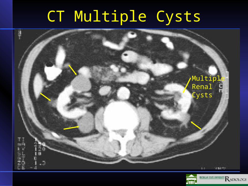
CT Multiple Cysts
MultipleRenal Cysts
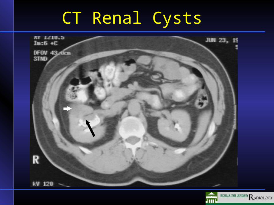
CT Renal Cysts
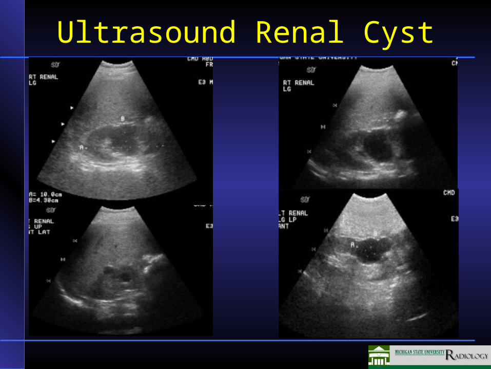
Ultrasound Renal Cyst
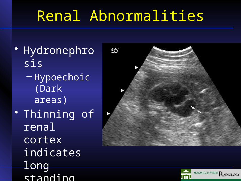
Renal Abnormalities
• Hydronephrosis– Hypoechoic
(Dark areas)• Thinning of
renal cortex indicates long standing process
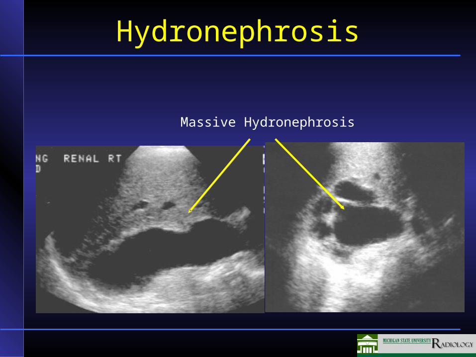
Hydronephrosis
Massive Hydronephrosis