FESS AND WHAT THE RADIOLOGIST NEEDS TO KNOW · FESS AND WHAT THE RADIOLOGIST NEEDS TO KNOW...
18
FESS AND WHAT THE RADIOLOGIST NEEDS TO KNOW Introduction: • Advances made in FESS (functional endoscopic sinus surgery) requires a thorough pre-op understanding of patients sino-nasal anatomy by ENT surgeon • CT scanning and subsequent evaluation of the patients anatomy with special attention to anatomical variants that may predispose to complications during surgery are important to reduce morbidity and mortality • OMU and the ethmoid sinus in particular are intimately associated and the anatomy of both must be thoroughly understood to accurately interpret CT scans • Normal variants predisposing to complications during FESS should be understood • Imaging evaluation of patients presenting with complications of FESS NORMAL ANATOMY: Nasal septum • Nasal septum extends the entire length of the nose and is comprised of a hard and soft part • Hard portion: perpendicular plate of ethmoid (sup) (posterior) vomer (post-inf) septal cartilage • Soft mobile septum: medial crus of alar cartilage (anterior) skin and soft tissue Anatomy of lateral wall of the nose • 3 Turbinates project from lateral nasal wall, I.e. superior, middle and inferior turbinates (concha). • As each turbinate curls inferolaterally, 3 distinct air channels are formed in the AP dimension, I.e superior, middle and inferior meati respectively. Created with novaPDF Printer (www.novaPDF.com)
Transcript of FESS AND WHAT THE RADIOLOGIST NEEDS TO KNOW · FESS AND WHAT THE RADIOLOGIST NEEDS TO KNOW...
FESS AND WHAT THE RADIOLOGIST NEEDS TO KNOW
Introduction:
• Advances made in FESS (functional endoscopic sinus surgery) requires a thorough pre-op understanding of patients sino-nasal anatomy by ENT surgeon
• CT scanning and subsequent evaluation of the patients anatomy with special attention to anatomical variants that may predispose to complications during surgery are important to reduce morbidity and mortality
• OMU and the ethmoid sinus in particular are intimately associated and the anatomy of both must be thoroughly understood to accurately interpret CT scans
• Normal variants predisposing to complications during FESS should be understood
• Imaging evaluation of patients presenting with complications of FESS NORMAL ANATOMY: Nasal septum
• Nasal septum extends the entire length of the nose and is comprised of a hard and soft part
• Hard portion: perpendicular plate of ethmoid (sup) (posterior) vomer (post-inf) septal cartilage
• Soft mobile septum: medial crus of alar cartilage (anterior) skin and soft tissue Anatomy of lateral wall of the nose
• 3 Turbinates project from lateral nasal wall, I.e. superior, middle and inferior turbinates (concha).
• As each turbinate curls inferolaterally, 3 distinct air channels are
formed in the AP dimension, I.e superior, middle and inferior meati respectively.
Created with novaPDF Printer (www.novaPDF.com)
• Various ostia drain into the meati
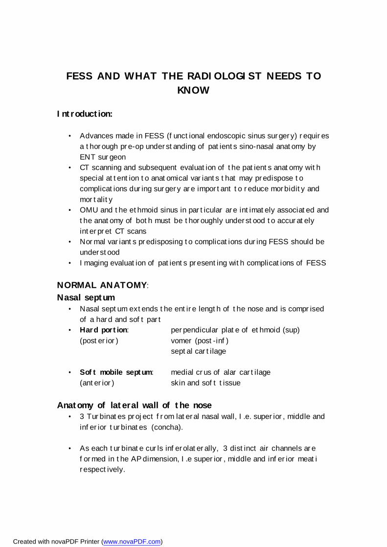
Superior turbinate and meatus • Part of ethmoid bone • Smallest turbinate, overlies the superior meatus • Posterior ethmoidal air cells drain into superior meatus via multiple
ostia • Spheno-ethmoidal recess lies on postero-superior aspect of superior
turbinate, between the anterior wall of the sphenoid sinus and the posterior wall of the ethmoid air cells.
• Easily seen on axials; on coronals more difficult – usually seen where sup turbinate ends posteriorly
• Sphenoethmoidal recess drains the sphenoid sinus through the sphenoid ostium
SPHENOETHMOIDAL RECESS NS = nasal septum E = ethmoid sinuses S = sphenoid sinus C = ICA ON = optic nerve
Created with novaPDF Printer (www.novaPDF.com)
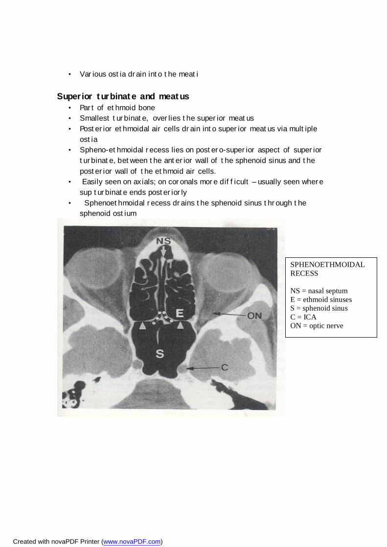
Middle turbinate and meatus • Middle turbinate (ethmoid bone) overlies the middle meatus, which is
the most important anatomic area in the lateral wall of the nose • Superiorly attached to lateral aspect of the cribriform plate; between
cribriform plate and fovea ethmoidalis • Lateral attachment, called the basal lamella, attaches to the thin
lamina papyracea of the lateral ethmoidal wall. Posteriorly, the basal lamella curves superiorly to become
coronally orientated. This coronal part, separates the anterior and middle from the posterior ethmoidal air cells.
• Pneumatization of the middle turbinate occurs as a normal variant and is called concha bullosa
BASAL LAMELLA (long white arrow) attachment of middle turbinate to the lamina papyracea
(black arrowhead)
B = Bulla ethmoidalis
Created with novaPDF Printer (www.novaPDF.com)
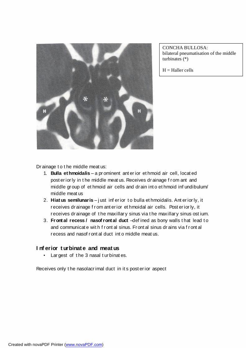
Drainage to the middle meatus:
1. Bulla ethmoidalis – a prominent anterior ethmoid air cell, located posteriorly in the middle meatus. Receives drainage from ant and middle group of ethmoid air cells and drain into ethmoid infundibulum/ middle meatus
2. Hiatus semilunaris – just inferior to bulla ethmoidalis. Anteriorly, it receives drainage from anterior ethmoidal air cells. Posteriorly, it receives drainage of the maxillary sinus via the maxillary sinus ostium.
3. Frontal recess / nasofrontal duct –defined as bony walls that lead to and communicate with frontal sinus. Frontal sinus drains via frontal recess and nasofrontal duct into middle meatus.
Inferior turbinate and meatus
• Largest of the 3 nasal turbinates. Receives only the nasolacrimal duct in its posterior aspect
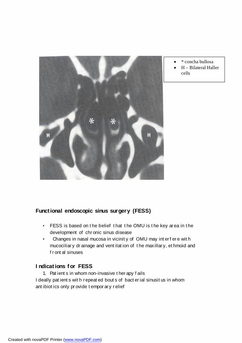
CONCHA BULLOSA: bilateral pneumatisation of the middle turbinates (*) H = Haller cells
Created with novaPDF Printer (www.novaPDF.com)
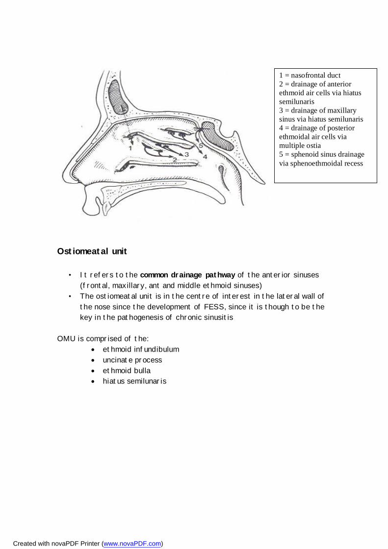
Ostiomeatal unit
• It refers to the common drainage pathway of the anterior sinuses (frontal, maxillary, ant and middle ethmoid sinuses)
• The ostiomeatal unit is in the centre of interest in the lateral wall of the nose since the development of FESS, since it is though to be the key in the pathogenesis of chronic sinusitis
OMU is comprised of the:
ethmoid infundibulum uncinate process ethmoid bulla hiatus semilunaris
1 = nasofrontal duct 2 = drainage of anterior ethmoid air cells via hiatus semilunaris 3 = drainage of maxillary sinus via hiatus semilunaris 4 = drainage of posterior ethmoidal air cells via multiple ostia 5 = sphenoid sinus drainage via sphenoethmoidal recess
Created with novaPDF Printer (www.novaPDF.com)
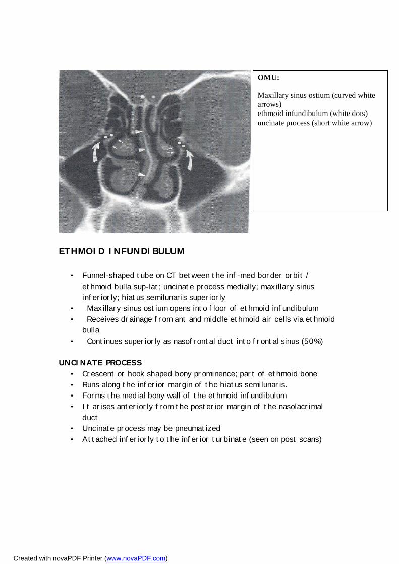
ETHMOID INFUNDIBULUM
• Funnel-shaped tube on CT between the inf-med border orbit / ethmoid bulla sup-lat; uncinate process medially; maxillary sinus inferiorly; hiatus semilunaris superiorly
• Maxillary sinus ostium opens into floor of ethmoid infundibulum • Receives drainage from ant and middle ethmoid air cells via ethmoid
bulla • Continues superiorly as nasofrontal duct into frontal sinus (50%)
UNCINATE PROCESS • Crescent or hook shaped bony prominence; part of ethmoid bone • Runs along the inferior margin of the hiatus semilunaris. • Forms the medial bony wall of the ethmoid infundibulum • It arises anteriorly from the posterior margin of the nasolacrimal
duct • Uncinate process may be pneumatized • Attached inferiorly to the inferior turbinate (seen on post scans)
OMU: Maxillary sinus ostium (curved white arrows) ethmoid infundibulum (white dots) uncinate process (short white arrow)
Created with novaPDF Printer (www.novaPDF.com)
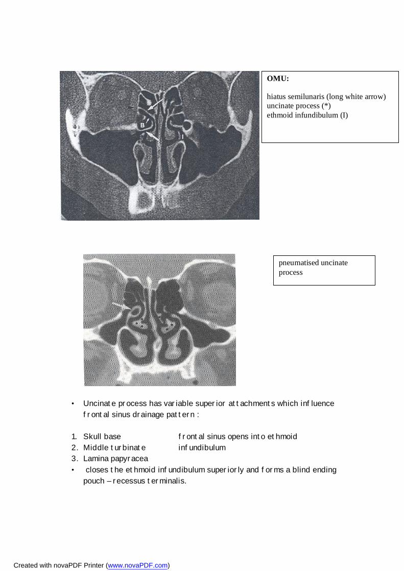
• Uncinate process has variable superior attachments which influence frontal sinus drainage pattern :
1. Skull base frontal sinus opens into ethmoid 2. Middle turbinate infundibulum 3. Lamina papyracea • closes the ethmoid infundibulum superiorly and forms a blind ending
pouch – recessus terminalis.
OMU: hiatus semilunaris (long white arrow) uncinate process (*) ethmoid infundibulum (I)
pneumatised uncinate process
Created with novaPDF Printer (www.novaPDF.com)
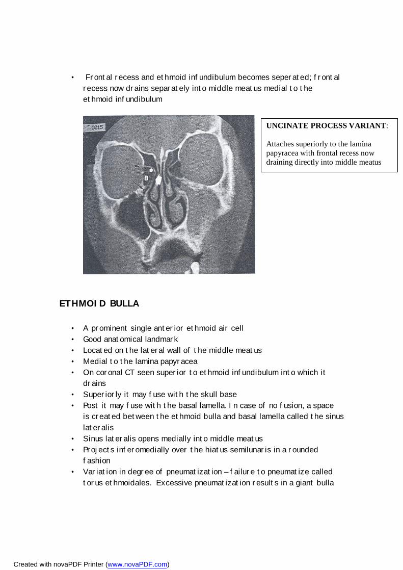
• Frontal recess and ethmoid infundibulum becomes seperated; frontal recess now drains separately into middle meatus medial to the ethmoid infundibulum
ETHMOID BULLA
• A prominent single anterior ethmoid air cell • Good anatomical landmark • Located on the lateral wall of the middle meatus • Medial to the lamina papyracea • On coronal CT seen superior to ethmoid infundibulum into which it
drains • Superiorly it may fuse with the skull base • Post it may fuse with the basal lamella. In case of no fusion, a space
is created between the ethmoid bulla and basal lamella called the sinus lateralis
• Sinus lateralis opens medially into middle meatus • Projects inferomedially over the hiatus semilunaris in a rounded
fashion • Variation in degree of pneumatization – failure to pneumatize called
torus ethmoidales. Excessive pneumatization results in a giant bulla
UNCINATE PROCESS VARIANT: Attaches superiorly to the lamina papyracea with frontal recess now draining directly into middle meatus
Created with novaPDF Printer (www.novaPDF.com)
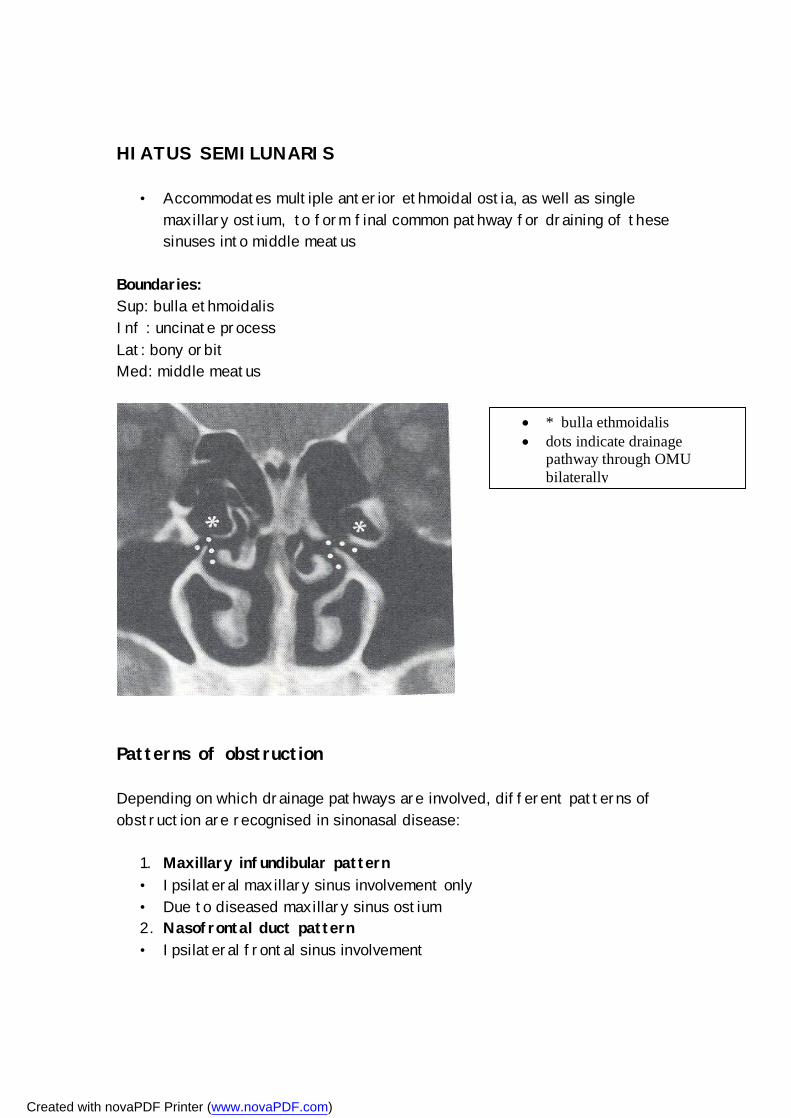
HIATUS SEMILUNARIS
• Accommodates multiple anterior ethmoidal ostia, as well as single maxillary ostium, to form final common pathway for draining of these sinuses into middle meatus
Boundaries: Sup: bulla ethmoidalis Inf : uncinate process Lat: bony orbit Med: middle meatus
Patterns of obstruction Depending on which drainage pathways are involved, different patterns of obstruction are recognised in sinonasal disease:
1. Maxillary infundibular pattern • Ipsilateral maxillary sinus involvement only • Due to diseased maxillary sinus ostium 2. Nasofrontal duct pattern • Ipsilateral frontal sinus involvement
* bulla ethmoidalis dots indicate drainage
pathway through OMU bilaterally
Created with novaPDF Printer (www.novaPDF.com)
• Due to obstrustion of nasofrontal duct / frontal recess 3. OMU pattern • Opacification of the middle meatus • Secondary inflammatory change in maxillary, ant and middle ethmoid,
and frontal sinus 4. Sphenoethmoidal recess pattern • Sphenoid and ipsilateral post ethmoid sinus involvement • Due to obstruction at level of spheno-ethmoidal recess 5. Sinonasal polyposis • Inflammatory polyps fills the nasal cavity and sinuses bilaterally – see
mixture of previously mentioned patterns 6. Sporadic pattern • Random inflammatory changes
Ethmoid sinus • Key sinus in FESS is the ethmoid sinus; knowledge of its anatomy is
essential for understanding pathological processes and in prevention of surgical complications.
“If the ethmoid were placed in any other part of the body it would be an insignificant and harmless collection of bony cells. In the place where nature has put it, it has major relationships so that diseases and surgery of the labyrinth often leads to tragedy. Any surgery in this region should be simple but has proven one of the easiest ways to kill a patient” MOSHER 1929
• Ethmoid bone delicate and complex; articulates with 13 other bones • Divided into 4 parts: cribriform plate
perpendicular plate (part of bony nasal septum) 2 laterally located ethmoid labyrinths Cribriform plate
• 2-3 mm wide plate • each has approximately 20 foramina – transmit olfactory nerve fibres • forms ant part of roof of nose
Ethmoid labyrinth
Created with novaPDF Printer (www.novaPDF.com)
• each side consist of 3-18 highly variable thin-walled air cells • in disarticulated bone – air cells open superiorly – closed by fovea
ethmoidalis of frontal bone (lat to cribriform plate) • air cells arranged in ant, middle and post groups • 3 air cell groups included in ant group 1. Frontal recess cells - most anteriorly
extends ant-sup toward frontal bone frontal sinus develop from these
2. Infundibular air cells – gives rise to agger nasi cells (extramural air cell)
3. Bulla air cells / bulla ethmoidalis – part of OMU
Late pneumatization of the ethmoid sinus lead to normal variants / extramural air cells:
1. Agger nasi cells • extension of pneumatization of ant ethmoid air cells into frontal
process of the maxilla / lacrimal bone • Located ant and inf to frontal recess • Forms ant part of floor of frontal sinus • Closeness to frontal recess make them excellent surgical landmarks –
often opened to view nasofrontal duct • Drain into ethmoid infundibulum 2. Concha bullosa • Pneumatization of middle turbinate from middle ethmoid air cells • 34 % incidence • May cause obstruction of ethmoid infundibulum 3. Haller cell • Air cell located below ethmoid bulla, along roof of maxillary sinus and
below lowest point of lamina papyracea • If large may obstruct maxillary sinus ostium 4. Onodi cell • Post-lat extension of post ethmoid air cell to surround optic nerve
Created with novaPDF Printer (www.novaPDF.com)
Functional endoscopic sinus surgery (FESS)
• FESS is based on the belief that the OMU is the key area in the development of chronic sinus disease
• Changes in nasal mucosa in vicinity of OMU may interfere with mucociliary drainage and ventilation of the maxillary, ethmoid and frontal sinuses
Indications for FESS
1. Patients in whom non-invasive therapy fails Ideally patients with repeated bouts of bacterial sinusitus in whom antibiotics only provide temporary relief
* concha bullosa H – Bilateral Haller
cells
Created with novaPDF Printer (www.novaPDF.com)
2. Chronic hyperplastic rhinosinusitis (extensive disease throughout paranasal sinuses) or polyps
3. Mucocele 4. Peri-orbital cellulitis secondary to ethmoiditis
Role of radiologist in FESS 1. Should be familiar with principles of FESS 2. Should carefully evaluate the paranasal sinuses, with particular
attention to the ethmoid bone and OMU 3. Should be familiar with normal variations that predispose to
complications of FESS 4. Should ensure correct patient preparation and correct technique of
CT evaluation Technique of FESS
1. FESS attempts to restore normal sinus drainage via an intranasal fiberoptic endoscope
2. Extent of surgery depends on the pattern of obstruction identified on CT scans
3. Involves minimal resection of sinus walls and mucosa 4. Success rate is comparable to that of older more invasive techniques
CT Technique for evaluation of OMU / sinuses
1. Coronal CT preferred – simulates plane seen by endoscopist • patient prone with neck hyperextended • 3mm slice increments
2. Axial plane (5mm slices) allows better evaluation of • Ant and post walls of frontal sinuses • Relationship between post ethmoid and sphenoid sinuses • Relationship of optic nerve to post ethmoid and sphenoid sinuses 2. Wide window widths (4000H/750H), ?soft tissue settings –
advantages - mucosal debris, microcalcifications, incidental findings in globe and orbit
Complications of FESS
• Complications vary from minor and temporary to that causing permanent injury and even death.
Created with novaPDF Printer (www.novaPDF.com)
• Anatomical variants predispose certain structures to injury during FESS
• Radiologist can minimise risk for complications by recognising normal variants and alerting surgeon
COMPLICATIONS:
• Recurrent inflammatory disease • Orbital injuries • Hemorrhage • CSF leak • Intracranial complications
Recurrent inflammatory disease • Factors predisposing to poor symptom relief after FESS: 1. Diffuse sinonasal polyposis 2. Asthma 3. Prior surgery 4. Tenacious sinus secretions Complications of FESS can predispose to recurrent infection: 1. Synechiae /adhesions - b/t lat nasal wall + lat aspect middle
turbinate; thus causing inadequate drainage through stenotic middle meatus
2. Closure of natural sinus ostia – common in sinonasal polyposis 3. Incomplete ethmoidectomy
Orbital complications: These include:
• Orbital edema, hematoma of lower eyelid and orbital emphysema due to violation of lamina papyracea
• Epiphora – injury to nasolacrimal duct – located just ant to maxillary sinus ostium
• Diplopia – d.t. extra-ocular muscle injury most commonly MR, SR and SO which are injured/transected during ethmoidectomy
• Blindness – Acute intra-orbital hematoma with resultant pressure on optic nerve leading to ischaemia (ant ethmoid artery
Created with novaPDF Printer (www.novaPDF.com)
• Blindness can also be result of direct optic nerve injury (rare) • Post-op orbital abcess – can lead to cavernous sinus thrombosis
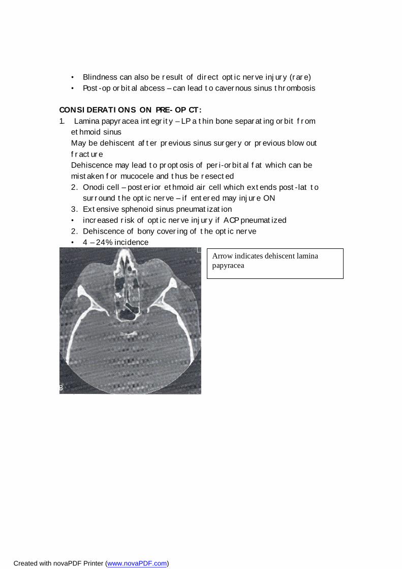
CONSIDERATIONS ON PRE-OP CT: 1. Lamina papyracea integrity – LP a thin bone separating orbit from ethmoid sinus
May be dehiscent after previous sinus surgery or previous blow out fracture Dehiscence may lead to proptosis of peri-orbital fat which can be mistaken for mucocele and thus be resected 2. Onodi cell – posterior ethmoid air cell which extends post-lat to
surround the optic nerve – if entered may injure ON 3. Extensive sphenoid sinus pneumatization • increased risk of optic nerve injury if ACP pneumatized 2. Dehiscence of bony covering of the optic nerve • 4 – 24% incidence
Arrow indicates dehiscent lamina papyracea
Created with novaPDF Printer (www.novaPDF.com)
Post-op evaluation of suspected orbital complications
• Post-operative evaluation should be conducted in both coronal and axial planes; usually unenhanced only
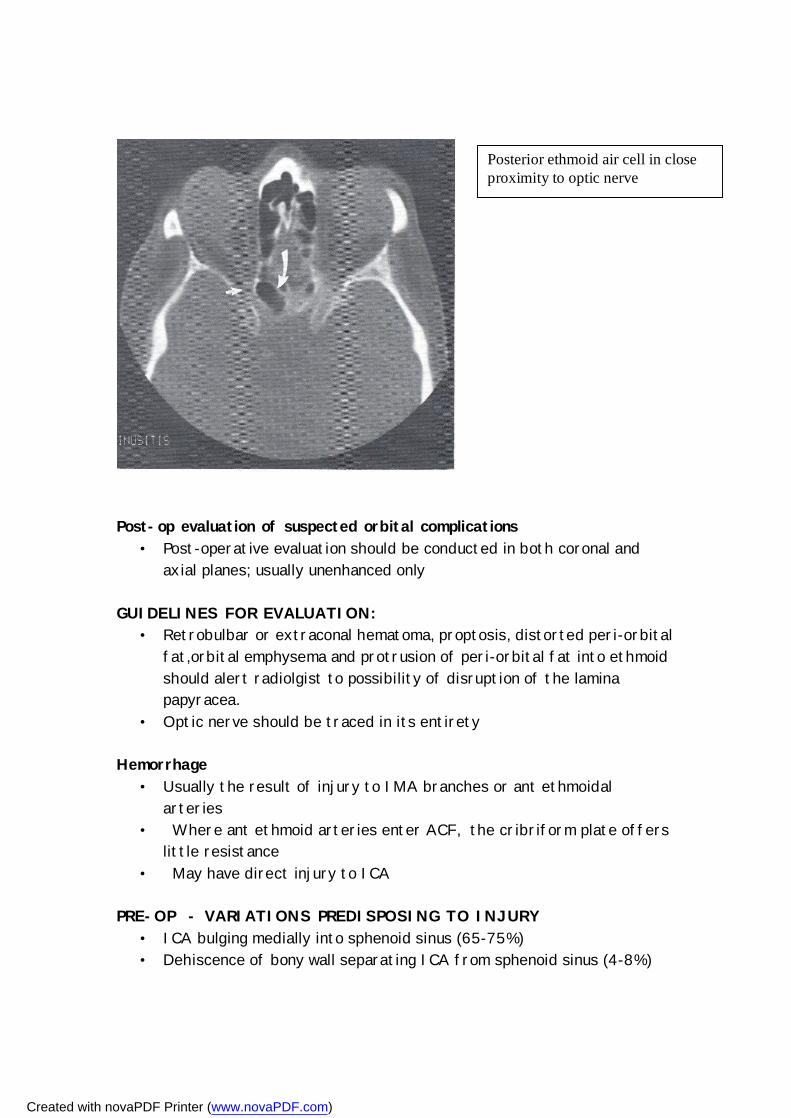
GUIDELINES FOR EVALUATION:
• Retrobulbar or extraconal hematoma, proptosis, distorted peri-orbital fat,orbital emphysema and protrusion of peri-orbital fat into ethmoid should alert radiolgist to possibility of disruption of the lamina papyracea.
• Optic nerve should be traced in its entirety Hemorrhage
• Usually the result of injury to IMA branches or ant ethmoidal arteries
• Where ant ethmoid arteries enter ACF, the cribriform plate offers little resistance
• May have direct injury to ICA PRE-OP - VARIATIONS PREDISPOSING TO INJURY
• ICA bulging medially into sphenoid sinus (65-75%) • Dehiscence of bony wall separating ICA from sphenoid sinus (4-8%)
Posterior ethmoid air cell in close proximity to optic nerve
Created with novaPDF Printer (www.novaPDF.com)
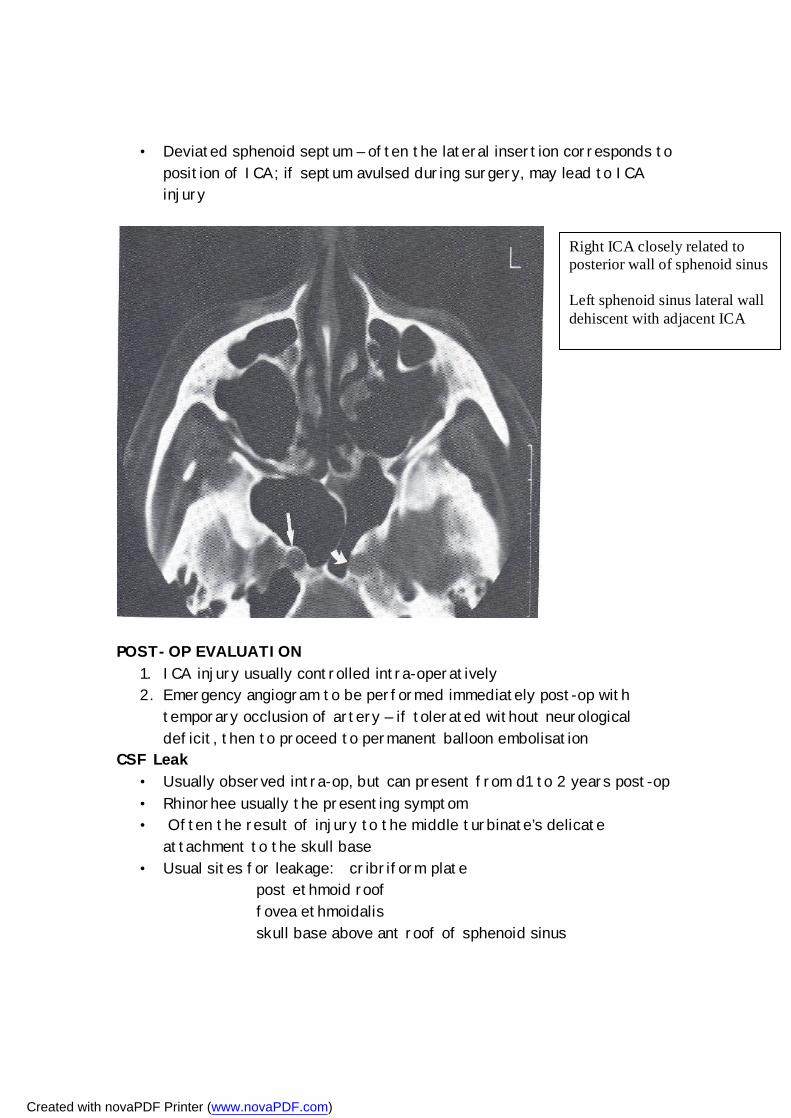
• Deviated sphenoid septum – often the lateral insertion corresponds to position of ICA; if septum avulsed during surgery, may lead to ICA injury
POST-OP EVALUATION
1. ICA injury usually controlled intra-operatively 2. Emergency angiogram to be performed immediately post-op with
temporary occlusion of artery – if tolerated without neurological deficit, then to proceed to permanent balloon embolisation
CSF Leak • Usually observed intra-op, but can present from d1 to 2 years post-op • Rhinorhee usually the presenting symptom • Often the result of injury to the middle turbinate’s delicate
attachment to the skull base • Usual sites for leakage: cribriform plate
post ethmoid roof fovea ethmoidalis skull base above ant roof of sphenoid sinus
Right ICA closely related to posterior wall of sphenoid sinus Left sphenoid sinus lateral wall dehiscent with adjacent ICA
Created with novaPDF Printer (www.novaPDF.com)
PRE-OP EVALUATION 1. Evaluate the above mentioned areas for dehiscence/ thinning
patients at increased risk if previous facial or skull base trauma; or previous sinus surgery
2. Low fovea ethmoidalis 3. Shallow olfactory fossa 4. Supra-orbital extension of ethmoid air cells
POST-OP EVALUATION OF CSF LEAK
• Unenhanced CT scan in coronal (3mm) and axial (5mm) plane • If any dehiscence seen – assumed to be the site of leakage • If no defect is seen – then proceed to perform CT cisternography
after injection of 5 ml of LOCM into lumbar SAS Intracranial complications
• Extremely rare • Not usually the result of anatomical variants • Documented complications include:
intracerebral hematomas cerebritis/ abcess encephalocoeles injury to ACA / ACoA and their branches pneumocephalus
CONCLUSION: • FESS and its complications has made it increasingly critical for the
radiologist to have a thorough understanding of sinonasal anatomy • In particular, one needs to be aware of the many normal variants of
especially the OMU and the ethmoid sinus. • It is the role of the radiologist to alert the surgeon of variants that
could put the patient at increased risk of complications during surgery.
• As a team, the radiologist and ENT surgeon should aim to optimise the benefits of FESS while minimising its risks
Created with novaPDF Printer (www.novaPDF.com)