Femoral Shaft Fractures Shaft...Femoral Shaft Fractures Robert F. Ostrum, MD Cooper University...
100
Femoral Shaft Fractures Robert F. Ostrum, MD Cooper University Hospital Camden, New Jersey Created March 2004; Revised June 2006: Revised 11/09
Transcript of Femoral Shaft Fractures Shaft...Femoral Shaft Fractures Robert F. Ostrum, MD Cooper University...

Femoral Shaft Fractures
Robert F. Ostrum, MD
Cooper University Hospital Camden, New Jersey
Created March 2004; Revised June 2006: Revised 11/09

Femur Fractures • Common injury due to major violent trauma • 1 femur fracture/ 10,000 people • More common in people < 25 yo or >65 yo • Femur fracture leads to reduced activity for 107
days, the average length of hospital stay is 25 days • Motor vehicle, motorcycle, auto-pedestrian,
aircraft, and gunshot wound accidents are most frequent causes

Anatomy • Long tubular bone, anterior bow, flair at femoral
condyles • Blood supply
– Metaphyseal vessels – Single nutrient artery in diaphysis enters through the
linea aspera – Nutrient artery communicates with medullary arteries
in intramedullary canal – Medullary arteries supply 2/3 of endosteal blood
supply

Blood Supply • Reaming destroys intramedullary endosteal blood
supply
• Periosteal blood flow increases
• Medullary blood supply is re-established over 8-12 weeks if spaces left in canal by implant
• Unreamed intramedullary nailing decreases blood flow less; restoration of endosteal blood flow earlier but equal to reamed canal at 12 weeks

Femur Fracture Classification
AO/OTA Femur Diaphysis - Bone segment 32

Femur Fracture Classification
• Type 0 - No comminution • Type 1 - Insignificant butterfly fragment
with transverse or short oblique fracture • Type 2 - Large butterfly of less than
50% of the bony width, > 50% of cortex intact
• Type 3 - Larger butterfly leaving less than 50% of the cortex in contact
• Type 4 - Segmental comminution » Winquist and Hansen 66A, 1984
Axial and rotational stability

Femur Fracture Management
• Piriformis fossa intact, lesser trochanter intact
• Can you nail this ?
• Should you nail this ?

Femur Fracture Management
• Initial traction with portable traction splint or transosseous pin and balanced suspension
• Evaluation of knee to determine pin placement
• Timing of surgery is dependent on: – Resuscitation of patient – Other injuries - abdomen, chest, brain – Isolated femur fracture

Bending moment = F x D F = Force
D
D = distance from force to implant
F = Force
D
The bending moment for the plate is greater due to the force being applied over a larger distance
IM Nail
Plate

Femur Fracture Management
• Diaphyseal fractures are managed by intramedullary nailing through an antegrade or retrograde insertion site
• Proximal or distal 1/3 fractures MAY be managed best with a plate or an intramedullary nail depending on the location and morphology of the fracture

Hare traction splint for initial reduction of femur fractures prior
to OR or skeletal traction
Femoral IM Nailing To Ream ?
Hypothesis: Femoral reaming increases fatty emboli to the
lungs and potentially increases pulmonary complications

Femur Fracture Reaming
• Reaming advantages: – Nail will not get incarcerated – Higher union rates – More durable fracture/nail construct – Earlier weight bearing
• Unreamed nails - still generate fat embolism with opening of piriformis fossa and probably higher pressure with unreamed nail insertion

Femur Fracture Reaming
• Reaming of the femoral shaft fracture – Multiple studies demonstrate that the thoracic
injury is the major determinant of pulmonary complications, NOT the use of a reamed IM nail
• Charash J Trauma 1994 • Van Os J Trauma 1994 • Ziran J Trauma 1997 • Bone Clin Orthop 1998 • Bosse JBJS 79A 1997

Femur Fracture Reaming
• Reaming of the femoral shaft fracture – Only Pape (J Trauma 1993) has shown a
deleterious pulmonary effect to immediate reamed intramedullary nailing in acute femur fracture patients with pulmonary trauma
– In both a retrospective analysis and multiple animal studies (Pape , J Trauma 1992)
– However, other animal studies refute these results • Wolinsky, J Orthop Tr 1998 • Duwelius, JBJS 79A 1997
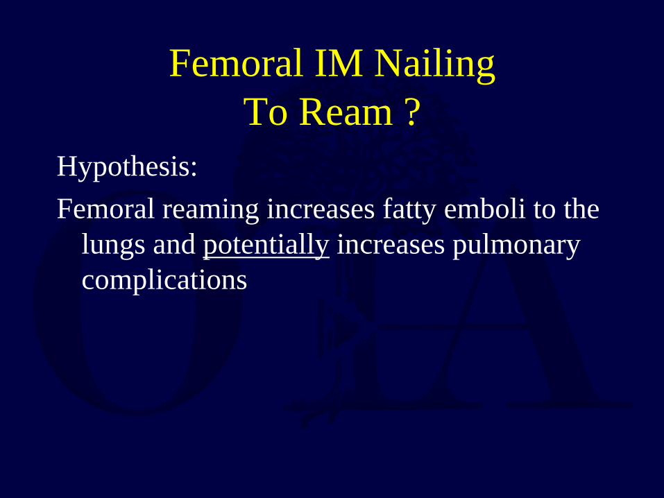
Femur Fracture Reaming Pressures
awl
9mm reaming guide pin
9.5mm first reamer 13mm reamer with larger shaft
NO increase pressure with nail insertion
No difference in pressures generated by head design
- Muller, Injury 1993
Injury + Patient
POLYTRAUMA • Early stabilization beneficial
» Seibel Ann Surg 1985 » Bone, JBJS 1989 » Goris , J Trauma 1982 » Johnson, J Trauma 1985 » Behrman, J Trauma 1990 » Bone, J Trauma 1994
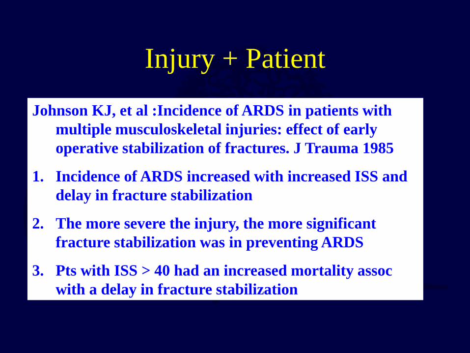
Johnson KJ, et al :Incidence of ARDS in patients with multiple musculoskeletal injuries: effect of early operative stabilization of fractures. J Trauma 1985
1. Incidence of ARDS increased with increased ISS and delay in fracture stabilization
2. The more severe the injury, the more significant fracture stabilization was in preventing ARDS
3. Pts with ISS > 40 had an increased mortality assoc with a delay in fracture stabilization

Damage Control Orthopaedics
Select group of critically injured or “borderline” patients may not tolerate extensive procedures or blood loss

External Fixator for Femoral Shaft Fracture
Multiply injured patient
Complex distal femur fracture
Dirty open fracture
Vascular injury
Exchange Nailing in the femur is safe and yields high union and low infection rates Nowotarski JBJS 2000

Injury + Patient
Practice management guidelines Recommendations-Polytrauma • Level II-no improvement in survival
- some patients fewer complications - no detrimental effect of early fixation - early fixation preferable
Dunham J Trauma 2001

Head Injury + Femur Fx • Early fixation of long bone
fractures does NOT promote secondary brain injury which may increase mortality, BUT hypoxia, hypotension, and increased ICP DO Poole J Trauma 1992
Schmeling CORR 1995 McKee J Trauma 1997 Velmahos Am J Surg 1998 Scalea J Trauma 1999

Chest Injury + Femur Fx CHEST INJURY • Increased pulmonary
morbidity (ARDS, fat embolism)
• Early long bone stabilization questioned in patients with significant pulmonary injury
Thoracic trauma ITSELF is the major determinant of morbidity and mortality, NOT IM NAILING Bone CORR 1995
Bosse JBJS 1997

Timing of femur fracture fixation: effect on outcome in patients with thoracic and head
injuries Brundage SI, J Trauma 2002
Data showed that early femur fracture fixation (< 24 hours) is associated with an improved outcome, even in patients with coexistent head and/or chest trauma.
Fixation of femur fractures at 2 to 5 days was associated with a significant increase in pulmonary
complications, particularly with concomitant head or chest trauma, and length of stay. Chest and head
trauma are not contraindications to early fixation with reamed intramedullary nailing.

Delayed IM Nailing of Femur Fractures Reduces Mortality
• 3069 patients, ISS> 15 • serious abdominal injury (AIS >3) had most benefit from resuscitation • delay > 12 hours DECREASED mortality by 50% in multisystem trauma patients
• Morshed, JBJS 2009

Comparison of Reamed vs Unreamed IM Nails 224 patients multiply injured patients Risk of nonunion was 5x greater in unreamed group 80% of nonunions could have been prevented by reaming
NO increase in ARDS with reaming !!
Powell and COA, JOT 2006
Conclusion:
REAM
Femoral Nailing Course # 101
1. Femoral Nail Design 2. Ream vs Unreamed 3. Nails available, treatment options
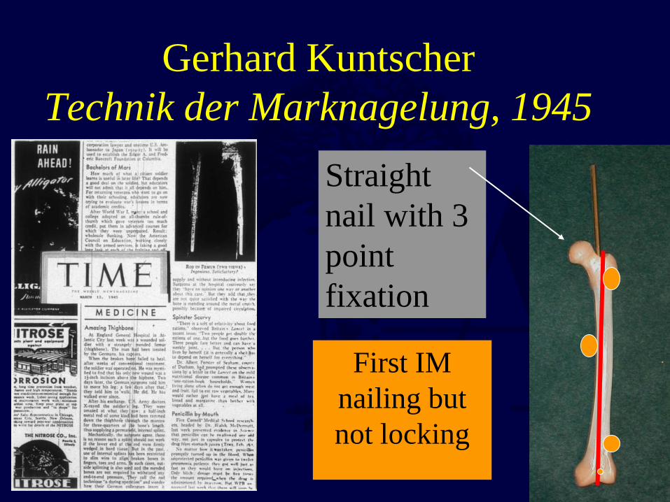
Gerhard Kuntscher Technik der Marknagelung, 1945
First IM nailing but not locking
Straight nail with 3 point fixation

Klemm K, Schellman WD: Veriegelung des marnagels, 1972
Kempf I, Grosse A: Closed Interlocking Intramedullary Nailing. Its Application to Comminuted fractures of the femur, 1985
Locking IM nails in the 1980’s

IM Nail Variables
• Stainless steel vs Titanium • Wall Thickness • Cannulation • Slotted vs Non-slotted • Radius of Curvature • ? To Ream

Stiffness Modulus of Elasticity
0 5 10 15 20 25 30 35 40
PMMA
cortex bone
titanium
316L stainless
cobalt
X 10 8 PSI
Metallurgy less important than other
parameters for stiffness of IM Nail

Wall Thickness
Large determinant of stiffness

Slotted vs Non-slotted
Anterior slot - improved flexibility
Posterior slot - increased bending strength
Non-slotted - increased torsional stiffness, increased strength in smaller sizes, ? comminution

Radius of Curvature of femur averages 120 cm
• Current femoral nails radius of curvature ranges from 150-300 cm
• IM nails are straighter (larger radius) than the femoral canal

Femur Fracture Management
• Antegrade nailing is still the gold standard – Highest union rates with reamed nails – Extraarticular starting point – Refined technique
• Antegrade nailing problems: – Varus alignment of proximal fractures – Trendelenburg gait – Can be difficult with obese or multiply injured patients
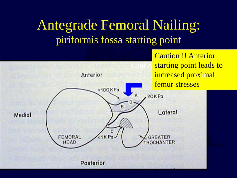
Antegrade Femoral Nailing: piriformis fossa starting point
Caution !! anterior
Caution !! Anterior starting point leads to increased proximal femur stresses

Minimally Invasive Nail Insertion Technique (MINIT)
1 2
3 4 Courtesy T.A. Russell, M.D.

Antegrade Femoral Nailing starting point
Posterior - loss of proximal fixation
Piriformis fossa- proper starting point
Anterior - generates huge forces, can lead to bursting of proximal femur

Femur Fractures
Gluteal muscles
Iliopsoas leads to flexion of the proximal fragment
Adductor muscles shorten the femur
These muscle forces must be overcome to reduce and intramedullary nail the femur

Static Locking of All Femoral IM Nails !!!
• Brumback- 1988 – 98% union with Statically Locked Rod

Immediate Weight Bearing • Mythical 70 Kg Man
– Axial Load to Failure 300% • 75% Stiffness in Bending • 50% Stiffness in torsion
– Withstand 500,000 cycle at loads of 3X body
– 28 Winquist type 4 fractures • 27 Healed primarily • No Locking Bolt or Rod Fatigue
» Brumback JBJS 1999

Antegrade Nailing Fracture Table or Not ?
Supine - better for multiply injured patients, tough starting point Lateral - easier piriformis fossa starting point, difficult set up, ? rotation Without a fracture table, length, distal lock first and slap nail
Lateral Supine with bolster under torso
Manual traction and rotation

Femur Fracture Management
• Retrograde nailing has advantages – Easier in large patients to find starting point – Better for combined fracture patterns (ipsilateral
femoral neck, tibia,acetabulum) – Union approaching antegrade nails when reamed
• Retrograde nailing has its problems: – Union rates are slightly lower, more dynamizing
with small diameter nails – Intra-articular starting point

Femur Fracture Technique
• Retrograde Intramedullary Nailing – Supine - flex the knee 50° to allow access to
Blumensaat’s line
Percutaneous with fluoro OR
Limited open technique

Center guide pin on AP and Lateral
Especially important for distal 1/3 fractures
Above Blumensaat’s Line
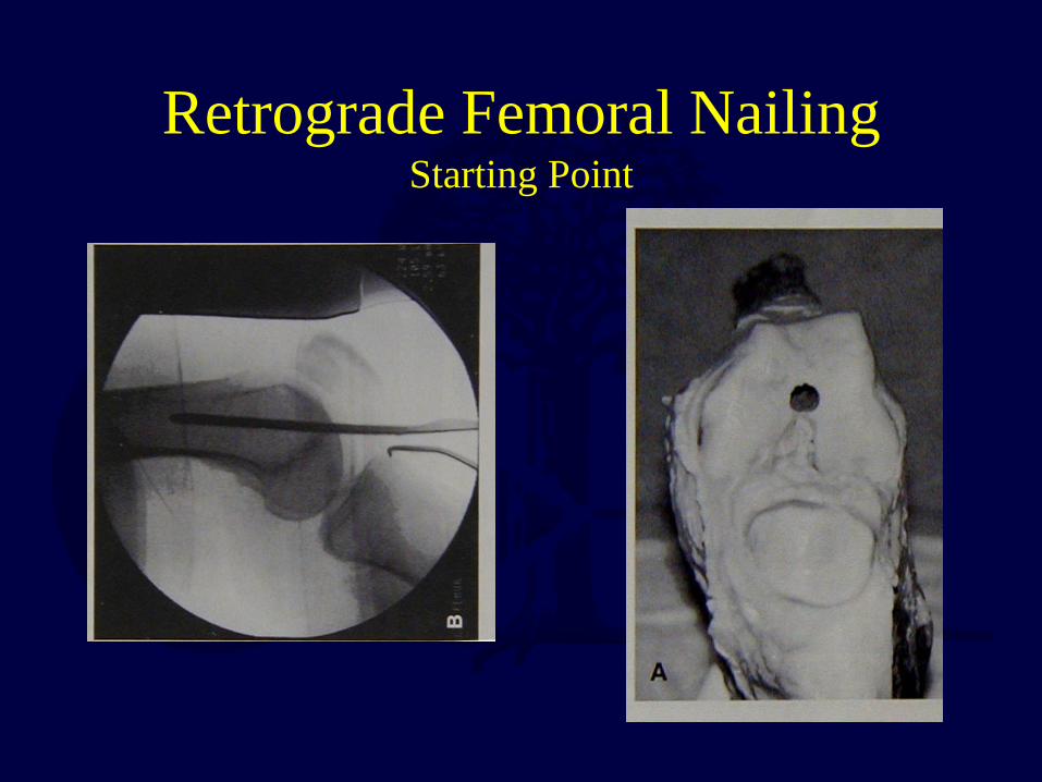
Retrograde Femoral Nailing Starting Point

Mean Contact Area
020406080
100120140160180200
90 degrees 120 degrees
ControlInFlushOut
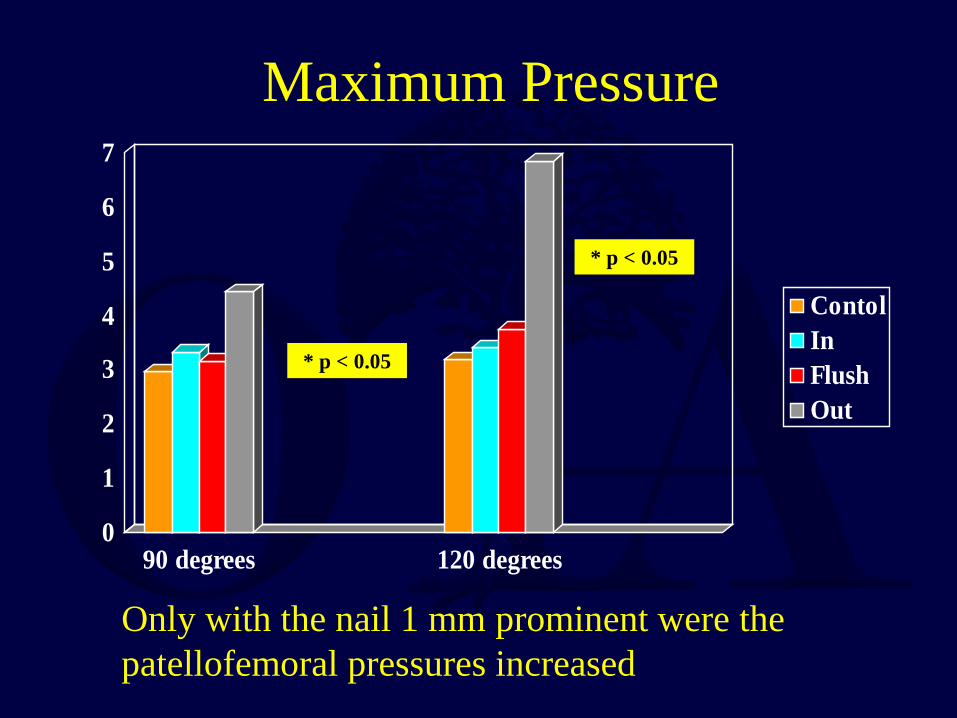
Maximum Pressure
0
1
2
3
4
5
6
7
90 degrees 120 degrees
ContolInFlushOut
* p < 0.05
* p < 0.05
Only with the nail 1 mm prominent were the patellofemoral pressures increased


Retrograde Femoral Nailing
• A cadaveric study using Fuji film demonstrated NO deleterious effects on the patello-femoral joint with a properly inserted retrograde IM nail
• The orthopaedic literature does NOT support decreased knee motion or increase knee pain with a retrograde nail

Bilateral femur fractures nailed retrograde
Less comminuted fracture nailed first to assess length
for segmental fracture
1 2

Retrograde IM Nail Femur Fractures
• 42 yo male C2 femur, Gr 2 open ipsilateral tibia fx

Retrograde IM Nail Femur Fractures
• Immediate post-op with treatment through a limited 4cm knee incision

Femur Fracture Management
• Retrograde Nailing – Union rates lower with unreamed nails – Higher dynamization with non canal sized nails – Better union rates equal to antegrade with reamed canal
sized nails • Moed JBJS 1995, J Orthop Trauma 1998 • Ostrum J Orthop Trauma 1998, 2000
– Advantages for ipsilateral acetabulum or femoral neck and shaft fracture, floating knees, obese patients, supracondylar fractures including those around total knee replacements

Retrograde Nailing is Beneficial for Floating Knee Injuries

Shortening after Retrograde Nail Insertion
Backslap after distal locking

Retrograde Nail: Long or Short ?
• 9 human matched cadaver femurs, gap model
• 36 cm vs 20 cm • Coronal and sagittal testing • 75 Newtons applied in 3 point bending • Locked with 1 or 2 proximal screws

Retrograde Nail: Long or Short ?
20cm 36cm 2 prox,sagittal 7.2* 1.8* 2 prox,coronal 6.3 4.3 1 prox,sagittal 7.6* 2.2* 1 prox,coronal 13.6* 4.4* Longer nails provide improved stability !!! * statistically significant at p<0.05

Femur Fracture Technique
• Antegrade Intramedullary Nailing – Supine - better for multiply injured patients – Lateral - easier piriformis fossa starting point,
difficult set up, rotation concerns – Without a fracture table
• Retrograde Intramedullary Nailing – Supine - flex the knee 50° to allow access to
Blumensaat’s line

Antegrade v Retrograde Comparisons
Equal union rates
Tornetta, JBJS (B), 2000 Ricci, JOT, 2001
Ostrum, JOT, 2000 • ANTEGRADE
– More hip and proximal thigh pain
– Greater incidence of Trendelenburg gait
• RETROGRADE – More symptomatic distal
hardware – Higher dynamization rates
with small diameter nails

Obesity Antegrade v Retrograde
Obese BMI >30
Non-Obese BMI <30
Ante OR Time 94 62 P<.003
Retro OR Time 67 62 nss
Ante Fluoro 247 135 P<.03
Retro Fluoro 76 63 nss
Tucker M. JOT 2007
Retrograde nailing is easier in obese patients !!

Comparison of Knee function after Antegrade and Retrograde IM Nailing
with Isokinetic Evaluation No differences in : • knee range of motion • Lysholm Scores • isokinetic knee evaluation • time to union • secondary surgeries (including hardware removal) - Daglar, JOT 2009

Antegrade Femoral Nailing: Piriformis vs Trochanteric
• Reduction and starting point are still the keys !! • Problems arise with subtrochanteric fractures • Inappropriate starting point leads to malreduction

Piriformis Nail: Poor Technique

Piriformis Nail: Poor Technique

three different starting points were used
Tip of Trochanter
2-3 mm medial to tip
2-3 mm lateral to tip

Femur # 9 Gamma
Holland TAN
TFN
Medial Lateral
Medial Lateral
Medial Lateral
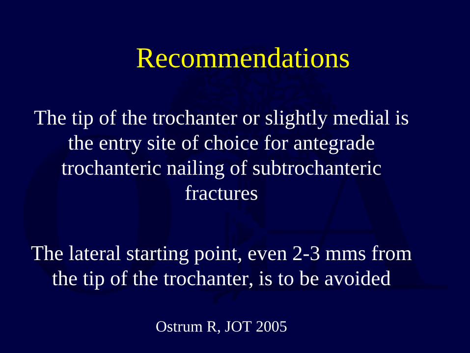
Recommendations
The tip of the trochanter or slightly medial is the entry site of choice for antegrade
trochanteric nailing of subtrochanteric fractures
The lateral starting point, even 2-3 mms from
the tip of the trochanter, is to be avoided
Ostrum R, JOT 2005

Lateral to tip of GT is OK for shaft fractures
Medial to the tip of the GT for subtrochanteric fractures
Lateral starting point with varus !

Reduction with medial tip starting point

Medial Trochanteric Portal
Perez E, Russell TA. JOT 2007

Starting point
Reduction

• Assessing rotation in the lateral position
• Without changing rotation of the C-arm
• A true AP of the hip and knee

• 17 mm entry hole in trochanter • 15-50% disruption of gluteus medius tendon • ? Functional sequelae
• McConnell T, Clin Orthop 2003
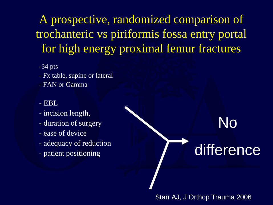
A prospective, randomized comparison of trochanteric vs piriformis fossa entry portal for high energy proximal femur fractures
-34 pts - Fx table, supine or lateral - FAN or Gamma
- EBL - incision length, - duration of surgery - ease of device - adequacy of reduction - patient positioning
No
difference
Starr AJ, J Orthop Trauma 2006

A prospective, randomized comparison of trochanteric vs piriformis fossa entry portal for high energy proximal femur fractures
- NO difference in : Hip Scores, RTW, Ambulation, Hip/Knee ROM
- Varus > 5 degrees - Recon = 2 - Gamma = 4 - BMI significantly linked to duration of OR and
length of incision, NOT EBL
Starr AJ, J Orthop Trauma 2006

Femur Fracture Complications
• Hardware failure • Nonunion - less than 1-2% • Malunion - shortening, malrotation,
angulation • Infection • Neurologic, vascular injury • Heterotopic ossification
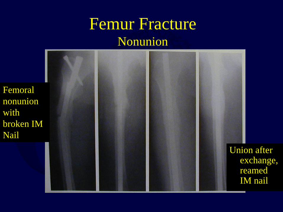
Femur Fracture Nonunion
Femoral nonunion with broken IM Nail
Union after exchange, reamed IM nail

Hypertrophic Nonunion • Problem with smaller diameter nails • Don’t Dynamize EXCHANGE !! • Has a blood supply, WANTS MORE
STABILITY

Plating of femoral nonunions after IM Nail
• 23 pts, nonunion of femur after IM nail • nail removal, PLATING, soft tissue preservation • 21/23 healed, avg 12 weeks • avg OR time 164 minutes (120-240) • avg EBL = 340 ml (200-700)
•Bellabarba, JOT 2001

Exchange Nailing of femoral Nounions
• 42 pts, closed exchange nailing • 7 posititve cultures • 36 (86%) healed, avg 4 mos after OR • Lack of immediate weight bearing, open fractures assoc
with nonunion after 1st OR • Atrophic/oligotrophic nonunions, and infection were
associated with treatment failure after exchange nail • A second nail larger by 2 mm or more than the original
nail was associated with a higher success rate • Shroeder, JOT 2009

Femur Fracture Subtrochanteric Fracture Management
• Possible to perform intramedullary nail if the piriformis fossa is intact
• Choice of nail type depends on if the lesser trochanter is intact
• Varus seen with proximal femur intramedullary nailing
• Plating is also an option with/without an intact starting point

Subtrochanteric fractures are from the base of the lesser trochanter to 5 cm distal

Low Subtroch Fx’s
Most low subtrochanteric fractures with an intact piriformis fossa can be treated with a 1st gen IM Nail

When piriformis fossa is not involved and the lesser trochanter is fractured, a 2nd generation nail may be used

Nail or… Plate

Indirect Reduction: Technique

Indirect Reduction
Step 1- Approximate closed reduction with
fracture table in BOTH planes
Step 2 - Percutaneous insertion of guide pins

Step 3 - Placement of lag screw and percutaneous
plate placement
Head
Knee
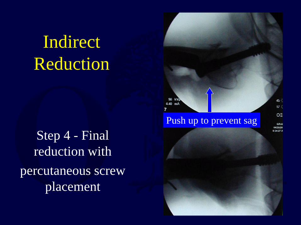
Indirect Reduction
Step 4 - Final reduction with
percutaneous screw placement
Push up to prevent sag

head
knee
Screw Placement

Final films after percutaneous Indirect Reduction of a Subtrochanteric femur fracture

Ipsilateral Femoral Neck & Shaft Fractures
• Optimum fixation of the femoral neck should be the goal
• Varus malunion of the femoral neck is not uncommon, osteotomies can lead to poor results
• Vertical femoral neck fracture seen in 26-59% of cases (Pauwel’s angle > 70°)
• Rate of avascular necrosis is low, 3%, even when missed

Ipsilateral Femoral Neck & Shaft Fractures
• Type 1 - nondisplaced femoral neck/hip fractures
• When found prior to nailing can be treated with screws or a sliding hip screw then retrograde or antegrade nail

Ipsilateral Femoral Neck & Shaft Fractures
• Type 2 - missed femoral neck fracture
• Insertion of screws around the nail
• Low AVN rate even when missed
• Vertical fractures not iatrogenic

Ipsilateral Femoral Neck & Shaft Fractures
• Type 3 - displaced femoral neck fractures • Treat with implant appropriate for neck fracture FIRST • Treat femoral shaft fracture with retrograde nail

Femoral Shaft Fracture with Vascular Injury
• Quick external fixation with restoration of length
• Fasciotomies

Femoral Shaft Fracture with Vascular Injury
• Exchange femoral nail either in same setting or in a few days
• When found early plating or rodding of femur is rarely possible first
• Do NOT perform IM nailing after arterial repair without initial length restoration

Open Femur Fracture Antegrade IM Nail is Safe
• Reamed , Antegrade Intramedullary Nailing has been shown to be effective
• A high union rate, low complications • Perhaps stage Grade 3B fractures after
debridement and skeletal traction – Brumback, JBJS 71A, 1989 – Lhowe, Hansen JBJS 70A, 198

Open Femur Fracture Antegrade IM Nail is Safe

IM Nailing of the Femoral Shaft
• Choice TO nail depends on fracture configuration, especially at proximal and distal ends
• Choice OF nail depends on fracture location, associated musculoskeletal injuries, obesity
• Think before IM Nailing of femur
Return to Lower Extremity
Index
E-mail OTA about
Questions/Comments
If you would like to volunteer as an author for the Resident Slide Project or recommend updates to any of the following slides, please send an e-mail to [email protected]