February 2019 Peripheral intravenous catheters: A review ... · Peripheral intravenous catheters: A...
76
Peripheral intravenous catheters: A review of guidelines and research 1 TRIM: D19-9938 February 2019 Peripheral intravenous catheters: A review of guidelines and research Prof Samantha Keogh and Dr Saira Mathew from the Queensland University of Technology have prepared this report on behalf of the Australian Commission on Safety and Quality in Health Care. Support was also provided by the following authors in preparing this report: Dr Evan Alexandrou, A/Prof Andrew C. Bulmer, Prof Marie Cooke, Dr Linda Coventry, Alexandra Hawthorn, Tricia Kleidon, Emily Larsen, Nicole Marsh, Anthony Marshall, Dr Gillian Ray-Barruel, Stephen Sinclair, Eugene Slaughter, Alana St John, Kerry Taliaferro, A/Prof Amanda Ullman, and Prof Claire M. Rickard from the Australian Vascular Access Society (AVAS).
Transcript of February 2019 Peripheral intravenous catheters: A review ... · Peripheral intravenous catheters: A...
Peripheral intravenous catheters: A review of guidelines and research 1
TRIM: D19-9938
February 2019
Peripheral intravenous catheters: A review of guidelines and research
Prof Samantha Keogh and Dr Saira Mathew from the Queensland University of Technology have prepared this report on behalf of the Australian Commission on Safety and Quality in Health Care.
Support was also provided by the following authors in preparing this report:
Dr Evan Alexandrou, A/Prof Andrew C. Bulmer, Prof Marie Cooke, Dr Linda Coventry, Alexandra Hawthorn, Tricia Kleidon, Emily Larsen, Nicole Marsh, Anthony Marshall, Dr Gillian Ray-Barruel, Stephen Sinclair, Eugene Slaughter, Alana St John, Kerry Taliaferro, A/Prof Amanda Ullman, and Prof Claire M. Rickard from the Australian Vascular Access Society (AVAS).
Peripheral intravenous catheters: A review of guidelines and research 2
Published by the Australian Commission on Safety and Quality in Health Care Level 5, 255 Elizabeth Street, Sydney NSW 2000
Phone: (02) 9126 3600 Fax: (02) 9126 3613
Email: [email protected] Website: www.safetyandquality.gov.au
ISBN: 978-1-925948-03
© Australian Commission on Safety and Quality in Health Care 2019
All material and work produced by the Australian Commission on Safety and Quality in Health Care (the Commission) is protected by copyright. The Commission reserves the right to set out the terms and conditions for the use of such material.
As far as practicable, material for which the copyright is owned by a third party will be clearly labelled. The Commission has made all reasonable efforts to ensure that this material has been reproduced in this publication with the full consent of the copyright owners.
With the exception of any material protected by a trademark, any content provided by third parties and where otherwise noted, all material presented in this publication is licensed under a Creative Commons Attribution–NonCommercial–NoDerivatives 4.0 International licence.
Enquiries about the licence and any use of this publication are welcome and can be sent to [email protected].
The Commission’s preference is that you attribute this publication (and any material sourced from it) using the following citation:
Keogh S, Mathew S. Peripheral intravenous catheters: A review of guidelines and research. Sydney: ACSQHC; 2019
Disclaimer
The content of this document is published in good faith by the Commission for information purposes. The document is not intended to provide guidance on particular healthcare choices. You should contact your health care provider for information or advice on particular healthcare choices.
This document includes the views or recommendations of its authors and third parties. Publication of this document by the Commission does not necessarily reflect the views of the Commission, or indicate a commitment to a particular course of action. The Commission does not accept any legal liability for any injury, loss or damage incurred by the use of, or reliance on, this document.
Peripheral intravenous catheters: A review of guidelines and research 3
Preface The insertion of a peripheral intravenous catheter (PIVC) is one of the most common clinical procedures performed. About 30 million are used in Australia each year, with up to 70% of hospitalised patients requiring a PIVC at some point during their hospital stay. However studies estimate that 4% to 28% of PIVCs inserted are not actually needed, placing the patient unnecessarily at risk of infection. Despite frequency in PIVC use, complications are reported to be as high as 70%. They can be prone to blockage and dislodgment, cause inflammation of the vein and infection. Nearly half of all first insertion attempts also fail, causing undue pain and anxiety for patients as a result of multiple failed attempts.
To reduce rates of PIVC-related complications, a number of evidence-based strategies have been suggested. Best practice guidelines recommend a range of strategies to reduce risk of complications and increase chances of PIVC success. Despite this, data from Australia and internationally suggest that a significant proportion of patients do not receive care as recommended to optimise use of PIVCs. A clinical care standard on peripheral intravenous access will aim to support national consistency of best practice for the insertion and management of PIVCs. To inform development of this clinical care standard two literature reviews were undertaken. The Commission engaged Professor Samantha Keogh and Dr Saira Mathew from the Queensland University of Technology (QUT) to conduct a literature review to better understand the current clinical environment of the techniques used for preventing and managing adverse events associated with the insertion and use of PIVCs.
Key findings This report provides a broad summary of the quality of current guidelines and recommendations regarding the insertion, management and removal of PIVCs in paediatric and adult patients.
The report highlights a number of similarities between guidelines including adherence to basic infection control measures through maintenance of hand hygiene and aseptic technique; skin decontamination prior to insertion; ongoing assessment for vascular access needed and removal when no longer required. Differences between guidelines were related to escalation pathways for patients with difficult vascular access; needless decontamination; flushing frequency, and device replacement schedule.
Gaps in evidence were reported in relation to the potential merits of having vascular access specialists (such as a service or team of clinicians), innovative catheter designs, optimal dressings and securement, and port or hub decontaminations. Other areas that require further research were noted, which included clinical monitoring of the device and the patient experience.
Recommendations of the report The authors of the report have made a number of recommendations in relation to the findings from this review. These include: I. Development of a national clinical care standard aimed at reducing PIVC related complications and failure.
Peripheral intravenous catheters: A review of guidelines and research 4
II. Education and training of nursing and medical staff that focus on contemporary and evidence based PIVC insertion and maintenance care. III. Evidence based assessment of patient’s need for vascular access and device type. IV. Early referral to vascular access specialist and use of ultrasound to minimise insertion trauma in patents identified as having difficult vascular access. V. Routine use of analgesic agents or strategies to minimise pain associated with PIVC insertion. VI. Evidence based clinical assessment of PIVC site and function VII. Ongoing monitoring and reporting of PIVC use and outcomes to facilitate benchmarking and drive quality improvement VIII. Inclusion of patients’ views in all future research in to the optimal PIVC insertion and maintenance practices and products.
Next steps for the Commission The Commission will consider the report’s recommendations in the development of the Peripheral Intravenous Access Clinical Care Standard.
Peripheral intravenous catheters: A review of guidelines and research 5
Executive summary Peripheral intravenous catheters are the most commonly used medical device nationally and globally. Up to 80% of patients require a PIVC to provide essential medical treatment. Rates of failed insertion and post insertion complications are high, with nearly half of all first insertion attempts and average of 40% of devices failing. Such failure rates for an essential device are unacceptable, especially as most failure is preventable with good insertion and maintenance practice. Therefore the Australian Commission on Safety and Quality in Health Care commissioned this review to appraise guidelines currently influencing clinical practice locally and internationally, analyse and synthesise recent trial research, and to better understand the current clinical environment for preventing PIVC associated complications and failure. Eighteen guidelines from professional colleges, membership associations and guideline agencies, as well as state health authorities were identified via systematic search strategy and appraised on their scope, quality and rigour using the appraisal of Guidelines for Research and Evaluation II (AGREE) tool. Most of the jurisdictional guidelines were ranked low against the tool, particularly in the area of rigour of development. There were a number of similarities between guidelines including adherence to basic infection control measures through maintenance of hand hygiene and aseptic technique; skin decontamination prior to insertion; ongoing assessment for vascular access needed and removal when no longer required. The main differences were related to escalation pathway for patients with difficult vascular access; needless decontamination; flushing frequency, and device replacement schedule. In total, 40 trials were identified, appraised, data extracted, meta analysed where possible or summarised narratively. The majority of trials evaluated different insertion practices and products, predominately focusing on the merits of visual aids (e,g, ultrasound or near infrared), and analgesia to minimise pain on insertion. Studies addressing post insertion care evaluated the impact of different dressing and securement products, flushing techniques and solutions, and device replacement schedules had on PIVC complications and failure. Gaps in evidence were identified in relation to the potential merits of vascular access specialist (service or team), innovative catheter designs, optimal dressing and securement, and port or hub decontaminations. Other areas of research that require further research included clinical monitoring of the device and patient experience. A number of recommendations are made in relation to the findings from this review. These include:
I. Development of a national clinical care standard aimed at reducing PIVC related complications and failure.
II. Education and training of nursing and medical staff that focus on contemporary and evidence based PIVC insertion and maintenance care.
III. Evidence based assessment of patient’s need for vascular access and device type. IV. Early referral to vascular access specialist and use of ultrasound to minimise
insertion trauma in patents identified as having difficult vascular access. V. Routine use of analgesic agents or strategies to minimise pain associated with PIVC
insertion. VI. Evidence based clinical assessment of PIVC site and function
VII. Ongoing monitoring and reporting of PIVC use and outcomes to facilitate benchmarking and drive quality improvement
VIII. Inclusion of patients’ views in all future research in to the optimal PIVC insertion and maintenance practices and products.
Peripheral intravenous catheters: A review of guidelines and research 6
Table of Contents Abbreviations ........................................................................................................................ 8
1. Introduction and background ............................................................................................. 9
1.1 Research questions ................................................................................................... 12
2. Method ............................................................................................................................ 13
2.1 Systematic Review of Guidelines ............................................................................... 13
2.1.1. Search methods ................................................................................................. 13
2.1.2 Inclusion and exclusion criteria ............................................................................ 13
2.1.3 Quality appraisal ................................................................................................. 14
2.1.4 Data extraction .................................................................................................... 14
2.1.5 Data synthesis .................................................................................................... 14
2.2 Systematic Review of trials ........................................................................................ 15
2.2.1 Search methods .................................................................................................. 15
2.2.2 Inclusion criteria .................................................................................................. 15
2.2.3 Study selection .................................................................................................... 15
3. Results ............................................................................................................................ 17
3.1 Systematic review of guidelines ................................................................................. 17
3.1.1 Characteristics of guidelines ............................................................................... 17
3.1.2 Appraiser agreement ........................................................................................... 21
3.2 Systematic review of trials ......................................................................................... 22
3.2.1 Characteristics of studies .................................................................................... 22
3.2.2 Quality appraisal ................................................................................................. 23
3.2.3 Outcomes............................................................................................................ 23
4. Discussion ...................................................................................................................... 31
4.1 Guidelines ................................................................................................................. 31
4.2 Intervention trials ....................................................................................................... 33
4.3 Gaps in identified literature ........................................................................................ 35
4.4 Limitations ................................................................................................................. 36
5. Implications and recommendations ................................................................................. 37
6. Conclusion ...................................................................................................................... 37
7. References ..................................................................................................................... 38
8. APPENDICES ................................................................................................................. 47
Appendix A: Detailed search strategy for guideline review ............................................... 47
Appendix B: Detailed search strategy for intervention review .......................................... 48
Appendix C: Table of excluded guidelines ....................................................................... 50

Peripheral intravenous catheters: A review of guidelines and research 7
Appendix E: Table of guideline quality assessment scores .............................................. 53
Appendix F: Table of included intervention studies .......................................................... 57
Appendix G: Quality appraisal of intervention studies (Risk of bias graph and summary) 75
Peripheral intravenous catheters: A review of guidelines and research 8
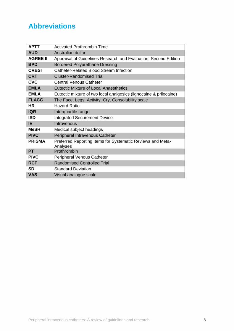
Abbreviations APTT Activated Prothrombin Time AUD Australian dollar AGREE II Appraisal of Guidelines Research and Evaluation, Second Edition BPD Bordered Polyurethane Dressing CRBSI Catheter-Related Blood Stream Infection CRT Cluster-Randomised Trial CVC Central Venous Catheter EMLA Eutectic Mixture of Local Anaesthetics EMLA Eutectic mixture of two local analgesics (lignocaine & prilocaine) FLACC The Face, Legs, Activity, Cry, Consolability scale HR Hazard Ratio IQR Interquartile range ISD Integrated Securement Device IV Intravenous MeSH Medical subject headings PIVC Peripheral Intravenous Catheter PRISMA Preferred Reporting Items for Systematic Reviews and Meta-
Analyses PT Prothrombin PIVC Peripheral Venous Catheter RCT Randomised Controlled Trial SD Standard Deviation VAS Visual analogue scale
Peripheral intravenous catheters: A review of guidelines and research 9
1. Introduction and background The majority of patients presenting to hospitals require at least one peripheral intravenous catheter (PIVC) for the delivery of intravenous fluids and medications or blood sampling. Despite the ubiquity of peripheral catheters in hospital care (~80%), the rate of complications associated with the insertion and use of these devices is reported to be unacceptably high. Nearly half of all first insertion attempts fail in adults and children 1-3. Device failure due to other complications (dislodgement, infiltration, occlusion, local inflammation) can be up to 69% 4-7. The rate of PIVC CRBSI is relatively low compared to that of central vascular access devices (from <0.01% to 0.18% 8-11), however the far greater number of PIVCs in use means that the absolute infection rates for PIVCs increase the overall absolute infection rates 12. This underscores the need for greater investment in PIVC research to reduce associated patient discomfort, delays in vital medical treatments, and waste of healthcare resources. The aim of this overall review is to identify and review current guidelines for PIVC insertion and maintenance as well as appraise evidence of interventions that prevent PIVC failure and make recommendations for policy and practice. Understanding the different PIVC related complications and possible risk factors associated with difficult insertion and post insertion failure is the first step in the development of practice and products that may mitigate this risk. PIVC complications and failure The rates for PIVC failure were derived from published research. Currently there is no regular surveillance or monitoring PIVC outcomes. Challenges with gaining successful and patent peripheral vascular access start at the moment of insertion. First attempt PIVC insertion failure rates can be up to 35-40% for adults 1,2 and between 50–65% for children 1,3; with a concerning 10% of adults and a quarter of all children experiencing more than four attempts at insertion1. The most frequently cited reasons for PIVC failure before the completion of prescribed treatment are dislodgment, occlusion, infiltration and phlebitis5-7. These may occur in isolation or in combination, indeed existence of one may be precursor to another complication. Dislodgement of the PIVC out of the vein, partial or complete, occurs when there is poor securement of the catheter to the skin, or with patient or operator (staff) interference 13,14. Dislodgement or accidental removal reportedly accounts for between 6% and 20% of catheter failures 11,15,16. This may be a contributing factor to localised irritation and inflammation (phlebitis) through micro motion (pistoning) of the device in the vein, further heightening the risk of failure. A poorly secured catheter often causes the patient discomfort and result in catheter failure, delaying intravenous (IV) therapy and requiring insertion of a new IV device. Occlusion, either partial or complete blockage, results in the inability to infuse or inject fluids or medications through the lumen of the PIVC5,17. Occlusion can be mechanical (e.g kinking), thrombotic or medication related in origin. Occlusion can also occur from irritation or trauma to the cannulated vein wall, leading to a release of thromboplastic substances and platelets18. This process promotes the clotting of blood and can result in narrowing or complete occlusion of the cannulated vein. Occlusion of the catheter lumen can be a precursor to infiltration as the IV fluid or medications leak into the surrounding tissue19. Infiltration is the defined as the leakage of a non-vesicant solution into surrounding tissues20. This may be related to dislodgment of the device from the vein or occlusion of the lumen. Signs and symptoms of infiltration can include oedema, stretched or blanched skin, localised
Peripheral intravenous catheters: A review of guidelines and research 10
cool skin, or visible leakage of IV fluids around the site19. This may or may not be accompanied with discomfort or pain. Although injury due to infiltration is often considered minor and usually resolves without any intervention, there is a risk of significant morbidity related to localised compression or even tissue damage21. Extravasation is traditionally distinct from infiltration as it specifically refers to the leakage of vesicant fluids or drugs (including contrast media or cytotoxic agents)19,20. The incidence of infiltration and extravasation is hard to determine because of limited reporting; however, extravasation injury from cancer chemotherapy is reported to be 11% in children and 22% in adults19. One study found that, of all the complications associated with peripheral cannulas, 34% occurred as a result of infiltration21. Given the root cause (vesicant substance) and related tissue damage, the presenting signs and symptoms of extravasation are similar to infiltration, but accompanied with more visible tissue damage and pain22. In recognition of the risk associated with their treatment Imaging and Cancer Care specialities usually have specific policies and procedure for dealing with extravasation to minimise damage23,24. Phlebitis is defined as localised irritation or inflammation of the vein wall has been the focus of much discussion and research in relation to PIVC complications25,26. It can have either a mechanical, chemical, or bacterial origin, and it can occur in isolation or in combination with any of the other known PIVC complications25,26. Reported phlebitis rates vary widely between studies (between 2% and 80%)11,27-29. However this may be reflective of the variation in the tools used to measure phlebitis rather than the prevalence of the condition itself25. Generally, phlebitis is characterised by a combination of tenderness/pain, erythema, oedema, purulent discharge, or a palpable cord, and results in failure and removal of the device30. However, even one sign, (e.g. erythema) can be an indication of underlying phlebitis26,30. PIVC associated infections are a relative rare but serious complication and occur when micro-organisms track along the insertion site and into the cannulated vein, irritating the vessel wall, contaminating the catheter and then the bloodstream12,31. These microbes may be from the patient’s skin, contaminated disinfectant or healthcare workers’ hands. The process may happen on insertion if the catheter is contaminated and then introduced into the patient or via microbial migration at any time while the catheter is in situ12. This may give rise to biofilm formation, proliferation of bacteria, and lead to blood stream infection32,33.The most common signs and symptoms of a local PVC-related infection are pain, erythema, pus, and palpable venous cord, whereas the more serious catheter related blood stream infection presents with fever, chills, headache, tachycardia, and nausea/vomiting20,31. Infection rates associated with PIVCs are much lower than those reported for central venous catheters (CVC), (0.2–0.7 episodes per 1000 calendar days)8,34. Whatever the root cause of failure of the PIVC it has significant implications for the patient’s treatment, hospital experience and healthcare budget. Failed PIVCs need to be removed and replaced, which means repeated painful needlesticks for the patient and interruptions to vital therapy. The mean cost of PIVC replacement has been costed at $70.00 AUD per episode of IV treatment35. At the current rate of failure, this costs the Australian healthcare system approximately $700 million each year. Repeated PIVC replacements can lead to venous access difficulties, increasing the need for more frequent replacements and the possibility of requiring a central venous access device Risk factors for PIVC complications To reduce the incidence of catheter failure and avoid preventable IV replacements, a clear understanding of why catheters fail is required. Previous research has identified that catheter gauge,7,27,36 insertion site,37-39 and inserter skill,7,40 have an impact on PIV failure. Limitations of existing research are small study sizes, retrospective design, or secondary analysis of an existing data set; all potentially introducing sampling or reporting bias41,42. However, a large prospective study with rigorous analysis was recently published6. The
Peripheral intravenous catheters: A review of guidelines and research 11
study identified that one in three PIVCs failed before the completion of prescribed therapy. This mirrored results observed in trials in similar settings7,11,43. Phlebitis was the main complication observed at a rate of 17%, followed by occlusion and/or infiltration (14%) and dislodgement (10%)6. Factors that were associated with PIVC failure included the PIVC gauge, insertion site, poor securement, type of IV medication administered and number of IV accesses generally. Specifically in Marsh and colleagues’ study, 22-gauge catheters were more likely to fail from occlusion/infiltration than other sizes (Hazard Ratio (HR) 1.43). The PIVC gauge has been previously been identified as a risk factor for catheter failure: in particular, small gauge catheters (≤ 22 gauge) and large gauge catheters (≥ 18 gauge)7,27,36. The issue of PIVC gauge may also be related to the catheter to vein ratio which is rarely measured or reported on in relation to PIVC insertion. Further, risk may not be because related to gauge alone. The level of intravascular purchase (degree to which catheter sits in the vein) associated with a shorter length may also influence PIVC performance6. These considerations challenge the recommendation to insert the smallest gauge peripheral catheter possible44,45. Marsh and colleagues’ study demonstrated that insertion in the patient’s dominant side was associated with phlebitis (HR 1.39)6. PIVCs inserted in the hand, anterior cubital fossa or over a joint have also been associated with an increased risk of catheter failure7,16,38,39. The placement of a PIVC over an articulated joint increases the risk micro movement which in turn increases the risk of local irritation and dislodgment of the PIVC within the vein. Patients have reported significant discomfort and pain associated with this also46. Multiple different products are available to facilitate PIVC insertion site dressing and securement. A recent four-armed trial did not demonstrate superiority of any of the major product types in use. Overall failure was approximately 40% in all groups43. However, the trial and cohort study did show that additional (secondary) securement was significantly associated with reduced risk of dislodgement (HR 0.44)6. However ad hoc use of non-sterile tape and joint immobilisation products (e.g. elasticised tubular bandage) may contaminate site or reduce visibility and assessment44. Multiple studies have reported certain IV medication and IV antibiotics as risk factors for PIV failure7,27,47,48. Marsh and colleague’s study specifically identified IV Flucloxacillin with a 2-fold increase in occlusion/infiltration and phlebitis6. The overall osmolarity and pH of the final reconstituted drug is what determines the compatibility with peripheral access48. Apart from the potentially vesicant nature of the drug, medication associated PIVC failure may be related to poor drug reconstitution, too rapid administration, or inadequate post drug administration flushing49. The risk of device failure was augmented by the number of accesses per day. This and similar results from other trial work underscore the fragility of peripheral access when subjected to repeated injection and infusion50. The association between PIVC use and failure may indicate that many of these patients were not suitable for a PIVC, and alternative devices and access needed to be considered (e.g. deep peripheral or central venous access devices)6. Inserter skill and training have been associated with rates of PIVC insertion success and performance51-54. However, there are limitations to these studies based on date, size, and design of study that make it hard to draw firm conclusions or apply findings to contemporary practice. Wallis and colleagues’ (2014) retrospective cohort study showed a reduced risk of dislodgement associated with cannulas inserted by an IV service (HR 1.69)7. The study also showed an increased risk of failure associated with second and subsequent PIVCs, underscoring the need to optimise first insertion success and maintenance practice for the duration of treatment. There is a need for good‐quality randomised controlled trials (RCTs) to evaluate the efficacy of a vascular access specialist team approach for vascular access device insertion and care for the prevention of failure55.
Peripheral intravenous catheters: A review of guidelines and research 12
PIVC failure has also been associated with insertion setting (emergency or community). Analysis in a cohort study identified an increased risk of dislodgement (HR 1.78) with PIVCs inserted by paramedics6. A prospective study conducted in two tertiary hospitals found a 20% higher rate of PIVC-associated Staphylococcus aureus infections with PIVCs inserted in the emergency department compared to those inserted in the wards56. So, the quality of the conditions of insertion need to be considered when assessing need for removal and replacement of the device. There are also a number of other important, albeit non-modifiable risk factors that clinicians need to consider prior to PIVC insertion. Other risk factors for PIVC insertion and maintenance include age (the very young and elderly)57,58, gender (female)7,50, obesity59,60, some medications (anticoagulants and corticosteroids) and patients with chronic diseases such as cancer, diabetes, or cardiovascular disease61,62. Knowledge of these risk factors can assist clinicians in their assessment of vascular access insertion and device choices. In summary, PIVCs are important devices in modern medicine. They are the most commonly used medical device with approximately 1.8 billion used globally each year and 25 million in Australia63,64. Despite their essential widespread use, multiple international and national guidelines, PIVC associated complications rates persist at an unacceptably high rate. Given the recent spotlight on PIVC complications the Australian Vascular Access Society and the Australian Commission for Safety and Quality in Healthcare sought a structured, evidence-based literature review to better understand the current clinical environment for preventing and managing PIVC related complications. Well-designed and executed randomised controlled trials (RCTs) provide reliable evidence with minimal bias compared to other study designs and are therefore considered the “gold standard” for evaluating the effectiveness of interventions 65,66. Systematic reviews evaluate the combined results of RCTs, analyse for bias, and provide an even higher level of evidence 67. Clinical guideline developers and health care staff rely on quality RCTs and systematic reviews to guide decision-making in clinical practice, however the guideline development and updating process can be so unwieldy that the timely inclusion of evidence in contemporary practice is hindered 68.
1.1 Research questions 1. What guidelines, standards for practice, policies and procedures relevant to the PIVC
insertion and maintenance are available in Australia and internationally? 2. What do current guidelines recommend for management of PIVC insertion and
maintenance? 3. What evidence is there regarding current clinical practice in Australia for the
prevention of PIVC related complications? 4. What indicators are currently used to measure or report adverse outcomes (e.g
routine monitoring, audits or quality improvement activities)? 5. What contributes to variations in complication rates and other adverse outcomes
associated with PIVCs, including patient groups with higher risk? What are the evidence gaps?
6. What is the literature on interventions to reduce PIVC related complications? What is the effectiveness of those interventions? What are the evidence gaps?
Information on the monitoring of PIVCs, the prevalence of PIVC complications, failure and associated risk factors have been covered in the introduction and background. The remainder of this document focuses on systematic reviews of PIVC guidelines and trial research.
Peripheral intravenous catheters: A review of guidelines and research 13
2. Method This review addresses two main areas: (1) a review of national and international guidelines; and (2) a review of the published literature. We have included publications that have been published in English in the previous five years (2013-2018) and focused on relevant guidelines, and standards of practice, policy and procedure documents (Australian and international) available from the internet, and in addition randomised clinical trials available through recognised electronic databases and relevant grey literature. This systematic reviews followed the Preferred Reporting Items for Systematic Reviews and Meta-Analyses (PRISMA) guidelines 69. Each review protocol was registered at PROSPERO (CRD42019120013 and CRD42019120011).
2.1 Systematic Review of Guidelines
2.1.1. Search methods
The literature was searched using MeSH terms and combinations of key terms in four databases: CINAHL, PubMed, Medline and google advanced search. The search was limited to English language publications from 2013 to 18 December 2018. The last five years were included to focus on the most recent national and international guidelines and standards of practice on peripheral intravenous device insertion, management and removal. The search syntax for each database are presented in the Appendix A. The search strategies were developed with the assistance of a health librarian using subject headings or text words relevant to peripheral intravenous catheters, peripheral intravenous devices, clinical practice guidelines, management guidelines, standards of practice with associated Boolean logic (and, or). Reference lists of all retrieved and relevant publications identified by these strategies were hand-searched. The selection of articles consisted of three stages of screening (titles, abstracts, and full text). Two reviewers (SK, SM) independently screened titles and abstracts, excluding studies that did not meet the inclusion criteria. Two authors also did full-text screening (SK, SM), with a third author (CMR) arbitrating any disagreement arising at any stage of the screening process. Results were imported, and duplicates removed with the help of reference management software (EndNote).
2.1.2 Inclusion and exclusion criteria
2.1.2.1 Types of studies Guidelines and practice standards specifically aimed at guiding and assisting healthcare professionals and patients in making decisions about insertion, management and removal of PIVCs were included in the review. Individual hospital guidelines, expert consensus (with process), and discussion papers on guidelines were excluded from the review. If the guidelines had been updated, only the most recent version was assessed.
2.1.2.2 Types of participants Adults or children requiring insertion, maintenance and removal of a PIVC in any acute or community healthcare setting.
Peripheral intravenous catheters: A review of guidelines and research 14
2.1.2.3 Outcomes The primary outcome of this review was to identify the national and international guidelines on the insertion and maintenance of PIVCs appraise the scope quality and rigour of these and synthesise findings.
2.1.3 Quality appraisal
The AGREE II tool was used to assess methodological quality of the guidelines70. The tool examines rigour and transparency of reporting. It was also used to determine whether the guidelines were evidence-based and whether they reported search strategies, and data extraction, to classify/grade the strength of their recommendations. The AGREE II consists of 23 items categorised into six domains and two overall assessment items. The first domain (Scope and purpose) addresses the objective, target population and health question addressed by the guideline. The second domain (Stakeholder involvement) assesses the involvement of stakeholders’ and consumers’ views and preferences on the development of the guideline. The third domain (Rigour of development) evaluates the process of collecting and synthesizing evidence, formulating recommendations, and developing methods to update the guidelines in future. The fourth domain (Clarity of presentation) covers the language, structure and presentation of the guideline. The fifth domain (Applicability) evaluates the potential barriers and facilitators to implementation and strategies to improve the application of the guidelines. The sixth domain (Editorial independence) assesses whether the guideline reported any conflict of interest and funding for the development of the guideline. The two additional assessment items (Overall Guideline Assessment) rate the overall quality of the guideline and whether the guideline is recommended for use in practice with or without any modifications, or should not be used at all. Each AGREE item is rated on a 7-point Likert scale ranging from ‘strongly disagree’ to ‘strongly agree’. Four appraisers (initials LC, SK, SK, AU) assessed each guideline to increase the reliability of the assessment according to AGREE II. The scores of the four appraisers were used to calculate an average for each domain and total scores were presented as percentages. Scores were checked and, where appropriate, moderated if there was a delta of three or more between scores. Once scores were finalised, a quality score was calculated for each domain as per the AGREE II tool recommended formula 70. This comprised of calculating the maximum and minimum score possible for each domain, calculated as a composite of the maximum or minimum score multiplied by the number of items in the domain and the number of appraisers. These values were then used in the final formula that generated a percentage reflecting the overall domain score. Cohen’s Kappa coefficient was used to evaluate the agreement among authors on the overall recommendation for use in practice.
2.1.4 Data extraction
The authors extracted data from all the included guidelines and standards using an Excel® (Microsoft)-based data extraction form designed for this review. The following data was extracted: clinical practice guideline, title, year, country and scope as defined by authors.
2.1.5 Data synthesis
Due to the nature of this review, a structured narrative synthesis was undertaken. Characteristics of the guidelines and standards are summarised and presented in the results.

Peripheral intravenous catheters: A review of guidelines and research 15
2.2 Systematic Review of trials
2.2.1 Search methods
The literature was searched using MeSH terms and combinations of key terms in four databases: CINAHL (Cumulative Index to Nursing and Allied Health Literature), PubMed, Medline and Cochrane library. The search was limited to English language publications from 2013 to current. The last five years were included to focus on the most recent publications on peripheral intravenous device insertion, management and removal to ensure relevance to current clinical practice. The search syntax for each database is presented in the Appendix B. The search strategy was developed with the assistance of a health librarian using subject headings, Mesh terms or text words relevant to peripheral intravenous catheter, peripheral intravenous devices, insertion site, securement, dressings, occlusive dressings, flushing, antiseptics, removal strategies, connectors, regimen, risk factors, infection, phlebitis, morbidity, mortality, dwell time, device failure, device malfunction, occlusion, blockage, infiltration, extravasation, dislodgement, removal, leakage, inserter skill, inserter characteristics, multiple insertion attempts, multiple cannulation, attempts, insertion difficulty, difficult vascular access, difficult intravenous access, cumulative, impact of multiple insertion attempts, escalation, with associated Boolean logic (and, or). Reference lists of all retrieved and relevant publications identified by these strategies were hand searched.
2.2.2 Inclusion criteria
2.2.2.1 Types of studies This review only included randomised controlled trials (RCTs) and cluster-randomised trials (CRTs).
2.2.2.2 Types of participants All age groups, including neonates, requiring insertion, maintenance or removal of a PIVC within a healthcare setting.
2.2.2.3 Outcomes The primary outcomes for insertion trials were first insertion success and time to insertion. Secondary outcomes included PIVC failure, local and primary infection, dwell time, level of patient reported pain relief, patient satisfaction and cost. The primary outcomes for maintenance trials were PIVC failure as a composite measure of occlusion; infiltration; dislodgement, and phlebitis. Secondary outcomes included local infections, bloodstream infections (primary), as well as dwell time, patient satisfaction and cost.
2.2.3 Study selection
The selection of studies consisted of three stages of screening (titles, abstracts, and full text). Two teams of paired reviewers (SK and SM plus AH and ES) independently screened titles and abstracts and excluded those studies that did not meet the inclusion criteria. Two authors then did full text screening (SM and AH) and a third author (SK) arbitrated any disagreement arising at any stage of the screening process. Results were imported, and
Peripheral intravenous catheters: A review of guidelines and research 16
duplicates removed with the help of reference management software (EndNote, Clarivate Analytics).
2.2.4 Data extraction
The results and characteristics of each study were extracted by the following authors (EA, AB, MC, SK, TK, EL, NM, SM, GRB, ES, AStJ, KT) and a second experienced researcher checked for accuracy and completeness (SK and SM). The authors extracted data from all the included studies using a data extraction form designed for this review. The data was extracted for the following items: author, date (year), study design, country, setting, participants and sample size, plus intervention and primary outcomes. When the information presented in the studies were unclear or incomplete, an attempt was made to contact the study author for further data or clarification.
2.2.5 Data synthesis
Clinical (sample or intervention characteristics), methodological and statistical heterogeneity was considered. Where appropriate, data was pooled using meta-analysis with Review Manager (RevMan) version 5.3. A random effects model was considered due to the clinical and statistical heterogeneity present in the studies. A structured narrative summary of all the studies included in the review was also conducted. Heterogeneity measures the variability among the combined studies; if there is considerable variation in the combined or pooled results, it may be misleading to report a combined summary measure. The chi-square test and I2 statistic were used to assess heterogeneity. The chi-square statistic was used to test the null hypothesis that there is no heterogeneity, with a P value < .05 indicating heterogeneity. The I2 statistic describes the variability in effect measurements that is due to heterogeneity rather than sampling error. The I2 value was interpreted as follows: Less than 40% might not be important, 30% to 60% may represent moderate heterogeneity, 50% to 90% may represent substantial heterogeneity, and 75% to 100% represents considerable heterogeneity. The pooled result was considered heterogeneous if the I2 statistic was >40% and the P value was <0.05. Data from cluster trials were transformed to effective sample size by calculating the design effect using the Rao and Scott (1992) calculation to avoid unit‐of‐analysis error with data 71. The average cluster size and the intraclass coefficient (ICC) was calculated (using either the ICC from the actual study or a reliable estimate from previous publications). The design effect for the study as a whole was calculated and number of participants and the number experiencing the event were adjusted for the categorical data meta-analysis. For continuous data, only the sample size was reduced; means and standard deviations remained unchanged.
2.2.6 Quality appraisal
Two independent reviewers (SM and ES) applied the Cochrane Risk of Bias (RoB2) for RCTs to assess the methodological quality of the studies included in this review 72. Each study was assessed on the five bias domains (selection, performance, detection, attrition and reporting bias). The risk of bias was assessed as low risk, high risk or unclear risk. A third reviewer (SK) arbitrated discrepancies at any stage of the quality assessment.
Peripheral intravenous catheters: A review of guidelines and research 17
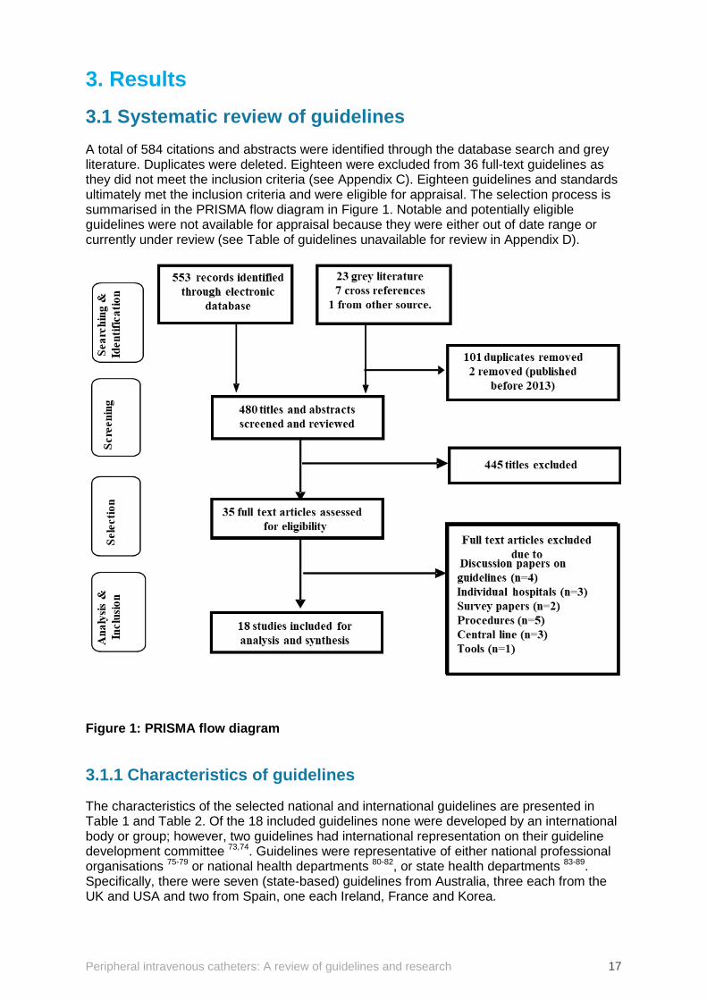
3. Results 3.1 Systematic review of guidelines A total of 584 citations and abstracts were identified through the database search and grey literature. Duplicates were deleted. Eighteen were excluded from 36 full-text guidelines as they did not meet the inclusion criteria (see Appendix C). Eighteen guidelines and standards ultimately met the inclusion criteria and were eligible for appraisal. The selection process is summarised in the PRISMA flow diagram in Figure 1. Notable and potentially eligible guidelines were not available for appraisal because they were either out of date range or currently under review (see Table of guidelines unavailable for review in Appendix D).
Figure 1: PRISMA flow diagram
3.1.1 Characteristics of guidelines
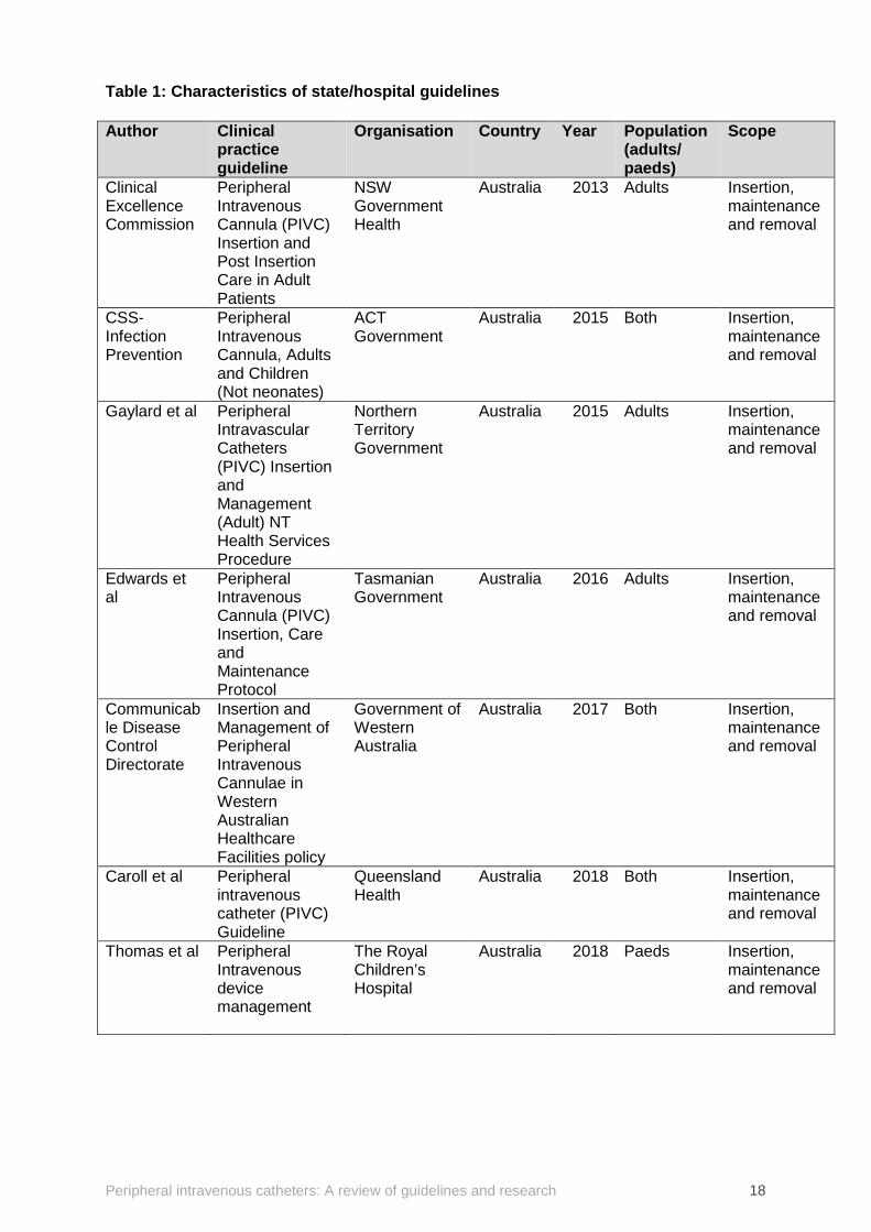
The characteristics of the selected national and international guidelines are presented in Table 1 and Table 2. Of the 18 included guidelines none were developed by an international body or group; however, two guidelines had international representation on their guideline development committee 73,74. Guidelines were representative of either national professional organisations 75-79 or national health departments 80-82, or state health departments 83-89. Specifically, there were seven (state-based) guidelines from Australia, three each from the UK and USA and two from Spain, one each Ireland, France and Korea.
Peripheral intravenous catheters: A review of guidelines and research 18
Table 1: Characteristics of state/hospital guidelines Author Clinical
practice guideline
Organisation Country Year Population (adults/ paeds)
Scope
Clinical Excellence Commission
Peripheral Intravenous Cannula (PIVC) Insertion and Post Insertion Care in Adult Patients
NSW Government Health
Australia 2013 Adults Insertion, maintenance and removal
CSS-Infection Prevention
Peripheral Intravenous Cannula, Adults and Children (Not neonates)
ACT Government
Australia 2015 Both Insertion, maintenance and removal
Gaylard et al
Peripheral Intravascular Catheters (PIVC) Insertion and Management (Adult) NT Health Services Procedure
Northern Territory Government
Australia 2015 Adults Insertion, maintenance and removal
Edwards et al
Peripheral Intravenous Cannula (PIVC) Insertion, Care and Maintenance Protocol
Tasmanian Government
Australia 2016 Adults Insertion, maintenance and removal
Communicable Disease Control Directorate
Insertion and Management of Peripheral Intravenous Cannulae in Western Australian Healthcare Facilities policy
Government of Western Australia
Australia 2017 Both Insertion, maintenance and removal
Caroll et al Peripheral intravenous catheter (PIVC) Guideline
Queensland Health
Australia 2018 Both Insertion, maintenance and removal
Thomas et al Peripheral Intravenous device management
The Royal Children’s Hospital
Australia 2018 Paeds Insertion, maintenance and removal
Peripheral intravenous catheters: A review of guidelines and research 19
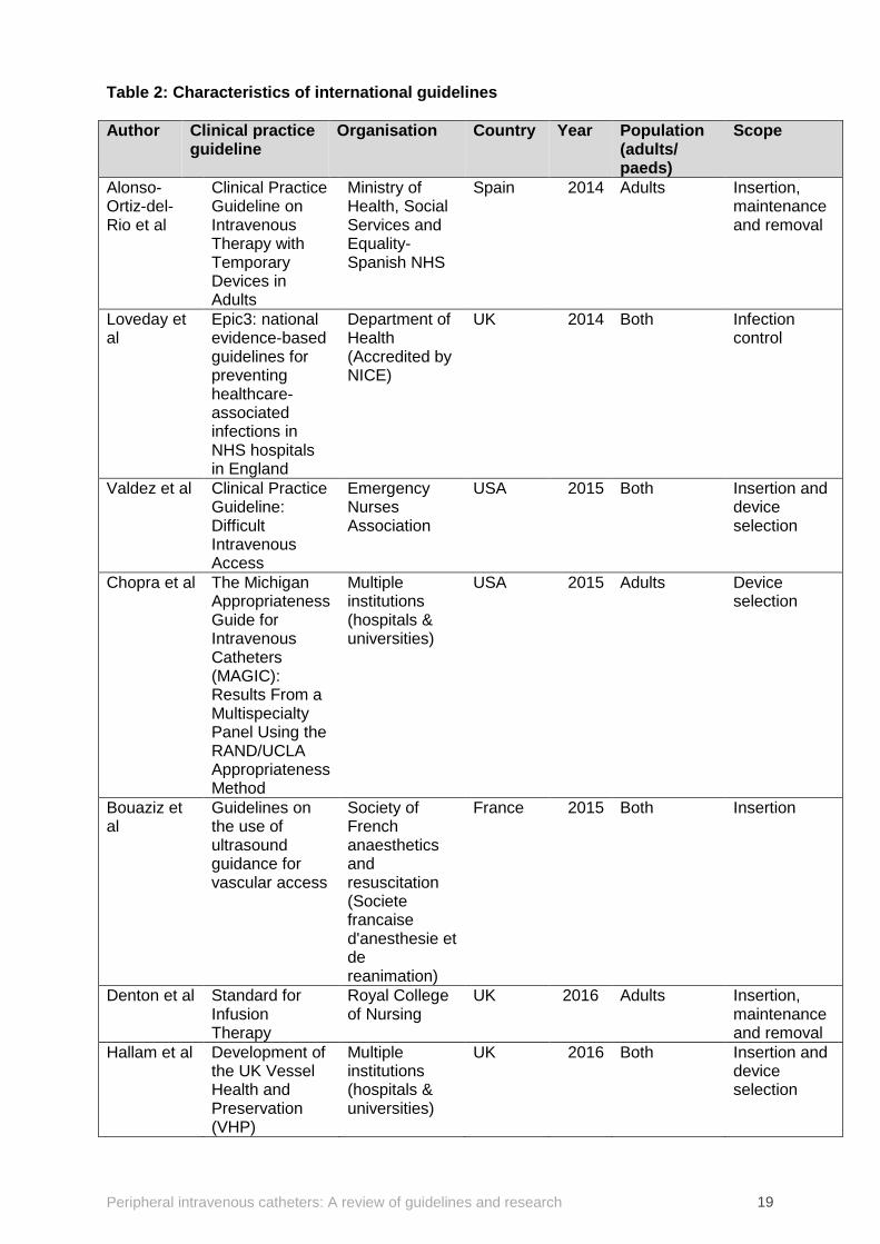
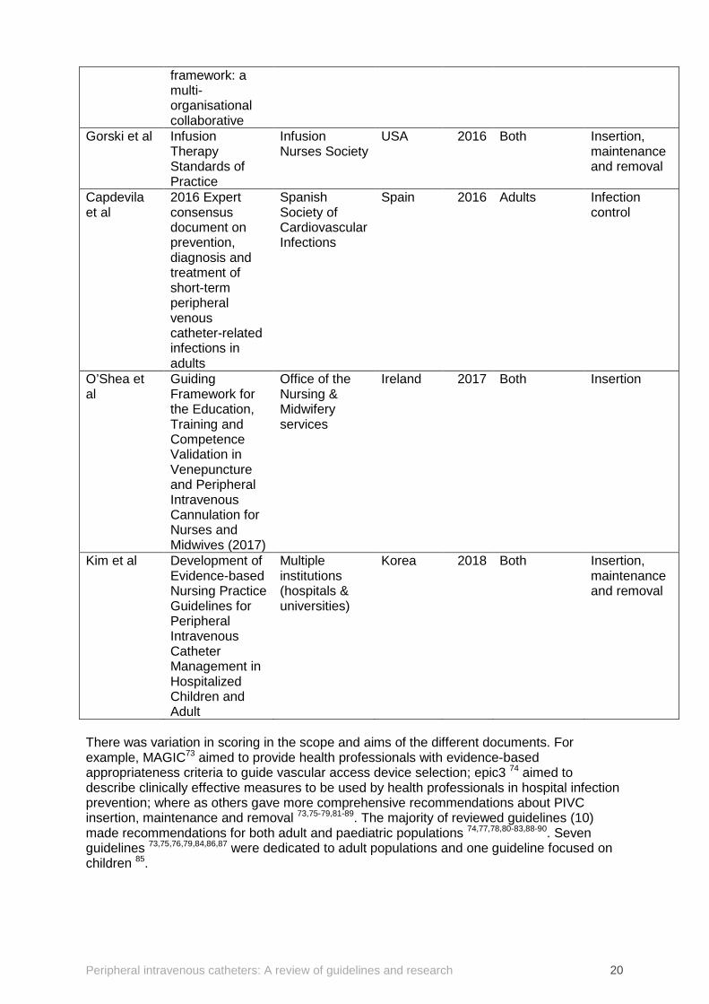
Table 2: Characteristics of international guidelines Author Clinical practice
guideline Organisation Country Year Population
(adults/ paeds)
Scope
Alonso-Ortiz-del-Rio et al
Clinical Practice Guideline on Intravenous Therapy with Temporary Devices in Adults
Ministry of Health, Social Services and Equality- Spanish NHS
Spain 2014 Adults Insertion, maintenance and removal
Loveday et al
Epic3: national evidence-based guidelines for preventing healthcare-associated infections in NHS hospitals in England
Department of Health (Accredited by NICE)
UK 2014 Both Infection control
Valdez et al Clinical Practice Guideline: Difficult Intravenous Access
Emergency Nurses Association
USA 2015 Both Insertion and device selection
Chopra et al The Michigan Appropriateness Guide for Intravenous Catheters (MAGIC): Results From a Multispecialty Panel Using the RAND/UCLA Appropriateness Method
Multiple institutions (hospitals & universities)
USA 2015 Adults Device selection
Bouaziz et al
Guidelines on the use of ultrasound guidance for vascular access
Society of French anaesthetics and resuscitation (Societe francaise d'anesthesie et de reanimation)
France 2015 Both Insertion
Denton et al Standard for Infusion Therapy
Royal College of Nursing
UK 2016 Adults Insertion, maintenance and removal
Hallam et al Development of the UK Vessel Health and Preservation (VHP)
Multiple institutions (hospitals & universities)
UK 2016 Both Insertion and device selection
Peripheral intravenous catheters: A review of guidelines and research 20
framework: a multi-organisational collaborative
Gorski et al Infusion Therapy Standards of Practice
Infusion Nurses Society
USA 2016 Both Insertion, maintenance and removal
Capdevila et al
2016 Expert consensus document on prevention, diagnosis and treatment of short-term peripheral venous catheter-related infections in adults
Spanish Society of Cardiovascular Infections
Spain 2016 Adults Infection control
O’Shea et al
Guiding Framework for the Education, Training and Competence Validation in Venepuncture and Peripheral Intravenous Cannulation for Nurses and Midwives (2017)
Office of the Nursing & Midwifery services
Ireland 2017 Both Insertion
Kim et al Development of Evidence-based Nursing Practice Guidelines for Peripheral Intravenous Catheter Management in Hospitalized Children and Adult
Multiple institutions (hospitals & universities)
Korea 2018 Both Insertion, maintenance and removal
There was variation in scoring in the scope and aims of the different documents. For example, MAGIC73 aimed to provide health professionals with evidence-based appropriateness criteria to guide vascular access device selection; epic3 74 aimed to describe clinically effective measures to be used by health professionals in hospital infection prevention; where as others gave more comprehensive recommendations about PIVC insertion, maintenance and removal 73,75-79,81-89. The majority of reviewed guidelines (10) made recommendations for both adult and paediatric populations 74,77,78,80-83,88-90. Seven guidelines 73,75,76,79,84,86,87 were dedicated to adult populations and one guideline focused on children 85.
Peripheral intravenous catheters: A review of guidelines and research 21
3.1.2 Appraiser agreement
The overall agreement was moderate (K = 0.58; 95% CI 0.36–0.79), with a substantial intra-category concordance (K = 0.66) for guidelines classified “Yes, recommended for use without modification”. The evaluators also demonstrated substantial agreement (K = 0.68) on those guidelines that were not recommended to be used in clinical practice. A moderate level of agreement (K = 0.41) was demonstrated among appraisers on the guidelines that required modification in its use.
3.1.2.1 Overall scores Domain scores and the overall assessment of the appraised guidelines are summarised in Appendix E. Highly skewed scores are acknowledged; however, both mean (SD) and median (range) are reported due to multiple scores of ‘zero’ across domains and across guidelines 91. Overall scores ranged from 25% to 96% (median 58%), with a mean (SD) of 56% (21). Only four guidelines 73,74,76,78 were accepted without modifications and had high overall scores. Seven guidelines 75,79-82,85,90 were ranked moderately (50%–80%) and would be accepted with modifications. The remaining seven guidelines 77,83,84,86-89 were not accepted and scored poorly overall (< 50%).
3.1.2.2 Domain scores Domain 1 (Scope and purpose) The mean (SD) overall score for the scope and purpose domain was 78% (22), (median: 86%; range: 8%-99%). Six guidelines received scores greater than 90% 74-76,81,82,86 and one received a score below 50% 87. The high scores were due to good reporting of objective and intent of guideline. Domain 2 (Stakeholder involvement) For this domain, the mean (SD) overall score was 45% (21), (median: 37%; range: 22%–89%). Two guidelines scored highly at 89% 73,74 and 10 guidelines received scores below 50% 75,77,80,81,83,84,86,88-90. The low scores were given mainly due to the guidelines’ lack of focus on the views and preferences of the target audience. Domain 3 (Rigour of development) The mean (SD) score for this domain was 43% (31) (median: 42%; range: 7%–97%). Two guidelines received very high scores (> 90%) 74,78, and nine received scores below 50% 79,82-
89, with three scoring less than 10% 83,84,89. The low scores in this domain were largely due to the lack of reported systematic searching of the literature, selection and appraisal of evidence. The recommendations provided lacked any supporting evidence and external appraisal. Domain 4 (Clarity of presentation) The mean (SD) score for the clarity of presentation domain was 67% (16) (median: 68%; range 36%–90%). One guideline scored over 90% 74 with two guidelines scoring less than 50% 77,79. This domain was relatively well addressed, as most made clear and specific recommendations for practice. Domain 5 (Applicability) The mean (SD) score for the applicability domain was 30% (26) (median: 25, range: 0%–84%). Two guidelines received a high score (> 85%) 74,76. Most guidelines scored below 50%, while six scored 10% or lower 77,84-86,88,90. These low scores were due to the lack of
Peripheral intravenous catheters: A review of guidelines and research 22
provision of tools or advice, and/or a lack of demonstrated understanding of the facilitators, barriers or resource implications of their recommendations. Domain 6 (Editorial independence) The mean (SD) score for this domain was 37% (42) (median: 15%; range: 0%–100%). Three guidelines received the highest possible score (100%) 73,74,80, and nine guidelines received scores of less than 5% 77,83-88,90. These low scores were due to lack of reporting.
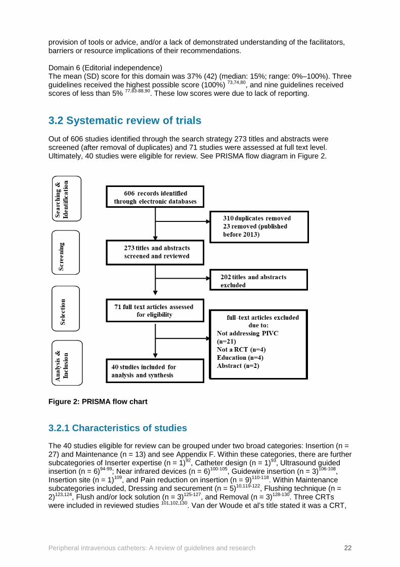
3.2 Systematic review of trials Out of 606 studies identified through the search strategy 273 titles and abstracts were screened (after removal of duplicates) and 71 studies were assessed at full text level. Ultimately, 40 studies were eligible for review. See PRISMA flow diagram in Figure 2.
Figure 2: PRISMA flow chart
3.2.1 Characteristics of studies
The 40 studies eligible for review can be grouped under two broad categories: Insertion (n = 27) and Maintenance (n = 13) and see Appendix F. Within these categories, there are further subcategories of Inserter expertise (n = 1)92, Catheter design (n = 1)93, Ultrasound guided insertion (n = 6)94-99; Near infrared devices (n = 6)100-105, Guidewire insertion (n = 3)106-108, Insertion site (n = 1)109, and Pain reduction on insertion (n = 9)110-118. Within Maintenance subcategories included, Dressing and securement (n = 5)10,119-122, Flushing technique (n = 2)123,124, Flush and/or lock solution (n = 3)125-127, and Removal (n = 3)128-130. Three CRTs were included in reviewed studies 101,102,130. Van der Woude et al’s title stated it was a CRT,
Peripheral intravenous catheters: A review of guidelines and research 23
however examination of method and results indicated that the trial was conducted like a traditional RCT randomising at individual level105. The 40 studies reviewed had a total of 15,335 participants (range 40 - 1807), and an age range from neonate to 90 years of age (10,789 adults, 4,273 paediatrics, 273 neonates). Studies were conducted in following settings: General medical and surgical n= 1310,92,93,98,107,112,117,121-124,128,130, Emergency Department n = 995,97,99,100,104,105,113,115,119, Intensive Care Unit n = 596,111,120,125,129, Surgical unit, n= 394,114,127, Anaesthetic and Operating Room n = 4101-103,118, Medical unit n= 2116,126, and 1 study each from Medical Imaging110, Obstetrics109, Out-patients106 and healthy volunteers in simulation unit108. However, this population was spread across the 12 sub categories (Insertion (n = 7), Maintenance (n = 4). Detailed information on the populations within individualised studies is presented in Table 1. Most of the studies were conducted in the USA (n = 11) and Australia (n = 7). Other countries represented included France (n = 3), Netherlands (n = 4), Italy and China (n = 3 each), Brazil and India (n = 2 each) and 1 each from Canada, Denmark, Malaysia, Spain and Turkey.
3.2.2 Quality appraisal
We analysed seven domains of potential risk of bias in the included studies (see Appendix G). Most studies avoided selection bias by using computerised randomisation methods. Allocation concealment was poorly reported across most studies. We rated the performance risk of bias as high for four studies 95,108,127,128 and unclear for 9 studies 93,96-98,104,106,112,118,119 with the remaining 27 studies at a low risk of bias. The main reason for performance bias was lack of blinding of both participants and personnel to intervention. This is not surprising given the challenge of blinding study participants and staff to study products and/or practice in the clinical setting. However, many studies did employ blinding of outcome assessors to minimise detection bias, though not all. There was minimal attrition and reporting bias. However, discrepancies between tabulated and textual data were noted in some studies thereby introducing other possible sources of bias93,108,114,125,128.
3.2.3 Outcomes
The findings of the review are listed under their relevant outcomes. Meta-analysis could not be undertaken for all outcomes due to variable intervention or outcome reporting. A narrative summary of these studies is presented.
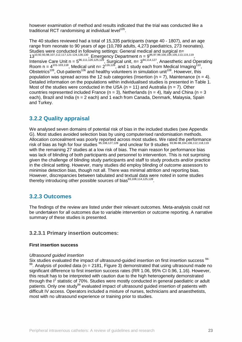
3.2.3.1 Primary insertion outcomes: First insertion success Ultrasound guided insertion Six studies evaluated the impact of ultrasound-guided insertion on first insertion success 94-
99. Analysis of pooled data (n = 2181, Figure 3) demonstrated that using ultrasound made no significant difference to first insertion success rates (RR 1.06, 95% CI 0.96, 1.16). However, this result has to be interpreted with caution due to the high heterogeneity demonstrated through the I2 statistic of 70%. Studies were mostly conducted in general paediatric or adult patients. Only one study99 evaluated impact of ultrasound guided insertion of patients with difficult IV access. Operators included a mixture of nurses, technicians and anaesthetists, most with no ultrasound experience or training prior to studies.
Peripheral intravenous catheters: A review of guidelines and research 24
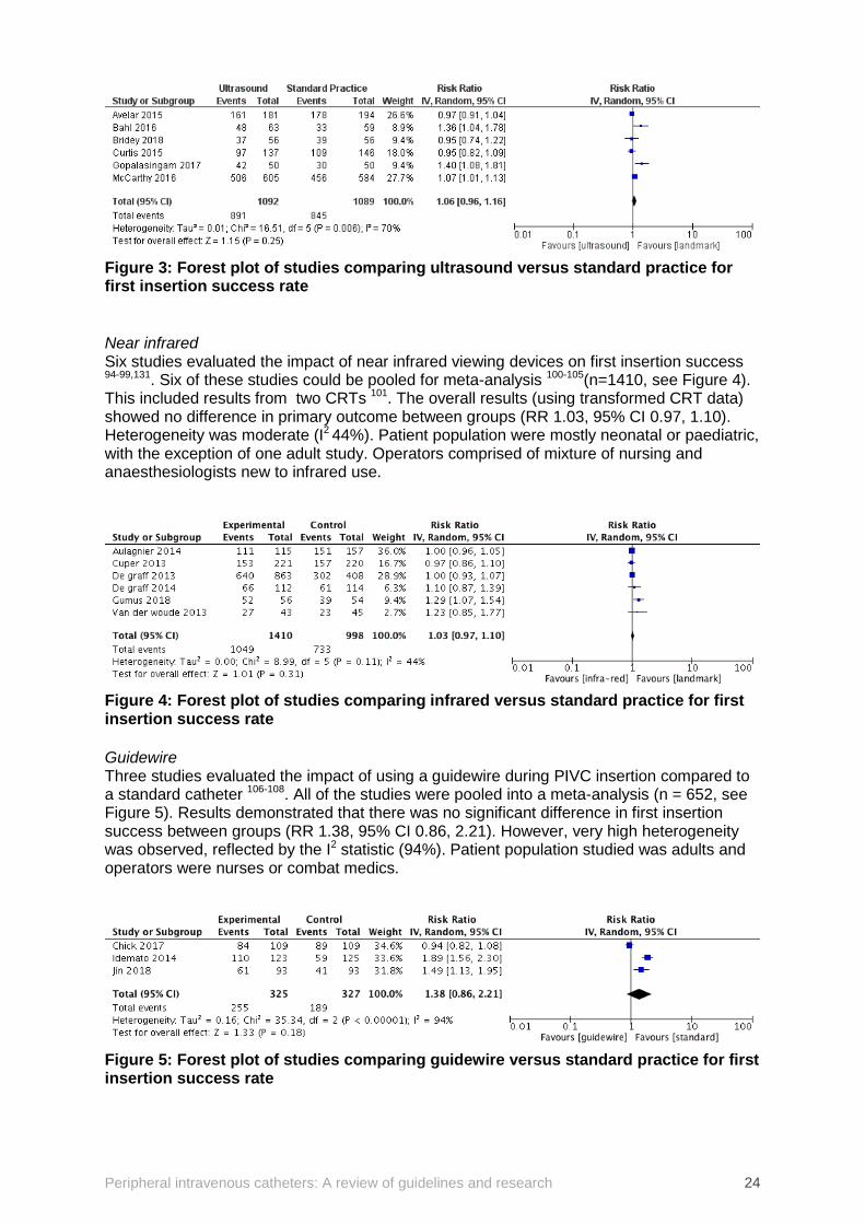
Figure 3: Forest plot of studies comparing ultrasound versus standard practice for first insertion success rate Near infrared Six studies evaluated the impact of near infrared viewing devices on first insertion success 94-99,131. Six of these studies could be pooled for meta-analysis 100-105(n=1410, see Figure 4). This included results from two CRTs 101. The overall results (using transformed CRT data) showed no difference in primary outcome between groups (RR 1.03, 95% CI 0.97, 1.10). Heterogeneity was moderate (I2 44%). Patient population were mostly neonatal or paediatric, with the exception of one adult study. Operators comprised of mixture of nursing and anaesthesiologists new to infrared use.
Figure 4: Forest plot of studies comparing infrared versus standard practice for first insertion success rate Guidewire Three studies evaluated the impact of using a guidewire during PIVC insertion compared to a standard catheter 106-108. All of the studies were pooled into a meta-analysis (n = 652, see Figure 5). Results demonstrated that there was no significant difference in first insertion success between groups (RR 1.38, 95% CI 0.86, 2.21). However, very high heterogeneity was observed, reflected by the I2 statistic (94%). Patient population studied was adults and operators were nurses or combat medics.
Figure 5: Forest plot of studies comparing guidewire versus standard practice for first insertion success rate
Peripheral intravenous catheters: A review of guidelines and research 25
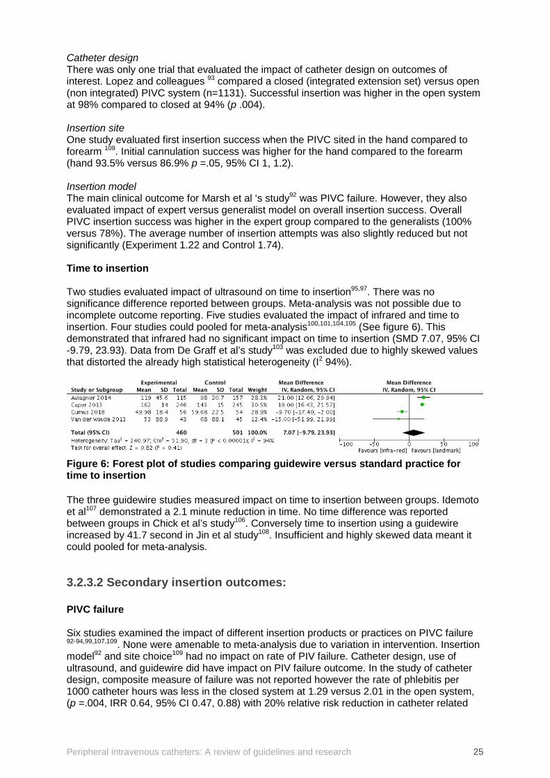
Catheter design There was only one trial that evaluated the impact of catheter design on outcomes of interest. Lopez and colleagues 93 compared a closed (integrated extension set) versus open (non integrated) PIVC system (n=1131). Successful insertion was higher in the open system at 98% compared to closed at 94% (p .004). Insertion site One study evaluated first insertion success when the PIVC sited in the hand compared to forearm 109. Initial cannulation success was higher for the hand compared to the forearm (hand 93.5% versus 86.9% p =.05, 95% CI 1, 1.2). Insertion model The main clinical outcome for Marsh et al ‘s study92 was PIVC failure. However, they also evaluated impact of expert versus generalist model on overall insertion success. Overall PIVC insertion success was higher in the expert group compared to the generalists (100% versus 78%). The average number of insertion attempts was also slightly reduced but not significantly (Experiment 1.22 and Control 1.74). Time to insertion Two studies evaluated impact of ultrasound on time to insertion95,97. There was no significance difference reported between groups. Meta-analysis was not possible due to incomplete outcome reporting. Five studies evaluated the impact of infrared and time to insertion. Four studies could pooled for meta-analysis100,101,104,105 (See figure 6). This demonstrated that infrared had no significant impact on time to insertion (SMD 7.07, 95% CI -9.79, 23.93). Data from De Graff et al’s study103 was excluded due to highly skewed values that distorted the already high statistical heterogeneity (I2 94%).
Figure 6: Forest plot of studies comparing guidewire versus standard practice for time to insertion The three guidewire studies measured impact on time to insertion between groups. Idemoto et al107 demonstrated a 2.1 minute reduction in time. No time difference was reported between groups in Chick et al’s study106. Conversely time to insertion using a guidewire increased by 41.7 second in Jin et al study108. Insufficient and highly skewed data meant it could pooled for meta-analysis.
3.2.3.2 Secondary insertion outcomes: PIVC failure Six studies examined the impact of different insertion products or practices on PIVC failure 92-94,99,107,109. None were amenable to meta-analysis due to variation in intervention. Insertion model92 and site choice109 had no impact on rate of PIV failure. Catheter design, use of ultrasound, and guidewire did have impact on PIV failure outcome. In the study of catheter design, composite measure of failure was not reported however the rate of phlebitis per 1000 catheter hours was less in the closed system at 1.29 versus 2.01 in the open system, (p =.004, IRR 0.64, 95% CI 0.47, 0.88) with 20% relative risk reduction in catheter related
Peripheral intravenous catheters: A review of guidelines and research 26
infection93. In McCarthy et al’s study infiltration was lower in difficult access group (USG 2.1% versus Landmark 3.2%)99. In Avelar et al’s study the proportion of patient’s experiencing infiltration was higher in the experimental group than the control (USG 34 (74%) versus landmark 23 (51%) p=.025. In Idemoto et al’s study107, use of guidewire was also associated with reduced PIV failure (PIVC failure Guidewire 44% versus Standard 12% p < .001). Additionally, infiltration and phlebitis were higher in the control group (Infiltration: Experiment 3(2%) versus Control 33(26%); Phlebitis: Experiment 1(<1%) versus Control 11 (9%)). Infection (local or primary) Idemoto and colleagues and Gonzalez et al93,107 were the only two insertion studies that reported infection outcome. This was negligible, (i.e. 0 in the experimental group and 1 case in the control group) in the Idemato study. The results were also negligible in Gonzalez study 13/529 in closed integrated system and 11/574 in the open PIVC system (p =.635) Dwell time Two studies evaluated the impact of insertion practice or product on dwell time. Use of integrated catheter design was associated with longer dwell time at 137.1 hours compared to open at 96 hours (p < .001)93. In Idemoto et al’s guidewire insertion study dwell time was higher for patients in the experimental group (mean time and (SD), guidewire 105 hours (61) versus control 35hours (25))107. Patient reported pain Nine studies evaluated various forms of analgesia for PIVC insertion 110-118. Only two studies 113,115 were amenable to meta-analysis due to similar interventions and outcomes, however reported outcome values (median and range), therefore, data could not be pooled 132. These two studies evaluated the effect of vapocoolant compared to placebo on pain upon PIVC insertion in an emergency department. Patient perception of pain was no different in Edwards and colleagues study 113 (Median pain score (range): Vapocoolant 2 (0-9) versus Placebo 2.5 (0-10, p =.33)). However, a statistically significant difference of 2 points, where the median pain on the Numeric Pain Scale (NRS) pain scale was less for vapocoolant group was observed between groups in Mace and colleagues’ (p <.001). A number of studies evaluated Eutectic Mixture of Local Anaesthetics (EMLA) as an intervention and control 110,112,114,118. Balanyuk and colleagues’ 110 study showed a significant improvement with distraction compared to EMLA. Mean pain score Distraction 0.69 (SD1.26) versus EMLA 1.86 (173), p < .001) with respect to the local anaesthetic in reducing pain perception. Cozzi and colleagues 112 evaluated EMLA versus warm lidocaine and tetracaine patch on pain during insertion. No significant difference in first insertion success or pain score (Self-reported Wong Baker pain score >4/10, Lidocaine 18/172 (11%) compared to EMLA 15/167 (9%) p =.65)). Stolz and colleagues 118 evaluated J-tip needleless delivered lidocaine intradermal versus EMLA on children’s pain scores for PIVC insertion. Results showed EMLA had lower procedural pain compared to intervention (Mean pain score J-tip lidocaine 2.99 (SD 2.58) versus EMLA 1.63 (SD1.65), p < .001). Gupta and colleagues114 conducted a four-arm RCT evaluating EMLA and Capsaicin (chilli pepper extract with analgesic properties) alone and in combination versus plain lubricant cream (placebo) on pains scores. The authors reported that the incidence of no pain was higher in the EMLA group compared to all others. However, statistical reporting was poor for this study and this conclusion could not be verified. One final study using active analgesia compared topical powdered lidocaine to sham placebo in a paediatric population 117. The substance was well tolerated and the Wong-Baker FACES Pain scores were significantly reduced in the powdered lidocaine group (Mean pain score Lidocaine 1.28 (SD 0.09) versus Sham placebo 1.67 (SD 0.9), p =.002).
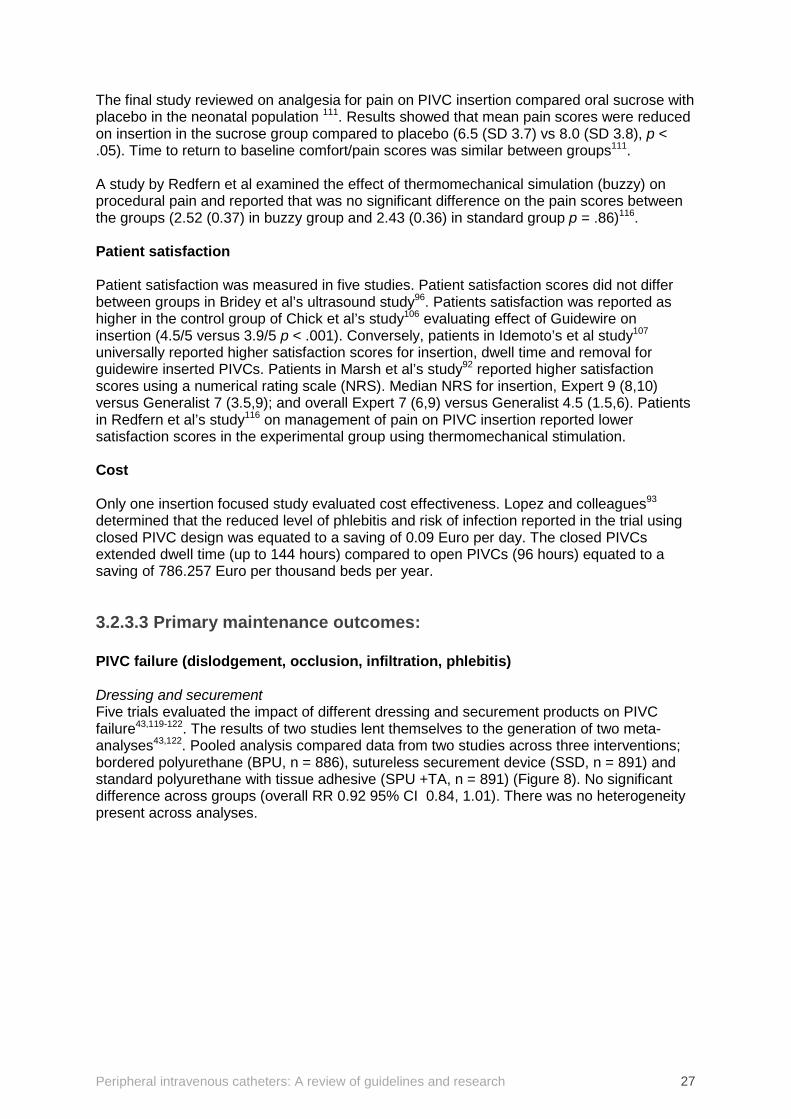
Peripheral intravenous catheters: A review of guidelines and research 27
The final study reviewed on analgesia for pain on PIVC insertion compared oral sucrose with placebo in the neonatal population 111. Results showed that mean pain scores were reduced on insertion in the sucrose group compared to placebo (6.5 (SD 3.7) vs 8.0 (SD 3.8), p < .05). Time to return to baseline comfort/pain scores was similar between groups111. A study by Redfern et al examined the effect of thermomechanical simulation (buzzy) on procedural pain and reported that was no significant difference on the pain scores between the groups (2.52 (0.37) in buzzy group and 2.43 (0.36) in standard group p = .86)116. Patient satisfaction Patient satisfaction was measured in five studies. Patient satisfaction scores did not differ between groups in Bridey et al’s ultrasound study96. Patients satisfaction was reported as higher in the control group of Chick et al’s study106 evaluating effect of Guidewire on insertion (4.5/5 versus 3.9/5 p < .001). Conversely, patients in Idemoto’s et al study107 universally reported higher satisfaction scores for insertion, dwell time and removal for guidewire inserted PIVCs. Patients in Marsh et al’s study92 reported higher satisfaction scores using a numerical rating scale (NRS). Median NRS for insertion, Expert 9 (8,10) versus Generalist 7 (3.5,9); and overall Expert 7 (6,9) versus Generalist 4.5 (1.5,6). Patients in Redfern et al’s study116 on management of pain on PIVC insertion reported lower satisfaction scores in the experimental group using thermomechanical stimulation. Cost Only one insertion focused study evaluated cost effectiveness. Lopez and colleagues93 determined that the reduced level of phlebitis and risk of infection reported in the trial using closed PIVC design was equated to a saving of 0.09 Euro per day. The closed PIVCs extended dwell time (up to 144 hours) compared to open PIVCs (96 hours) equated to a saving of 786.257 Euro per thousand beds per year.
3.2.3.3 Primary maintenance outcomes: PIVC failure (dislodgement, occlusion, infiltration, phlebitis) Dressing and securement Five trials evaluated the impact of different dressing and securement products on PIVC failure43,119-122. The results of two studies lent themselves to the generation of two meta-analyses43,122. Pooled analysis compared data from two studies across three interventions; bordered polyurethane (BPU, n = 886), sutureless securement device (SSD, n = 891) and standard polyurethane with tissue adhesive (SPU +TA, n = 891) (Figure 8). No significant difference across groups (overall RR 0.92 95% CI 0.84, 1.01). There was no heterogeneity present across analyses.
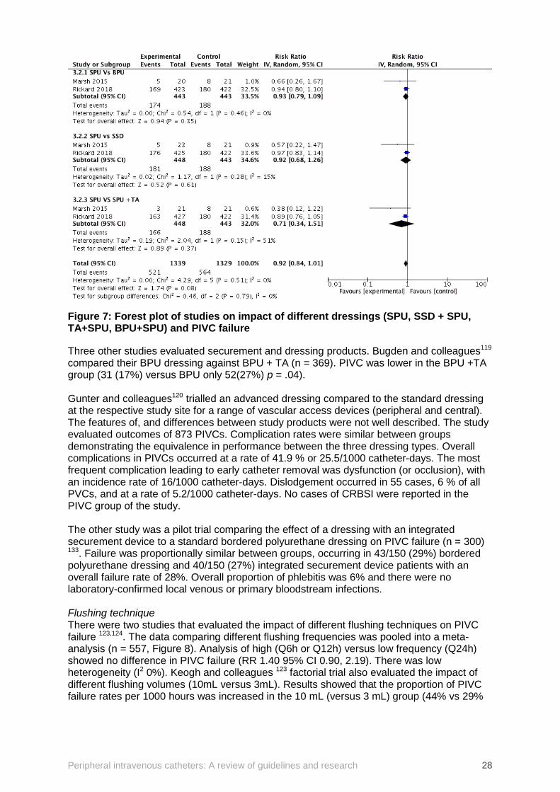
Peripheral intravenous catheters: A review of guidelines and research 28
Figure 7: Forest plot of studies on impact of different dressings (SPU, SSD + SPU, TA+SPU, BPU+SPU) and PIVC failure Three other studies evaluated securement and dressing products. Bugden and colleagues119 compared their BPU dressing against BPU + TA (n = 369). PIVC was lower in the BPU +TA group (31 (17%) versus BPU only 52(27%) p = .04). Gunter and colleagues120 trialled an advanced dressing compared to the standard dressing at the respective study site for a range of vascular access devices (peripheral and central). The features of, and differences between study products were not well described. The study evaluated outcomes of 873 PIVCs. Complication rates were similar between groups demonstrating the equivalence in performance between the three dressing types. Overall complications in PIVCs occurred at a rate of 41.9 % or 25.5/1000 catheter-days. The most frequent complication leading to early catheter removal was dysfunction (or occlusion), with an incidence rate of 16/1000 catheter-days. Dislodgement occurred in 55 cases, 6 % of all PVCs, and at a rate of 5.2/1000 catheter-days. No cases of CRBSI were reported in the PIVC group of the study. The other study was a pilot trial comparing the effect of a dressing with an integrated securement device to a standard bordered polyurethane dressing on PIVC failure (n = 300) 133. Failure was proportionally similar between groups, occurring in 43/150 (29%) bordered polyurethane dressing and 40/150 (27%) integrated securement device patients with an overall failure rate of 28%. Overall proportion of phlebitis was 6% and there were no laboratory-confirmed local venous or primary bloodstream infections. Flushing technique There were two studies that evaluated the impact of different flushing techniques on PIVC failure 123,124. The data comparing different flushing frequencies was pooled into a meta-analysis (n = 557, Figure 8). Analysis of high (Q6h or Q12h) versus low frequency (Q24h) showed no difference in PIVC failure (RR 1.40 95% CI 0.90, 2.19). There was low heterogeneity (I2 0%). Keogh and colleagues 123 factorial trial also evaluated the impact of different flushing volumes (10mL versus 3mL). Results showed that the proportion of PIVC failure rates per 1000 hours was increased in the 10 mL (versus 3 mL) group (44% vs 29%
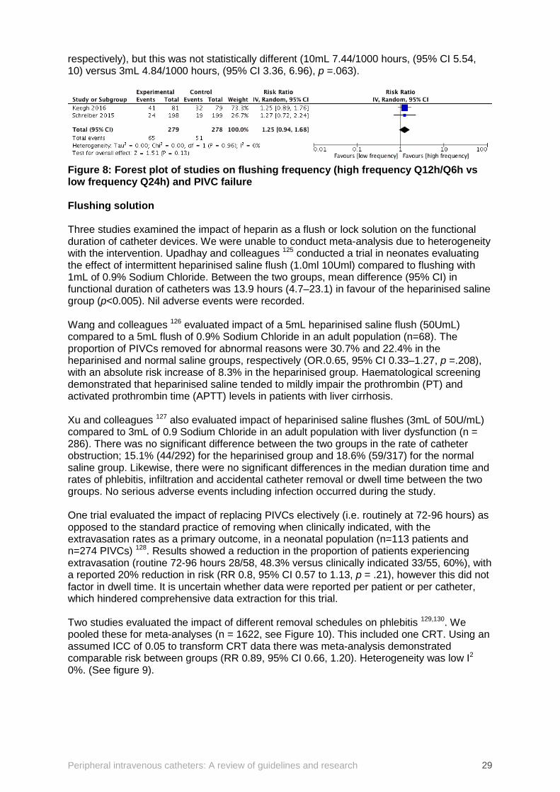
Peripheral intravenous catheters: A review of guidelines and research 29
respectively), but this was not statistically different (10mL 7.44/1000 hours, (95% CI 5.54, 10) versus 3mL 4.84/1000 hours, (95% CI 3.36, 6.96), p =.063).
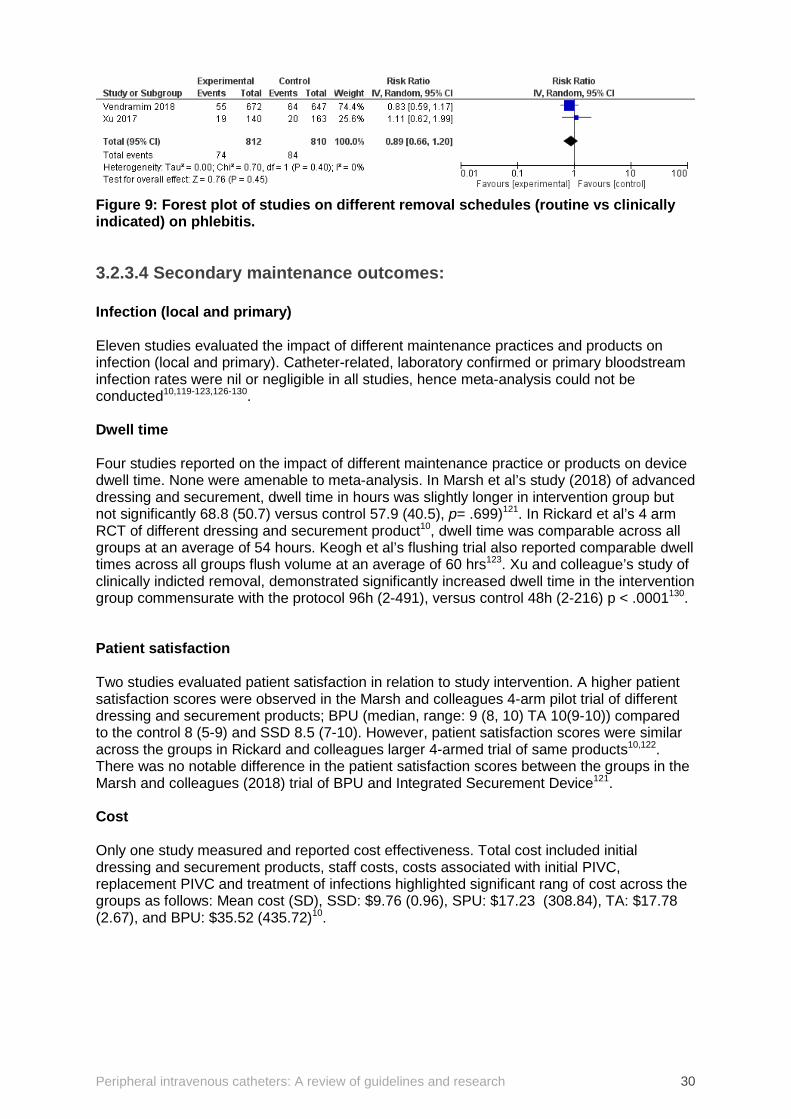
Figure 8: Forest plot of studies on flushing frequency (high frequency Q12h/Q6h vs low frequency Q24h) and PIVC failure Flushing solution Three studies examined the impact of heparin as a flush or lock solution on the functional duration of catheter devices. We were unable to conduct meta-analysis due to heterogeneity with the intervention. Upadhay and colleagues 125 conducted a trial in neonates evaluating the effect of intermittent heparinised saline flush (1.0ml 10Uml) compared to flushing with 1mL of 0.9% Sodium Chloride. Between the two groups, mean difference (95% CI) in functional duration of catheters was 13.9 hours (4.7–23.1) in favour of the heparinised saline group (p<0.005). Nil adverse events were recorded. Wang and colleagues 126 evaluated impact of a 5mL heparinised saline flush (50UmL) compared to a 5mL flush of 0.9% Sodium Chloride in an adult population (n=68). The proportion of PIVCs removed for abnormal reasons were 30.7% and 22.4% in the heparinised and normal saline groups, respectively (OR.0.65, 95% CI 0.33–1.27, p =.208), with an absolute risk increase of 8.3% in the heparinised group. Haematological screening demonstrated that heparinised saline tended to mildly impair the prothrombin (PT) and activated prothrombin time (APTT) levels in patients with liver cirrhosis. Xu and colleagues 127 also evaluated impact of heparinised saline flushes (3mL of 50U/mL) compared to 3mL of 0.9 Sodium Chloride in an adult population with liver dysfunction (n = 286). There was no significant difference between the two groups in the rate of catheter obstruction; 15.1% (44/292) for the heparinised group and 18.6% (59/317) for the normal saline group. Likewise, there were no significant differences in the median duration time and rates of phlebitis, infiltration and accidental catheter removal or dwell time between the two groups. No serious adverse events including infection occurred during the study. One trial evaluated the impact of replacing PIVCs electively (i.e. routinely at 72-96 hours) as opposed to the standard practice of removing when clinically indicated, with the extravasation rates as a primary outcome, in a neonatal population (n=113 patients and n=274 PIVCs) 128. Results showed a reduction in the proportion of patients experiencing extravasation (routine 72-96 hours 28/58, 48.3% versus clinically indicated 33/55, 60%), with a reported 20% reduction in risk (RR 0.8, 95% CI 0.57 to 1.13, p = .21), however this did not factor in dwell time. It is uncertain whether data were reported per patient or per catheter, which hindered comprehensive data extraction for this trial. Two studies evaluated the impact of different removal schedules on phlebitis 129,130. We pooled these for meta-analyses (n = 1622, see Figure 10). This included one CRT. Using an assumed ICC of 0.05 to transform CRT data there was meta-analysis demonstrated comparable risk between groups (RR 0.89, 95% CI 0.66, 1.20). Heterogeneity was low I2 0%. (See figure 9).
Peripheral intravenous catheters: A review of guidelines and research 30
Figure 9: Forest plot of studies on different removal schedules (routine vs clinically indicated) on phlebitis.
3.2.3.4 Secondary maintenance outcomes: Infection (local and primary) Eleven studies evaluated the impact of different maintenance practices and products on infection (local and primary). Catheter-related, laboratory confirmed or primary bloodstream infection rates were nil or negligible in all studies, hence meta-analysis could not be conducted10,119-123,126-130. Dwell time Four studies reported on the impact of different maintenance practice or products on device dwell time. None were amenable to meta-analysis. In Marsh et al’s study (2018) of advanced dressing and securement, dwell time in hours was slightly longer in intervention group but not significantly 68.8 (50.7) versus control 57.9 (40.5), p= .699)121. In Rickard et al’s 4 arm RCT of different dressing and securement product10, dwell time was comparable across all groups at an average of 54 hours. Keogh et al’s flushing trial also reported comparable dwell times across all groups flush volume at an average of 60 hrs123. Xu and colleague’s study of clinically indicted removal, demonstrated significantly increased dwell time in the intervention group commensurate with the protocol 96h (2-491), versus control 48h (2-216) p < .0001130. Patient satisfaction Two studies evaluated patient satisfaction in relation to study intervention. A higher patient satisfaction scores were observed in the Marsh and colleagues 4-arm pilot trial of different dressing and securement products; BPU (median, range: 9 (8, 10) TA 10(9-10)) compared to the control 8 (5-9) and SSD 8.5 (7-10). However, patient satisfaction scores were similar across the groups in Rickard and colleagues larger 4-armed trial of same products10,122. There was no notable difference in the patient satisfaction scores between the groups in the Marsh and colleagues (2018) trial of BPU and Integrated Securement Device121. Cost Only one study measured and reported cost effectiveness. Total cost included initial dressing and securement products, staff costs, costs associated with initial PIVC, replacement PIVC and treatment of infections highlighted significant rang of cost across the groups as follows: Mean cost (SD), SSD: $9.76 (0.96), SPU: $17.23 (308.84), TA: $17.78 (2.67), and BPU: $35.52 (435.72)10.
Peripheral intravenous catheters: A review of guidelines and research 31
4. Discussion 4.1 Guidelines The aim of this systematic review was to appraise the scope, quality and rigour of contemporary clinical guidelines and practice standards for insertion, maintenance and removal of peripheral intravenous catheters in adult and paediatric hospital populations. We analysed the guidelines and standards published since 2013 using the AGREE II tool. Variability in the methodological quality of the retrieved guidelines and standards was high. Only one guideline (the UK’s epic3134) scored consistently high (> 80%) across all domains, except for a score of 72% in Domain 5 (Applicability). Three other guidelines scored consistently over 50%,73,76,78 with variation in quality in Domain 5 (applicability). The remaining guidelines were highly variable or rate consistently poor against the AGREE II tool categories. Most guidelines performed adequately or well in Scope and Clarity of presentation; however, the most poorly adhered to domain was rigour of development. The development of evidence-based guidelines requires a well-resourced and representative committee, who have the skills and knowledge to search and appraise relevant research and make recommendations for clinical practice135. A number of large international and national bodies are resourced to generate and maintain quality guidelines, and this was reflected in their AGREE scoring (e.g. MAGIC, epic3, INS). The Centers for Disease Control (CDC) Guidelines for Prevention of Intravascular Catheter-Related Infections (2011) did not meet search criteria because of date limitations31. The CDC is a well-resourced independent body that convened a large and representative healthcare team to generate these guidelines based on the best available evidence sourced through systematic search and appraisal. Thereby demonstrating many of the key desirable characteristics of quality guidelines. Since the original publication date of these in 2011 the CDC has stated that they will only make updates when significant new evidence becomes available136. An example of this is the recommendation to use chlorhexidine impregnated dressings for short term non tunnelled central venous catheters in 2017. Guidelines were found from all major states in Australia. National guidelines for the prevention and control of infection in healthcare from 2010 were outside the date limits and unavailable as currently under review137. The guidelines generated by local bodies reviewed did not perform well against the AGREE II scoring categories. It is unclear as to whether the issue is with quality of reporting of these documents or the development process itself, but the reviewer and indeed the user has only the content published to appraise. Similarities and differences between guidelines Much of the content of the guidelines are similar especially in regards to standard and transmission based precautions, hand hygiene and aseptic technique. These are largely based on the CDC and epic3 Guidelines. Other commonalities include:
• Assessment (at basic level) of need and type of IV device, and catheter site selection • Skin decontamination with 2% Chlorohexidine in 70% alcohol solution (unless
contraindicated) • Limiting of insertion attempts to two (2) per practitioner • Use of a transparent, semi permeable dressing to cover IV insertion site and secure
catheter • Decontamination of any ports or hubs prior to access • Use of O.9% sodium chloride flushes pre and post drug administration
Peripheral intravenous catheters: A review of guidelines and research 32
• Removal and replacement of IV device if (a) no longer device no longer working (i.e. occluded) or (b) patient shows signs of local or systemic infection (i.e. local redness, pain or swelling or fever and malaise.
• Removal of device when no longer required There are a number of differences between and unresolved issues in the guidelines. These include:
• Quality and detail of patient assessment determining the type of and site location for the PIVC. For example, MAGIC guidelines, INS and epic3 give detailed instructions on device selection or site choice based patient and treatment factors and harm minimisation.
• Detailed assessment of quality of patient’s vessel integrity. The UK’s Vessel Health Preservation framework and the US’s Emergency Nurse’s Clinical Practice Guidelines are only documents to provide decision matrix to guide and strategies for patients with difficult vascular access
• Escalation of insertion process if first insertion attempts unsuccessful. Most guidelines limited each individual to two attempts but few offered a formal pathway to optimise insertion thereafter (other than more blind attempts)
• If and when to use ultrasound guided vessel location and insertion (as above) • Use of analgesia agents or pain relieving strategies. Few recommended routine pain
relief pre insertion despite significant evidence and patient reports in favour of this • Specific type of dressing and securement device(s). Generic desirable characteristics
of dressing and securement device listed but no specific product types named. • Type of needleless connector. No specific product characteristic or type
recommended. • Type and technique of port or hub decontamination. There were varied
recommendations for decontamination solution (70% alcohol versus 2% Chlorhexidine in alcohol), decontamination timing (times of 5, 15 and 30 second cited), as well as technique (active scrubbing versus passive cap).
• Frequency and technique for IV flushing. Varied frequencies cited for flushing of PIVCs not in continuous use (e.g. Q24h, Q8h, Q6h and PRN).
• Frequency of IV administration set changes. Disagreement between key guidelines on frequency of IV administration set changes (for solutions not containing blood or lipid). CDC recommend change every seven days and epic3 recommend change every four days.
• Criteria for ongoing assessment of PIVC site and function. There was a general recommendation for management of dressing and patency of PIVC and removal if showing site or patient exhibiting signs of infection, but no comprehensive criteria for assessment or recommendation of frequency of assessment.
• Criteria for replacement of PIVCs. Disagreement between key national and international reviews. The UK, Spanish and Irish guidelines all recommend replacement of PIVCs as clinically indicated. The CDC (2011) recommend removal of PIVCs no more frequently than 72-96 hours and regard clinically indicated as unresolved (except in children). All local (Australian) guidelines recommend PIVC replacement at 72 hours.
In summary, many of the national and international guidelines exhibited consensus on the key recommendations regarding PIVC insertion and maintenance. Several guidelines focused on optimising device selection and insertion, reflecting the growing research and commentary around vessel health preservation. Most cited the level and source of evidence that supported these recommendations. Areas of practice that remained unresolved were largely due to lack of high quality evidence (e.g. needless decontamination). There was significant variation in reporting and quality of guidelines at a local level, which invites the question of whether state and local bodies would do better to endorse relevant national and international guideline and then provide staff with local work statements to implement these.
Peripheral intravenous catheters: A review of guidelines and research 33
Clinicians rely on guidelines and standards to create hospital policies designed to aid decision-making at the bedside and drive quality practice. If the guidelines themselves are of poor or variable quality, this raises concerns for patient outcomes.
4.2 Intervention trials Forty trials assessing PIVC-related interventions in the last five years were identified. These differed in quality, size and scope. The aim was to identify high-level evidence to guide PIVC insertion and maintenance practice. Insertion The majority of trials identified in this review focused on insertion products and practice. This is commensurate with the results of a Scoping Review in 2014138. Most of the studies focused on evaluation of visual aids to optimise first insertion success and time to insertion. Findings from the meta-analysis of eligible studies did not demonstrate a significant difference in this review. However, these results should be viewed with caution due to the high statistical heterogeneity observed across studies as well as the significant clinical variation with population, settings and operators. Most studies only offered new operators a short course (theory and practice) as a requirement for training before they entered the study. Therefore, aids like ultrasound may not have demonstrated superiority due to the complexity of the procedure and training required, versus the usual landmark technique. Operator experience does have an impact on the rate and quality of PIVC cannulation139. So standardising and optimising education related to PIVC insertion (with and without ultrasound) is important for patient outcomes. Previously published systematic reviews and meta-analysis of studies found that in patients with difficult IV access the use of ultrasound-guidance for peripheral venous access improved first time success rates compared to traditional technique140-143. The studies did not demonstrate that ultrasound guided insertion had an impact on time to successful cannulation or the number of skin punctures though. However, two paediatric studies found that time to successful cannulation was reduced and fewer attempts were required to achieve success for patients randomised to ultrasound guided insertion compared with the traditional method144,145. Further, a systematic review and meta-analysis found that ultrasound guided peripheral IV cannulation significantly reduces number of attempts and procedure time in paediatric patients146. Based on systematic reviews of the literature, routine use of ultrasound-guided peripheral venous cannulation is not recommended, however, for patients with known or suspected difficult venous access ultrasound-guided techniques improve success rates in a timely manner with improved patient satisfaction. Research for peripheral catheter insertion has also focused on pre insertion analgesia. Analgesia for catheter insertion featured in 32% of PIVC trials identified in a scoping review of RCTs in peripheral vascular access devices138. No meta-analysis of studies was possible in this review due to variation in interventions. However, individual studies did demonstrate reduced patient reported pain scores with analgesia intervention115,118. Three systematic reviews of studies that pre dated this review demonstrated the impact of some sort of analgesia over no treatment147-149. Meta-analysis of studies did not demonstrate which anaesthetic was superior to another147, and Vapocoolant was not found to be as effective in children148. However overall, pain is less is using any local anaesthetic or numbing agent than when unattenuated during cannulation and some sort of pain relief should be routinely offered to patients prior to PIVC insertion. Other studies on inserter model and catheter design were limited, but considering the increased acuity and comorbidities of patients requiring vascular access demand further investment in quality research to inform future practice and product use.
Peripheral intravenous catheters: A review of guidelines and research 34
Maintenance Given the challenges of acquiring successful peripheral vascular access it is vital to maintain the integrity and performance of the device. Although many RCTs examined catheter insertion strategies there were significantly less studies focused on post insertion care and maintenance of peripheral catheters. This mirrors the findings of a prior scoping review in this area138. Just one third of studies reviewed evaluated different interventions related to post PIVC insertion care and maintenance. The largest subcategory was evaluation of dressing and securement products on PIVC complications and failure (n=5 trials). Only one small study in the Emergency Department yielded a significant result favouring the intervention (Bordered dressing plus tissue adhesive)119. Just two studies were amenable to meta-analysis and neither this nor the remaining studies demonstrated a superior product for dressing and securement. Guidelines are currently unable to make firm recommendations for dressing and securement practice and products due to the lack of evidence. The extensive use of PIVCs, high rates of failure, and substantial costs to healthcare for dressing and securement products highlights the need for further investment and innovation to develop effective products43. Other studies within Maintenance tested the impact of different flushing techniques and solutions. Meta-analysis of two small studies of different flushing frequency demonstrated that less frequent flushing was not associated with increased risk of PIVC failure and could therefore be considered as a safe alternative to high frequency flushing regimens prescribed in many guidelines. However, the mechanisms of how IV flushing, medication and fluid administration impact on the cannula, vessel endothelium and blood components are poorly understood and require further explication. We need to explore how syringe and infusion technology and method of administration can make a difference to PIVC outcomes18,50. Three studies compared heparinised saline versus saline flush alone on PIVC function and dwell time. Meta-analysis was not possible due to clinical variation in the intervention (i.e. heparin concentration). One study reported a statistical difference of 14 hours duration. Previous trials and systematic reviews have failed to demonstrate a benefit of heparin over saline as flush or lock solution in peripheral or central IV devices in adults150-154. The use of anticoagulant (usually heparin) to prevent occlusion of IVs has been traditionally used in the past, based on a not unreasonable suspicion of effectiveness. However, the process of in vivo catheter occlusion is complex and multi factorial, not simply based on blood clotting or deposit of blood proteins or cells 155. Thrombotic occlusion only accounts for only 58% of occlusions. Intraluminal occlusion can also be secondary to mechanical (e.g. kinks, pinches) and chemical causes (e.g. drug precipitate, lipid build up)156. Leading guideline bodies do not recommend the routine use of anticoagulant lock or flush solutions, with the exception for central IVs in paediatrics 44,134,157. Although this recommendation lacks a firm evidence base. Moving forward, researchers should consider rigorous evaluation of other lock solutions (e.g. tetrasodium citrate, thrombin inhibitors, plasmin activators). The impact of different PIVC replacement schedules was the focus of three trials evaluated in this review. They all evaluated the impact of routine compared to clinically indicated removal of PIVCs. Meta-analysis of two of the studies demonstrated equivalent risk of phlebitis between groups. Cases of catheter related bloodstream infection across groups and overall was nil or negligible. The result aligns with that of a current systematic review of seven trials158. The authors concluded that there was moderate certainty evidence of no clear difference in rates of CRBSI, thrombophlebitis, all-cause blood stream infection, mortality or pain between clinically indicated or routine replacement of PIVC (ibid). Adopting the practice of replacement of PIVCs as clinically indicated would provide significant cost savings35, spare patients the unnecessary pain of routine re-sites in the absence of clinical indications and would reduce time spent by busy clinicians on this intervention. Replacement of PIVCs as clinically indicated in children is standard recommended practice in all
Peripheral intravenous catheters: A review of guidelines and research 35
guidelines. As noted earlier in this review a number of leading national guidelines no longer recommend routine removal of PIVCs44,76,134,159. The CDC regards the issue of clinically indicated removal as unresolved, However, this statement predates the publication a major trial and systematic reviews addressing this issue136. To minimise PIVC related complications, clinical staff should regularly assess the insertion site and remove the catheter if signs of pain, inflammation, infiltration, occlusion, or infection are present, or if the catheter is no longer needed for therapy. This underscores the need for pragmatic and evidence based tools to guide clinical assessment. Just seven studies (17%) evaluated patient satisfaction or feedback on practice or product under evaluation. These were in relation to ultrasound guided insertion, catheter design, distraction therapy (for pain relief) and type of dressing or securement. The role of the consumer was placed front and centre in the recently updated in the Australian National Quality Healthcare Standards160. Partnering with consumers was described as a broad concept that could involve consumer engagement and participation in care decision as well as consumer centred care itself (ibid). This can extend to research groups and guideline development bodies. “The purpose of these partnerships is to improve the outcomes, experiences and the delivery of care by drawing on the knowledge, skills and experiences of people who are using, have used or may use the health service.”(NSQHC Standard 2: Partnering with consumers) In summary, of the 40 trials identified in the five years only one large, high quality PIVC trial was conducted and reported in the last five years. Billions of devices are used in healthcare settings globally, with billions of dollars spent on related products and staff resources. Yet there is such little investment in quality research to inform product and practice choices. Clinicians and policy makers largely operate in an evidence vacuum. This clearly contributes to practice inconsistency, inefficiency and ineffectiveness of some practice and products. Note though, that where there is evidence, there is poor uptake. (For example, use of ultrasound and analgesia for insertion, flushing solution, and device removal). There is an onus on government and the healthcare industry to invest in vascular access research; a further onus on vascular access researchers to conduct and report on quality and meaningful research. Finally, there is an onus on clinicians as health professionals to value and implement findings to improve patient outcomes and reduce waste of healthcare resources.
4.3 Gaps in identified literature This literature review identified several gaps in the evidence base, which need to be met to facilitate development of strong evidence-based recommendations. In particular, there is a need for adequately powered and efficiently designed randomised control trials to assess:
• Impact of vascular access specialist (team or service) on patient and PIVC outcomes • Clinical and cost effectiveness of innovative catheter design on insertion and function • Optimal dressing and securement product(s) to reduce PIVC failure • Needleless connector type on PIVC function and infection outcomes • Needleless connector denomination solution, timing and technique and infection
outcomes • Impact of infusion and flushing regimens on maintaining PIVC function
Other areas where more research is needed include:
• Criteria for clinical assessment to monitor PIVCs • Patient experience of PIVC insertion and different maintenance practices
Peripheral intravenous catheters: A review of guidelines and research 36
4.4 Limitations The review of national and international guidelines and local policies was limited by access to the materials online. It is possible that many hospitals in Australia (if not all) have a policy on PIVC insertion and maintenance, however these will often be behind institutional firewalls and therefore we were unable to access. However the guidelines reviewed appeared to be representative of key local, national and international agencies. Limitations with the systematic review of PIVC intervention trials are related to the quality and heterogeneity of the trials. The risk of bias analysis demonstrated that the most of the trials (two thirds) had a low risk of bias. However, the process of data extraction revealed small sample sized and inconsistencies and variation in reporting that meant meta-analysis was not possible. The review was also limited to the last five years. However, the aim was to capture trial research that reflected contemporary practice and products. Other significant trials and systematic reviews were referenced in the discussion and inform the final recommendation
Peripheral intravenous catheters: A review of guidelines and research 37
5. Implications and recommendations This review highlighted a number of inconsistencies between Australian guidelines and leading international bodies. Universally, there are areas of PIVC practice that do not reflect the most recent or highest level of evidence available. (For example, use of ultrasound and analgesia for insertion, flushing solution, and device removal.). The review highlighted gaps in evidence that make it challenging for guidelines developers to construct definitive recommendations about specific practice or products to optimise PIVC outcome and the patient’s IV experience. Many PIVC complications are avoidable and investment in quality evidence generation and implementation is urgently warranted. Given the enormous number of PIVCs used, even small differences in clinical and cost-effectiveness of different practices and products could generate significant improvement in patient outcomes and cost savings for hospital budgets. A number of recommendations are made in relation to the findings from this review. These include:
I. Development of a national clinical care standard aimed at reducing PIVC related complications and failure.
II. Education and training of nursing and medical staff that focus on contemporary and evidence based PIVC insertion and maintenance care.
III. Evidence based assessment of patient’s need for vascular access and device type. IV. Early referral to vascular access specialist and use of ultrasound to minimise
insertion trauma in patents identified as having difficult vascular access. V. Routine use of analgesic agents or strategies to minimise PIVC insertion
associated pain. VI. Evidence based clinical assessment of PIVC site and function
VII. Ongoing monitoring and reporting of PIVC use and outcomes to facilitate benchmarking and drive quality improvement
VIII. Inclusion of patients’ views in all future research in to the optimal PIVC insertion and maintenance practices and products.
6. Conclusion This structured evidence-based literature review was undertaken to better understand the current clinical environment for peripheral intravenous catheter insertion and care, and to identify issues or gaps that may be addressed by clinical experts at a clinical roundtable. The review includes an analysis of international, national and state guidelines as well as a review of 40 randomised clinical trials. There was a high degree of variation in guideline quality and recommendations. This may be contributing to inconsistency in practice and the unacceptable rates of PIVC failure reported in studies. The development of a national clinical care standard should assist in bringing clinicians and health services together to agree upon the way forward to improve outcomes for all patients who rely on safe and patent vascular access for the delivery of vital medical treatment.
Peripheral intravenous catheters: A review of guidelines and research 38
7. References 1. Cooke M, Ullman AJ, Ray-Barruel G, Wallis M, Corley A, Rickard CM. Not "just" an intravenous line: Consumer perspectives on peripheral intravenous cannulation (PIVC). An international cross-sectional survey of 25 countries. PLoS One 2018;13:e0193436. 2. Jacobson AF, Winslow EH. Variables influencing intravenous catheter insertion difficulty and failure: an analysis of 339 intravenous catheter insertions. Heart Lung 2005;34:345-59. 3. Malyon L, Ullman AJ, Phillips N, et al. Peripheral intravenous catheter duration and failure in paediatric acute care: A prospective cohort study. Emerg Med Australas 2014;26:602-8. 4. Bausone-Gazda D, Lefaiver CA, Walters SA. A randomized controlled trial to compare the complications of 2 peripheral intravenous catheter-stabilization systems. J Infus Nurs 2010;33:371-84. 5. Helm RE, Klausner JD, Klemperer JD, Flint LM, Huang E. Accepted but unacceptable: peripheral IV catheter failure. J Infus Nurs 2015;38:189-203. 6. Marsh N, Webster J, Larson E, Cooke M, Mihala G, Rickard CM. Observational Study of Peripheral Intravenous Catheter Outcomes in Adult Hospitalized Patients: A Multivariable Analysis of Peripheral Intravenous Catheter Failure. J Hosp Med 2018;13:83-9. 7. Wallis MC, McGrail M, Webster J, et al. Risk factors for peripheral intravenous catheter failure: a multivariate analysis of data from a randomized controlled trial. Infection control and hospital epidemiology 2014;35:63-8. 8. Maki DG, Kluger DM, Crnich CJ. The risk of bloodstream infection in adults with different intravascular devices: a systematic review of 200 published prospective studies. Mayo Clin Proc 2006;81:1159-71. 9. Mermel LA. Short-term Peripheral Venous Catheter-Related Bloodstream Infections: A Systematic Review. Clin Infect Dis 2017;65:1757-62. 10. Rickard CM, N.;Webster, J.;Runnegar, N.;Larsen, E.;McGrail, M. R.;Fullerton, F.;Bettington, E.;Whitty, J. A.;Choudhury, M. A.;Tuffaha, H.;Corley, A.;McMillan, D. J.; Fraser, J. F.;Marshall, A. P.;Playford, E. G. Dressings and securements for the prevention of peripheral intravenous catheter failure in adults (SAVE): a pragmatic, randomised controlled, superiority trial. Lancet 2018;392:419-30. 11. Rickard CM, Webster J, Wallis MC, et al. Routine versus clinically indicated replacement of peripheral intravenous catheters: a randomised controlled equivalence trial. Lancet 2012;380:1066-74. 12. Zhang L, Cao S, Marsh N, et al. Infection risks associated with peripheral vascular catheters. J Infect Prev 2016;17:207-13. 13. Royer T. Improving short peripheral IV outcomes: A clincal trial of two securement methods. Journal of the Associtaion fro Vascular Access 2003;8:45-9. 14. Campbell C, Bowden T. Peripheral vascular access devices: care and maintenance. British Journal of Cardiac Nurisng 2011;6:132. 15. Bolton D. Improving peripheral cannulation parctice at an NHS Trust. British Journal of Nursing 2010;19:1346-50. 16. Dillon MF, Curran J, Martos R, et al. Factors that affect longevity of intravenous cannulas: a prospective study. QJM 2008;101:731-5. 17. Keogh S, Flynn J, Marsh N, Higgins N, Davies K, Rickard CM. Nursing and midwifery practice for maintenance of vascular access device patency. A cross-sectional survey. International journal of nursing studies 2015;52:1678-85. 18. Hawthorn A, Bulmer AC, Mosawy S, Keogh S. Implications for maintaining vascular access device patency and performance: Application of science to practice. J Vasc Access 2019:1129729818820200. 19. Hadaway LC. Preventing and managing peripheral extravasation. Nursing 2004;34:66-7.

Peripheral intravenous catheters: A review of guidelines and research 39
20. Dychter SS, Gold DA, Carson D, Haller M. Intravenous therapy: a review of complications and economic considerations of peripheral access. J Infus Nurs 2012;35:84-91. 21. Dougherty L. IV therapy: recognizing the differences between infiltration and extravasation. Br J Nurs 2008;17:896, 8-901. 22. Doellman D, Hadaway L, Bowe-Geddes LA, et al. Infiltration and extravasation: update on prevention and management. J Infus Nurs 2009;32:203-11. 23. Kreidieh FY, Moukadem HA, El Saghir NS. Overview, prevention and management of chemotherapy extravasation. World J Clin Oncol 2016;7:87-97. 24. Nicola R, Shaqdan KW, Aran S, Prabhakar AM, Singh AK, Abujudeh HH. Contrast Media Extravasation of Computed Tomography and Magnetic Resonance Imaging: Management Guidelines for the Radiologist. Curr Probl Diagn Radiol 2016;45:161-4. 25. Ray-Barruel G, Polit DF, Murfield JE, Rickard CM. Infusion phlebitis assessment measures: a systematic review. J Eval Clin Pract 2014;20:191-202. 26. Tagalakis V, Kahn SR, Libman M, Blostein M. The epidemiology of peripheral vein infusion thrombophlebitis: a critical review. Am J Med 2002;113:146-51. 27. Catney MR, Hillis S, Wakefield B, et al. Relationship between peripheral intravenous catheter Dwell time and the development of phlebitis and infiltration. J Infus Nurs 2001;24:332-41. 28. Malach T, Jerassy Z, Rudensky B, et al. Prospective surveillance of phlebitis associated with peripheral intravenous catheters. Am J Infect Control 2006;34:308-12. 29. Uslusoy E, Mete S. Predisposing factors to phlebitis in patients with peripheral intravenous catheters: a descriptive study. J Am Acad Nurse Pract 2008;20:172-80. 30. Mihala G, Ray-Barruel G, Chopra V, et al. Phlebitis Signs and Symptoms With Peripheral Intravenous Catheters: Incidence and Correlation Study. J Infus Nurs 2018;41:260-3. 31. O'Grady NP, Alexander M, Burns LA, et al. Guidelines for the prevention of intravascular catheter-related infections. Am J Infect Control 2011;39:S1-34. 32. Beveridge TJ, Makin SA, Kadurugamuwa JL, Li Z. Interactions between biofilms and the environment. FEMS Microbiol Rev 1997;20:291-303. 33. Donlan RM. Biofilms and device-associated infections. Emerg Infect Dis 2001;7:277-81. 34. Lolom I, Deblangy C, Capelle A, et al. [Effect of a long-term quality improvement program on the risk of infection related to peripheral venous catheters]. Presse Med 2009;38:34-42. 35. Tuffaha HW, Rickard CM, Webster J, et al. Cost-effectiveness analysis of clinically indicated versus routine replacement of peripheral intravenous catheters. Appl Health Econ Health Policy 2014;12:51-8. 36. Abolfotouh MA, Salam M, Bani-Mustafa A, White D, Balkhy HH. Prospective study of incidence and predictors of peripheral intravenous catheter-induced complications. Ther Clin Risk Manag 2014;10:993-1001. 37. Barbut F, Pistone T, Guiguet M, et al. [Complications due to peripheral venous catheterization. Prospective study]. Presse Med 2003;32:450-6. 38. Cicolini G, Bonghi AP, Di Labio L, Di Mascio R. Position of peripheral venous cannulae and the incidence of thrombophlebitis: an observational study. J Adv Nurs 2009;65:1268-73. 39. Saini R, Agnihotri M, Gupta A, Walia I. Epidemiology of infiltartion and phelbitis. Nursing AN d Midwifery Research Journal 2011;7:22-33. 40. Palefski SS, Stoddard GJ. The infusion nurse and patient complication rates of peripheral-short catheters. A prospective evaluation. J Intraven Nurs 2001;24:113-23. 41. Hulley SB, Newman MD, Cummings SR. Designing Clincial Research. In: Hulley SB, Cummings SR, Browner WS, D.G. G, Newan MD, eds. Designing Clincial Research. 4th edition ed. Philadelphia: Williams and Wilkins; 2013:85-96. 42. Elwood M, ed. Critical Appraisal of Epidemiological Studies and Clinical Trials. 4th edition ed. Oxford: Oxford University Press; 2017.
Peripheral intravenous catheters: A review of guidelines and research 40
43. Rickard CM, Marsh N, Webster J, et al. Dressings and securements for the prevention of peripheral intravenous catheter failure in adults (SAVE): a pragmatic, randomised controlled, superiority trial. Lancet 2018;392:419-30. 44. Infusion Nurses Society. Infusion Nursing Standards of Practice. Journal of Infusion Nursing 2016;39. 45. Intravenous Nursing New Zealand. Infusion Therapy Standards of Practice. Wellington, NZ.: IVN NZ; 2012. 46. Larsen E, Keogh S, Marsh N, Rickard C. Experiences of peripheral IV insertion in hospital: a qualitative study. Br J Nurs 2017;26:S18-S25. 47. do Rego Furtado LC. Maintenance of peripheral venous access and its impact on the development of phlebitis: a survey of 186 catheters in a general surgery department in Portugal. J Infus Nurs 2011;34:382-90. 48. Gorski LA, Hagle ME, Bierman S. Intermittently delivered IV medication and pH: reevaluating the evidence. J Infus Nurs 2015;38:27-46. 49. Keogh S, Shelverton C, Flynn J, Davies K, Marsh N, Rickard C. An observational study of nurses' intravenous flush and medication practice in the clnical setting. Vascular Access 2017;3:3-10. 50. Keogh S, Flynn J, Marsh N, Mihala G, Davies K, Rickard C. Varied flushing frequency and volume to prevent peripheral intravenous catheter failure: a pilot, factorial randomised controlled trial in adult medical-surgical hospital patients. Trials 2016;17:348. 51. Carr PJ, Glynn RW, Dineen B, Kropmans TJ. A pilot intravenous cannulation team: an Irish perspective. Br J Nurs 2010;19:S19-27. 52. da Silva GA, Priebe S, Dias FN. Benefits of establishing an intravenous team and the standardization of peripheral intravenous catheters. J Infus Nurs 2010;33:156-60. 53. Meier PA, Fredrickson M, Catney M, Nettleman MD. Impact of a dedicated intravenous therapy team on nosocomial bloodstream infection rates. Am J Infect Control 1998;26:388-92. 54. Soifer NE, Borzak S, Edlin BR, Weinstein RA. Prevention of peripheral venous catheter complications with an intravenous therapy team: a randomized controlled trial. Arch Intern Med 1998;158:473-7. 55. Carr PJ, Higgins NS, Cooke ML, Mihala G, Rickard CM. Vascular access specialist teams for device insertion and prevention of failure. Cochrane Database Syst Rev 2018;3:CD011429. 56. Stuart RL, Cameron DR, Scott C, et al. Peripheral intravenous catheter-associated Staphylococcus aureus bacteraemia: more than 5 years of prospective data from two tertiary health services. Med J Aust 2013;198:551-3. 57. Gabriel J. Venepuncture and cannulation: considering the ageing vein. British Journal of Nursing 2012;21 (Suppl 1 IV Therapy):S22-S8. 58. Larsen P, Eldridge D, Brinkley J, et al. Pediatric peripheral intravenous access: does nursing experience and competence really make a difference? J Infus Nurs 2010;33:226-35. 59. Nafiu OO, Burke C, Cowan A, Tutuo N, Maclean S, Tremper KK. Comparing peripheral venous access between obese and normal weight children. Paediatr Anaesth 2010;20:172-6. 60. Sebbane M, Claret PG, Lefebvre S, et al. Predicting peripheral venous access difficulty in the emergency department using body mass index and a clinical evaluation of venous accessibility. J Emerg Med 2013;44:299-305. 61. Dougherty L. Intravenous therapy in older patients. Nursing Standard 2013;28:50-8. 62. LeVasseur N, Stober C, Daigle K, et al. Optimizing vascular access for patients receiving intravenous systemic therapy for early-stage breast cancer-a survey of oncology nurses and physicians. Curr Oncol 2018;25:e298-e304. 63. Alexandrou E, Ray-Barruel G, Carr PJ, et al. Use of Short Peripheral Intravenous Catheters: Characteristics, Management, and Outcomes Worldwide. J Hosp Med 2018;13. 64. Australian Institute of Health and Welfare. Australian hospital statistics 2012-2013. Canberra: AIHW.; 2014. 65. Akobeng AK. Evidence in practice. Arch Dis Child 2005;90:849-52.
Peripheral intravenous catheters: A review of guidelines and research 41
66. Cochrane AL. Effectiveness and efficiency: random reflection on helath services. London: Nuffield Provincial Hospitals Trust; 1973. 67. Sackett DL, Rosenberg WM, Gray JA, Haynes RB, Richardson WS. Evidence based medicine: what it is and what it isn't. BMJ 1996;312:71-2. 68. Morris ZS, Wooding S, Grant J. The answer is 17 years, what is the question: understanding time lags in translational research. J R Soc Med 2011;104:510-20. 69. Shamseer L, Moher D, Clarke M, et al. Preferred reporting items for systematic review and meta-analysis protocols (PRISMA-P) 2015: elaboration and explanation. 2015;349:g7647. 70. Brouwers MC, Kho ME, Browman GP, et al. AGREE II: advancing guideline development, reporting and evaluation in health care. J Clin Epidemiol 2010;63:1308-11. 71. Rao J, Scott AJB. A simple method for the analysis of clustered binary data. 1992:577-85. 72. Higgins JP, Altman DG, Gøtzsche PC, et al. The Cochrane Collaboration’s tool for assessing risk of bias in randomised trials. 2011;343:d5928. 73. Chopra V, Flanders SA, Saint S, et al. The Michigan Appropriateness Guide for Intravenous Catheters (MAGIC): Results From a Multispecialty Panel Using the RAND/UCLA Appropriateness Method. Ann Intern Med 2015;163:S1-40. 74. Loveday H, Wilson J, Pratt R, et al. epic3: national evidence-based guidelines for preventing healthcare-associated infections in NHS hospitals in England. Journal of Hospital infection 2014;86:S1-S70. 75. Capdevila JA, Guembe M, Barberán J, et al. 2016 Expert consensus document on prevention, diagnosis and treatment of short-term peripheral venous catheter-related infections in adults. 2016;23:192-8. 76. Clinical Practice Guideline on Intravenous Therapy with Temporary Devices in Adults. 2014. (Accessed 16 Dec, 2018, at http://www.guiasalud.es/GPC/GPC_541_Terapia_intravenosa_AETSA_compl_en.pdf.) 77. Kim Y-J, Lee S-M, Park H-R, Sohng K-Y, Kim S-J. Development of Evidence-based Nursing Practice Guidelines for Peripheral Intravenous Catheter Management in Hospitalized Children and Adult2017. 78. Gorski LA, Lynn; H, Mary; H, Mary; M, Marsha; O, Darcy D. Infusion nursing standards of practice. Journal of Infusion Nursing 2016;30:151-2. 79. Standards of infusion therapy 2016. (Accessed 18 Dec, 2018, at https://www.rcn.org.uk/professional-development/publications/pub-005704.) 80. Hallam C, Weston V, Denton A, et al. Development of the UK Vessel Health and Preservation (VHP) framework: a multi-organisational collaborative. 2016;17:65-72. 81. Bouaziz H, Zetlaoui PJ, Pierre S, et al. Guidelines on the use of ultrasound guidance for vascular access. Anaesthesia, critical care & pain medicine 2015;34:65-9. 82. Guiding Framework for the Education, Training and Competence Validation in Venepuncture and Peripheral Intravenous Cannulation for Nurses and Midwives (2017). 2017. (Accessed 18 Dec, 2018, at file:///C:/Users/sairaann/Downloads/Venepuncture_and_Cannulation_Guiding_Framework.pdf.) 83. Insertion and Management of Peripheral Intravenous Cannulae in Western Australian Healthcare Facilities Policy. 2017. at https://kemh.health.wa.gov.au/~/media/Files/Hospitals/WNHS/For%20health%20professionals/Clinical%20guidelines/Pharmacy/guidelines/Intravenous%20Medications%20-%20Procedure%20for%20Reconstitution%20and%20Administration.pdf.) 84. Peripheral Intravascular Catheters (PIVC) Insertion and Management (Adult) NT Health Services Procedure. 2015. 85. Peripheral intravenous (IV) device management- Clinical guideline. 2018. (Accessed 16 Dec, 2018, at https://www.rch.org.au/rchcpg/hospital_clinical_guideline_index/Peripheral_Intravenous_IV_Device_Management/.)
Peripheral intravenous catheters: A review of guidelines and research 42
86. Peripheral Intravenous Cannula (PIVC) Insertion and Post Insertion Care in Adult Patients. 2013. (Accessed 16 Dec, 2018, at https://www1.health.nsw.gov.au/pds/ActivePDSDocuments/GL2013_013.pdf.) 87. Peripheral Intravenous Cannula (PIVC) Insertion, Care and Maintenance Protocol 2018. 88. Peripheral Intravenous Cannula, Adults and Children (Not neonates). 2015. (Accessed 18 Dec, 2018, at https://health.act.gov.au/sites/default/files//new_policy_and_plan/Peripheral%20Intravenous%20Cannula,%20Adults%20and%20Children%20(Not%20neonates).docx.) 89. Peripheral intravenous catheter (PIVC) Guideline 2018 (Accessed 18 Dec, 2018, at https://www.health.qld.gov.au/__data/assets/pdf_file/0025/444490/icare-pivc-guideline.pdf.) 90. CLINICAL PRACTICE GUIDELINE: Difficult Intravenous Access. 2015. (Accessed 16 Dec, 2018, at https://www.ena.org/docs/default-source/resource-library/practice-resources/cpg/difficultivaccesscpg.pdf?sfvrsn=9944da58_12.) 91. Helsel DR. Less than obvious-statistical treatment of data below the detection limit. 1990;24:1766-74. 92. Marsh N, Webster J, Larsen E, et al. Expert versus generalist inserters for peripheral intravenous catheter insertion: a pilot randomised controlled trial. 2018;19:564. 93. Gonzalez Lopez JLAV, A.;Fernandez del Palacio, E.;Olivares Corral, J.;Benedicto Marti, C.;Herrera Portal, P. Indwell times, complications and costs of open vs closed safety peripheral intravenous catheters: a randomized study. The Journal of hospital infection 2014;86:117-26. 94. Avelar AFMP, Maria Angélica Sorgini;da Luz Gonçalves Pedreira, Mavilde. Ultrasonography-Guided Peripheral Intravenous Access in Children: A Randomized Controlled Trial. Journal Of Infusion Nursing: The Official Publication Of The Infusion Nurses Society 2015;38:320-7. 95. Bahl AP, Ananda Vishnu;Tucker, Jared;Bagan, Michael. A randomized controlled trial assessing the use of ultrasound for nurse-performed IV placement in difficult access ED patients. American Journal of Emergency Medicine 2016;34:1950-4. 96. Bridey CT, N.;Lefevre, T.;Maire-Richard, A.;Morel, M.;Levy, B.;Girerd, N.;Kimmoun, A. Ultrasound-guided versus landmark approach for peripheral intravenous access by critical care nurses: a randomised controlled study. BMJ open 2018;8. 97. Curtis SJC, William R.;Logue, Erin;Vandermeer, Ben;Hanson, Amanda;Klassen, Terry. Ultrasound or near-infrared vascular imaging to guide peripheral intravenous catheterization in children: a pragmatic randomized controlled trial. CMAJ: Canadian Medical Association Journal = Journal De L'association Medicale Canadienne 2015;187:563-70. 98. Gopalasingam NO, D. S.;Kristensen, B. S.;Lundgaard, P.;Veien, M.;Gjedsted, J.;Sloth, E.;Juhl-Olsen, P. Ultrasound-guidance outperforms the palpation technique for peripheral venous catheterisation in anaesthetised toddlers: a randomised study. Acta anaesthesiologica Scandinavica 2017;61:601-8. 99. McCarthy MLS, Hamid;Boniface, Keith S.;Eggelton, Russell;Lowey, Andrew;Lim, Kelvin;Shesser, Robert;Li, Ximin;Zeger, Scott L. Ultrasonography Versus Landmark for Peripheral Intravenous Cannulation: A Randomized Controlled Trial. Annals Of Emergency Medicine 2016;68:10-8. 100. Aulagnier JH, C.;Mathieu, E.;Dreyfus, J. F.;Fischler, M.;Le Guen, M. Efficacy of AccuVein to facilitate peripheral intravenous placement in adults presenting to an emergency department: a randomized clinical trial. Academic emergency medicine : official journal of the Society for Academic Emergency Medicine 2014;21:858-63. 101. Cuper NJdG, Jurgen C.;Verdaasdonk, Rudolf M.;Kalkman, Cor J. Near-infrared imaging in intravenous cannulation in children: a cluster randomized clinical trial. Pediatrics 2013;131:e191-e7. 102. de Graaff JCC, N. J.;Mungra, R. A. A.;Vlaardingerbroek, K.;Numan, S. C.;Kalkman, C. J. Near-infrared light to aid peripheral intravenous cannulation in children: a cluster randomised clinical trial of three devices. Anaesthesia 2013;68:835-45. 103. de Graaff JCC, Natascha J.;van Dijk, Atty T. H.;Timmers-Raaijmaakers, Brigitte C. M. S.;van der Werff, Désirée B. M.;Kalkman, Cor J. Evaluating NIR vascular imaging to
Peripheral intravenous catheters: A review of guidelines and research 43
support intravenous cannulation in awake children difficult to cannulate; a randomized clinical trial. Paediatric Anaesthesia 2014;24:1174-9. 104. Gumus MB, Z. Efficacy of Veinlite PEDI in Pediatric Peripheral Intravenous Access: A Randomized Controlled Trial. Pediatric emergency care 2018. 105. van der Woude OCPC, Natascha J.;Getrouw, Chavalleh;Kalkman, Cor J.;de Graaff, Jurgen C. The effectiveness of a near-infrared vascular imaging device to support intravenous cannulation in children with dark skin color: a cluster randomized clinical trial. Anesthesia And Analgesia 2013;116:1266-71. 106. Chick JFBR, S. N.;Chen, J. X.;Sammarco, T.;Chittams, J.;Trerotola, S. O. A randomized comparison between an intravenous catheter system with a retractable guidewire and conventional intravenous catheters. J Vasc Access 2017;18:530-4. 107. Idemoto BKR, J. R.;Reynolds, J. D.;Hickman Jr, R. L. The accucath intravenous catheter system with retractable coiled tip guidewire and conventional peripheral intravenous catheters: a prospective, randomized, controlled comparison. JAVA - journal of the association for vascular access 2014;19:94‐102. 108. Jin LMM, Sarah;Ruley, James;Riddle, Mark;Aden, James. "Guidewire Intravenous Catheter Systems Do Not Improve First-Pass Success Rates for Peripheral Access When Placed By Army Combat Medics (68W) in a Pre-hospital Setting." A Prospective, Randomized Controlled Trial with Crossover Study Design. Military Medicine 2018;183:e730-e4. 109. Tan PCM, A.;Khong, S. Y.;Omar, S. Z.;Noor Azmi, M. A. Peripheral Intravenous Catheterisation in Obstetric Patients in the Hand or Forearm Vein: A Randomised Trial. Sci Rep 2016;6:23223. 110. Balanyuk IL, G.;Provenzano, M.;Bianco, R.;Meroni, C.;Ferri, P.;Bonetti, L. Distraction Technique for pain reduction in Peripheral Venous Catheterization: randomized, controlled trial. Acta bio-medica : Atenei Parmensis 2018;89:55-63. 111. Cook LMN-D, Jacinth;Damani, Samina;Lawrence, Vickie;Layson, Sandra;Mitchell, Diane;Muhammad, Shaqunna;Samaniego-Yamin, Lourdes;Talley, Jacquelyn Walters;VanNatta, Barbara;Higgins, Melinda;Cooley, Kim. Randomized Clinical Trial of 24% Oral Sucrose to Decrease Pain Associated With Peripheral Intravenous Catheter Insertion in Preterm and Term Newborns. Advances In Neonatal Care: Official Journal Of The National Association Of Neonatal Nurses 2017;17:E3-E11. 112. Cozzi GB, Fabio;Benini, Franca;Neri, Elena;Rusalen, Francesca;Celentano, Loredana;Zanon, Davide;Schreiber, Silvana;Ronfani, Luca;Barbi, Egidio. First-time success with needle procedures was higher with a warm lidocaine and tetracaine patch than an eutectic mixture of lidocaine and prilocaine cream. Acta Paediatrica (Oslo, Norway: 1992) 2017;106:773-8. 113. Edwards CN, C. A Randomized, Double-Blind Trial to Determine if Vapocoolant in the Adult Population Improves Patient Perception of Pain With Peripheral Intravascular Access. Advanced emergency nursing journal 2017;39:288-94. 114. Gupta D, Kishore K, Rastogi S, Singh PK, Agarwal A, Singh U. A Comparative Evaluation of Local Application of the Combination of Eutectic Mixture of Local Anesthetics and Capsaicin for Attenuation of Venipuncture Pain. 2013;116:568-71. 115. Mace SE. Prospective, double blind, randomized, controlled trial comparing vapocoolant spray versus placebo spray in adults undergoing intravenous cannulation. Scandinavian journal of pain 2017;17:8-15. 116. Redfern REM, Jennifer;Sievert, Deana;Chen, John T. Effects of Thermomechanical Stimulation During Intravenous Catheter Insertion in Adults: A Prospective Randomized Study. Journal of Infusion Nursing 2018;41:294-300. 117. Schmitz ML, Zempsky WT, Meyer JMJCt. Safety and efficacy of a needle-free powder lidocaine delivery system in pediatric patients undergoing venipuncture or peripheral venous cannulation: randomized double-blind COMFORT-004 trial. 2015;37:1761-72. 118. Stoltz PM, Renee C. B. Comparison of Children's Venipuncture Fear and Pain: Randomized Controlled Trial of EMLA® and J-Tip Needleless Injection System®. Journal of Pediatric Nursing 2017;37:91-6.
Peripheral intravenous catheters: A review of guidelines and research 44
119. Bugden SS, Karla;Scott, Mark;Mihala, Gabor;Clark, Sean;Johnstone, Christopher;Fraser, John F.;Rickard, Claire M. Skin Glue Reduces the Failure Rate of Emergency Department-Inserted Peripheral Intravenous Catheters: A Randomized Controlled Trial. Annals Of Emergency Medicine 2016;68:196-201. 120. Günther SS, Carole;Hamidfar-Roy, Rebecca;Bonadona, Agnès;Lugosi, Maxime;Ara-Somohano, Claire;Minet, Clémence;Potton, Leïla;Cartier, Jean-Charles;Vésin, Aurelien;Chautemps, Magalie;Styfalova, Lenka;Ruckly, Stephane;Timsit, Jean-François;Günther, Silvia Calviño;Bonadona, Agnès;Minet, Clémence;Potton, Leïla;Vésin, Aurelien;Timsit, Jean-François. Complications of intravascular catheters in ICU: definitions, incidence and severity. A randomized controlled trial comparing usual transparent dressings versus new-generation dressings (the ADVANCED study). Intensive Care Medicine 2016;42:1753-65. 121. Marsh NL, E.;Genzel, J.;Mihala, G.;Ullman, A. J.;Kleidon, T.;Cadigan, S.;Rickard, C. M. A novel integrated dressing to secure peripheral intravenous catheters in an adult acute hospital: a pilot randomised controlled trial. Trials 2018;19:596. 122. Marsh NW, J.;Flynn, J.;Mihala, G.;Hewer, B.;Fraser, J.;Rickard, C. M. Securement methods for peripheral venous catheters to prevent failure: a randomised controlled pilot trial. Journal of vascular access 2015;16:237‐44. 123. Keogh SF, J.;Marsh, N.;Mihala, G.;Davies, K.;Rickard, C. Varied flushing frequency and volume to prevent peripheral intravenous catheter failure: a pilot, factorial randomised controlled trial in adult medical-surgical hospital patients. Trials 2016;17:348. 124. Schreiber SZ, Chiara;Ronfani, Luca;Delise, Anna;Corbelli, Alessandra;Bortoluzzi, Rosamaria;Taddio, Andrea;Barbi, Egidio. Normal saline flushes performed once daily maintain peripheral intravenous catheter patency: a randomised controlled trial. Archives of disease in childhood 2015;100:700-3. 125. Upadhyay AV, K. K.;Lal, P.;Chawla, D.;Sreenivas, V. Heparin for prolonging peripheral intravenous catheter use in neonates: a randomized controlled trial. Journal of Perinatology 2015;35:274-7. 126. Wang RZ, M. G.;Luo, O.;He, L.;Li, J. X.;Tang, Y. J.;Luo, Y. L.;Zhou, M.;Tang, L.;Zhang, Z. X.;et al.,. Heparin saline versus normal saline for flushing and locking peripheral venous catheters in decompensated liver cirrhosis patients: a randomized controlled trial. Medicine (united states) 2015;94. 127. Xu LH, Y.;Huang, X.;Fu, J.;Zhang, J. Heparinized saline versus normal saline for maintaining peripheral venous catheter patency in China: An open-label, randomized controlled study. The Journal of international medical research 2017;45:471-80. 128. Chin LYW, T. A.;Van Haltren, K.;Hayden, L.;Davies-Tuck, M.;Malhotra, A. Elective replacement of intravenous cannula in neonates-a randomised trial. European journal of pediatrics 2018;177:1719-26. 129. Webster J, Osborne S, Rickard CM, New K. Clinically‐indicated replacement versus routine replacement of peripheral venous catheters. Cochrane Database of Systematic Reviews 2015. 130. Xu LH, Y.;Huang, X.;Fu, J.;Zhang, J. Clinically indicated replacement versus routine replacement of peripheral venous catheters in adults: A nonblinded, cluster-randomized trial in China. International journal of nursing practice 2017;23. 131. Durand-Bailloud LA, L. S.;Savoldelli, G.;Ecarnot, F.;Girard, C.;Benkhadra, M. Non-dominant hand quicker to insert peripheral venous catheters under echographic guidance: a randomised trial. Anaesthesia, critical care & pain medicine 2017;36:291‐6. 132. Green S, Higgins J. Cochrane handbook for systematic reviews of interventions. Version; 2005. 133. Marsh NW, J.;Flynn, J.;Hewer, B.;Mihala, G.;Rickard, C. Securing Peripheral Venous Catheters in Hospitals: can we do Better at Preventing Catheter Failure? Journal of vascular access 2014:204. 134. Loveday HP, Wilson JA, Pratt RJ, et al. epic3: national evidence-based guidelines for preventing healthcare-associated infections in NHS hospitals in England. The Journal of hospital infection 2014;86 Suppl 1:S1-70.

Peripheral intravenous catheters: A review of guidelines and research 45
135. Chen Y, Yang K, Marusic A, et al. A Reporting Tool for Practice Guidelines in Health Care: The RIGHT Statement. Ann Intern Med 2017;166:128-32. 136. O’Grady NP. Updates to CDC guidelines for prevention of intrvascular catheter related infections. In: Meeitng doAS, ed. reposnse to question about ststuts of 2011 verison of guidelines ed2017. 137. National Health and Medical Research Council (NHMRC). Australian guidelines for the prevention and control of infection in healthcare. In: NHMRC, ed. Canberra: Commonwealth of Australia; 2010. 138. Takashima M, Ray-Barruel G, Keogh S, Rickard C. Randomised controlled trials in peripheral vascular access catheters: a scoping review. Vascular Access 2015;1:10-37. 139. Schoenfeld E, Boniface K, Shokoohi H. ED technicians can successfully place ultrasound-guided intravenous catheters in patients with poor vascular access. The American journal of emergency medicine 2011;29:496-501. 140. Egan G, Healy D, O'Neill H, Clarke-Moloney M, Grace PA, Walsh SR. Ultrasound guidance for difficult peripheral venous access: systematic review and meta-analysis. Emerg Med J 2013;30:521-6. 141. Liu YT, Alsaawi A, Bjornsson HM. Ultrasound-guided peripheral venous access: a systematic review of randomized-controlled trials. Eur J Emerg Med 2014;21:18-23. 142. Stolz LA, Stolz U, Howe C, Farrell IJ, Adhikari S. Ultrasound-guided peripheral venous access: a meta-analysis and systematic review. J Vasc Access 2015;16:321-6. 143. van Loon FHJ, Buise MP, Claassen JJF, Dierick-van Daele ATM, Bouwman ARA. Comparison of ultrasound guidance with palpation and direct visualisation for peripheral vein cannulation in adult patients: a systematic review and meta-analysis. Br J Anaesth 2018;121:358-66. 144. Benkhadra M, Collignon M, Fournel I, et al. Ultrasound guidance allows faster peripheral IV cannulation in children under 3 years of age with difficult venous access: a prospective randomized study. Paediatr Anaesth 2012;22:449-54. 145. Doniger SJ, Ishimine P, Fox JC, Kanegaye JT. Randomized controlled trial of ultrasound-guided peripheral intravenous catheter placement versus traditional techniques in difficult-access pediatric patients. Pediatric emergency care 2009;25:154-9. 146. Heinrichs J, Fritze Z, Vandermeer B, Klassen T, Curtis S. Ultrasonographically guided peripheral intravenous cannulation of children and adults: a systematic review and meta-analysis. Ann Emerg Med 2013;61:444-54 e1. 147. Bond M, Crathorne L, Peters J, et al. First do no harm: pain relief for the peripheral venous cannulation of adults, a systematic review and network meta-analysis. BMC Anesthesiol 2016;16:81. 148. Griffith RJ, Jordan V, Herd D, Reed PW, Dalziel SR. Vapocoolants (cold spray) for pain treatment during intravenous cannulation. Cochrane Database Syst Rev 2016;4:CD009484. 149. Hogan ME, Smart S, Shah V, Taddio A. A systematic review of vapocoolants for reducing pain from venipuncture and venous cannulation in children and adults. J Emerg Med 2014;47:736-49. 150. Bertolino G, Pitassi A, Tinelli C, et al. Intermittent flushing with heparin versus saline for maintenance of peripheral intravenous catheters in a medical department: a pragmatic cluster-randomized controlled study. Worldviews Evid Based Nurs 2012;9:221-6. 151. Lopez-Briz E, Ruiz Garcia V, Cabello JB, Bort-Marti S, Carbonell Sanchis R, Burls A. Heparin versus 0.9% sodium chloride intermittent flushing for prevention of occlusion in central venous catheters in adults. Cochrane Database Syst Rev 2014:CD008462. 152. Lopez-Briz E, Ruiz Garcia V, Cabello JB, Bort-Marti S, Carbonell Sanchis R, Burls A. Heparin versus 0.9% sodium chloride locking for prevention of occlusion in central venous catheters in adults. Cochrane Database Syst Rev 2018;7:CD008462. 153. Mitchell MD, Anderson BJ, Williams K, Umscheid CA. Heparin flushing and other interventions to maintain patency of central venous catheters: a systematic review. J Adv Nurs 2009;65:2007-21.
Peripheral intravenous catheters: A review of guidelines and research 46
154. Randolph AG, Cook DJ, Gonzales CA, Andrew M. Benefit of heparin in peripheral venous and arterial catheters: systematic review and meta-analysis of randomised controlled trials. BMJ 1998;316:969-75. 155. Pittiruti M, Bertoglio S, Scoppettuolo G, et al. Evidence-based criteria for the choice and the clinical use of the most appropriate lock solutions for central venous catheters (excluding dialysis catheters): a GAVeCeLT consensus. J Vasc Access 2016;17:453-64. 156. Candian Vascular Access Association. Occlusion Management Guideline for Central Venous Access Devices. Vascular Access 2013;7:1-36. 157. Monagle P, Chan AKC, Goldenberg NA, et al. Antithrombotic therapy in neonates and children: Antithrombotic Therapy and Prevention of Thrombosis, 9th ed: American College of Chest Physicians Evidence-Based Clinical Practice Guidelines. Chest 2012;141:e737S-e801S. 158. Webster J, Osborne S, Rickard CM, Marsh N. Clinically-indicated replacement versus routine replacement of peripheral venous catheters. Cochrane Database Syst Rev 2019;1:CD007798. 159. Bodenham Chair A, Babu S, Bennett J, et al. Association of Anaesthetists of Great Britain and Ireland: Safe vascular access 2016. Anaesthesia 2016;71:573-85. 160. Australian Commission on Safety and Quality in Health Care. National Safety and Quality Health Service Standards. 2nd edition ed. Sydney: ACSQHC; 2017.
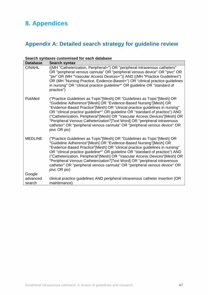
Peripheral intravenous catheters: A review of guidelines and research 47
8. Appendices
Appendix A: Detailed search strategy for guideline review Search syntaxes customised for each database Database Search syntax CINAHL ((MH "Catheterization, Peripheral+") OR "peripheral intravenous catheters"
OR "peripheral venous cannula" OR "peripheral venous device" OR "pivc" OR "piv" OR (MH "Vascular Access Devices+")) AND ((MH "Practice Guidelines") OR (MH "Nursing Practice, Evidence-Based+") OR “clinical practice guidelines in nursing" OR "clinical practice guideline*" OR guideline OR "standard of practice")
PubMed ("Practice Guidelines as Topic"[Mesh] OR "Guidelines as Topic"[Mesh] OR "Guideline Adherence"[Mesh] OR "Evidence-Based Nursing"[Mesh] OR "Evidence-Based Practice"[Mesh] OR “clinical practice guidelines in nursing" OR "clinical practice guideline*" OR guideline OR "standard of practice") AND ("Catheterization, Peripheral"[Mesh] OR "Vascular Access Devices"[Mesh] OR "Peripheral Venous Catheterization"[Text Word] OR "peripheral intravenous catheter" OR "peripheral venous cannula" OR "peripheral venous device" OR pivc OR piv)
MEDLINE ("Practice Guidelines as Topic"[Mesh] OR "Guidelines as Topic"[Mesh] OR "Guideline Adherence"[Mesh] OR "Evidence-Based Nursing"[Mesh] OR "Evidence-Based Practice"[Mesh] OR “clinical practice guidelines in nursing" OR "clinical practice guideline*" OR guideline OR "standard of practice") AND ("Catheterization, Peripheral"[Mesh] OR "Vascular Access Devices"[Mesh] OR "Peripheral Venous Catheterization"[Text Word] OR "peripheral intravenous catheter" OR "peripheral venous cannula" OR "peripheral venous device" OR pivc OR piv)
Google advanced search
clinical practice guidelines AND peripheral intravenous catheter insertion (OR maintenance)
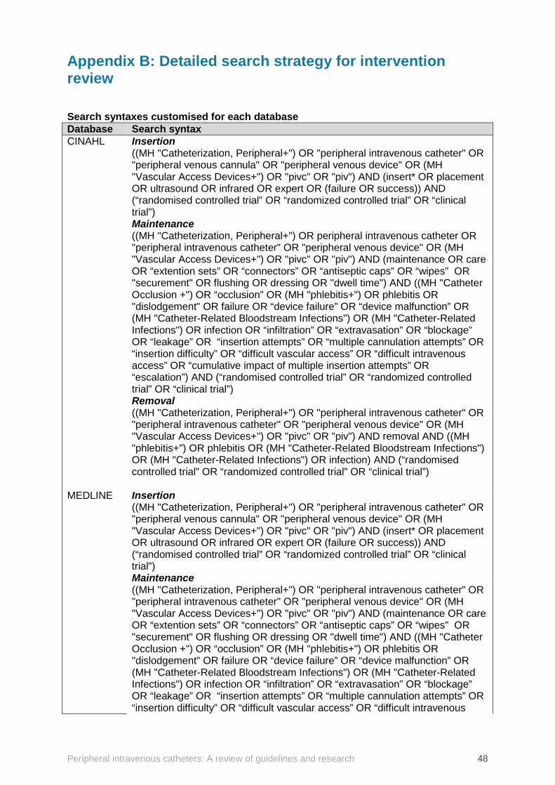
Peripheral intravenous catheters: A review of guidelines and research 48
Appendix B: Detailed search strategy for intervention review Search syntaxes customised for each database Database Search syntax CINAHL Insertion
((MH "Catheterization, Peripheral+") OR "peripheral intravenous catheter" OR "peripheral venous cannula" OR "peripheral venous device" OR (MH "Vascular Access Devices+") OR "pivc" OR "piv") AND (insert* OR placement OR ultrasound OR infrared OR expert OR (failure OR success)) AND (“randomised controlled trial” OR “randomized controlled trial” OR “clinical trial”) Maintenance ((MH "Catheterization, Peripheral+") OR peripheral intravenous catheter OR "peripheral intravenous catheter" OR "peripheral venous device" OR (MH "Vascular Access Devices+") OR "pivc" OR "piv") AND (maintenance OR care OR “extention sets” OR “connectors” OR “antiseptic caps” OR “wipes” OR "securement" OR flushing OR dressing OR "dwell time") AND ((MH "Catheter Occlusion +") OR “occlusion” OR (MH "phlebitis+") OR phlebitis OR "dislodgement" OR failure OR “device failure” OR “device malfunction” OR (MH "Catheter-Related Bloodstream Infections") OR (MH "Catheter-Related Infections") OR infection OR “infiltration” OR “extravasation” OR “blockage” OR “leakage” OR “insertion attempts” OR “multiple cannulation attempts” OR “insertion difficulty” OR “difficult vascular access” OR “difficult intravenous access” OR “cumulative impact of multiple insertion attempts” OR “escalation”) AND (“randomised controlled trial” OR “randomized controlled trial” OR “clinical trial”) Removal ((MH "Catheterization, Peripheral+") OR "peripheral intravenous catheter" OR "peripheral intravenous catheter" OR "peripheral venous device" OR (MH "Vascular Access Devices+") OR "pivc" OR "piv") AND removal AND ((MH "phlebitis+") OR phlebitis OR (MH "Catheter-Related Bloodstream Infections") OR (MH "Catheter-Related Infections") OR infection) AND (“randomised controlled trial” OR “randomized controlled trial” OR “clinical trial”)
MEDLINE Insertion ((MH "Catheterization, Peripheral+") OR "peripheral intravenous catheter" OR "peripheral venous cannula" OR "peripheral venous device" OR (MH "Vascular Access Devices+") OR "pivc" OR "piv") AND (insert* OR placement OR ultrasound OR infrared OR expert OR (failure OR success)) AND (“randomised controlled trial” OR “randomized controlled trial” OR “clinical trial”) Maintenance ((MH "Catheterization, Peripheral+") OR "peripheral intravenous catheter" OR "peripheral intravenous catheter" OR "peripheral venous device" OR (MH "Vascular Access Devices+") OR "pivc" OR "piv") AND (maintenance OR care OR “extention sets” OR “connectors” OR “antiseptic caps” OR “wipes” OR "securement" OR flushing OR dressing OR "dwell time") AND ((MH "Catheter Occlusion +") OR “occlusion” OR (MH "phlebitis+") OR phlebitis OR "dislodgement" OR failure OR “device failure” OR “device malfunction” OR (MH "Catheter-Related Bloodstream Infections") OR (MH "Catheter-Related Infections") OR infection OR “infiltration” OR “extravasation” OR “blockage” OR “leakage” OR “insertion attempts” OR “multiple cannulation attempts” OR “insertion difficulty” OR “difficult vascular access” OR “difficult intravenous
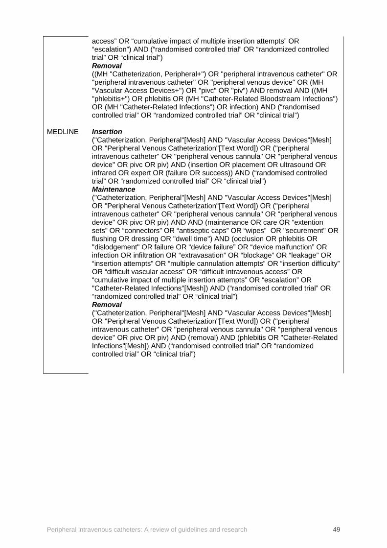
Peripheral intravenous catheters: A review of guidelines and research 49
access” OR “cumulative impact of multiple insertion attempts” OR “escalation”) AND (“randomised controlled trial” OR “randomized controlled trial” OR “clinical trial”) Removal ((MH "Catheterization, Peripheral+") OR "peripheral intravenous catheter" OR "peripheral intravenous catheter" OR "peripheral venous device" OR (MH "Vascular Access Devices+") OR "pivc" OR "piv") AND removal AND ((MH "phlebitis+") OR phlebitis OR (MH "Catheter-Related Bloodstream Infections") OR (MH "Catheter-Related Infections") OR infection) AND (“randomised controlled trial” OR “randomized controlled trial” OR “clinical trial”)
MEDLINE Insertion ("Catheterization, Peripheral"[Mesh] AND "Vascular Access Devices"[Mesh] OR "Peripheral Venous Catheterization"[Text Word]) OR ("peripheral intravenous catheter" OR "peripheral venous cannula" OR "peripheral venous device" OR pivc OR piv) AND (insertion OR placement OR ultrasound OR infrared OR expert OR (failure OR success)) AND (“randomised controlled trial” OR “randomized controlled trial” OR “clinical trial”) Maintenance ("Catheterization, Peripheral"[Mesh] AND "Vascular Access Devices"[Mesh] OR "Peripheral Venous Catheterization"[Text Word]) OR ("peripheral intravenous catheter" OR "peripheral venous cannula" OR "peripheral venous device" OR pivc OR piv) AND AND (maintenance OR care OR “extention sets” OR “connectors” OR “antiseptic caps” OR “wipes” OR "securement" OR flushing OR dressing OR "dwell time") AND (occlusion OR phlebitis OR "dislodgement" OR failure OR “device failure” OR “device malfunction” OR infection OR infiltration OR “extravasation” OR “blockage” OR “leakage” OR “insertion attempts” OR “multiple cannulation attempts” OR “insertion difficulty” OR “difficult vascular access” OR “difficult intravenous access” OR “cumulative impact of multiple insertion attempts” OR “escalation” OR "Catheter-Related Infections"[Mesh]) AND (“randomised controlled trial” OR “randomized controlled trial” OR “clinical trial”) Removal ("Catheterization, Peripheral"[Mesh] AND "Vascular Access Devices"[Mesh] OR "Peripheral Venous Catheterization"[Text Word]) OR ("peripheral intravenous catheter" OR "peripheral venous cannula" OR "peripheral venous device" OR pivc OR piv) AND (removal) AND (phlebitis OR "Catheter-Related Infections"[Mesh]) AND (“randomised controlled trial” OR “randomized controlled trial” OR “clinical trial”)
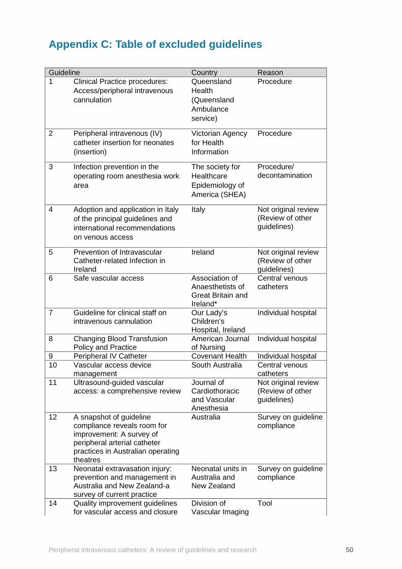
Peripheral intravenous catheters: A review of guidelines and research 50
Appendix C: Table of excluded guidelines Guideline Country Reason 1 Clinical Practice procedures:
Access/peripheral intravenous cannulation
Queensland Health (Queensland Ambulance service)
Procedure
2 Peripheral intravenous (IV) catheter insertion for neonates (insertion)
Victorian Agency for Health Information
Procedure
3 Infection prevention in the operating room anesthesia work area
The society for Healthcare Epidemiology of America (SHEA)
Procedure/ decontamination
4 Adoption and application in Italy of the principal guidelines and international recommendations on venous access
Italy Not original review (Review of other guidelines)
5 Prevention of Intravascular Catheter-related Infection in Ireland
Ireland Not original review (Review of other guidelines)
6 Safe vascular access Association of Anaesthetists of Great Britain and Ireland*
Central venous catheters
7 Guideline for clinical staff on intravenous cannulation
Our Lady’s Children’s Hospital, Ireland
Individual hospital
8 Changing Blood Transfusion Policy and Practice
American Journal of Nursing
Individual hospital
9 Peripheral IV Catheter Covenant Health Individual hospital 10 Vascular access device
management South Australia Central venous
catheters 11 Ultrasound-guided vascular
access: a comprehensive review Journal of Cardiothoracic and Vascular Anesthesia
Not original review (Review of other guidelines)
12 A snapshot of guideline compliance reveals room for improvement: A survey of peripheral arterial catheter practices in Australian operating theatres
Australia Survey on guideline compliance
13 Neonatal extravasation injury: prevention and management in Australia and New Zealand-a survey of current practice
Neonatal units in Australia and New Zealand
Survey on guideline compliance
14 Quality improvement guidelines for vascular access and closure
Division of Vascular Imaging
Tool
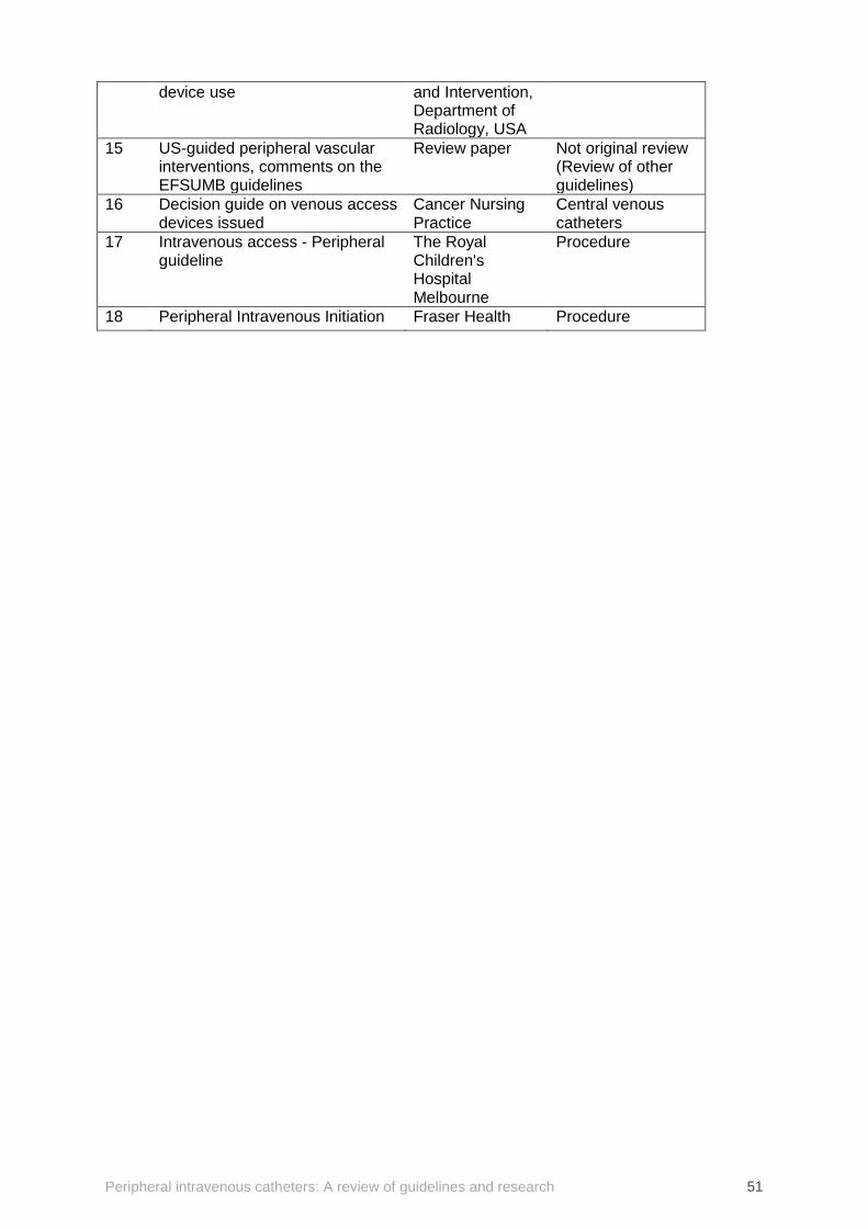
Peripheral intravenous catheters: A review of guidelines and research 51
device use and Intervention, Department of Radiology, USA
15 US-guided peripheral vascular interventions, comments on the EFSUMB guidelines
Review paper Not original review (Review of other guidelines)
16 Decision guide on venous access devices issued
Cancer Nursing Practice
Central venous catheters
17 Intravenous access - Peripheral guideline
The Royal Children's Hospital Melbourne
Procedure
18 Peripheral Intravenous Initiation Fraser Health Procedure
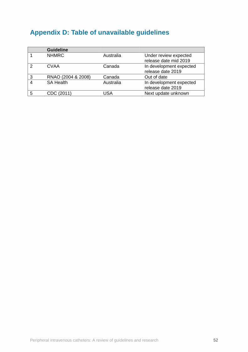
Peripheral intravenous catheters: A review of guidelines and research 52
Appendix D: Table of unavailable guidelines Guideline 1 NHMRC Australia Under review expected
release date mid 2019 2 CVAA Canada In development expected
release date 2019 3 RNAO (2004 & 2008) Canada Out of date 4 SA Health Australia In development expected
release date 2019 5 CDC (2011) USA Next update unknown
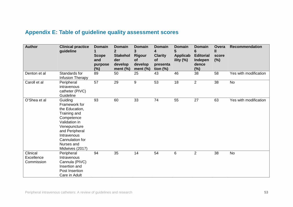
Peripheral intravenous catheters: A review of guidelines and research 53
Appendix E: Table of guideline quality assessment scores Author Clinical practice
guideline Domain 1 Scope and purpose (%)
Domain 2 Stakeholder development (%)
Domain 3 Rigour of development (%)
Domain 4 Clarity of presentation (%)
Domain 5 Applicability (%)
Domain 6 Editorial Independence (%)
Overall score (%)
Recommendation
Denton et al Standards for Infusion Therapy
89 50 25 43 46 38 58 Yes with modification
Caroll et al Peripheral intravenous catheter (PIVC) Guideline
57 29 9 53 18 2 38 No
O’Shea et al Guiding Framework for the Education, Training and Competence Validation in Venepuncture and Peripheral Intravenous Cannulation for Nurses and Midwives (2017)
93 60 33 74 55 27 63 Yes with modification
Clinical Excellence Commission
Peripheral Intravenous Cannula (PIVC) Insertion and Post Insertion Care in Adult
94 35 14 54 6 2 38 No
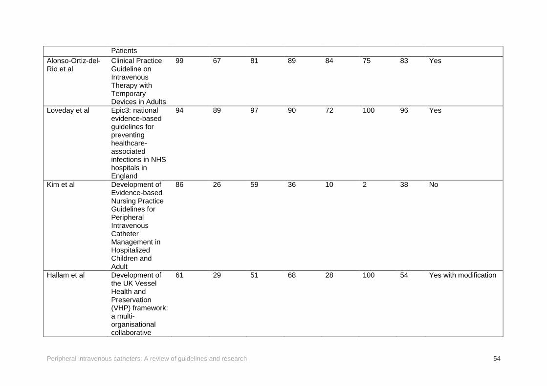
Peripheral intravenous catheters: A review of guidelines and research 54
Patients Alonso-Ortiz-del-Rio et al
Clinical Practice Guideline on Intravenous Therapy with Temporary Devices in Adults
99 67 81 89 84 75 83 Yes
Loveday et al Epic3: national evidence-based guidelines for preventing healthcare-associated infections in NHS hospitals in England
94 89 97 90 72 100 96 Yes
Kim et al Development of Evidence-based Nursing Practice Guidelines for Peripheral Intravenous Catheter Management in Hospitalized Children and Adult
86 26 59 36 10 2 38 No
Hallam et al Development of the UK Vessel Health and Preservation (VHP) framework: a multi-organisational collaborative
61 29 51 68 28 100 54 Yes with modification
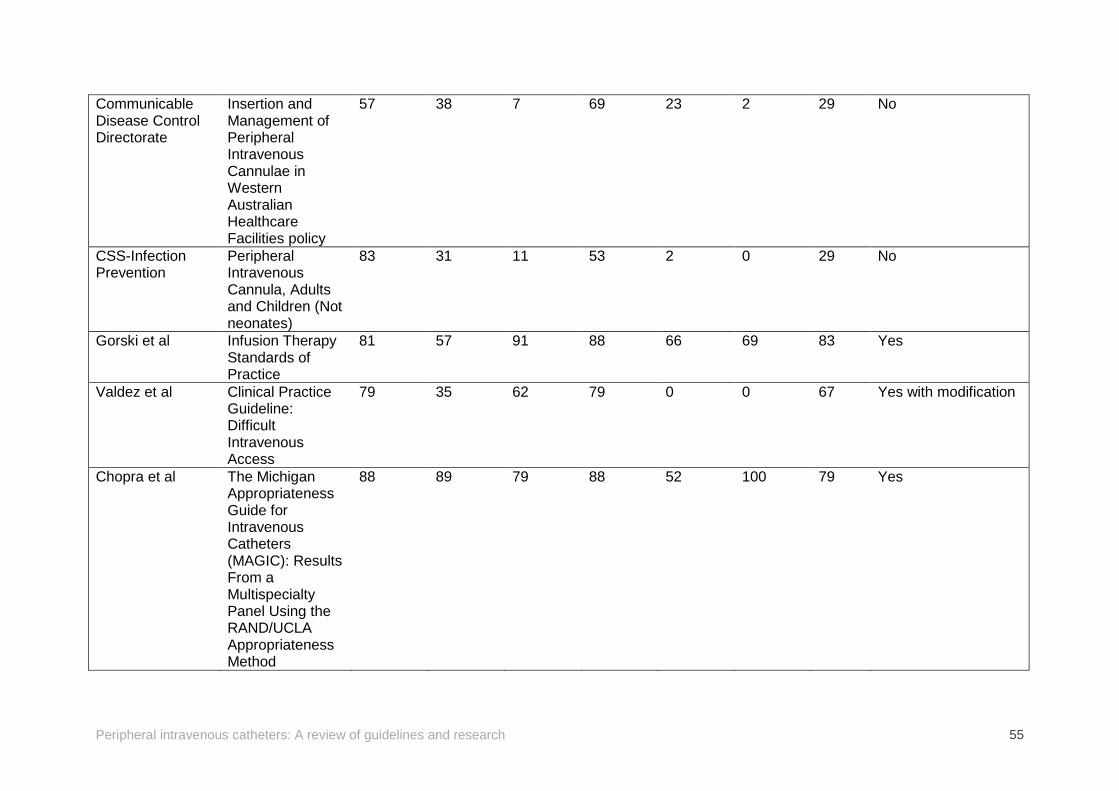
Peripheral intravenous catheters: A review of guidelines and research 55
Communicable Disease Control Directorate
Insertion and Management of Peripheral Intravenous Cannulae in Western Australian Healthcare Facilities policy
57 38 7 69 23 2 29 No
CSS-Infection Prevention
Peripheral Intravenous Cannula, Adults and Children (Not neonates)
83 31 11 53 2 0 29 No
Gorski et al Infusion Therapy Standards of Practice
81 57 91 88 66 69 83 Yes
Valdez et al Clinical Practice Guideline: Difficult Intravenous Access
79 35 62 79 0 0 67 Yes with modification
Chopra et al The Michigan Appropriateness Guide for Intravenous Catheters (MAGIC): Results From a Multispecialty Panel Using the RAND/UCLA Appropriateness Method
88 89 79 88 52 100 79 Yes
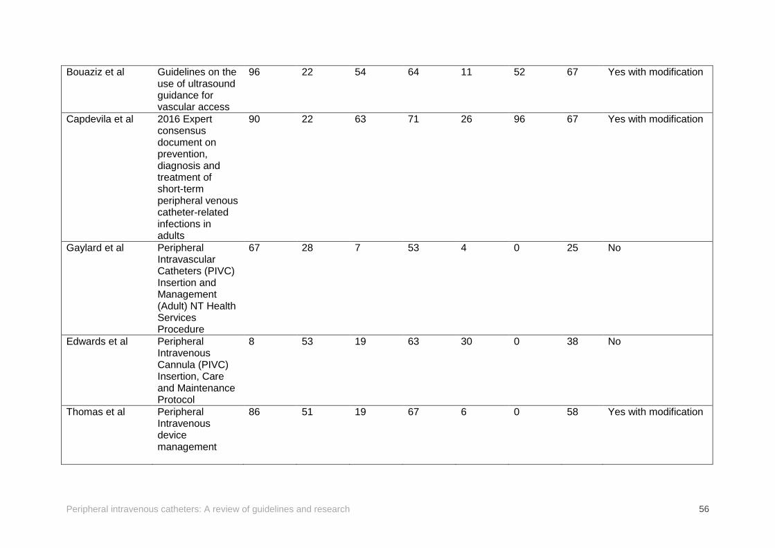
Peripheral intravenous catheters: A review of guidelines and research 56
Bouaziz et al Guidelines on the use of ultrasound guidance for vascular access
96 22 54 64 11 52 67 Yes with modification
Capdevila et al 2016 Expert consensus document on prevention, diagnosis and treatment of short-term peripheral venous catheter-related infections in adults
90 22 63 71 26 96 67 Yes with modification
Gaylard et al
Peripheral Intravascular Catheters (PIVC) Insertion and Management (Adult) NT Health Services Procedure
67 28 7 53 4 0 25 No
Edwards et al Peripheral Intravenous Cannula (PIVC) Insertion, Care and Maintenance Protocol
8 53 19 63 30 0 38 No
Thomas et al Peripheral Intravenous device management
86 51 19 67 6 0 58 Yes with modification
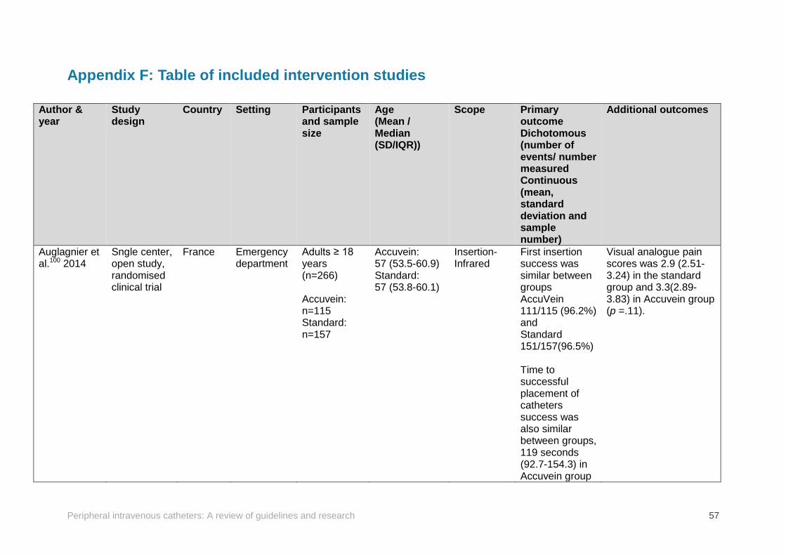
Peripheral intravenous catheters: A review of guidelines and research 57
Appendix F: Table of included intervention studies
Author & year
Study design
Country Setting Participants and sample size
Age (Mean / Median (SD/IQR))
Scope Primary outcome Dichotomous (number of events/ number measured Continuous (mean, standard deviation and sample number)
Additional outcomes
Auglagnier et al.100 2014
Sngle center, open study, randomised clinical trial
France Emergency department
Adults ≥ 18 years (n=266) Accuvein: n=115 Standard: n=157
Accuvein: 57 (53.5-60.9) Standard: 57 (53.8-60.1)
Insertion- Infrared
First insertion success was similar between groups AccuVein 111/115 (96.2%) and Standard 151/157(96.5%) Time to successful placement of catheters success was also similar between groups, 119 seconds (92.7-154.3) in Accuvein group
Visual analogue pain scores was 2.9 (2.51-3.24) in the standard group and 3.3(2.89-3.83) in Accuvein group (p =.11).
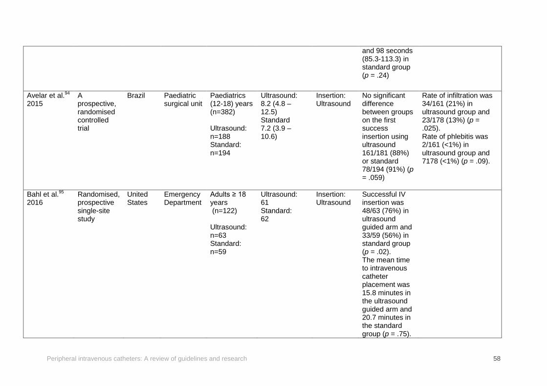
Peripheral intravenous catheters: A review of guidelines and research 58
and 98 seconds (85.3-113.3) in standard group (p = .24)
Avelar et al.94 2015
A prospective, randomised controlled trial
Brazil Paediatric surgical unit
Paediatrics (12-18) years (n=382) Ultrasound: n=188 Standard: n=194
Ultrasound: 8.2 (4.8 – 12.5) Standard 7.2 (3.9 – 10.6)
Insertion: Ultrasound
No significant difference between groups on the first success insertion using ultrasound 161/181 (88%) or standard 78/194 (91%) (p = .059)
Rate of infiltration was 34/161 (21%) in ultrasound group and 23/178 (13%) (p = .025). Rate of phlebitis was 2/161 (<1%) in ultrasound group and 7178 (<1%) (p = .09).
Bahl et al.95
2016 Randomised, prospective single-site study
United States
Emergency Department
Adults ≥ 18 years (n=122) Ultrasound: n=63 Standard: n=59
Ultrasound: 61 Standard: 62
Insertion: Ultrasound
Successful IV insertion was 48/63 (76%) in ultrasound guided arm and 33/59 (56%) in standard group (p = .02). The mean time to intravenous catheter placement was 15.8 minutes in the ultrasound guided arm and 20.7 minutes in the standard group (p = .75).
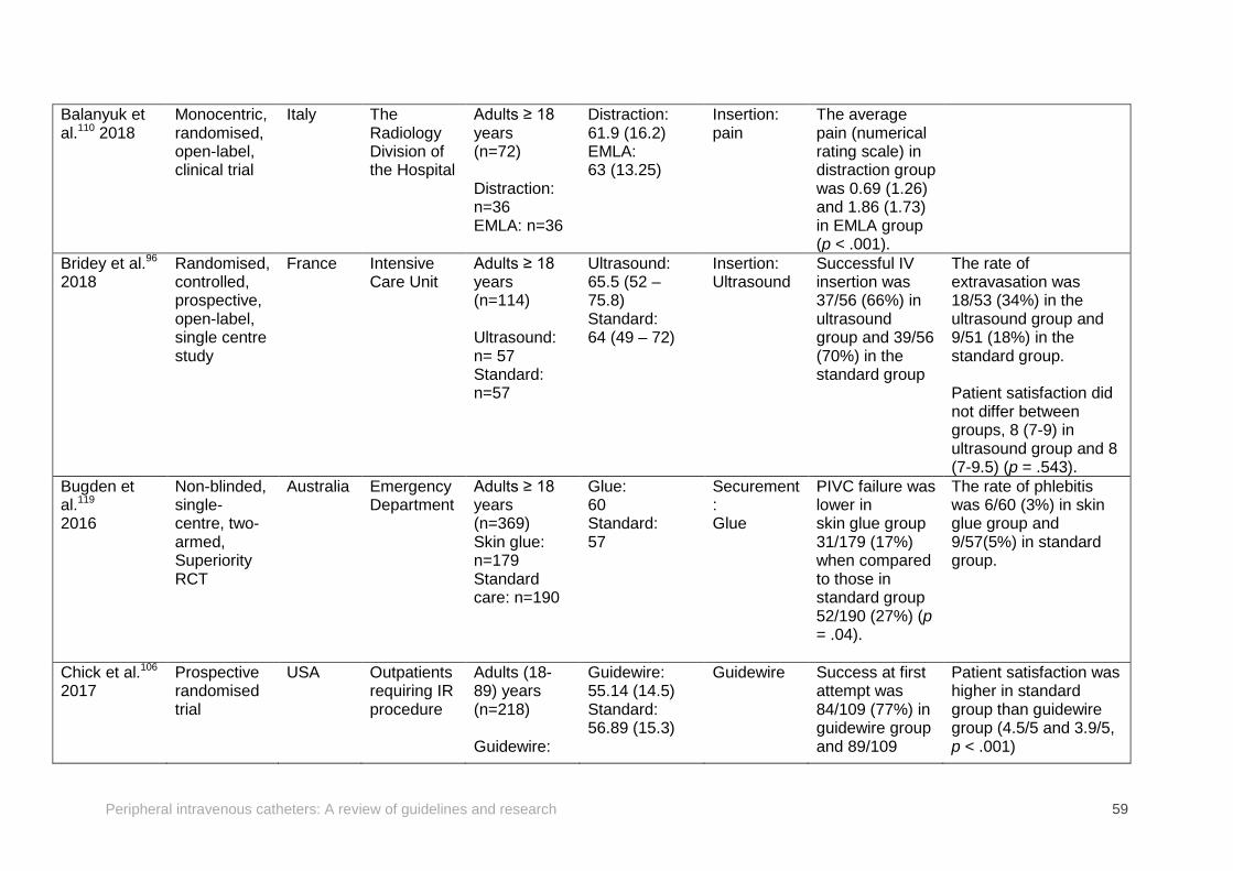
Peripheral intravenous catheters: A review of guidelines and research 59
Balanyuk et al.110 2018
Monocentric, randomised, open-label, clinical trial
Italy The Radiology Division of the Hospital
Adults ≥ 18 years (n=72) Distraction: n=36 EMLA: n=36
Distraction: 61.9 (16.2) EMLA: 63 (13.25)
Insertion: pain
The average pain (numerical rating scale) in distraction group was 0.69 (1.26) and 1.86 (1.73) in EMLA group (p < .001).
Bridey et al.96 2018
Randomised, controlled, prospective, open-label, single centre study
France Intensive Care Unit
Adults ≥ 18 years (n=114) Ultrasound: n= 57 Standard: n=57
Ultrasound: 65.5 (52 – 75.8) Standard: 64 (49 – 72)
Insertion: Ultrasound
Successful IV insertion was 37/56 (66%) in ultrasound group and 39/56 (70%) in the standard group
The rate of extravasation was 18/53 (34%) in the ultrasound group and 9/51 (18%) in the standard group. Patient satisfaction did not differ between groups, 8 (7-9) in ultrasound group and 8 (7-9.5) (p = .543).
Bugden et al.119
2016
Non-blinded, single-centre, two-armed, Superiority RCT
Australia Emergency Department
Adults ≥ 18 years (n=369) Skin glue: n=179 Standard care: n=190
Glue: 60 Standard: 57
Securement: Glue
PIVC failure was lower in skin glue group 31/179 (17%) when compared to those in standard group 52/190 (27%) (p = .04).
The rate of phlebitis was 6/60 (3%) in skin glue group and 9/57(5%) in standard group.
Chick et al.106 2017
Prospective randomised trial
USA Outpatients requiring IR procedure
Adults (18-89) years (n=218) Guidewire:
Guidewire: 55.14 (14.5) Standard: 56.89 (15.3)
Guidewire Success at first attempt was 84/109 (77%) in guidewire group and 89/109
Patient satisfaction was higher in standard group than guidewire group (4.5/5 and 3.9/5, p < .001)
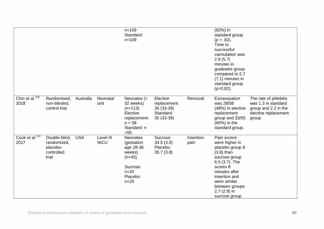
Peripheral intravenous catheters: A review of guidelines and research 60
n=109 Standard: n=109
(82%) in standard group (p = .82). Time to successful cannulation was 2.9 (5.7) minutes in guidewire group compared to 2.7 (7.1) minutes in standard group (p=0.82).
Chin et al.128 2018
Randomised, non-blinded, control trial
Australia Neonatal unit
Neonates (> 32 weeks) (n=113) Elective replacement: n = 58 Standard: n =55
Elective replacement: 36 (33-39) Standard: 35 (33-39)
Removal Extravasation was 28/58 (48%) in elective replacement group and 33/55 (60%) in the standard group.
The rate of phlebitis was 1.3 in standard group and 2.2 in the elective replacement group.
Cook et al.111 2017
Double-blind, randomized, placebo-controlled trial
USA Level III NICU
Neonates (gestation age 28-36 weeks) (n=40) Sucrose: n=20 Placebo: n=20
Sucrose: 34.5 (3.9) Placebo: 35.7 (3.8)
Insertion: pain
Pain scores were higher in placebo group 8 (3.8) than sucrose group 6.5 (3.7). The scores 8 minutes after insertion and were similar between groups 2.7 (2.8) in sucrose group
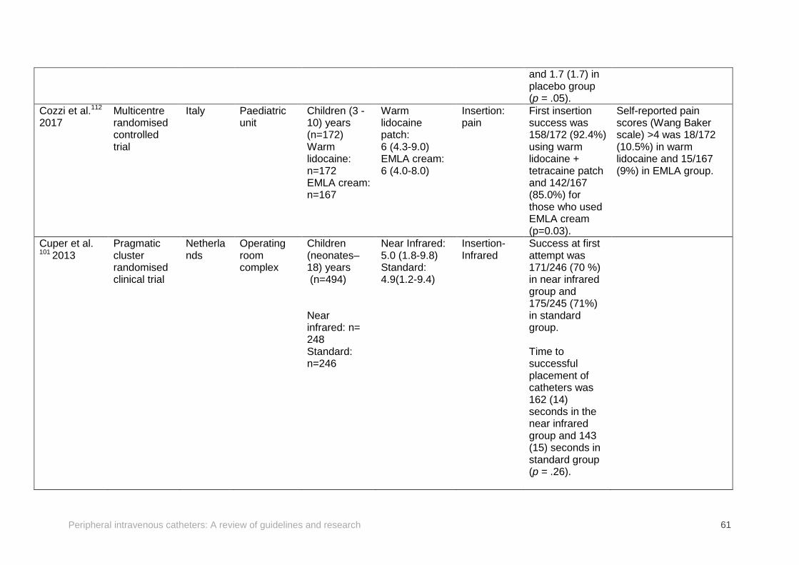
Peripheral intravenous catheters: A review of guidelines and research 61
and 1.7 (1.7) in placebo group (p = .05).
Cozzi et al.112
2017 Multicentre randomised controlled trial
Italy Paediatric unit
Children (3 -10) years (n=172) Warm lidocaine: n=172 EMLA cream: n=167
Warm lidocaine patch: 6 (4.3-9.0) EMLA cream: 6 (4.0-8.0)
Insertion: pain
First insertion success was 158/172 (92.4%) using warm lidocaine + tetracaine patch and 142/167 (85.0%) for those who used EMLA cream (p=0.03).
Self-reported pain scores (Wang Baker scale) >4 was 18/172 (10.5%) in warm lidocaine and 15/167 (9%) in EMLA group.
Cuper et al. 101 2013
Pragmatic cluster randomised clinical trial
Netherlands
Operating room complex
Children (neonates–18) years (n=494) Near infrared: n= 248 Standard: n=246
Near Infrared: 5.0 (1.8-9.8) Standard: 4.9(1.2-9.4)
Insertion- Infrared
Success at first attempt was 171/246 (70 %) in near infrared group and 175/245 (71%) in standard group. Time to successful placement of catheters was 162 (14) seconds in the near infrared group and 143 (15) seconds in standard group (p = .26).
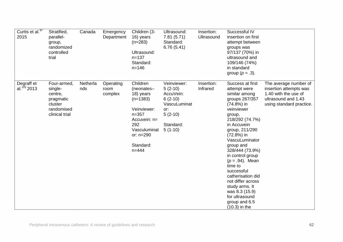
Peripheral intravenous catheters: A review of guidelines and research 62
Curtis et al.97
2015 Stratified, parallel-group, randomized controlled trial
Canada Emergency Department
Children (3-16) years (n=283) Ultrasound: n=137 Standard: n=146
Ultrasound: 7.81 (5.71) Standard: 6.76 (5.41)
Insertion: Ultrasound
Successful IV insertion on first attempt between groups was 97/137 (70%) in ultrasound and 109/146 (74%) in standard group (p = .3).
Degraff et al.102 2013
Four-armed, single-centre, pragmatic cluster randomised clinical trial
Netherlands
Operating room complex
Children (neonates–18) years (n=1383) Veinviewer: n=357 Accuvein: n= 292 Vasculuminator: n=290 Standard: n=444
Veinviewer: 5 (2-10) AccuVein: 6 (2-10) VascuLuminator: 5 (2-10) Standard: 5 (1-10)
Insertion: Infrared
Success at first attempt were similar among groups 267/357 (74.8%) in veinviewer group, 218/292 (74.7%) in Accuvein group, 211/290 (72.8%) in VascuLuminator group and 328/444 (73.9%) in control group (p = .94). Mean time to successful catherisation did not differ across study arms. It was 8.3 (15.9) for ultrasound group and 6.5 (10.3) in the
The average number of insertion attempts was 1.40 with the use of ultrasound and 1.43 using standard practice.
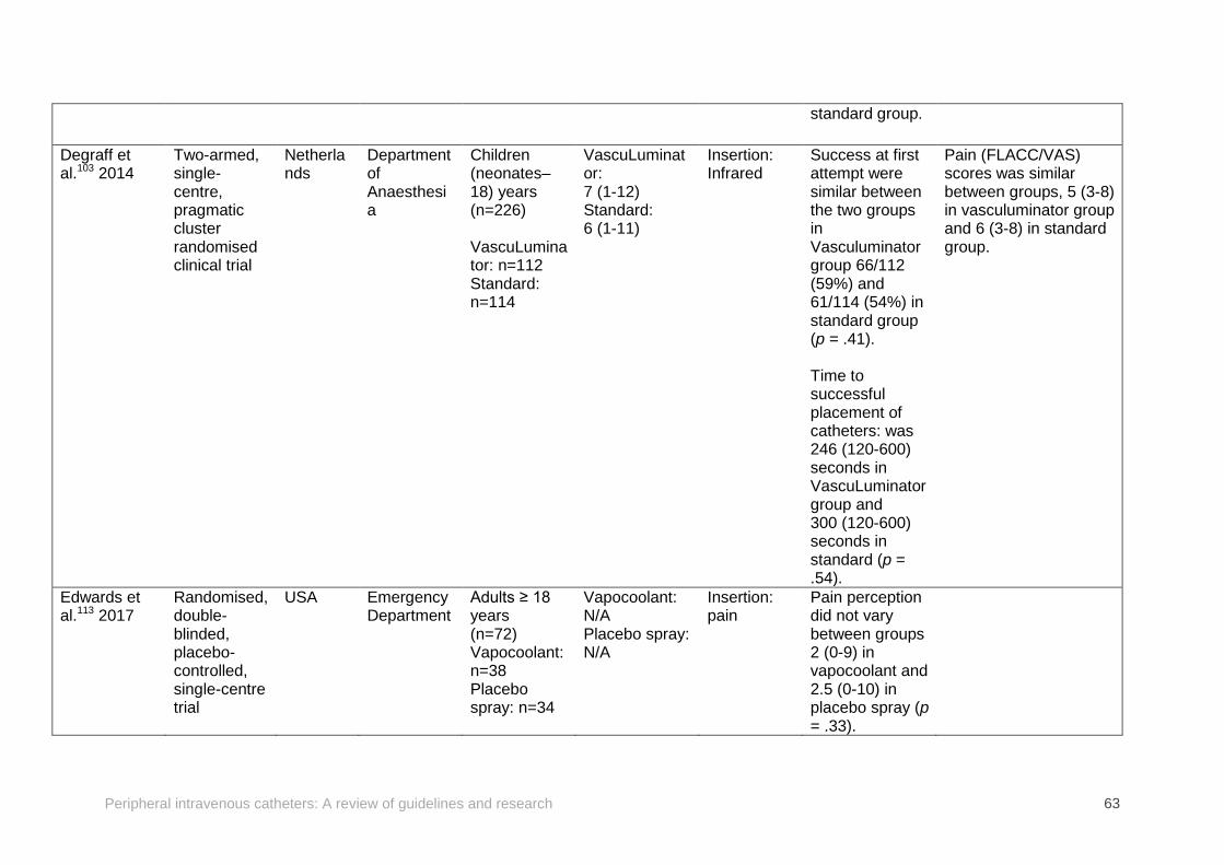
Peripheral intravenous catheters: A review of guidelines and research 63
standard group.
Degraff et al.103 2014
Two-armed, single-centre, pragmatic cluster randomised clinical trial
Netherlands
Department of Anaesthesia
Children (neonates–18) years (n=226) VascuLuminator: n=112 Standard: n=114
VascuLuminator: 7 (1-12) Standard: 6 (1-11)
Insertion: Infrared
Success at first attempt were similar between the two groups in Vasculuminator group 66/112 (59%) and 61/114 (54%) in standard group (p = .41). Time to successful placement of catheters: was 246 (120-600) seconds in VascuLuminator group and 300 (120-600) seconds in standard (p = .54).
Pain (FLACC/VAS) scores was similar between groups, 5 (3-8) in vasculuminator group and 6 (3-8) in standard group.
Edwards et al.113 2017
Randomised, double-blinded, placebo-controlled, single-centre trial
USA Emergency Department
Adults ≥ 18 years (n=72) Vapocoolant: n=38 Placebo spray: n=34
Vapocoolant: N/A Placebo spray: N/A
Insertion: pain
Pain perception did not vary between groups 2 (0-9) in vapocoolant and 2.5 (0-10) in placebo spray (p = .33).
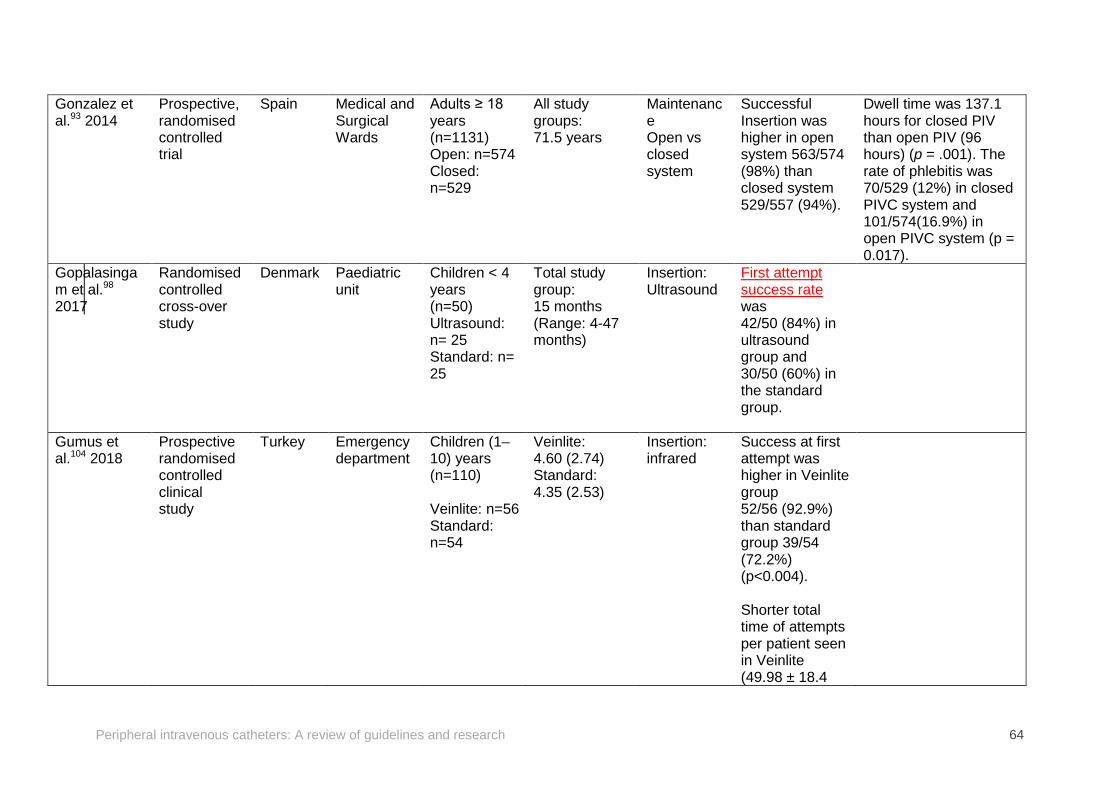
Peripheral intravenous catheters: A review of guidelines and research 64
Gonzalez et al.93 2014
Prospective, randomised controlled trial
Spain Medical and Surgical Wards
Adults ≥ 18 years (n=1131) Open: n=574 Closed: n=529
All study groups: 71.5 years
Maintenance Open vs closed system
Successful Insertion was higher in open system 563/574 (98%) than closed system 529/557 (94%).
Dwell time was 137.1 hours for closed PIV than open PIV (96 hours) (p = .001). The rate of phlebitis was 70/529 (12%) in closed PIVC system and 101/574(16.9%) in open PIVC system (p = 0.017).
Gopalasingam et al.98 2017
Randomised controlled cross-over study
Denmark Paediatric unit
Children < 4 years (n=50) Ultrasound: n= 25 Standard: n= 25
Total study group: 15 months (Range: 4-47 months)
Insertion: Ultrasound
First attempt success rate was 42/50 (84%) in ultrasound group and 30/50 (60%) in the standard group.
Gumus et al.104 2018
Prospective randomised controlled clinical study
Turkey Emergency department
Children (1–10) years (n=110) Veinlite: n=56 Standard: n=54
Veinlite: 4.60 (2.74) Standard: 4.35 (2.53)
Insertion: infrared
Success at first attempt was higher in Veinlite group 52/56 (92.9%) than standard group 39/54 (72.2%) (p<0.004). Shorter total time of attempts per patient seen in Veinlite (49.98 ± 18.4
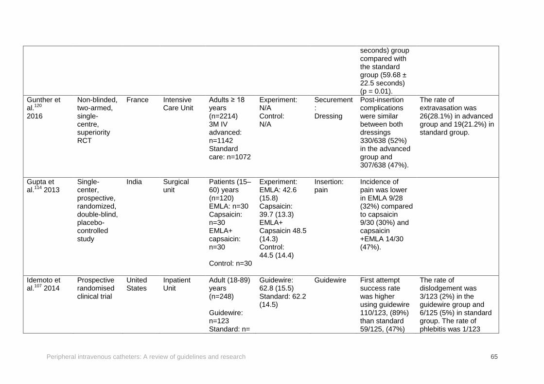
Peripheral intravenous catheters: A review of guidelines and research 65
seconds) group compared with the standard group (59.68 ± 22.5 seconds) (p = 0.01).
Gunther et al.120
2016
Non-blinded, two-armed, single-centre, superiority RCT
France Intensive Care Unit
Adults ≥ 18 years (n=2214) 3M IV advanced: n=1142 Standard care: n=1072
Experiment: N/A Control: N/A
Securement: Dressing
Post-insertion complications were similar between both dressings 330/638 (52%) in the advanced group and 307/638 (47%).
The rate of extravasation was 26(28.1%) in advanced group and 19(21.2%) in standard group.
Gupta et al.114 2013
Single-center, prospective, randomized, double-blind, placebo-controlled study
India
Surgical unit
Patients (15–60) years (n=120) EMLA: n=30 Capsaicin: n=30 EMLA+ capsaicin: n=30 Control: n=30
Experiment: EMLA: 42.6 (15.8) Capsaicin: 39.7 (13.3) EMLA+ Capsaicin 48.5 (14.3) Control: 44.5 (14.4)
Insertion: pain
Incidence of pain was lower in EMLA 9/28 (32%) compared to capsaicin 9/30 (30%) and capsaicin +EMLA 14/30 (47%).
Idemoto et al.107 2014
Prospective randomised clinical trial
United States
Inpatient Unit
Adult (18-89) years (n=248) Guidewire: n=123 Standard: n=
Guidewire: 62.8 (15.5) Standard: 62.2 (14.5)
Guidewire First attempt success rate was higher using guidewire 110/123, (89%) than standard 59/125, (47%)
The rate of dislodgement was 3/123 (2%) in the guidewire group and 6/125 (5%) in standard group. The rate of phlebitis was 1/123
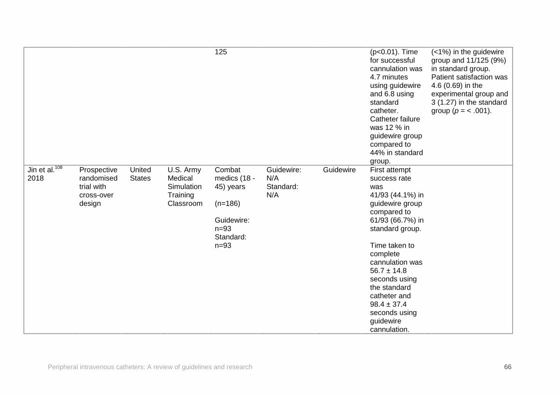
Peripheral intravenous catheters: A review of guidelines and research 66
125 (p<0.01). Time for successful cannulation was 4.7 minutes using guidewire and 6.8 using standard catheter. Catheter failure was 12 % in guidewire group compared to 44% in standard group.
(<1%) in the guidewire group and 11/125 (9%) in standard group. Patient satisfaction was 4.6 (0.69) in the experimental group and 3 (1.27) in the standard group (p = < .001).
Jin et al.108
2018 Prospective randomised trial with cross-over design
United States
U.S. Army Medical Simulation Training Classroom
Combat medics (18 -45) years (n=186) Guidewire: n=93 Standard: n=93
Guidewire: N/A Standard: N/A
Guidewire First attempt success rate was 41/93 (44.1%) in guidewire group compared to 61/93 (66.7%) in standard group. Time taken to complete cannulation was 56.7 ± 14.8 seconds using the standard catheter and 98.4 ± 37.4 seconds using guidewire cannulation.
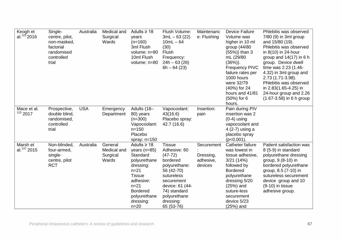
Peripheral intravenous catheters: A review of guidelines and research 67
Keogh et al.123 2016
Single-centre, pilot, non-masked, factorial randomised controlled trial
Australia Medical and Surgical Wards
Adults ≥ 18 years (n=160) 3ml Flush volume: n=80 10ml Flush volume: n=80
Flush Volume: 3mL – 63 (22) 10mL – 64 (30) Flush Frequency: 24h – 63 (26) 6h – 64 (23)
Maintenance: Flushing
Device Failure Volume was higher in 10 ml group (44/80 (55%)) than 3 mL (29/80 (36%)). Frequency PIVC failure rates per 1000 hours were 32/79 (40%) for 24 hours and 41/81 (50%) for 6 hours.
Phlebitis was observed 7/80 (9) in 3ml group and 15/80 (19). Phlebitis was observed in 8(10) in 24-hour group and 14(17) in 6 h group. Device dwell time was 2.23 (1.46-4.32) in 3ml group and 2.73 (1.71-3.98). Phlebitis was observed in 2.83(1.65-4.25) in 24-hour group and 2.26 (1.67-3.58) in 6 h group
Mace et al. 115 2017
Prospective, double blind, randomised, controlled trial
USA Emergency Department
Adults (18–80) years (n=300) Vapocoolant: n=150 Placebo spray: n=150
Vapocoolant: 43(16.6) Placebo spray: 42.7 (16.6)
Insertion: pain
Pain during PIV insertion was 2 (0-4) using vapocoolant and 4 (2-7) using a placebo spray (p<0.001).
Marsh et al.122 2015
Non-blinded, four-armed, single-centre, pilot RCT
Australia General Medical and Surgical Wards
Adults ≥ 18 years (n=85) Standard polyurethane dressing: n=21 Tissue adhesive: n=21 Bordered polyurethane dressing: n=20
Tissue Adhesive: 60 (47-72) bordered polyurethane: 56 (42-70) sutureless securement device: 61 (44-74) standard polyurethane dressing: 65 (53-76)
Securement: Dressing, adhesive, devices
Catheter failure was lowest in tissue adhesive, 3/21 (14%) followed by Bordered polyurethane dressing 5/20 (25%) and suture-less securement device 5/23 (25%) and
Patient satisfaction was 8 (5-9) in standard polyurethane dressing group, 9 (8-10) in bordered polyurethane group, 8.5 (7-10) in sutureless securement device group and 10 (9-10) in tissue adhesive group.
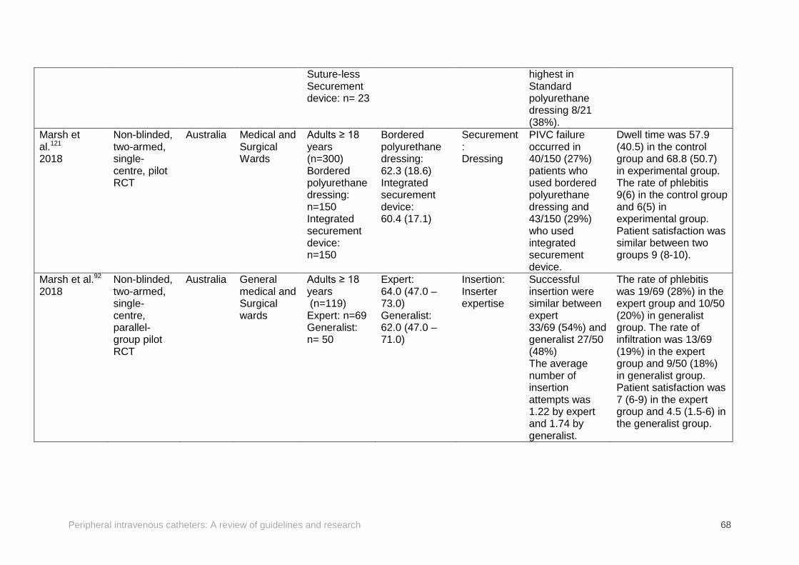
Peripheral intravenous catheters: A review of guidelines and research 68
Suture-less Securement device: n= 23
highest in Standard polyurethane dressing 8/21 (38%).
Marsh et al.121
2018
Non-blinded, two-armed, single-centre, pilot RCT
Australia Medical and Surgical Wards
Adults ≥ 18 years (n=300) Bordered polyurethane dressing: n=150 Integrated securement device: n=150
Bordered polyurethane dressing: 62.3 (18.6) Integrated securement device: 60.4 (17.1)
Securement: Dressing
PIVC failure occurred in 40/150 (27%) patients who used bordered polyurethane dressing and 43/150 (29%) who used integrated securement device.
Dwell time was 57.9 (40.5) in the control group and 68.8 (50.7) in experimental group. The rate of phlebitis 9(6) in the control group and 6(5) in experimental group. Patient satisfaction was similar between two groups 9 (8-10).
Marsh et al.92 2018
Non-blinded, two-armed, single-centre, parallel-group pilot RCT
Australia General medical and Surgical wards
Adults ≥ 18 years (n=119) Expert: n=69 Generalist: n= 50
Expert: 64.0 (47.0 – 73.0) Generalist: 62.0 (47.0 – 71.0)
Insertion: Inserter expertise
Successful insertion were similar between expert 33/69 (54%) and generalist 27/50 (48%) The average number of insertion attempts was 1.22 by expert and 1.74 by generalist.
The rate of phlebitis was 19/69 (28%) in the expert group and 10/50 (20%) in generalist group. The rate of infiltration was 13/69 (19%) in the expert group and 9/50 (18%) in generalist group. Patient satisfaction was 7 (6-9) in the expert group and 4.5 (1.5-6) in the generalist group.
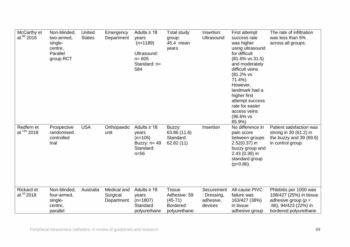
Peripheral intravenous catheters: A review of guidelines and research 69
McCarthy et al.99 2016
Non-blinded, two-armed, single-centre, Parallel group RCT
United States
Emergency Department
Adults ≥ 18 years (n=1189) Ultrasound: n= 605 Standard: n= 584
Total study group: 45.4 mean years
Insertion: Ultrasound
First attempt success rate was higher using ultrasound for difficult (81.6% vs 31.5) and moderately difficult veins (81.2% vs 71.4%). However, landmark had a higher first attempt success rate for easier access veins (96.6% vs 85.9%)
The rate of infiltration was less than 5% across all groups.
Redfern et al.116 2018
Prospective randomised controlled trial
USA Orthopaedic unit
Adults ≥ 18 years (n=105) Buzzy: n= 49 Standard: n=56
Buzzy: 63.86 (11.6) Standard: 62.82 (11)
Insertion No difference in pain score between groups 2.52(0.37) in buzzy group and 2.43 (0.36) in standard group (p=0.86).
Patient satisfaction was strong in 30 (61.2) in the buzzy and 39 (69.6) in control group.
Rickard et al.10 2018
Non-blinded, four-armed, single-centre, parallel
Australia Medical and Surgical Department
Adults ≥ 18 years (n=1807) Standard polyurethane
Tissue Adhesive: 59 (45-71) Bordered polyurethane:
Securement: Dressing, adhesive, devices
All-cause PIVC failure was 163/427 (38%) in tissue adhesive group
Phlebitis per 1000 was 108/427 (25%) in tissue adhesive group (p = .66), 94/423 (22%) in bordered polyurethane
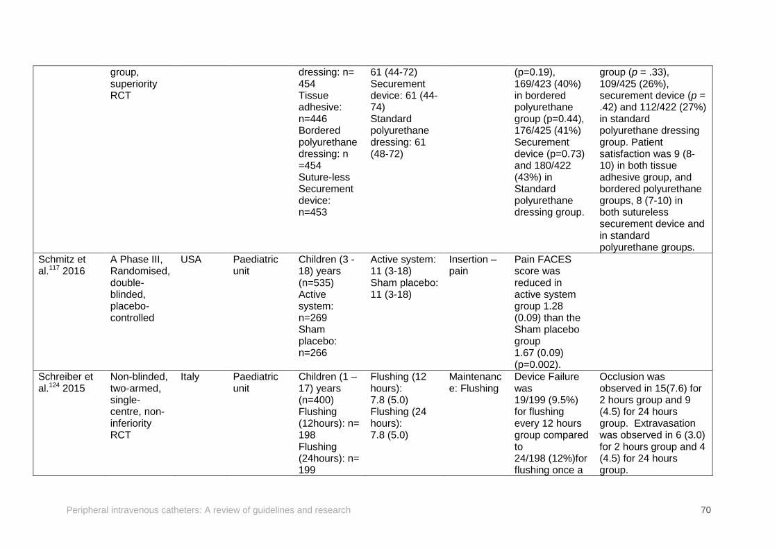
Peripheral intravenous catheters: A review of guidelines and research 70
group, superiority RCT
dressing: n= 454 Tissue adhesive: n=446 Bordered polyurethane dressing: n =454 Suture-less Securement device: n=453
61 (44-72) Securement device: 61 (44-74) Standard polyurethane dressing: 61 (48-72)
(p=0.19), 169/423 (40%) in bordered polyurethane group (p=0.44), 176/425 (41%) Securement device (p=0.73) and 180/422 (43%) in Standard polyurethane dressing group.
group (p = .33), 109/425 (26%), securement device (p = .42) and 112/422 (27%) in standard polyurethane dressing group. Patient satisfaction was 9 (8-10) in both tissue adhesive group, and bordered polyurethane groups, 8 (7-10) in both sutureless securement device and in standard polyurethane groups.
Schmitz et al.117 2016
A Phase III, Randomised, double-blinded, placebo-controlled
USA Paediatric unit
Children (3 -18) years (n=535) Active system: n=269 Sham placebo: n=266
Active system: 11 (3-18) Sham placebo: 11 (3-18)
Insertion –pain
Pain FACES score was reduced in active system group 1.28 (0.09) than the Sham placebo group 1.67 (0.09) (p=0.002).
Schreiber et al.124 2015
Non-blinded, two-armed, single-centre, non-inferiority RCT
Italy Paediatric unit
Children (1 – 17) years (n=400) Flushing (12hours): n= 198 Flushing (24hours): n= 199
Flushing (12 hours): 7.8 (5.0) Flushing (24 hours): 7.8 (5.0)
Maintenance: Flushing
Device Failure was 19/199 (9.5%) for flushing every 12 hours group compared to 24/198 (12%)for flushing once a
Occlusion was observed in 15(7.6) for 2 hours group and 9 (4.5) for 24 hours group. Extravasation was observed in 6 (3.0) for 2 hours group and 4 (4.5) for 24 hours group.
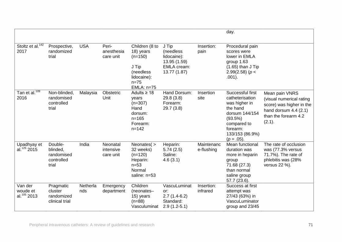
Peripheral intravenous catheters: A review of guidelines and research 71
day.
Stoltz et al.142
2017 Prospective, randomized trial
USA Peri-anesthesia care unit
Children (8 to 18) years (n=150) J Tip (needless lidocaine): n=75 EMLA: n=75
J Tip (needless lidocaine): 13.95 (1.59) EMLA cream: 13.77 (1.87)
Insertion: pain
Procedural pain scores were lower in EMLA group 1.63 (1.65) than J Tip 2.99(2.58) (p < .001).
Tan et al.109 2016
Non-blinded, randomised controlled trial
Malaysia Obstetric Unit
Adults ≥ 18 years (n=307) Hand dorsum: n=165 Forearm: n=142
Hand Dorsum: 29.8 (3.8) Forearm: 29.7 (3.8)
Insertion site
Successful first catheterisation was higher in the hand dorsum 144/154 (93.5%) compared to forearm: 133/153 (86.9%) (p = .05).
Mean pain VNRS (visual numerical rating score) was higher in the hand dorsum 4.4 (2.1) than the forearm 4.2 (2.1).
Upadhyay et al.125 2015
Double- blinded, randomised controlled trial
India Neonatal intensive care unit
Neonates( > 32 weeks) (n=120) Heparin: n=53 Normal saline: n=53
Heparin: 5.74 (2.5) Saline: 4.6 (3.1)
Maintenance-flushing
Mean functional duration was more in heparin group 71.68 (27.3) than normal saline group 57.7 (23.6).
The rate of occlusion was (77.3% versus 71.7%). The rate of phlebitis was (28% versus 22 %).
Van der woude et al.105 2013
Pragmatic cluster randomized clinical trial
Netherlands
Emergency department
Children (neonates–15) years (n=88) Vasculuminat
VascuLuminator: 2.7 (1.4-6.2) Standard: 2.9 (1.2-5.1)
Insertion: infrared
Success at first attempt was 27/43 (63%) in VascuLuminator group and 23/45
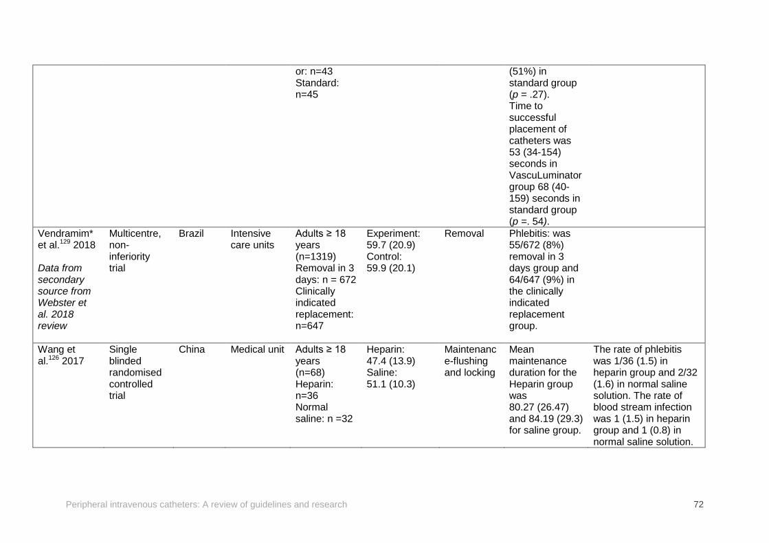
Peripheral intravenous catheters: A review of guidelines and research 72
or: n=43 Standard: n=45
(51%) in standard group (p = .27). Time to successful placement of catheters was 53 (34-154) seconds in VascuLuminator group 68 (40-159) seconds in standard group (p =. 54).
Vendramim* et al.129 2018 Data from secondary source from Webster et al. 2018 review
Multicentre, non-inferiority trial
Brazil Intensive care units
Adults ≥ 18 years (n=1319) Removal in 3 days: n = 672 Clinically indicated replacement: n=647
Experiment: 59.7 (20.9) Control: 59.9 (20.1)
Removal Phlebitis: was 55/672 (8%) removal in 3 days group and 64/647 (9%) in the clinically indicated replacement group.
Wang et al.126 2017
Single blinded randomised controlled trial
China Medical unit Adults ≥ 18 years (n=68) Heparin: n=36 Normal saline: n =32
Heparin: 47.4 (13.9) Saline: 51.1 (10.3)
Maintenance-flushing and locking
Mean maintenance duration for the Heparin group was 80.27 (26.47) and 84.19 (29.3) for saline group.
The rate of phlebitis was 1/36 (1.5) in heparin group and 2/32 (1.6) in normal saline solution. The rate of blood stream infection was 1 (1.5) in heparin group and 1 (0.8) in normal saline solution.
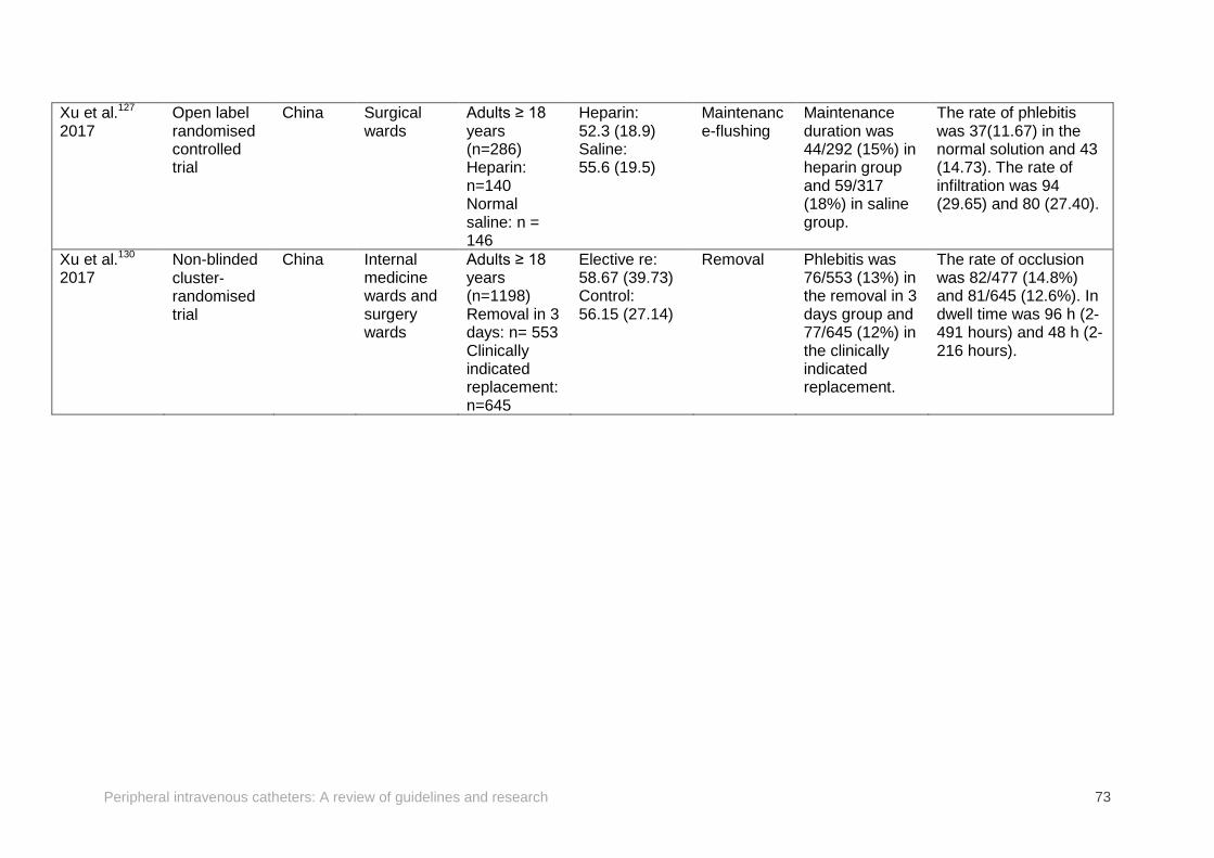
Peripheral intravenous catheters: A review of guidelines and research 73
Xu et al.127 2017
Open label randomised controlled trial
China Surgical wards
Adults ≥ 18 years (n=286) Heparin: n=140 Normal saline: n = 146
Heparin: 52.3 (18.9) Saline: 55.6 (19.5)
Maintenance-flushing
Maintenance duration was 44/292 (15%) in heparin group and 59/317 (18%) in saline group.
The rate of phlebitis was 37(11.67) in the normal solution and 43 (14.73). The rate of infiltration was 94 (29.65) and 80 (27.40).
Xu et al.130 2017
Non-blinded cluster‐randomised trial
China Internal medicine wards and surgery wards
Adults ≥ 18 years (n=1198) Removal in 3 days: n= 553 Clinically indicated replacement: n=645
Elective re: 58.67 (39.73) Control: 56.15 (27.14)
Removal Phlebitis was 76/553 (13%) in the removal in 3 days group and 77/645 (12%) in the clinically indicated replacement.
The rate of occlusion was 82/477 (14.8%) and 81/645 (12.6%). In dwell time was 96 h (2-491 hours) and 48 h (2-216 hours).

Peripheral intravenous catheters: A review of guidelines and research 74
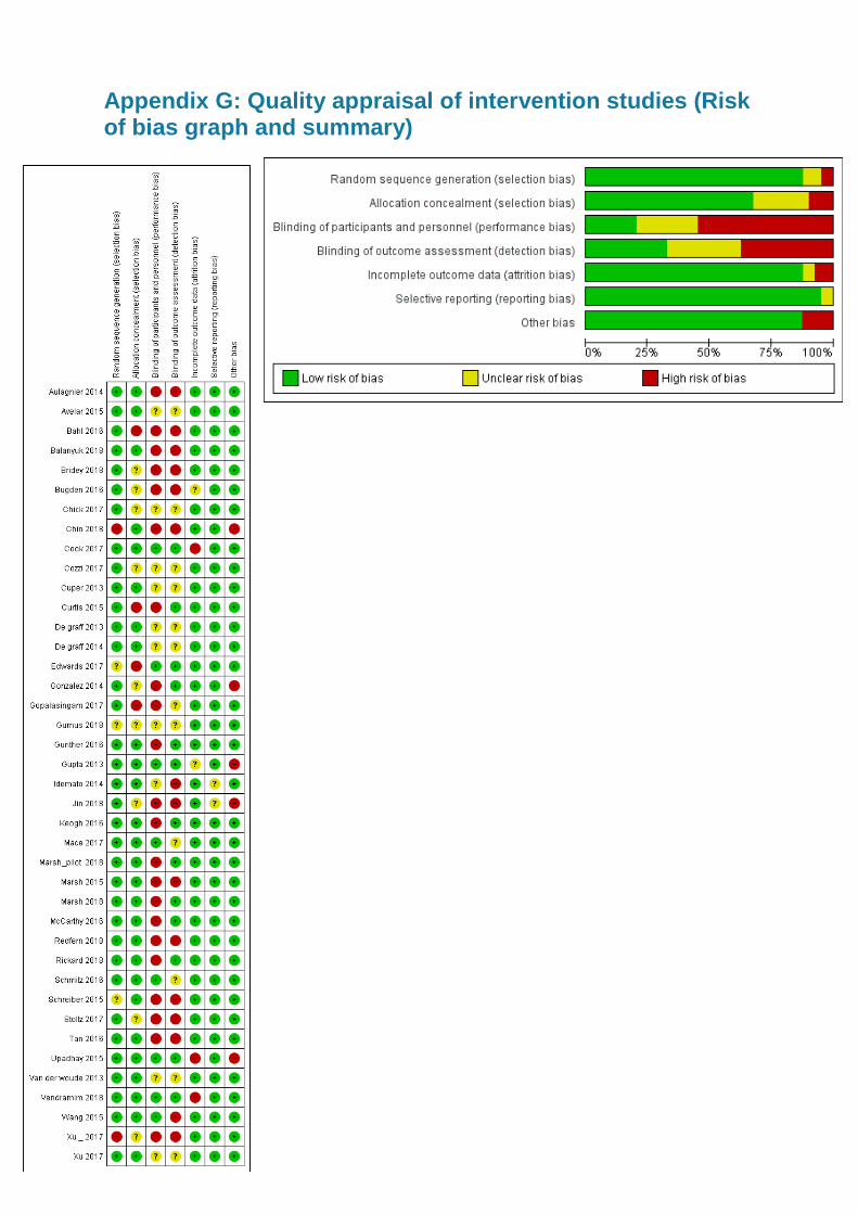
Appendix G: Quality appraisal of intervention studies (Risk of bias graph and summary)

Peripheral intravenous catheters: A review of guidelines and research 76
Level 5, 255 Elizabeth Street, Sydney NSW 2000 GPO Box 5480, Sydney NSW 2001
Phone: (02) 9126 3600 Fax: (02) 9126 3613
Email: [email protected] Website: www.safetyandquality.gov.au