Familial incidence of mammary gland tumours in the …vri.cz/docs/vetmed/58-8-442.pdf · Familial...
7
Case Report Veterinarni Medicina, 58, 2013: (8): 442–448 442 Familial incidence of mammary gland tumours in the Djungarian hamster ( Phodopus sungaros ): a case report F. Jelinek 1 , J. Felsberg 2 , M. Mestan 3 1 Veterinary Histopathological Laboratory, Prague, Czech Republic 2 Institute of Microbiology, Academy of Sciences of the Czech Republic, Prague, Czech Republic 3 Veterinary Surgery, Prague, Czech Republic ABSTRACT: Tumours of the mammary gland were diagnosed in one female Djungarian hamster (Phodopus sungaros) and in two of her daughters from two litters. Altogether, five tumours were diagnosed. Three of them were adenocarcinomas, one was adenoma with disseminated foci of adenocarcinoma and one was diagnosed as an atypical fibrosarcoma derived from cutaneous ganglion cell-like cells. This was a recurrent tumour in the proxim- ity of the previously extirpated adenocarcinoma. Using specific PCR primers for conserved regions of the mouse mammary tumour virus (MMTV) no endogenous provirus DNA could be detected in DNA samples isolated from formalin-fixed paraffin embedded tissue sections. Keywords: hamsters; neoplasias; atypical fibrosarcoma; mammary gland; clinic; pathology; molecular biology Hamsters are popular pet animals with Syrian hamsters (Mesocricetus auratus) and Djungarian hamsters ( Phodopus sungaros ) being among the most frequently kept species. Reports on sponta- neous tumours in domestic hamsters are scarce, and most are individual case reports (Kondo et al. 2008a). The incidence of tumours in these two spe- cies is quite different. In the analysis of tumours from 85 domestic hamsters 70 were Djungarian hamsters and 15 Syrian hamsters (Kondo et al . 2008a). In the integumentary system of Syrian hamsters melanomas and cutaneous lymphoma are the most frequently detected tumours, but both are low in incidence (Percy and Barthold 1993; Brown and Donnelly 2012). Tumours of the mam- mary gland in Syrian hamsters are rare and only a few cases have been described in the literature (e.g. Jelinek 1986a). However, in Djungarian ham- sters the incidence of cutaneous neoplasias is five times greater than in Syrian hamsters and these are predominantly represented by mammary tu- mours, atypical fibromas and papilomas (Brown and Donnelly 2012). Kondo el al. (2009) analysed mammary tumours from 12 domestic Djungarian hamsters. Eleven were female and one was male. Histopathology revealed three subtypes: simple ad- enoma, tubulopapillary carcinoma, and complex carcinoma. No angioinvasion was observed. It is the aim of this contribution to describe mammary neoplasias in a female of Djungarian hamster and her two daughters. Case description Clinical and gross pathology. The owners of the pets in question originally intended to acquire two female Djungarian hamsters as pets. However, in the pet shop they purchased, by mistake, one male and one female. Two months later the female gave birth to four young. After weaning, three young animals (two females and one male) and the father were sold back to the pet shop and the family re- tained the mother and her daughter. The mother was, however, fertilized before the weaning of off- spring and one month after the first parturition, bore a second litter of young comprised of two hamsters; one died shortly after birth while the other, female, survived. A tumour of the first left mammary gland was observed in the mother at the age of 15 months. At the time of extirpation its size was 2 × 1 × 1 cm
-
Upload
truongmien -
Category
Documents
-
view
215 -
download
1
Transcript of Familial incidence of mammary gland tumours in the …vri.cz/docs/vetmed/58-8-442.pdf · Familial...
Case Report Veterinarni Medicina, 58, 2013: (8): 442–448
442
Familial incidence of mammary gland tumours in the Djungarian hamster (Phodopus sungaros): a case report
F. Jelinek1, J. Felsberg2, M. Mestan3
1Veterinary Histopathological Laboratory, Prague, Czech Republic2Institute of Microbiology, Academy of Sciences of the Czech Republic, Prague, Czech Republic3Veterinary Surgery, Prague, Czech Republic
ABSTRACT: Tumours of the mammary gland were diagnosed in one female Djungarian hamster (Phodopus sungaros) and in two of her daughters from two litters. Altogether, five tumours were diagnosed. Three of them were adenocarcinomas, one was adenoma with disseminated foci of adenocarcinoma and one was diagnosed as an atypical fibrosarcoma derived from cutaneous ganglion cell-like cells. This was a recurrent tumour in the proxim-ity of the previously extirpated adenocarcinoma. Using specific PCR primers for conserved regions of the mouse mammary tumour virus (MMTV) no endogenous provirus DNA could be detected in DNA samples isolated from formalin-fixed paraffin embedded tissue sections.
Keywords: hamsters; neoplasias; atypical fibrosarcoma; mammary gland; clinic; pathology; molecular biology
Hamsters are popular pet animals with Syrian hamsters (Mesocricetus auratus) and Djungarian hamsters (Phodopus sungaros) being among the most frequently kept species. Reports on sponta-neous tumours in domestic hamsters are scarce, and most are individual case reports (Kondo et al. 2008a). The incidence of tumours in these two spe-cies is quite different. In the analysis of tumours from 85 domestic hamsters 70 were Djungarian hamsters and 15 Syrian hamsters (Kondo et al. 2008a). In the integumentary system of Syrian hamsters melanomas and cutaneous lymphoma are the most frequently detected tumours, but both are low in incidence (Percy and Barthold 1993; Brown and Donnelly 2012). Tumours of the mam-mary gland in Syrian hamsters are rare and only a few cases have been described in the literature (e.g. Jelinek 1986a). However, in Djungarian ham-sters the incidence of cutaneous neoplasias is five times greater than in Syrian hamsters and these are predominantly represented by mammary tu-mours, atypical fibromas and papilomas (Brown and Donnelly 2012). Kondo el al. (2009) analysed mammary tumours from 12 domestic Djungarian hamsters. Eleven were female and one was male. Histopathology revealed three subtypes: simple ad-
enoma, tubulopapillary carcinoma, and complex carcinoma. No angioinvasion was observed. It is the aim of this contribution to describe mammary neoplasias in a female of Djungarian hamster and her two daughters.
Case description
Clinical and gross pathology. The owners of the pets in question originally intended to acquire two female Djungarian hamsters as pets. However, in the pet shop they purchased, by mistake, one male and one female. Two months later the female gave birth to four young. After weaning, three young animals (two females and one male) and the father were sold back to the pet shop and the family re-tained the mother and her daughter. The mother was, however, fertilized before the weaning of off-spring and one month after the first parturition, bore a second litter of young comprised of two hamsters; one died shortly after birth while the other, female, survived.
A tumour of the first left mammary gland was observed in the mother at the age of 15 months. At the time of extirpation its size was 2 × 1 × 1 cm
Veterinarni Medicina, 58, 2013 (8): 442–448 Case Report
443
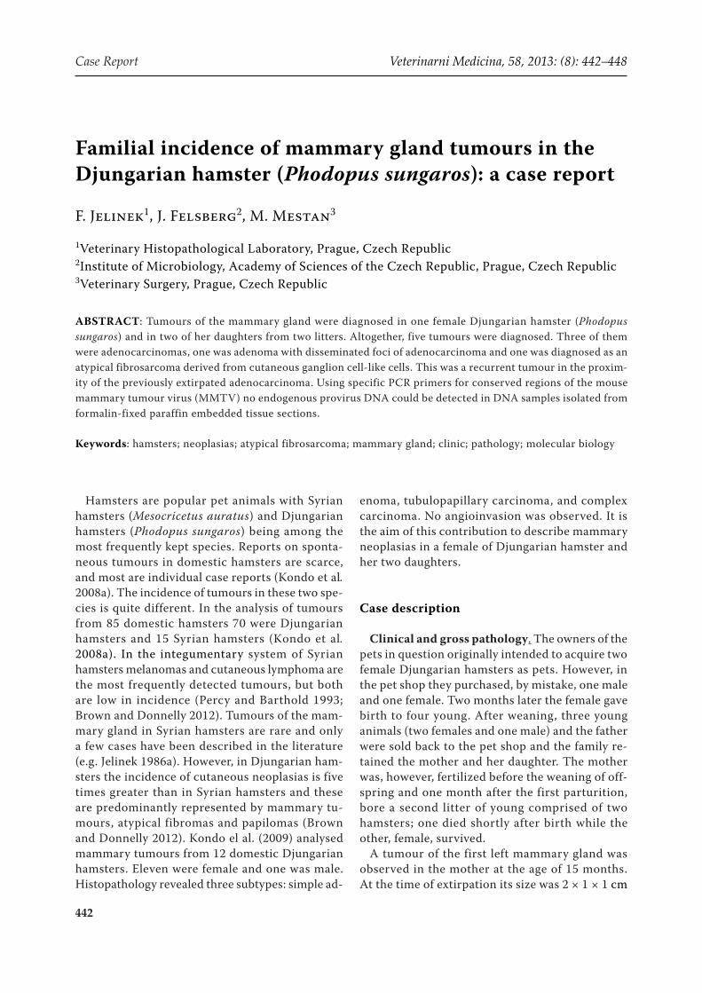
(Figure 1). It was well circumscribed and red-brown in colour. The cut surface was grey-brown with one cyst in the central region. The female survived for 13 months after the surgery without any health problems, i.e., until the age of at least 28 months.
In the first daughter a tumour of the first left mam-mary gland appeared at the age of nine months. At the time of surgery its size was 1.3 × 1.3 × 1.5 cm. The tumour was well circumscribed and the cut surface was pinkish with a small quantity of cloudy secretion. Three weeks after the operation the owner observed recurrence of the tumour. During the following three weeks (altogether six weeks after the extirpation) the globoid neoplasia reached 3 cm in diameter. In the first right mammary gland a tumour 1.5 × 1 × 1 cm in size simultaneously developed. The hamster was euthanized because of unfavourable prognosis. At post-mortem examination the body weight of the animal was 59 g and tumour weight was 19 g. The tumour had a smooth surface, greyish colour and surrounded the left thoracic limb (Figure 2). On the cut surface a large central necrosis circumscribed by homogeneous greyish tissue was apparent. All three siblings were healthy throughout the monitoring of their health status, i.e., up to 20 months of age.
In the second daughter three tumours developed at the age of 11 months. One was located in the first right mammary gland. It was globoid, 1.2 cm in diameter. In close contact with the tumour, two neoplastic formations were situated on the right side of the chest. The larger tumour was 1 cm in diam-eter, the smaller one measured 0.4 cm in diameter. All three tumours were solid, well circumscribed and of pinkish colour on cut surface. In the smallest tumour small cysts containing clear secretion was observed. The female died three months after the operation due to the development of other tumours and apathy. She was not examined either clinically or pathologically.
Histopathology and immunohistochemistry. Samples for histopathological examination were fixed in 10% neutral buffered formalin, embedded in paraffin wax, sectioned at 5 µm thick slices and stained with haematoxylin and eosin and in indicat-ed cases staining with Masson blue trichrome and PAS reaction was performed. The common immu-noperoxidase method was conducted on additional histological sections for detection of the antigens listed in Table 1. All antibodies were produced by DakoCytomation (Glostrup, DK). With exception of cytokeratins that were unmasked by digestion with Pronase (DakoCytomation, Glostrup, DK) for 10 min at room temperature, all remaining antigens were unmasked by boiling of the slides for 10 min in 0.1M citrate buffer, pH 6.0. Endogenous peroxi-dase activity was blocked with 3% H2O2 for 15 min. Binding of the primary antibodies was visualised
Figure 1. Tumour of the mammary gland in the mother
Figure 2. First daughter. Tumour arising at the site of the previously extirpated mammary carcinoma, surround-ing the left thoracic limb and the tumour of the first right mammary gland
Table 1. Specification of the antibodies and their dilution
Antibody Clone Dilution
Actin HHF35 1 : 50
Cytokeratin MNF116 1 : 50
CD 117 (c-kit) Rabbit polyclonal 1 : 100
HLA-DR TAL1B5 1 : 50
Chromogranin Purified IgG 1 : 100
Myeloid/histiocyte antigen MAC387 1 : 100
NSE (neuron-specific enolase) BBS/NC/VI-H14 1 : 100
PCNA (proliferating cell nuclear antigen) PC10 1 : 50
Vimentin V9 1 : 25
Case Report Veterinarni Medicina, 58, 2013: (8): 442–448
444
by means of the Streptavidin-Biotin Universal detection System (Ultratech HRP, Immunotech, Marseille) and using the DAB Chromogen Kit (Ultratech DAB, Immunotech, Marseille). The slides were counterstained with haematoxylin.
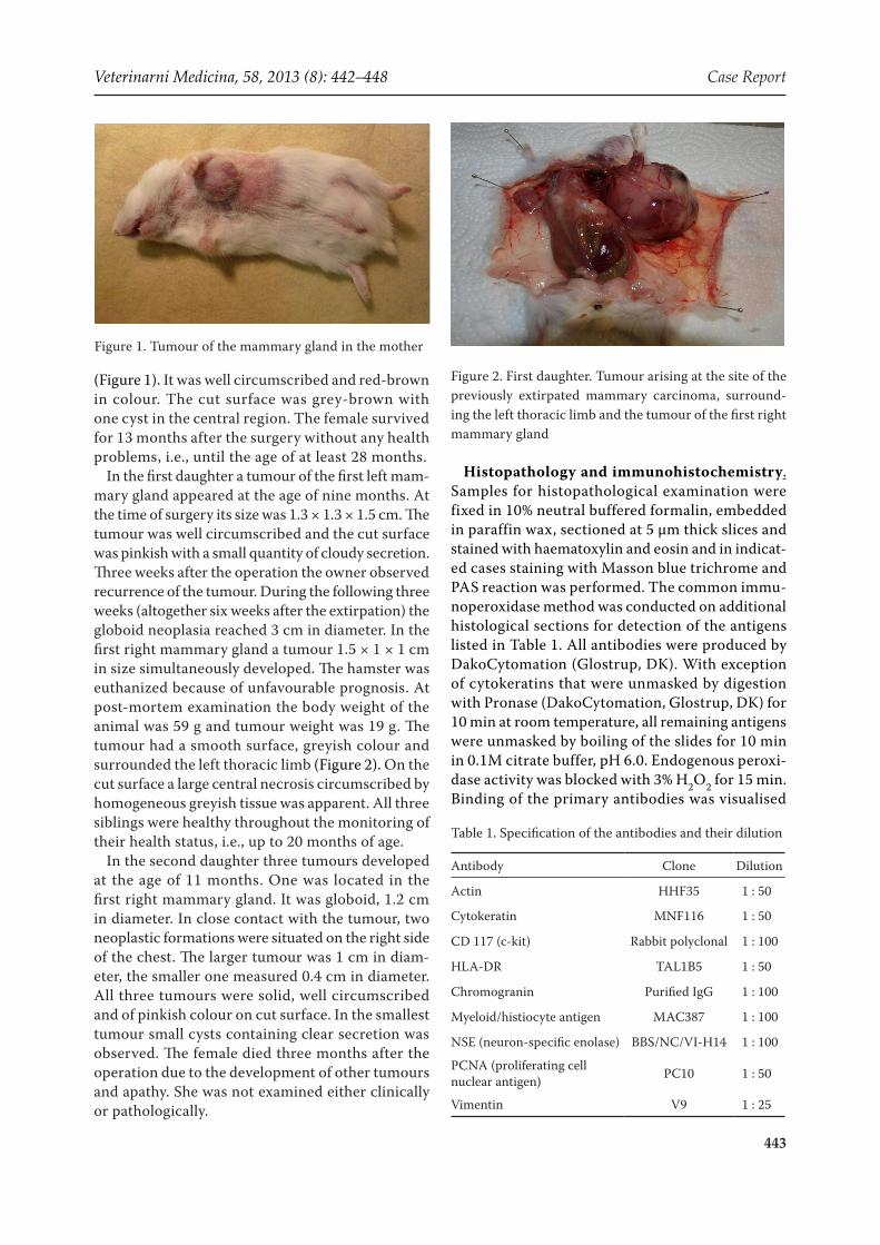
Tumour in the mother. The adenocarcinoma was arranged predominantly in tubular, alveolar or cystopapillary structures frequently lined by multi-layered epithelium. Neoplastic epithelial cells were characterised by anisocytosis, anisokaryosis and moderate mitotic activity (Figure 3). Multiplied my-oepithelial cells were identified by means of actin in part of neoplastic tubules (Figure 4), and regions of increased proliferative activity of epithelial cells were demonstrated using PCNA. Some small foci of necrosis and larger central necrosis with secondary
inflammatory cellularity were also present in the tumour. Angioinvasion was not observed.
Tumours in the first daughter. A predominantly well differentiated tubular adenocarcinoma with some dilated tubules or small cystic formations was diagnosed. Multiplied myoepithelial cells were observed in less differentiated regions. Necrosis in the central part of the tumour with moderate secondary inflammatory cellularity was present as well. Angioinvasion was not observed.
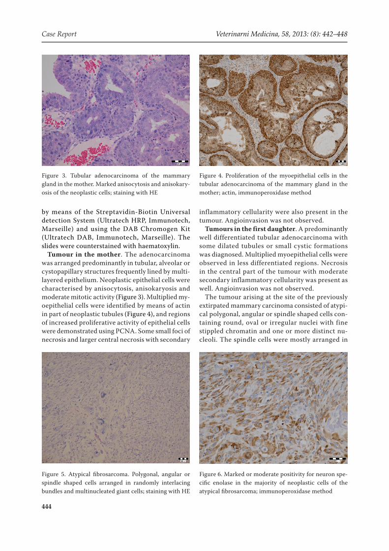
The tumour arising at the site of the previously extirpated mammary carcinoma consisted of atypi-cal polygonal, angular or spindle shaped cells con-taining round, oval or irregular nuclei with fine stippled chromatin and one or more distinct nu-cleoli. The spindle cells were mostly arranged in
Figure 3. Tubular adenocarcinoma of the mammary gland in the mother. Marked anisocytosis and anisokary-osis of the neoplastic cells; staining with HE
Figure 4. Proliferation of the myoepithelial cells in the tubular adenocarcinoma of the mammary gland in the mother; actin, immunoperoxidase method
Figure 6. Marked or moderate positivity for neuron spe-cific enolase in the majority of neoplastic cells of the atypical fibrosarcoma; immunoperoxidase method
Figure 5. Atypical fibrosarcoma. Polygonal, angular or spindle shaped cells arranged in randomly interlacing bundles and multinucleated giant cells; staining with HE
Veterinarni Medicina, 58, 2013 (8): 442–448 Case Report
445
randomly interlacing bundles and multinucleated giant cells were disseminated in the neoplastic tis-sue. Marked anisocytosis and anisokaryosis char-acterised the neoplastic cells (Figure 5). Mitotic activity was moderate – 13 mitotic figures were found in 10 high-power fields (40×), in the range of zero to three per one field. Angioinvasion was not found. Staining of histological sections with Masson blue trichrome revealed a mild to moder-ate quantity of delicate collagen fibrils produced by the neoplastic cells. Some foci of degenerated collagen fibres were disseminated in the neoplas-tic tissue. Immunohistochemistry was negativite for cytokeratin, myeloid/histiocyte antigen and HLA-DR antigens. Suspect to moderate positivity of actin was observed in less than 10% of neoplastic cells. Moderate positivity for chromogranin and CD 117 (c-kit) was detected in the cytoplasm of the majority of neoplastic cells. Marked positivity for vimentin was observed in all neoplastic cells, and the majority of them were also NSE-positive (Figure 6). Based on histological and immunohisto-chemical examinations, the tumour was diagnosed as an atypical fibrosarcoma derived from cutane-ous ganglion cell-like cells as described by Kondo et al. (2011).
The tumour in the first right mammary gland was an adenocarcinoma arranged predominantly in tubular structures. Cystic formations were present in part of the tumour. Many tubules consisted of epithelial and myoepithelial cels with vacuolated cytoplasm. A part of these cells contained nuclei of pyknotic appearance. On the basis of PAS reaction negativity, hydropic degeneration was presumed. Staining for neutral lipids could not be performed, because the whole tumour was processed using the paraffin technique. Angioinvasion was not present.
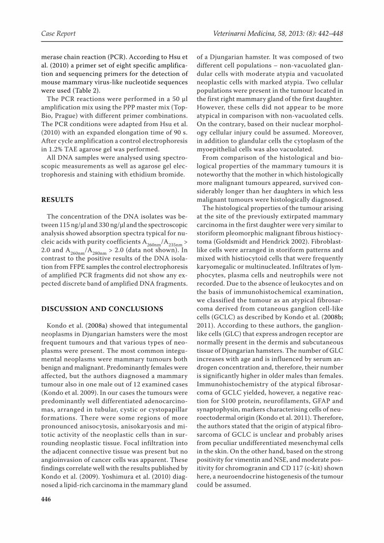
Tumours in the second daughter. In all three tumours tubular adenoma with cystic formations was diagnosed. In the adenomatous tissue there were, however, disseminated foci of adenocarci-noma, mostly in situ but in places with signs of invasion through the basal membrane (Figure 7).
Molecular biology. DNA from five samples of formalin-fixed paraffin-embedded tissue sec-tions (FFPE): B30/12 – 0.017 g; B83/12 – 0.018 g; B205/12 – 0.020 g; 5/2/12 – 0.014 g; and B34/13 as a negative control – 0.022 g was isolated using the High Pure PCR Template Prepartion Kit ver. 16.0 (Roche) with some modifications. Because of the small volume of the samples the final volume of elu-tion buffer was reduced to 100 µl. For the detection of endogenous Djungarian hamster provirus DNA (MRS-Ps) derived from mouse mammary tumour virus (MMTV) the DNA isolates were analysed by amplification of DNA fragments using the poly-
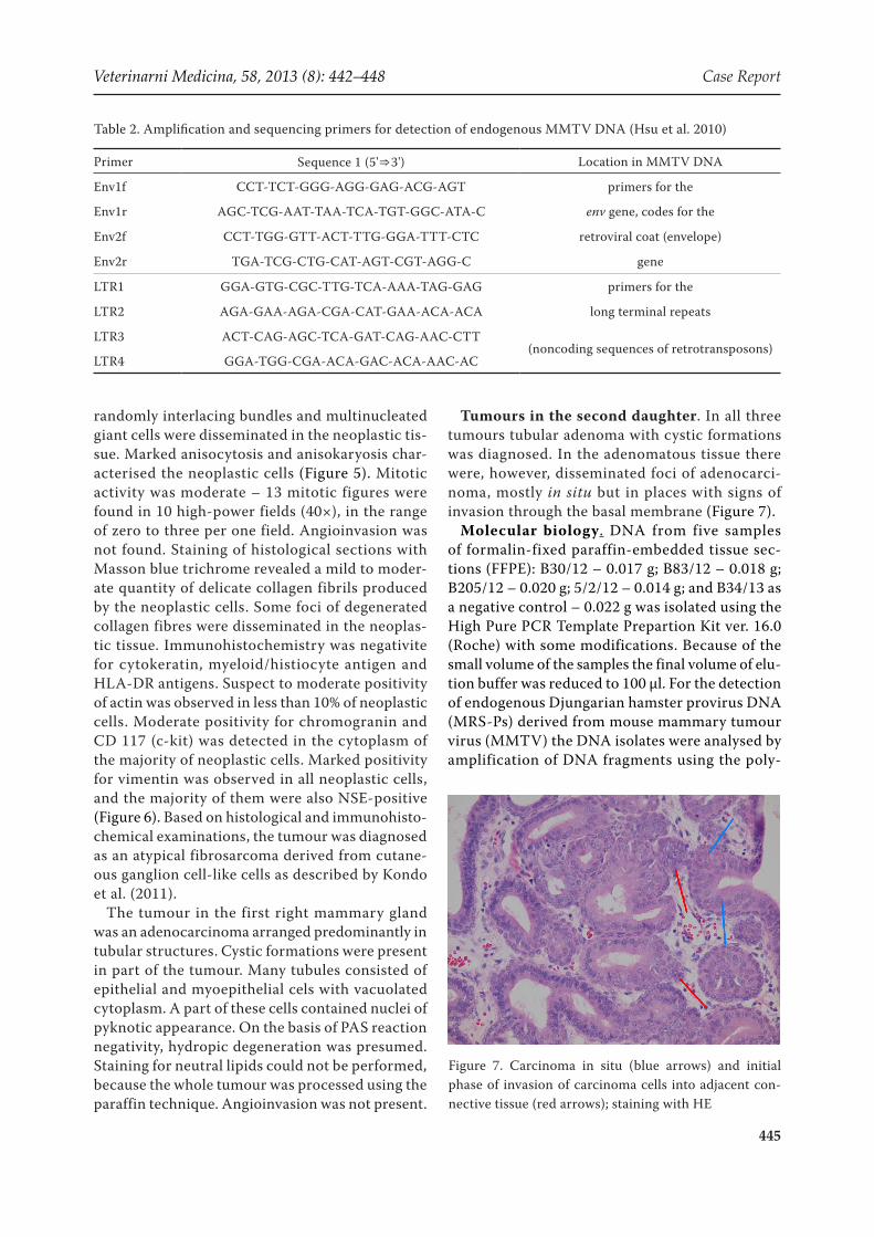
Table 2. Amplification and sequencing primers for detection of endogenous MMTV DNA (Hsu et al. 2010)
Primer Sequence 1 (5'⇒3') Location in MMTV DNA
Env1f CCT-TCT-GGG-AGG-GAG-ACG-AGT primers for the
Env1r AGC-TCG-AAT-TAA-TCA-TGT-GGC-ATA-C env gene, codes for the
Env2f CCT-TGG-GTT-ACT-TTG-GGA-TTT-CTC retroviral coat (envelope)
Env2r TGA-TCG-CTG-CAT-AGT-CGT-AGG-C gene
LTR1 GGA-GTG-CGC-TTG-TCA-AAA-TAG-GAG primers for the
LTR2 AGA-GAA-AGA-CGA-CAT-GAA-ACA-ACA long terminal repeats
LTR3 ACT-CAG-AGC-TCA-GAT-CAG-AAC-CTT(noncoding sequences of retrotransposons)
LTR4 GGA-TGG-CGA-ACA-GAC-ACA-AAC-AC
Figure 7. Carcinoma in situ (blue arrows) and initial phase of invasion of carcinoma cells into adjacent con-nective tissue (red arrows); staining with HE
Case Report Veterinarni Medicina, 58, 2013: (8): 442–448
446
merase chain reaction (PCR). According to Hsu et al. (2010) a primer set of eight specific amplifica-tion and sequencing primers for the detection of mouse mammary virus-like nucleotide sequences were used (Table 2).
The PCR reactions were performed in a 50 µl amplification mix using the PPP master mix (Top-Bio, Prague) with different primer combinations. The PCR conditions were adapted from Hsu et al. (2010) with an expanded elongation time of 90 s. After cycle amplification a control electrophoresis in 1.2% TAE agarose gel was performed.
All DNA samples were analysed using spectro-scopic measurements as well as agarose gel elec-trophoresis and staining with ethidium bromide.
RESULTS
The concentration of the DNA isolates was be-tween 115 ng/µl and 330 ng/µl and the spectroscopic analysis showed absorption spectra typical for nu-cleic acids with purity coefficients A260nm/A235nm > 2.0 and A260nm/A280nm > 2.0 (data not shown). In contrast to the positive results of the DNA isola-tion from FFPE samples the control electrophoresis of amplified PCR fragments did not show any ex-pected discrete band of amplified DNA fragments.
DISCUSSION AND CONCLUSIONS
Kondo et al. (2008a) showed that integumental neoplasms in Djungarian hamsters were the most frequent tumours and that various types of neo-plasms were present. The most common integu-mental neoplasms were mammary tumours both benign and malignant. Predominantly females were affected, but the authors diagnosed a mammary tumour also in one male out of 12 examined cases (Kondo et al. 2009). In our cases the tumours were predominantly well differentiated adenocarcino-mas, arranged in tubular, cystic or cystopapillar formations. There were some regions of more pronounced anisocytosis, anisokaryosis and mi-totic activity of the neoplastic cells than in sur-rounding neoplastic tissue. Focal infiltration into the adjacent connective tissue was present but no angioinvasion of cancer cells was apparent. These findings correlate well with the results published by Kondo et al. (2009). Yoshimura et al. (2010) diag-nosed a lipid-rich carcinoma in the mammary gland
of a Djungarian hamster. It was composed of two different cell populations – non-vacuolated glan-dular cells with moderate atypia and vacuolated neoplastic cells with marked atypia. Two cellular populations were present in the tumour located in the first right mammary gland of the first daughter. However, these cells did not appear to be more atypical in comparison with non-vacuolated cells. On the contrary, based on their nuclear morphol-ogy cellular injury could be assumed. Moreover, in addition to glandular cells the cytoplasm of the myoepithelial cells was also vacuolated.
From comparison of the histological and bio-logical properties of the mammary tumours it is noteworthy that the mother in which histologically more malignant tumours appeared, survived con-siderably longer than her daughters in which less malignant tumours were histologically diagnosed.
The histological properties of the tumour arising at the site of the previously extirpated mammary carcinoma in the first daughter were very similar to storiform pleomorphic malignant fibrous histiocy-toma (Goldsmidt and Hendrick 2002). Fibroblast-like cells were arranged in storiform patterns and mixed with histiocytoid cells that were frequently karyomegalic or multinucleated. Infiltrates of lym-phocytes, plasma cells and neutrophils were not recorded. Due to the absence of leukocytes and on the basis of immunohistochemical examination, we classified the tumour as an atypical fibrosar-coma derived from cutaneous ganglion cell-like cells (GCLC) as described by Kondo et al. (2008b; 2011). According to these authors, the ganglion-like cells (GLC) that express androgen receptor are normally present in the dermis and subcutaneous tissue of Djungarian hamsters. The number of GLC increases with age and is influenced by serum an-drogen concentration and, therefore, their number is significantly higher in older males than females. Immunohistochemistry of the atypical fibrosar-coma of GCLC yielded, however, a negative reac-tion for S100 protein, neurofilaments, GFAP and synaptophysin, markers characterising cells of neu-roectodermal origin (Kondo et al. 2011). Therefore, the authors stated that the origin of atypical fibro-sarcoma of GCLC is unclear and probably arises from peculiar undifferentiated mesenchymal cells in the skin. On the other hand, based on the strong positivity for vimentin and NSE, and moderate pos-itivity for chromogranin and CD 117 (c-kit) shown here, a neuroendocrine histogenesis of the tumour could be assumed.
Veterinarni Medicina, 58, 2013 (8): 442–448 Case Report
447
The development of mammary tumours in the mother and also in her two daughters suggests a genetic disposition. A viral aetiology of these car-cinomas cannot be reliably excluded. Viral aetiol-ogy of mammary carcinoma is well known in mice and Litvinov et al. (1985) described a stable cell line from a mammary gland cancer of a Djungarian hamster suitable for the study of oncogenic proper-ties of mouse mammary tumour virus.
The amplification of DNA fragments for the de-tection of endogenous MMTV using the primer sets according to Hsu et al. (2010) was negative. The presence of mouse mammary tumour virus-like retrotransposons in the genomes of Djungarian hamsters and other closely related animals was repeatedly demonstrated (Vasetskii et al. 1989; Tikhonenko et al. 1990; Vasetskii 1994; Lesser et al. 1995; Hsu et al. 2010; Xu et al. 2012). Thus, more experiments to detect and amplify MMTV sequences in DNA isolated from FFPE samples as described are necessary. In this direction, a greater variety of primer sets used and applied PCR condi-tions, including control amplifications of “house-keeping” genes must be tested.
As was already mentioned in the introduction, the incidence of tumours in Syrian hamsters is quite unique. Adrenocortical hyperplasia and ad-enoma were diagnosed repeatedly, mostly without symptoms of hyperadrenocorticism or Cushing disease (Brown and Donnelly 2012). Lymphoma, predominantly multicentric, is the most common neoplasm in older hamsters. Cutaneous lymphoma, resembling mycosis fungoides is the second varia-tion, and epizootic lymphoma in young hamsters caused by hamster polyomavirus is the third varia-tion of lymphomas in golden hamsters (Brown and Donnelly 2012). In one breeding colony of labora-tory Syrian hamsters, in which wet tail frequently appeared in young animals, adenocarcinoma of the small intestine and lymphoma arising from intesti-nal mucosa-associated lymphatic tissue (MALToma) were repeatedly diagnosed in adult hamsters (Jelinek 1986b, 1989). Only one case of a mammary carci-noma in a pet female was observed (Jelinek 1986a).
REFERENCES
Brown C, Donnelly TM (2012): Disease problems of small rodents. In: Quesenberry KE, Carpenter JW (eds.): Ferrets, Rabbits and Rodents. Clinical Medicine and Surgery. Elsevier Saunders, St. Louis. 354–372.
Goldschmidt MH, Hendrick MJ (2002): Tumors of the skin and soft tissues. 4th ed. In: Meuten DJ (ed.): Tu-mors of Domestic Animals. Iowa State Press A Balck-well Publishing Company, Iowa. 45–117.
Hsu WL, Lin HY, Chiou SS, Chang CC, Wang SP, Lin KH, Chulakasian S, Wong ML, Chang SC (2010): Mouse mammary tumor virus-like nucleotide se-quences in canine and feline mammary tumors. Jour-nal of Clinical Microbiology 48, 4354–4362.
Jelinek F (1986a): Tubular adenocarcinoma of the mam-mary gland in a golden hamster. Zeitschrift für Ver-suchstierkunde 28, 167–171.
Jelinek F (1986b): Adenocarcinomas of the small intes-tine in golden hamsters. Zeitschrift für Versuchst-ierkunde 28, 153–155.
Jelinek F (1989): Intestinal lymphoma in golden hamster (Mesocricetus auratus). Zeitschrift für Versuchstier- kunde 32, 123–128.
Kondo H, Onuma M, Shibuya H, Sato T (2008a): Spon-taneous tumors in domestic hamsters. Veterinary Pa-thology 45, 674–680.
Kondo H, Onuma M, Ito H, Shibuya H, Sato T (2008b): Spontaneous fibrosarcoma in a Djungarian hamster (Phodopus sungorus). Comparative Medicine 58, 294–296.
Kondo H, Onuma M, Shibuya H, Sato T (2009): Mor-phological and immunohistochemical studies of spon-taneous mammary tumours in Siberian hamsters (Phodopus sungorus). Journal of Comparative Pathol-ogy 140, 127–131.
Kondo H, Onuma M, Shibuya H, Sato H, Abbott JR (2011): Atypical fibrosarcomas derived from cutane-ous ganglion cell-like cells in 2 domestic Djungarian hamsters (Phodopus sungorus). Journal of American Laboratory Animal Science 50, 523–525.
Lesser J, Bittoun P, Emanoil-Ravier R, Peries J (1995): Biological and molecular studies on Syrian hamster intracisternal R-type particles. Archives of Virology 140, 95–109.
Litvinov SV, Sokova OJ, Kryukova IN (1985): A new cell system for the study of mouse mammary tumour virus. Acta Virologica 29, 83–86.
Percy DH, Barthold SW (1993): Pathology of Laboratory Rodents and Rabbits. Iowa State University Press, Ames. 229 pp.
Tikhonenko AT, Vassetzky III NS, Golovkina TV, Gud-kov AV (1990): Molecular cloning and primary struc-ture analysis of the mouse mammar y tumor virus-related element from Dwarf hamster genome. Virus Genes 3, 259–261.
Vasetskii NS, Tikhonenko AT, Golovkina TV, Gudkov AV (1989): Cloning and analysis of the primary struc-
Case Report Veterinarni Medicina, 58, 2013: (8): 442–448
448
ture of an element, related to the murine mammary cancer virus, from the genome of the Djungarian ham-ster (in Russian). Molekuliarnaia Genetika, Mikrobio-logia i Virusologia 10, 16–19.
Vasetskii NS (1994): Analysis of the sequence of the en-dogenous Djungarian hamster provirus (MRS-Ps) related to the murine ma mammary cancer virus (in Russian). Moleclar Biology (Moscow) 28, 900–908.
Xu X, Nagarajan H, Lewis NE, Pan S, Cai Z, Liu X, Chen W, Xie M, Wang W, Hammond S, Andersen MR, Neff N, Passarelli B, Koh W, Fan HC, Wang J, Gui Y, Lee
KH, Betenbaugh MJ, Quake SR, Famili I., Palsson BO, Wang J (2012): The genomic sequence of the Chinese hamster ovary (CHO)-K1 cell line. Nature Biotechnol-ogy 29, 735–741.
Yoshimura H, Kimura N, Nakahira R, Mischishita M, Ohkusu-Tsukada K, Takahashi K (2010): Lipid-rich carcinoma on the mammary gland of a Djungarian hamster (Phodopus sungorus). Journal of Veterinary Diagnostic Investigation 22, 305–309.
Received: 2013–07–30Accepted after corrections: 2013–08–27
Corresponding Author:
Prof. MVDr. Frantisek Jelinek, CSc., Dipl. ECVP, Veterinary Histopathological Laboratory, Sojovicka 16, 197 00 Prague 9, Czech RepublicE-mail: [email protected]