Failure to Launch -...
14
TRANSLATIONAL PERSPECTIVES Failure to Launch Targeting Inflammation in Acute Coronary Syndromes Jennifer A. Rymer, MD, MBA, L. Kristin Newby, MD, MHS SUMMARY The importance of inflammation and inflammatory pathways in atherosclerotic disease and acute coronary syndromes (ACS) is well established. The success of statin therapy rests not only on potently reducing levels of low-density lipoprotein cholesterol, but also on the many beneficial, pleiotropic effects statin therapy has on various inflammatory mechanisms in atherosclerotic disease, from reducing endothelial dysfunction to attenuating levels of serum C-reactive protein. Due to the growing awareness of the importance of inflammation in ACS, investigators have attempted to develop novel therapies against known markers of inflammation for several decades. Targeted pathways have ranged from inhibiting C5 cleavage with a high-affinity monoclonal antibody against C5 to inhibiting the activation of the p38 mitogen-activated protein kinase signaling cascades. In each of these instances, despite promising early preclinical and mechanistic studies and phase 2 trials suggesting a potential benefit in reducing post-MI complications or restenosis, these novel therapies have failed to show benefits during large, phase 3 clinical outcomes trials. This review discusses several examples of novel anti-inflammatory therapies that failed to show significant improvement on clinical outcomes when tested in large, randomized trials and highlights potential explanations for why targeted therapies against known markers of inflammation in ACS have failed to launch. (J Am Coll Cardiol Basic Trans Science 2017;2:484–97) © 2017 The Authors. Published by Elsevier on behalf of the American College of Cardiology Foundation. This is an open access article under the CC BY-NC-ND license (http://creativecommons.org/licenses/by-nc-nd/4.0/). O ver the past 2 decades, there has been increasing interest in discovering novel therapeutic agents for reducing residual risk among patients with acute coronary syndromes (ACS), including ST-segment elevation myocardial infarction (STEMI), non–ST-segment myocardial infarction (NSTEMI), and unstable angina. Each year, roughly 1.1 million patients are hospitalized with an ACS event in the United States (1). Although the overall incidence of ACS appears to be declining, the direct and indirect costs associated with treating patients with ACS and its downstream sequelae, including congestive heart failure and repeat revascularization, remain a medical and economic burden (2–4). Atherosclerotic disease leading to an ACS event is a complex process of atheroma formation and eventual plaque rupture. The 30-day rate of recurrent events post-ACS is estimated to be around 2% (5,6). High- sensitivity C-reactive protein (hsCRP) (a marker of inflammation), can be elevated for several months after an ACS event, and is a marker of risk for sub- sequent development of heart failure and increased mortality (7). These observations led to a focus on inflammatory pathways as participants in the forma- tion, proliferation, and rupture of atherosclerotic plaques (8). The intersection of ACS pathobiology, inflammation, and high rates of recurrent events post-ACS has generated interest in development of From the Division of Cardiology, Department of Medicine, and Duke Clinical Research Institute, Duke University School of Medicine, Durham, North Carolina. Both authors have reported that they have no relationships relevant to the contents of this paper to disclose. All authors attest they are in compliance with human studies committees and animal welfare regulations of the authors’ institutions and Food and Drug Administration guidelines, including patient consent where appropriate. For more information, visit the JACC: Basic to Translational Science author instructions page. Manuscript received July 12, 2017; accepted July 12, 2017. JACC: BASIC TO TRANSLATIONAL SCIENCE VOL. 2, NO. 4, 2017 ª 2017 THE AUTHORS. PUBLISHED BY ELSEVIER ON BEHALF OF THE AMERICAN COLLEGE OF CARDIOLOGY FOUNDATION. THIS IS AN OPEN ACCESS ARTICLE UNDER THE CC BY-NC-ND LICENSE ( http://creativecommons.org/licenses/by-nc-nd/4.0/ ). ISSN 2452-302X http://dx.doi.org/10.1016/j.jacbts.2017.07.001
-
Upload
truongtuyen -
Category
Documents
-
view
216 -
download
0
Transcript of Failure to Launch -...
J A C C : B A S I C T O T R A N S L A T I O N A L S C I E N C E V O L . 2 , N O . 4 , 2 0 1 7
ª 2 0 1 7 T H E A U T H O R S . P U B L I S H E D B Y E L S E V I E R O N B E H A L F O F T H E AM E R I C A N
C O L L E G E O F C A R D I O L O G Y F O UN DA T I O N . T H I S I S A N O P E N A C C E S S A R T I C L E U N D E R
T H E C C B Y - N C - N D L I C E N S E ( h t t p : / / c r e a t i v e c o mm o n s . o r g / l i c e n s e s / b y - n c - n d / 4 . 0 / ) .
I S S N 2 4 5 2 - 3 0 2 X
h t t p : / / d x . d o i . o r g / 1 0 . 1 0 1 6 / j . j a c b t s . 2 0 1 7 . 0 7 . 0 0 1
TRANSLATIONAL PERSPECTIVES
Failure to LaunchTargeting Inflammation in Acute Coronary Syndromes
Jennifer A. Rymer, MD, MBA, L. Kristin Newby, MD, MHS
SUMMARY
Fro
Me
pa
All
ins
vis
Ma
The importance of inflammation and inflammatory pathways in atherosclerotic disease and acute coronary syndromes
(ACS) is well established. The success of statin therapy rests not only on potently reducing levels of low-density
lipoprotein cholesterol, but also on the many beneficial, pleiotropic effects statin therapy has on various inflammatory
mechanisms in atherosclerotic disease, from reducing endothelial dysfunction to attenuating levels of serum C-reactive
protein. Due to the growing awareness of the importance of inflammation in ACS, investigators have attempted to
develop novel therapies against known markers of inflammation for several decades. Targeted pathways have ranged
from inhibiting C5 cleavage with a high-affinity monoclonal antibody against C5 to inhibiting the activation of the p38
mitogen-activated protein kinase signaling cascades. In each of these instances, despite promising early preclinical
and mechanistic studies and phase 2 trials suggesting a potential benefit in reducing post-MI complications or
restenosis, these novel therapies have failed to show benefits during large, phase 3 clinical outcomes trials. This review
discusses several examples of novel anti-inflammatory therapies that failed to show significant improvement on clinical
outcomes when tested in large, randomized trials and highlights potential explanations for why targeted therapies
against knownmarkers of inflammation in ACS have failed to launch. (J Am Coll Cardiol Basic Trans Science 2017;2:484–97)
© 2017 The Authors. Published by Elsevier on behalf of theAmerican College of Cardiology Foundation. This is an open access
article under the CC BY-NC-ND license (http://creativecommons.org/licenses/by-nc-nd/4.0/).
O ver the past 2 decades, there has beenincreasing interest in discovering noveltherapeutic agents for reducing residual
risk among patients with acute coronary syndromes(ACS), including ST-segment elevation myocardialinfarction (STEMI), non–ST-segment myocardialinfarction (NSTEMI), and unstable angina. Each year,roughly 1.1 million patients are hospitalized with anACS event in the United States (1). Although the overallincidence of ACS appears to be declining, the directand indirect costs associated with treating patientswith ACS and its downstream sequelae, includingcongestive heart failure and repeat revascularization,remain a medical and economic burden (2–4).
m the Division of Cardiology, Department of Medicine, and Duke Clin
dicine, Durham, North Carolina. Both authors have reported that they h
per to disclose.
authors attest they are in compliance with human studies committe
titutions and Food and Drug Administration guidelines, including patien
it the JACC: Basic to Translational Science author instructions page.
nuscript received July 12, 2017; accepted July 12, 2017.
Atherosclerotic disease leading to an ACS event is acomplex process of atheroma formation and eventualplaque rupture. The 30-day rate of recurrent eventspost-ACS is estimated to be around 2% (5,6). High-sensitivity C-reactive protein (hsCRP) (a marker ofinflammation), can be elevated for several monthsafter an ACS event, and is a marker of risk for sub-sequent development of heart failure and increasedmortality (7). These observations led to a focus oninflammatory pathways as participants in the forma-tion, proliferation, and rupture of atheroscleroticplaques (8). The intersection of ACS pathobiology,inflammation, and high rates of recurrent eventspost-ACS has generated interest in development of
ical Research Institute, Duke University School of
ave no relationships relevant to the contents of this
es and animal welfare regulations of the authors’
t consent where appropriate. For more information,
AB BR E V I A T I O N S
AND ACRONYM S
ACS = acute coronary
syndromes
CABG = coronary artery bypass
graft
CAD = coronary artery disease
HDL-C = high-density
lipoprotein cholesterol
hsCRP = high-sensitivity
C-reactive protein
IL = interleukin
LDL-C = low-density
lipoprotein cholesterol
Lp-PLA2 = lipoprotein-
associated phospholipase A2
MAPK = mitogen-activated
protein kinase
MI = myocardial infarction
NSTEMI = non–ST-segment
myocardial infarction
PCI = percutaneous coronary
intervention
PSGL = P-selectin glycoprotein
ligand
sPLA2 = secretory
phospholipase A2
STEMI = ST-segment elevation
myocardial infarction
SVG = saphenous vein grafts
TBR = tissue-to-background
ratio
J A C C : B A S I C T O T R A N S L A T I O N A L S C I E N C E V O L . 2 , N O . 4 , 2 0 1 7 Rymer and NewbyA U G U S T 2 0 1 7 : 4 8 4 – 9 7 Failure to Launch
485
therapeutics that target inflammatory pathways tomitigate the development of post-ACS complications.
In this review, we examine the fate of work overthe past decade to develop therapeutics that targetvarious inflammatory pathways and their compo-nents (Central Illustration). We will focus on thera-peutic agents directed at the complement pathwayand cleavage of C5, secretory phospholipase A2
(sPLA2), lipoprotein-associated phospholipase A2
(Lp-PLA2), interleukin-1, and the mitogen-activatedprotein kinase (MAPK) signaling cascades (Table 1).We examine their development from animal modelsand other preclinical investigations through early andlate-phase randomized clinical trials that tested theexperimental drugs in ACS patients. We then offerpotential explanations for why work targeting theseinflammatory components and pathways in ACS hasnot translated into therapeutic successes anddescribe current, promising novel therapies that arestill under investigation.
CASE STUDIES OF ANTI-INFLAMMATORY AGENTS
FOR CARDIOVASCULAR DISEASE
PEXELIZUMAB. Pexelizumab was an early exampleof an anti-inflammatory therapy for ACS, targetingthe C5 component of the complement cascade.Cleavage of the C5 component of the complementpathway results in formation of C5a and C5b-9, themembrane attack complex (9). C5a is proin-flammatory, and the membrane attack complex isassociated with activation of endothelial cells andleukocytes. Pexelizumab is a single-chain fragmentof a humanized monoclonal antibody that binds to C5with high affinity, preventing its cleavage. Earlyin vitro and human studies suggested that myocar-dial cell necrosis during an ACS event triggered therelease of subcellular membrane constituents, whichresulted in the activation of the complement system(10). For example, in a rabbit model of ischemiainduced by ligature to a large marginal branch of thecircumflex artery, there was accumulation of themembrane attack complex (C5b-C9) in infarctedmyocardium, and rapid activation of the complementpathway occurred during reperfusion (11). One groupof rabbits (n ¼ 17) underwent circumflex coronaryocclusion for variable time periods (from 0.5 to 29 h)without subsequent reperfusion, whereas the othergroup (n ¼ 23) underwent coronary occlusion (from0.5 to 6 h) with subsequent reperfusion. AlthoughC5b-C9 was detected in the infarcted myocardium 5to 6 h after occlusion without reperfusion, it wasdetected much earlier (at 30 min) in the group thatunderwent reperfusion, suggesting that the presence
of reperfusion rapidly activates the comple-ment pathway.
In addition to the association of comple-ment activation with infarction and reperfu-sion injury, in a rat model of myocardialinfarction (MI) and reperfusion, infusion ofmonoclonal antibodies against the ratC5 component prior to ligating the left anteriordescending artery for 30 min, followed byreperfusion, significantly reduced left ven-tricular polymorphonuclear leukocyte infil-tration, apoptosis and necrosis, and overallinfarct size (12). In another study ofmyocardialischemia and reperfusion injury in rats, injec-tion of C5 short hairpin ribonucleic acid 2 daysbefore induction of ischemia inhibited C5expression, significantly decreased the level oftroponin T, and reduced the infarct size by40% (13). Additionally, in a pigmodel, infusionof a monoclonal antibody to C5a (at the time ofonset of ischemia) followed by 50 min of oc-clusion of the left anterior descending arteryusing an occluder catheter and 3 h of reperfu-sion resulted in significant reduction in infarctsize and reperfusion injury (14).
Based on these promising animal data andother early studies, a phase 2 program waslaunched. The CARDINAL (Complement AndReDuction of INfarct size after Angioplasty orLytics) phase 2 program included 2 phase 2,parallel group, double-blind, placebo-
controlled trials of pexelizumab in STEMI patients.These trials tested the effect of pexelizumab (bolusplus infusion administered <6 h after symptomonset) on infarct size (creatine kinase–MB area underthe curve at 72 h) and clinical composite outcomesafter fibrinolytic reperfusion therapy in the COMPLY(COMPlement inhibition in myocardial infarctiontreated with thromboLYtics) trial (15) or primarypercutaneous coronary intervention (PCI) in theCOMMA (COMplement inhibition in Myocardialinfarction treated with Angioplasty) trial (16). TheCOMPLY investigators found no difference in infarctsize or 90-day composite clinical outcomes for pex-elizumab versus placebo among STEMI patients whoreceived fibrinolytic therapy. Similarly, in theCOMMA trial, among STEMI patients treated withprimary PCI, there was no difference in infarct size orthe 90-day primary composite of death, heart failure,shock, or stroke between patients treated with pla-cebo versus pexelizumab. However, 90-day mortalitywas significantly lower among patients who receivedpexelizumab compared with placebo (1.8% vs. 5.9%;p ¼ 0.014) (Figure 1). Interestingly, this mortality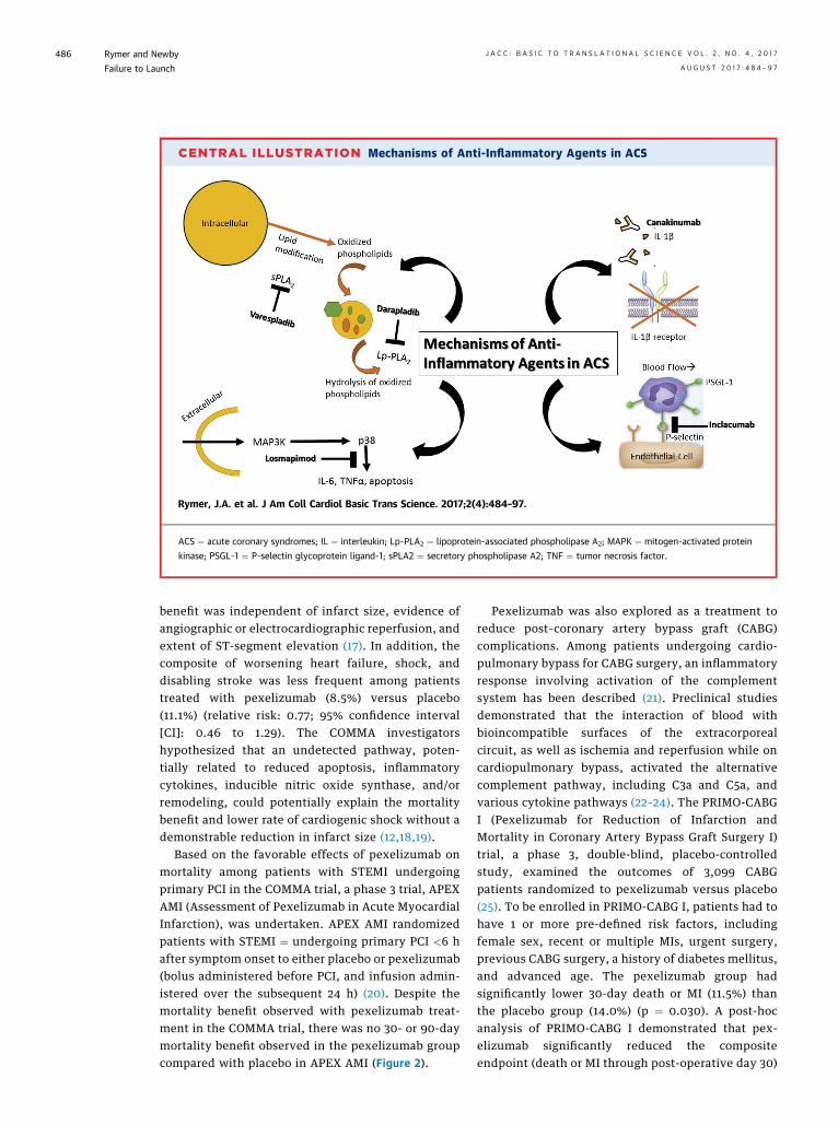
CENTRAL ILLUSTRATION Mechanisms of Anti-Inflammatory Agents in ACS
Rymer, J.A. et al. J Am Coll Cardiol Basic Trans Science. 2017;2(4):484–97.
ACS ¼ acute coronary syndromes; IL ¼ interleukin; Lp-PLA2 ¼ lipoprotein-associated phospholipase A2; MAPK ¼ mitogen-activated protein
kinase; PSGL-1 ¼ P-selectin glycoprotein ligand-1; sPLA2 ¼ secretory phospholipase A2; TNF ¼ tumor necrosis factor.
Rymer and Newby J A C C : B A S I C T O T R A N S L A T I O N A L S C I E N C E V O L . 2 , N O . 4 , 2 0 1 7
Failure to Launch A U G U S T 2 0 1 7 : 4 8 4 – 9 7
486
benefit was independent of infarct size, evidence ofangiographic or electrocardiographic reperfusion, andextent of ST-segment elevation (17). In addition, thecomposite of worsening heart failure, shock, anddisabling stroke was less frequent among patientstreated with pexelizumab (8.5%) versus placebo(11.1%) (relative risk: 0.77; 95% confidence interval[CI]: 0.46 to 1.29). The COMMA investigatorshypothesized that an undetected pathway, poten-tially related to reduced apoptosis, inflammatorycytokines, inducible nitric oxide synthase, and/orremodeling, could potentially explain the mortalitybenefit and lower rate of cardiogenic shock without ademonstrable reduction in infarct size (12,18,19).
Based on the favorable effects of pexelizumab onmortality among patients with STEMI undergoingprimary PCI in the COMMA trial, a phase 3 trial, APEXAMI (Assessment of Pexelizumab in Acute MyocardialInfarction), was undertaken. APEX AMI randomizedpatients with STEMI ¼ undergoing primary PCI <6 hafter symptom onset to either placebo or pexelizumab(bolus administered before PCI, and infusion admin-istered over the subsequent 24 h) (20). Despite themortality benefit observed with pexelizumab treat-ment in the COMMA trial, there was no 30- or 90-daymortality benefit observed in the pexelizumab groupcompared with placebo in APEX AMI (Figure 2).
Pexelizumab was also explored as a treatment toreduce post–coronary artery bypass graft (CABG)complications. Among patients undergoing cardio-pulmonary bypass for CABG surgery, an inflammatoryresponse involving activation of the complementsystem has been described (21). Preclinical studiesdemonstrated that the interaction of blood withbioincompatible surfaces of the extracorporealcircuit, as well as ischemia and reperfusion while oncardiopulmonary bypass, activated the alternativecomplement pathway, including C3a and C5a, andvarious cytokine pathways (22–24). The PRIMO-CABGI (Pexelizumab for Reduction of Infarction andMortality in Coronary Artery Bypass Graft Surgery I)trial, a phase 3, double-blind, placebo-controlledstudy, examined the outcomes of 3,099 CABGpatients randomized to pexelizumab versus placebo(25). To be enrolled in PRIMO-CABG I, patients had tohave 1 or more pre-defined risk factors, includingfemale sex, recent or multiple MIs, urgent surgery,previous CABG surgery, a history of diabetes mellitus,and advanced age. The pexelizumab group hadsignificantly lower 30-day death or MI (11.5%) thanthe placebo group (14.0%) (p ¼ 0.030). A post-hocanalysis of PRIMO-CABG I demonstrated that pex-elizumab significantly reduced the compositeendpoint (death or MI through post-operative day 30)
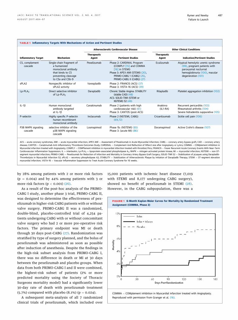
TABLE 1 Inflammatory Targets With Mechanisms of Action and Pertinent Studies
Inflammatory Target Mechanism
Atherosclerotic Cardiovascular Disease Other Clinical Conditions
TherapeuticAgent Pertinent Studies
TherapeuticAgent Indication/Pertinent Studies
C5, complementcascade
Single-chain fragment ofa humanizedmonoclonal antibodythat binds to C5preventing cleavageto C5a and C5b-9
Pexelizumab Phase 2: CARDINAL Program(COMPLY [15] and COMMA[16] in STEMI)
Phase 3: APEX AMI (STEMI) (20),PRIMO-CABG 1 (CABG) (25),PRIMO-CABG II (CABG) (27)
Eculizumab Atypical hemolytic uremic syndrome(99), pregnant patients withparoxysmal nocturnalhemoglobinuria (100), maculardegeneration (101)
sPLA2 Nonspecific inhibitor ofsPLA2 activity
Varespladib Phase 2: FRANCIS (ACS) (37)Phase 3: VISTA-16 (ACS) (39)
Lp-PLA2 Direct selective inhibitorof Lp-PLA2
Darapladib Chronic Stable Angina: STABILITY(stable CAD) (48)
ACS: SOLID-TIMI (STEMI orNSTEMI) 52 (49)
Rilapladib Platelet aggregation inhibition (102)
IL-1b Human monoclonalantibody targetedat IL-1b
Canakinumab Phase 2 (patients with highcardiovascular risk) (61)
Phase 3: CANTOS (post-ACS)
Anakinra(IL1-RA)
Recurrent pericarditis (103)Rheumatoid arthritis (104)Severe hidradenitis suppurativa (105)
P-selectin Highly specific P-selectinhuman recombinantmonoclonal antibody
Inclacumab Phase 2 (NSTEMI, CABG)(69,72)
Crizanlizumab Sickle cell pain (106)
P38 MAPK signalingcascade
selective inhibitor of thep38 MAPK signalingcascade
Losmapimod Phase 1b: (NSTEMI) (85)Phase 3: (acute MI) (86)
Doramapimod Active Crohn’s disease (107)
ACS ¼ acute coronary syndromes; AMI ¼ acute myocardial infarction; APEX AMI ¼ Assessment of Pexelizumab in Acute Myocardial Infarction; CABG ¼ coronary artery bypass graft; CAD ¼ coronary arterydisease; CANTOS ¼ Canakinumab Anti-Inflammatory Thrombosis Outcomes Study; CARDINAL ¼ Complement And ReDuction of INfarct size after Angioplasty or Lytics; COMMA ¼ COMplement inhibition inMyocardial infarction treated with Angioplasty; COMPLY ¼ COMPlement inhibition in myocardial infarction treated with thromboLYtics; FRANCIS ¼ Fewer Recurrent Acute Coronary Events With Near-TermCardiovascular Inflammation Suppression; IL ¼ interleukin; Lp-PLA2 ¼ lipoprotein-associated phospholipase A2; MAPK ¼ mitogen-activated protein kinase; MI ¼ myocardial infarction; NSTEMI ¼ non–ST-segment myocardial infarction; PRIMO-CABG ¼ Pexelizumab for Reduction of Infarction and Mortality in Coronary Artery Bypass Graft Surgery; SOLID-TIMI 52 ¼ Stabilization of pLaques usIng Darapladib-Thrombolysis In Myocardial Infarction 52; sPLA2 ¼ secretory phospholipase A2; STABILITY ¼ Stabilization of Atherosclerotic Plaque by Initiation of Darapladib Therapy; STEMI ¼ ST-segment elevationmyocardial infarction; VISTA-16 ¼ Vascular Inflammation Suppression to Treat Acute Coronary Syndrome for 16 weeks.
FIGURE 1 6-Month Kaplan-Meier Curves for Mortality by Randomized Treatment
Assignment (COMMA, Phase 2)
COMMA ¼ COMplement inhibition in Myocardial infarction treated with Angioplasty.
Reproduced with permission from Granger et al. (16).
J A C C : B A S I C T O T R A N S L A T I O N A L S C I E N C E V O L . 2 , N O . 4 , 2 0 1 7 Rymer and NewbyA U G U S T 2 0 1 7 : 4 8 4 – 9 7 Failure to Launch
487
by 28% among patients with 2 or more risk factors(p ¼ 0.004) and by 44% among patients with 3 ormore risk factors (p < 0.001) (26).
As a result of the post-hoc analysis of the PRIMO-CABG I study, another phase 3 trial, PRIMO-CABG II,was designed to determine the effectiveness of pex-elizumab in higher-risk CABG patients with or withoutvalve surgery. PRIMO-CABG II was a randomized,double-blind, placebo-controlled trial of 4,254 pa-tients undergoing CABG with or without concomitantvalve surgery who had 2 or more pre-operative riskfactors. The primary endpoint was MI or deaththrough 30 days post-CABG (27). Randomization wasstratified by type of surgery planned, and the bolus ofpexelizumab was administered as soon as possibleafter induction of anesthesia. Despite the findings inthe high-risk subset analysis from PRIMO-CABG I,there was no difference in death or MI at 30 daysbetween the pexelizumab and placebo groups. Whendata from both PRIMO-CABG I and II were combined,the highest-risk subset of patients (2% or morepredicted mortality using the Society of ThoracicSurgeons mortality model) had a significantly lower30-day rate of death with pexelizumab treatment(5.7%) compared with placebo (8.1%) (p ¼ 0.024).
A subsequent meta-analysis of all 7 randomizedclinical trials of pexelizumab, which included over
15,000 patients with ischemic heart disease (7,019with STEMI and 8,177 undergoing CABG surgery),showed no benefit of pexelizumab in STEMI (28).However, in the CABG subpopulation, there was a
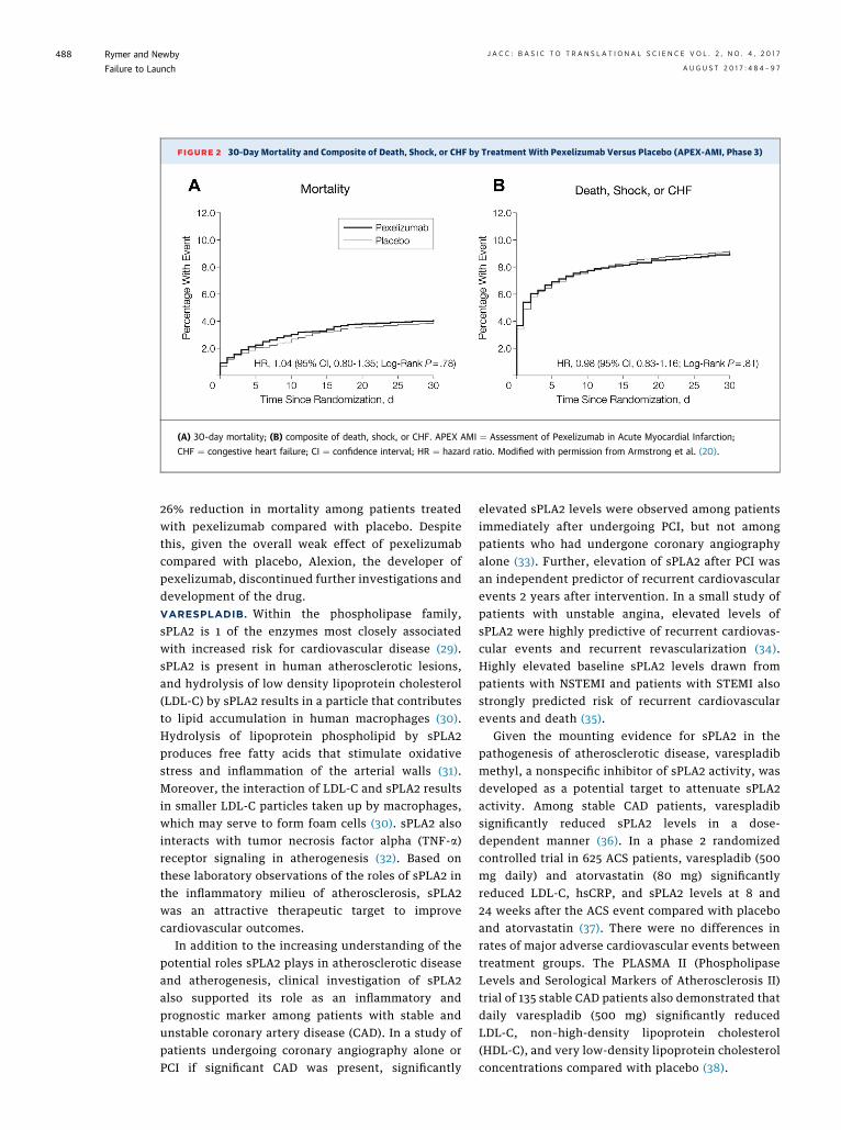
FIGURE 2 30-Day Mortality and Composite of Death, Shock, or CHF by Treatment With Pexelizumab Versus Placebo (APEX-AMI, Phase 3)
(A) 30-day mortality; (B) composite of death, shock, or CHF. APEX AMI ¼ Assessment of Pexelizumab in Acute Myocardial Infarction;
CHF ¼ congestive heart failure; CI ¼ confidence interval; HR ¼ hazard ratio. Modified with permission from Armstrong et al. (20).
Rymer and Newby J A C C : B A S I C T O T R A N S L A T I O N A L S C I E N C E V O L . 2 , N O . 4 , 2 0 1 7
Failure to Launch A U G U S T 2 0 1 7 : 4 8 4 – 9 7
488
26% reduction in mortality among patients treatedwith pexelizumab compared with placebo. Despitethis, given the overall weak effect of pexelizumabcompared with placebo, Alexion, the developer ofpexelizumab, discontinued further investigations anddevelopment of the drug.VARESPLADIB. Within the phospholipase family,sPLA2 is 1 of the enzymes most closely associatedwith increased risk for cardiovascular disease (29).sPLA2 is present in human atherosclerotic lesions,and hydrolysis of low density lipoprotein cholesterol(LDL-C) by sPLA2 results in a particle that contributesto lipid accumulation in human macrophages (30).Hydrolysis of lipoprotein phospholipid by sPLA2produces free fatty acids that stimulate oxidativestress and inflammation of the arterial walls (31).Moreover, the interaction of LDL-C and sPLA2 resultsin smaller LDL-C particles taken up by macrophages,which may serve to form foam cells (30). sPLA2 alsointeracts with tumor necrosis factor alpha (TNF-a)receptor signaling in atherogenesis (32). Based onthese laboratory observations of the roles of sPLA2 inthe inflammatory milieu of atherosclerosis, sPLA2was an attractive therapeutic target to improvecardiovascular outcomes.
In addition to the increasing understanding of thepotential roles sPLA2 plays in atherosclerotic diseaseand atherogenesis, clinical investigation of sPLA2also supported its role as an inflammatory andprognostic marker among patients with stable andunstable coronary artery disease (CAD). In a study ofpatients undergoing coronary angiography alone orPCI if significant CAD was present, significantly
elevated sPLA2 levels were observed among patientsimmediately after undergoing PCI, but not amongpatients who had undergone coronary angiographyalone (33). Further, elevation of sPLA2 after PCI wasan independent predictor of recurrent cardiovascularevents 2 years after intervention. In a small study ofpatients with unstable angina, elevated levels ofsPLA2 were highly predictive of recurrent cardiovas-cular events and recurrent revascularization (34).Highly elevated baseline sPLA2 levels drawn frompatients with NSTEMI and patients with STEMI alsostrongly predicted risk of recurrent cardiovascularevents and death (35).
Given the mounting evidence for sPLA2 in thepathogenesis of atherosclerotic disease, varespladibmethyl, a nonspecific inhibitor of sPLA2 activity, wasdeveloped as a potential target to attenuate sPLA2activity. Among stable CAD patients, varespladibsignificantly reduced sPLA2 levels in a dose-dependent manner (36). In a phase 2 randomizedcontrolled trial in 625 ACS patients, varespladib (500mg daily) and atorvastatin (80 mg) significantlyreduced LDL-C, hsCRP, and sPLA2 levels at 8 and24 weeks after the ACS event compared with placeboand atorvastatin (37). There were no differences inrates of major adverse cardiovascular events betweentreatment groups. The PLASMA II (PhospholipaseLevels and Serological Markers of Atherosclerosis II)trial of 135 stable CAD patients also demonstrated thatdaily varespladib (500 mg) significantly reducedLDL-C, non–high-density lipoprotein cholesterol(HDL-C), and very low-density lipoprotein cholesterolconcentrations compared with placebo (38).
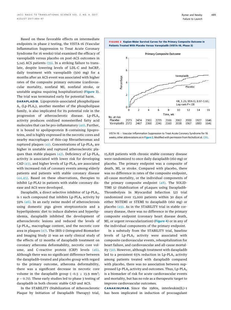
FIGURE 3 Kaplan-Meier Survival Curves for the Primary Composite Outcome in
Patients Treated With Placebo Versus Varespladib (VISTA-16, Phase 3)
VISTA-16 ¼ Vascular Inflammation Suppression to Treat Acute Coronary Syndrome for 16
weeks; other abbreviations as inFigure 2.Modifiedwith permission fromNicholls et al. (39).
J A C C : B A S I C T O T R A N S L A T I O N A L S C I E N C E V O L . 2 , N O . 4 , 2 0 1 7 Rymer and NewbyA U G U S T 2 0 1 7 : 4 8 4 – 9 7 Failure to Launch
489
Based on these favorable effects on intermediateendpoints in phase 2 testing, the VISTA-16 (VascularInflammation Suppression to Treat Acute CoronarySyndrome for 16 weeks) trial examined the efficacy ofvarespladib versus placebo on post-ACS outcomes in5,145 ACS patients (39). In a striking failure to trans-late, despite lowering levels of LDL-C and hsCRP,daily treatment with varespladib (500 mg) for 4months after an ACS event was associated with higherrates of the composite primary outcome (cardiovas-cular mortality, nonfatal MI, nonfatal stroke, orunstable angina requiring hospitalization) (Figure 3).The trial was terminated early for potential harm.DARAPLADIB. Lipoprotein-associated phospholipaseA2 (Lp-PLA2), another member of the phospholipasefamily, is also implicated for its potential role in theprogression of atherosclerotic disease. Lp-PLA2
activity produces oxidized nonesterified fatty acidmolecules that can be pro-inflammatory (40). Further,it is bound to apolipoprotein B–containing lipopro-teins, and is highly expressed in the necrotic cores andnearby macrophages of thin-cap fibroatheromas andruptured plaques (41). Concentrations of Lp-PLA2 arehigher in unstable and ruptured atherosclerotic pla-ques than stable plaques (42). Deficiency of Lp-PLA2
activity is associated with lower risk for developingCAD (43), and higher levels of Lp-PLA2 are associatedwith increased risk of coronary events among elderlypatients and patients with stable coronary disease(44,45). Based on these observations, therapies toinhibit Lp-PLA2 in patients with stable coronary dis-ease and ACS were developed.
Darapladib, a direct selective inhibitor of Lp-PLA2,is 1 such compound that inhibits Lp-PLA2 activity by59% (46). In an early swine model of atherosclerosisusing domestic pigs given streptozotocin and ahyperlipidemic diet to induce diabetes and hyperlip-idemia, darapladib inhibited the development ofatherosclerotic lesions and reduced the levels ofLp-PLA2, macrophage content, and the necrotic corearea in plaques (47). The IBIS-2 (Integrated Biomarkerand Imaging Study 2) was an early clinical study ofthe effects of 12 months of darapladib treatment oncoronary atheroma deformability, necrotic core vol-ume, and C-reactive protein (CRP) levels (46).Although there was no significant difference betweenthe darapladib-treated and placebo group with regardto the primary outcome, atheroma deformability,there was a significant decrease in necrotic corevolume in the darapladib group (�0.5 � 13.9 mm3;p ¼ 0.71). These early studies led to phase 3 testing ofdarapladib in both chronic stable CAD and ACS.
In the STABILITY (Stabilization of AtheroscleroticPlaque by Initiation of Darapladib Therapy) trial,
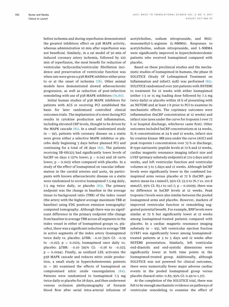
15,828 patients with chronic stable coronary diseasewere randomized to once daily darapladib (160 mg) orplacebo. The primary endpoint was a composite ofdeath, MI, or stroke. Compared with placebo, therewas no difference in rates of the composite endpoint,all-cause mortality, or the individual components ofthe primary composite endpoint (48). The SOLID-TIMI 52 (Stabilization of pLaques usIng Darapladib-Thrombolysis In Myocardial Infarction 52) trialrandomized over 13,000 patients within 30 days ofeither NSTEMI or STEMI to darapladib (160 mg) orplacebo (49). As in the STABILITY trial in stable cor-onary disease, there was no difference in the primarycomposite endpoint (coronary heart disease death,MI, or urgent revascularization) (Figure 4) or in any ofthe individual components of the primary endpoint.
In a substudy from the STABILITY trial, baselinelevels of Lp-PLA2 activity were associated withcomposite cardiovascular events, rehospitalization forheart failure, and cardiovascular and all-cause mortal-ity (50). However, although treatment with darapladibled to a persistent 65% reduction in Lp-PLA2 activityamong patients treated with darapladib comparedwith placebo, there was no association between sup-pressed Lp-PLA2 activity and outcomes. Thus, Lp-PLA2
is a biomarker of risk for acute cardiovascular eventsand mortality, but has no role as a therapeutic target toimprove cardiovascular outcomes.CANAKINUMAB. Since the 1980s, interleukin(IL)-1has been implicated in induction of procoagulant
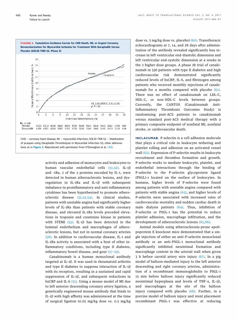
FIGURE 4 Cumulative Incidence Curves for CHD Death, MI, or Urgent Coronary
Revascularization for Myocardial Ischemia for Treatment With Darapladib Versus
Placebo (SOLID-TIMI 52, Phase 3)
CHD ¼ coronary heart disease; MI ¼ myocardial infarction; SOLID-TIMI 52 ¼ Stabilization
of pLaques usIng Darapladib-Thrombolysis In Myocardial Infarction 52; other abbrevia-
tions as in Figure 2. Reproduced with permission from O’Donoghue et al. (49).
Rymer and Newby J A C C : B A S I C T O T R A N S L A T I O N A L S C I E N C E V O L . 2 , N O . 4 , 2 0 1 7
Failure to Launch A U G U S T 2 0 1 7 : 4 8 4 – 9 7
490
activity and adhesion of monocytes and leukocytes tohuman vascular endothelial cells (51,52). IL-1band -1Ra, 2 of the 3 proteins encoded by IL-1, weredetected in human atherosclerotic lesions, and dys-regulation in IL-1Ra and IL-1b with subsequentimbalance in proinflammatory and anti-inflammatorycytokines has been hypothesized to promote athero-sclerotic disease (51,53,54). In clinical studies,patients with unstable angina had significantly higherlevels of IL-1Ra than patients with stable coronarydisease, and elevated IL-1Ra levels preceded eleva-tions in troponin and creatinine kinase in patientswith STEMI (55). IL-1b has been detected in theluminal endothelium and macrophages of athero-sclerotic lesions, but not in normal coronary arteries(56). In addition to cardiovascular disease, IL-1 andIL-1Ra activity is associated with a host of other in-flammatory conditions, including type II diabetes,inflammatory bowel disease, and gout (57–59).
Canakinumab is a human monoclonal antibodytargeted at IL-1b. It was used in rheumatoid arthritisand type II diabetes to suppress interaction of IL-1bwith its receptors, resulting in a sustained and rapidsuppression of IL-1b, and subsequent reductions inhsCRP and IL-6 (51). Using a mouse model of MI dueto left anterior descending coronary artery ligation, agenetically engineered mouse antibody that binds toIL-1b with high affinity was administered at the timeof surgical ligation (0.05 mg/kg dose vs. 0.5 mg/kg
dose vs. 5 mg/kg dose vs. placebo) (60). Transthoracicechocardiograms at 7, 14, and 28 days after adminis-tration of the antibody revealed significantly less in-crease in left ventricular end-diastolic dimension andleft ventricular end-systolic dimension at 4 weeks inthe 2 higher-dose groups. A phase 2b trial of canaki-numab in 556 patients with type II diabetes and highcardiovascular risk demonstrated significantlyreduced levels of hsCRP, IL-6, and fibrinogen amongpatients who received monthly injections of canaki-numab for 4 months compared with placebo (61).There was no effect of canakinumab on LDL-C,HDL-C, or non–HDL-C levels between groups.Currently, the CANTOS (Canakinumab Anti-Inflammatory Thrombosis Outcomes Study) israndomizing post-ACS patients to canakinumabversus standard post-ACS medical therapy with aprimary composite endpoint of nonfatal MI, nonfatalstroke, or cardiovascular death.
INCLACUMAB. P-selectin is a cell adhesion moleculethat plays a critical role in leukocyte tethering andplatelet rolling and adhesion on an activated vesselwall (62). Expression of P-selectin results in leukocyterecruitment and thrombus formation and growth.P-selectin works to mediate leukocyte, platelet, andendothelial interactions through the binding ofP-selectin to the P-selectin glycoprotein ligand(PSGL)-1 located on the surface of leukocytes. Inhumans, higher levels of P-selectin were foundamong patients with unstable angina compared withpatients with stable angina (63), and higher levels ofP-selectin were associated with increased rates ofcardiovascular mortality and sudden cardiac death inmale dialysis patients (64). Thus, inhibition ofP-selectin or PSGL-1 has the potential to reduceplatelet adhesion, macrophage infiltration, and thedevelopment of atherosclerotic lesions (65,66).
Animal models using atherosclerosis-prone apoli-poprotein E knockout mice demonstrated that a sin-gle injection of either an anti–P-selectin monoclonalantibody or an anti–PSGL-1 monoclonal antibodysignificantly inhibited neointimal formation andmacrophage content in the arterial wall when given3 h before carotid artery wire injury (67). In a pigmodel of balloon-mediated injury to the left anteriordescending and right coronary arteries, administra-tion of a recombinant immunoglobulin to PSGL-115 min before balloon injury significantly reducedneointimal hyperplasia and levels of TNF-a, IL-1b,and macrophages at the site of the ballooninjury compared with placebo (68). Further, in aporcine model of balloon injury and stent placementrecombinant PSGL-1 was effective at reducing
J A C C : B A S I C T O T R A N S L A T I O N A L S C I E N C E V O L . 2 , N O . 4 , 2 0 1 7 Rymer and NewbyA U G U S T 2 0 1 7 : 4 8 4 – 9 7 Failure to Launch
491
platelet-leukocyte reactions and rates of in-stentrestenosis (69). Pigs underwent angioplasty injury totheir coronary arteries, and 2 weeks later had stentsimplanted at the sites of injury 15 min after admin-istration of recombinant PSGL-1 versus placebo. Pigstreated with recombinant PSGL-1 had 3-fold largerresidual lumen 4 weeks later. Additionally, in amouse model of P-selectin activity, P-selectinknockout mice demonstrated inhibited migration ofsmooth muscle cells into the atherosclerotic lesioncompared with wild-type mice (70).
Given the promising results for P-selectin inhibi-tion in animal models of vascular injury, inclacumabwas developed as a highly specific human recombi-nant monoclonal antibody against P-selectin. In thephase 2 SELECT-ACS (Effects of the P-SelectinAntagonist Inclacumab on Myocardial Damage afterPercutaneous Coronary Intervention for Non-ST-Elevation Myocardial Infarction) trial, 544 patientswith NSTEMI who were scheduled to undergocoronary angiography were randomized to an infu-sion of placebo, a 5-mg/kg inclacumab infusion, or a20-mg/kg inclacumab infusion, between 1 and 24 hbefore PCI (71). The primary endpoint was change introponin I level from baseline at 16 and 24 h. Therewas no significant difference in change in placebo-adjusted geometric mean percent change introponin I levels at 16 and 24 h from baseline in thegroup who received the 5-mg/kg inclacumab infusioncompared with placebo. However, in the group thatreceived the 20-mg/kg inclacumab infusion comparedwith placebo, there was a significant difference inplacebo-adjusted geometric mean percent change introponin I levels at 24 h (decrease by 23.8%; p ¼ 0.05)but not at 16 h (decrease by 22.4%; p ¼ 0.07). Thedose-dependent effect of inclacumab on infarct sizeas assessed by change in troponin I levels at 24 h inSELECT-NSTEMI suggested that inhibition ofP-selectin with inclacumab could be a viable treat-ment for patients with NSTEMI. Ultimately, a largephase 3 trial will be needed to determine if the use ofinclacumab in patients with NSTEMI translates intoan actual improvement in clinical outcomes,including rates of post-MI heart failure and mortality.
Based on several preclinical studies, inclacumabhas also been studied in patients undergoing CABG.Failure of saphenous vein grafts (SVGs) remains anongoing concern for patients undergoing CABG and isbelieved to be mediated by the binding of PSGL-1to platelet P-selectin and platelet-leukocyteinteractions. SELECT-CABG (Effects of the P-selectinAntagonist Inclacumab in Patients Undergoing Coro-nary Artery Bypass Graft Surgery) was a phase 2 trialof inclacumab for SVG preservation. In this trial, 384
patients undergoing CABG (with placement of at least1 SVG) were randomized to receive a 20-mg/kg infu-sion of inclacumab or placebo (starting between 4 hand 6 weeks before CABG) and continuing at 4-weekintervals for 32 weeks (72). At the conclusion oftreatment, there was no significant difference in theper-protocol population in the percentage of patientswith $1 SVG with a diameter stenosis >50% (26.4%placebo group vs. 22.3% inclacumab group; adjustedodds ratio: 0.80; 95% CI: 0.47 to 1.38; p ¼ 0.43).Similarly, there was no significant difference in thepercentage of patients with $1 SVG with a diameterstenosis >50% in the intention-to-treat population at1 year. The results of the SELECT-CABG trial demon-strated little practical use for inclacumab in thepost-CABG population, despite promising results inanimal models of arterial injury, pre-clinical studies,and SELECT-ACS.LOSMAPIMOD. The intracellular MAPK signalingcascades have been investigated for their potential rolein the pathogenesis of atherosclerotic disease. The p38MAPK family is a part of the group of stress-activatedkinases that transduce extracellular signals that allowfor cellular adaptation (73). However, after atheroscle-rotic plaque rupture, ischemia, or vascular injury, thep38 MAPKs are activated in endothelial cells, myocar-dium, andmacrophages; regulate the transcription andtranslation of inflammatory molecules, such as TNF-aand interleukins 1, 6, and 8; and aid in the formation ofreactive oxygen species (5,73–75). Among patients withadvanced heart failure and ischemic cardiomyopathy,p38 MAPK activation has been associated with fibrosis,apoptosis, and hypertrophy, which have been linked tocardiac remodeling (76).
Losmapimod is a selective inhibitor of the p38MAPK signaling cascade, developed by GlaxoSmith-Kline (Brentford, United Kingdom). Various preclini-cal studies supported the investigation of losmapimodand p38 MAPK signaling in ACS. In a model of p38MAPK activation, rabbit hearts were excised andmounted on a Langendorff perfusion apparatus, withexposure to 30 min of global ischemia, with either 60(to test p38 MAPK levels) or 120 min (for myocardialnecrosis) of reperfusion time (77). The hearts werethen split into 4 groups: administration of normalsaline, administration of p38 MAPK inhibitor 10 minbefore ischemia and during reperfusion, administra-tion of p38 MAPK inhibitor at the time of reperfusionand throughout, or administration of p38 MAPKinhibitor 10 min after reperfusion began andthroughout. Overall, the administration of the p38MAPK inhibitor resulted in decreased myocardialapoptosis (14.7 � 3.2% vs. 30.6 � 3.5% placebo;p < 0.01). Importantly, administering the inhibitor
Rymer and Newby J A C C : B A S I C T O T R A N S L A T I O N A L S C I E N C E V O L . 2 , N O . 4 , 2 0 1 7
Failure to Launch A U G U S T 2 0 1 7 : 4 8 4 – 9 7
492
before ischemia and during reperfusion demonstratedthe greatest inhibitory effect on p38 MAPK activity,whereas administration 10 min after reperfusion wasnot beneficial. Similarly, in a rat model of 30 min ofinduced coronary artery ischemia, followed by 120min of reperfusion, the most benefit for reduction ofventricular tachycardia/ventricular fibrillation inci-dence and preservation of ventricular function waswhen rats were given a p38MAPK inhibitor either priorto or at the onset of ischemia (78). Other animalmodels have demonstrated slowed atheroscleroticprogression, as well as reduction of post-infarctionremodeling with use of p38 MAPK inhibitors (79,80).
Initial human studies of p38 MAPK inhibitors forpatients with ACS or receiving PCI established thebasis for later randomized controlled clinicaloutcomes trials. The implantation of a stent during PCIresults in cytokine production and inflammation,including elevated CRP levels, thought to be driven bythe MAPK cascade (81). In a small randomized study(n ¼ 92), patients with coronary disease on a statinwere given either a selective MAPK inhibitor or pla-cebo daily beginning 3 days before planned PCI andcontinuing for a total of 28 days (82). The patientsreceiving SB-681323 had significantly lower levels ofhsCRP on days 5 (37% lower; p ¼ 0.04) and 28 (40%lower; p ¼ 0.003) when compared with placebo. In astudy of the effect of losmapimod on vascular inflam-mation in the carotid arteries and aorta, 99 partici-pants with known atherosclerotic disease on a statinwere randomized to receive losmapimod 7.5 mg daily,7.5 mg twice daily, or placebo (83). The primaryendpoint was the change in baseline in the averagetissue-to-background ratio (TBR) of the index vessel(the artery with the highest average maximum TBR atbaseline) using FDG positron emission tomography/computed tomography. Although there was no signif-icant difference in the primary endpoint (the changefrom baseline in average TBR across all segments in theindex vessel in either of losmapimod groups vs. pla-cebo), there was a significant reduction in average TBRin active segments of the index artery (losmapimodtwice daily vs. placebo: DTBR: �0.10 [95% CI: �0.19to �0.02]; p ¼ 0.0125; losmapimod once daily vs.placebo: DTBR: �0.10 [95% CI: �0.18 to �0.02];p ¼ 0.0194). Finally, as oxidized LDL activates thep38 MAPK cascade and reduces nitric oxide produc-tion, a small study in hypercholesteremic patients(n ¼ 56) examined the effects of losmapimod oncompromised nitric oxide vasoregulation (84).Patients were randomized to losmapimod 7.5 mgtwice daily or placebo for 28 days, and then underwentvenous occlusion plethysmography of forearmblood flow after serial intra-arterial infusions of
acetylcholine, sodium nitroprusside, and N(G)-monomethyl-L-arginine (L-NMMA). Responses toacetylcholine, sodium nitroprusside, and L-NMMAwere significantly improved in hypercholesterolemicpatients who received losmapimod compared withplacebo.
Based on these preclinical studies and the mecha-nistic studies of losmapimod in humans, the phase 1bSOLSTICE (Study Of LoSmapimod Treatment onInflammation and infarCt sizE) was performed (85).SOLSTICE randomized over 500 patients with NSTEMIto treatment for 12 weeks with either losmapimod(either 7.5 or 15 mg loading dose followed by 7.5 mgtwice daily) or placebo within 18 h of presenting withan NSTEMI and at least 2 h prior to PCI to examine itsmechanistic effects. The coprimary outcomes wereinflammation (hsCRP concentration at 12 weeks) andinfarct size (area under the curve for troponin I over 72h or hospital discharge, whichever came first). Otheroutcomes included hsCRP concentrations at 14 weeks,IL-6 concentrations at 24 h and 12 weeks, infarct sizeby creatine kinase–MB area under the curve (AUC) andpeak troponin I concentration over 72 h or discharge,B-type natriuretic peptide levels at 72 h and 12 weeks,cardiac magnetic resonance imaging infarct size andLVEF (primary substudy endpoint) at 3 to 5 days and 12weeks, and left ventricular function and ventricularvolumes at 3 to 5 days and 12 weeks. Although hsCRPlevels were significantly lower in the combined los-mapimod arms versus placebo at 72 h (hsCRP: geo-metric mean 64.1 mmol/l, 95% CI: 53.0 to 76.7 vs. 110.8mmol/l, 95% CI: 83.1 to 147.7; p ¼ 0.0009), there wasno difference in hsCRP levels at 12 weeks. Peaktroponin I levels were also similar between the pooledlosmapimod arms and placebo. However, markers ofimproved ventricular function or remodeling sug-gested potential benefit. For example, BNP levels weresimilar at 72 h but significantly lower at 12 weeksamong losmapimod-treated patients compared withplacebo. In a cardiac magnetic resonance imagingsubstudy (n ¼ 93), left ventricular ejection fraction(LVEF) was significantly lower among losmapimod-treated patients at 3 to 5 days and 12 weeks afterNSTEMI presentation. Similarly, left ventricularend-diastolic and end-systolic dimensions weresignificantly lower at both time points in thelosmapimod-treated group. Additionally, althoughSOLSTICE was not powered for clinical outcomes,there were numerically fewer major adverse cardiacevents in the pooled losmapimod group versusplacebo (hazard ratio: 0.82; 95% CI: 0.49 to 1.37).
At the completion of the SOLSTICE trial, there wasfelt to be enoughmechanistic evidence on pathways ofventricular remodeling to examine the effect of
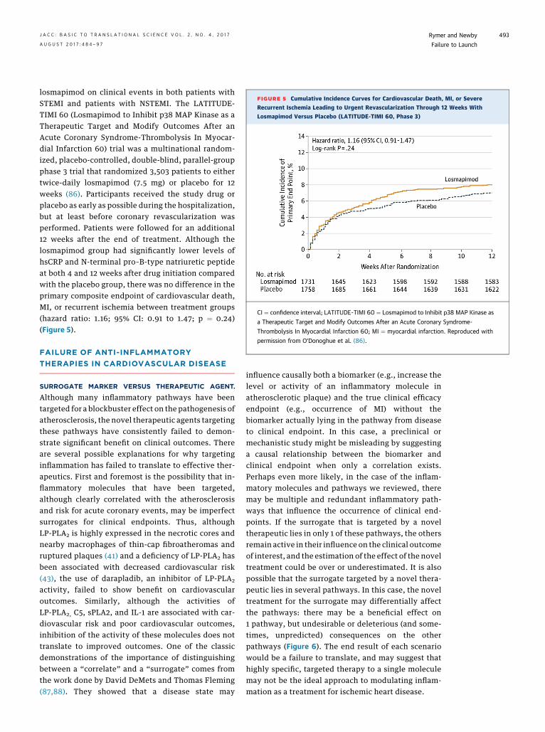
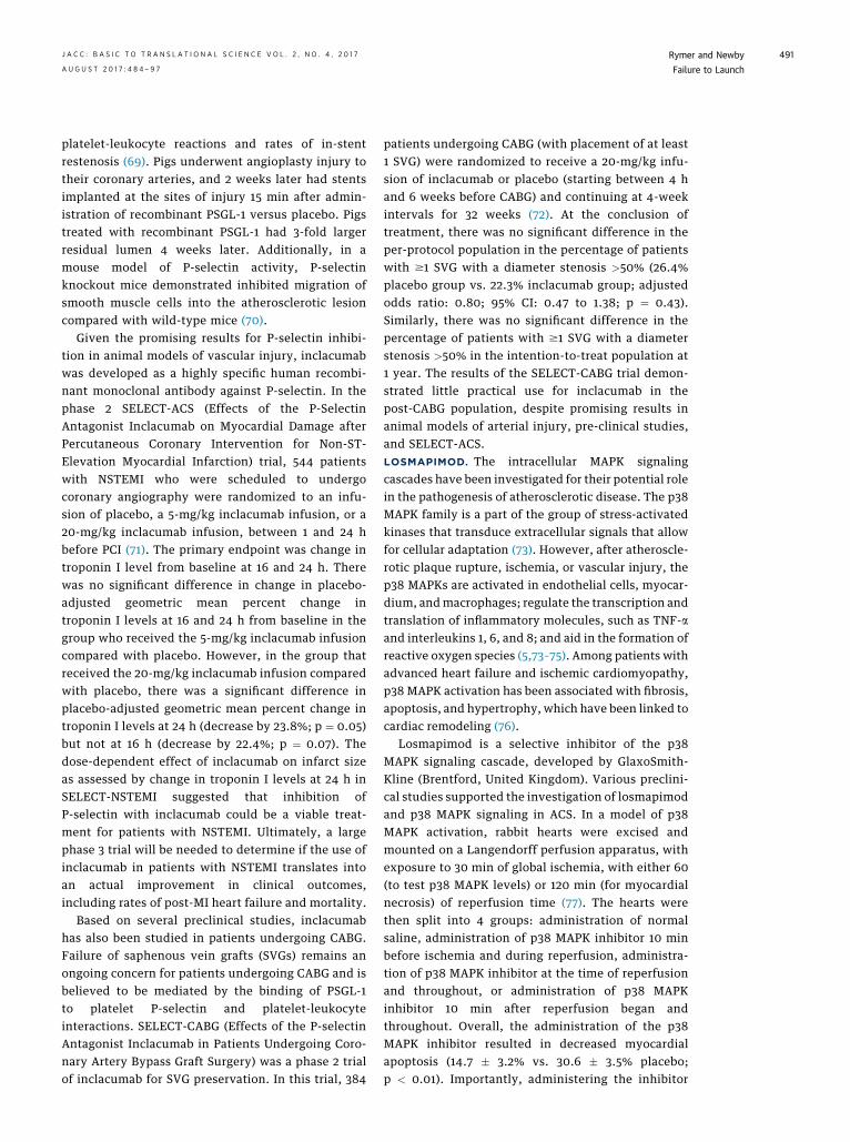
FIGURE 5 Cumulative Incidence Curves for Cardiovascular Death, MI, or Severe
Recurrent Ischemia Leading to Urgent Revascularization Through 12 Weeks With
Losmapimod Versus Placebo (LATITUDE-TIMI 60, Phase 3)
CI ¼ confidence interval; LATITUDE-TIMI 60 ¼ Losmapimod to Inhibit p38 MAP Kinase as
a Therapeutic Target and Modify Outcomes After an Acute Coronary Syndrome-
Thrombolysis In Myocardial Infarction 60; MI ¼ myocardial infarction. Reproduced with
permission from O’Donoghue et al. (86).
J A C C : B A S I C T O T R A N S L A T I O N A L S C I E N C E V O L . 2 , N O . 4 , 2 0 1 7 Rymer and NewbyA U G U S T 2 0 1 7 : 4 8 4 – 9 7 Failure to Launch
493
losmapimod on clinical events in both patients withSTEMI and patients with NSTEMI. The LATITUDE-TIMI 60 (Losmapimod to Inhibit p38 MAP Kinase as aTherapeutic Target and Modify Outcomes After anAcute Coronary Syndrome-Thrombolysis In Myocar-dial Infarction 60) trial was a multinational random-ized, placebo-controlled, double-blind, parallel-groupphase 3 trial that randomized 3,503 patients to eithertwice-daily losmapimod (7.5 mg) or placebo for 12weeks (86). Participants received the study drug orplacebo as early as possible during the hospitalization,but at least before coronary revascularization wasperformed. Patients were followed for an additional12 weeks after the end of treatment. Although thelosmapimod group had significantly lower levels ofhsCRP and N-terminal pro–B-type natriuretic peptideat both 4 and 12 weeks after drug initiation comparedwith the placebo group, there was no difference in theprimary composite endpoint of cardiovascular death,MI, or recurrent ischemia between treatment groups(hazard ratio: 1.16; 95% CI: 0.91 to 1.47; p ¼ 0.24)(Figure 5).
FAILURE OF ANTI-INFLAMMATORY
THERAPIES IN CARDIOVASCULAR DISEASE
SURROGATE MARKER VERSUS THERAPEUTIC AGENT.
Although many inflammatory pathways have beentargeted for a blockbuster effect on the pathogenesis ofatherosclerosis, the novel therapeutic agents targetingthese pathways have consistently failed to demon-strate significant benefit on clinical outcomes. Thereare several possible explanations for why targetinginflammation has failed to translate to effective ther-apeutics. First and foremost is the possibility that in-flammatory molecules that have been targeted,although clearly correlated with the atherosclerosisand risk for acute coronary events, may be imperfectsurrogates for clinical endpoints. Thus, althoughLP-PLA2 is highly expressed in the necrotic cores andnearby macrophages of thin-cap fibroatheromas andruptured plaques (41) and a deficiency of LP-PLA2 hasbeen associated with decreased cardiovascular risk(43), the use of darapladib, an inhibitor of LP-PLA2
activity, failed to show benefit on cardiovascularoutcomes. Similarly, although the activities ofLP-PLA2, C5, sPLA2, and IL-1 are associated with car-diovascular risk and poor cardiovascular outcomes,inhibition of the activity of these molecules does nottranslate to improved outcomes. One of the classicdemonstrations of the importance of distinguishingbetween a “correlate” and a “surrogate” comes fromthe work done by David DeMets and Thomas Fleming(87,88). They showed that a disease state may
influence causally both a biomarker (e.g., increase thelevel or activity of an inflammatory molecule inatherosclerotic plaque) and the true clinical efficacyendpoint (e.g., occurrence of MI) without thebiomarker actually lying in the pathway from diseaseto clinical endpoint. In this case, a preclinical ormechanistic study might be misleading by suggestinga causal relationship between the biomarker andclinical endpoint when only a correlation exists.Perhaps even more likely, in the case of the inflam-matory molecules and pathways we reviewed, theremay be multiple and redundant inflammatory path-ways that influence the occurrence of clinical end-points. If the surrogate that is targeted by a noveltherapeutic lies in only 1 of these pathways, the othersremain active in their influence on the clinical outcomeof interest, and the estimation of the effect of the noveltreatment could be over or underestimated. It is alsopossible that the surrogate targeted by a novel thera-peutic lies in several pathways. In this case, the noveltreatment for the surrogate may differentially affectthe pathways: there may be a beneficial effect on1 pathway, but undesirable or deleterious (and some-times, unpredicted) consequences on the otherpathways (Figure 6). The end result of each scenariowould be a failure to translate, and may suggest thathighly specific, targeted therapy to a single moleculemay not be the ideal approach to modulating inflam-mation as a treatment for ischemic heart disease.
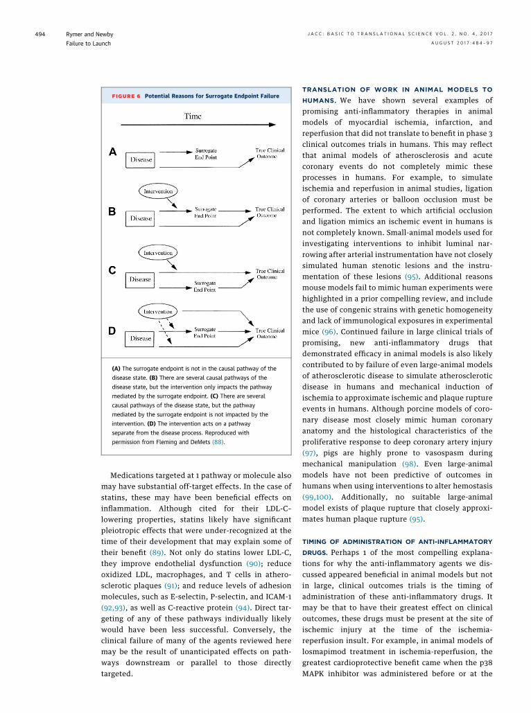
FIGURE 6 Potential Reasons for Surrogate Endpoint Failure
(A) The surrogate endpoint is not in the causal pathway of the
disease state. (B) There are several causal pathways of the
disease state, but the intervention only impacts the pathway
mediated by the surrogate endpoint. (C) There are several
causal pathways of the disease state, but the pathway
mediated by the surrogate endpoint is not impacted by the
intervention. (D) The intervention acts on a pathway
separate from the disease process. Reproduced with
permission from Fleming and DeMets (88).
Rymer and Newby J A C C : B A S I C T O T R A N S L A T I O N A L S C I E N C E V O L . 2 , N O . 4 , 2 0 1 7
Failure to Launch A U G U S T 2 0 1 7 : 4 8 4 – 9 7
494
Medications targeted at 1 pathway or molecule alsomay have substantial off-target effects. In the case ofstatins, these may have been beneficial effects oninflammation. Although cited for their LDL-C–lowering properties, statins likely have significantpleiotropic effects that were under-recognized at thetime of their development that may explain some oftheir benefit (89). Not only do statins lower LDL-C,they improve endothelial dysfunction (90); reduceoxidized LDL, macrophages, and T cells in athero-sclerotic plaques (91); and reduce levels of adhesionmolecules, such as E-selectin, P-selectin, and ICAM-1(92,93), as well as C-reactive protein (94). Direct tar-geting of any of these pathways individually likelywould have been less successful. Conversely, theclinical failure of many of the agents reviewed heremay be the result of unanticipated effects on path-ways downstream or parallel to those directlytargeted.
TRANSLATION OF WORK IN ANIMAL MODELS TO
HUMANS. We have shown several examples ofpromising anti-inflammatory therapies in animalmodels of myocardial ischemia, infarction, andreperfusion that did not translate to benefit in phase 3clinical outcomes trials in humans. This may reflectthat animal models of atherosclerosis and acutecoronary events do not completely mimic theseprocesses in humans. For example, to simulateischemia and reperfusion in animal studies, ligationof coronary arteries or balloon occlusion must beperformed. The extent to which artificial occlusionand ligation mimics an ischemic event in humans isnot completely known. Small-animal models used forinvestigating interventions to inhibit luminal nar-rowing after arterial instrumentation have not closelysimulated human stenotic lesions and the instru-mentation of these lesions (95). Additional reasonsmouse models fail to mimic human experiments werehighlighted in a prior compelling review, and includethe use of congenic strains with genetic homogeneityand lack of immunological exposures in experimentalmice (96). Continued failure in large clinical trials ofpromising, new anti-inflammatory drugs thatdemonstrated efficacy in animal models is also likelycontributed to by failure of even large-animal modelsof atherosclerotic disease to simulate atheroscleroticdisease in humans and mechanical induction ofischemia to approximate ischemic and plaque ruptureevents in humans. Although porcine models of coro-nary disease most closely mimic human coronaryanatomy and the histological characteristics of theproliferative response to deep coronary artery injury(97), pigs are highly prone to vasospasm duringmechanical manipulation (98). Even large-animalmodels have not been predictive of outcomes inhumans when using interventions to alter hemostasis(99,100). Additionally, no suitable large-animalmodel exists of plaque rupture that closely approxi-mates human plaque rupture (95).
TIMING OF ADMINISTRATION OF ANTI-INFLAMMATORY
DRUGS. Perhaps 1 of the most compelling explana-tions for why the anti-inflammatory agents we dis-cussed appeared beneficial in animal models but notin large, clinical outcomes trials is the timing ofadministration of these anti-inflammatory drugs. Itmay be that to have their greatest effect on clinicaloutcomes, these drugs must be present at the site ofischemic injury at the time of the ischemia-reperfusion insult. For example, in animal models oflosmapimod treatment in ischemia-reperfusion, thegreatest cardioprotective benefit came when the p38MAPK inhibitor was administered before or at the
J A C C : B A S I C T O T R A N S L A T I O N A L S C I E N C E V O L . 2 , N O . 4 , 2 0 1 7 Rymer and NewbyA U G U S T 2 0 1 7 : 4 8 4 – 9 7 Failure to Launch
495
onset of ischemia, with little to no benefit whenadministered at the onset of reperfusion (77,78).However, the occurrence of acute coronary events inhumans does not easily accommodate the adminis-tration of agents like losmapimod prior to the occlu-sion of a coronary artery (as in animal models ofcoronary ligation). Further, the design and logistics ofhuman studies often do not allow for controlled, earlyadministration at the time of reperfusion. InSOLSTICE, losmapimod could be administered anytime within 18 h of presentation with NSTEMI (85). At18 h after presentation with NSTEMI, ischemic injuryand the resultant inflammatory process is likely wellunderway, which may explain the discrepancy inresults between the preclinical and clinical studies.Similarly, although the recombinant immunoglobulinto PSGL-1 was administered 15 min prior to ballooninjury in an animal model (66), the administration ofinclacumab occurred periprocedurally in SELECT-ACS(69). Unfortunately, earlier administration of thesenovel agents prior to or at the onset of ischemia isimpractical in clinical practice.
CONCLUSIONS
Over the past several decades, many novel therapieshave targeted inflammatory pathways involved in
ACS and atherosclerotic disease with the hope ofimproving outcomes post-ACS. Although manyagents showed promising results in early animalmodels and preclinical studies, demonstratingreductions in levels of inflammatory markers andinfarct size, these results failed to translate to ther-apeutic benefit when tested in large randomizedclinical outcomes trials. There are many potentialexplanations for why these seemingly promisingtherapies failed in clinical testing, from incompleteunderstanding of the role of inflammation in CADand ACS to imperfect animal models of disease andtreatment. This does not mean that we shouldabandon investigation into modulation of inflamma-tion to improve cardiovascular outcomes. Rather,these experiences should challenge us scientificallyto more deeply understand the contribution ofinflammation in ACS (correlate or mediator), while atthe same time rethinking the animal modelconstructs we use in early drug development so thatthey as closely as possible mimic human clinicalconditions.
ADDRESS FOR CORRESPONDENCE: Dr. L. KristinNewby, Division of Cardiology, Duke Clinical ResearchInstitute, P.O. Box 17969, Durham, North Carolina27715-7969. E-mail: [email protected].
RE F E RENCE S
1. Go AS, Mozaffarian D, Roger VL, et al. Heartdisease and stroke statistics—2014 update: areport from the American Heart Association.Circulation 2014;129:e28–292.
2. Zhao Z, Winget M. Economic burden of illness ofacute coronary syndromes: medical and produc-tivity costs. BMC Health Services Research 2011;11:2–9.
3. Ghushchyan V, Nair KV, Page RL. Indirect anddirect costs of acute coronary syndromes withcomorbid atrial fibrillation, heart failure, or both.Vasc Health Risk Manag 2015;11:25–34.
4. Yeh RW, Sidney S, Chandra M, Sorel M,Selby JV, Go AS. Population trends in the inci-dence and outcomes of acute myocardial infarc-tion. N Engl J Med 2010;362:2155–65.
5. Tobbia P, Brodie BR, Witzenbichler B, et al.Adverse event rates following primary PCI forSTEMI at US and non-US hospitals: three-yearanalysis from the HORIZONS-AMI trial. Euro-Intervention 2013;8:1134–42.
6. Brieger D, Fox KA, Fitzgerald G, et al. Predictingfreedom from clinical events in non-ST-elevationacute coronary syndromes: the Global Registry ofAcute Coronary Events. Heart 2009;95:888.
7. Suleiman M, Khatib R, Agmon Y, et al. Earlyinflammation and risk of long-term developmentof heart failure and mortality in survivors of acute
myocardial infarction predictive role of C-reactiveprotein. J Am Coll Cardiol 2006;47:962–8.
8. Kragholm K, Newby LK, Melloni C. Emergingtreatment options to improve cardiovascular out-comes in patients with acute coronary syndrome:focus on losmapimod. Drug Des Devel Ther 2015;9:4279–86.
9. Thomas TC, Rollins SA, Rother RP, et al.Inhibition of complement activity by humanizedanti-C5 antibody and single-chain Fv. Mol Immu-nol 1996;33:1389–401.
10. Pinckard RN, Olson MS, Giclas PC, Terry R,Boyer JT, O’Rourke RA. Consumption of classicalcomplement components by heart subcellularmembranes in vitro and in patients after acutemyocardial infarction. J Clin Invest 1975;56:740–50.
11. Mathey D, Schofer J, Schafer HJ, et al. Earlyaccumulation of the terminal complement-complex in the ischaemic myocardium afterreperfusion. Eur Heart J 1994;15:418–23.
12. Vakeva AP, Agah A, Rollins SA, et al. Myocar-dial infarction and apoptosis after myocardialischemia and reperfusion: role of the terminalcomplement components and inhibition by anti-C5therapy. Circulation 1998;97:2259–67.
13. Tang K, Cheng Y, Wu S, Liu L, Cheng L. Pro-tective effect of C5 shRNA on myocardial
ischemia-reperfusion injury in rats. Can J PhysiolPharmacol 2012;90:1394–402.
14. Amsterdam EA, Stahl GL, Pan HL, Rendig SV,Fletcher MP, Longhurst JC. Limitation of reperfu-sion injury by a monoclonal antibody to c5a duringmyocardial infarction in pigs. Am J Physiol 1995;268:H448–57.
15. Mahaffey KW, Granger CB, Nicolau JC, et al.Effect of pexelizumab, an anti-C5 complementantibody, as adjunctive therapy to fibrinolysis inacute myocardial infarction: The COMPlementinhibition in myocardial infarction treated withthromboLYtics (COMPLY) trial. Circulation 2003;108:1176–83.
16. Granger CB, Mahaffey KW, Weaver WD, et al.Effect of pexelizumab, an anti-C5 complementantibody, as adjunctive therapy to primary percu-taneous intervention acute myocardial infarction:the COMplement inhibition in Myocardial infarc-tion treated with Angioplasty (COMMA) trial.Circulation 2003;108:1184–90.
17. Armstrong PW, Mahaffey KW, Chang WC, et al.Concerning the mechanism of pexelizumab’sbenefit in acute myocardial infarction. Am Heart J2006;151:787–90.
18. Stahl GL, Amsterdam EA, Symons JD, et al.Role of thromboxane A2 in the cardiovascularresponse to intracoronary C5a. Circ Res 1990;66:1103–11.
Rymer and Newby J A C C : B A S I C T O T R A N S L A T I O N A L S C I E N C E V O L . 2 , N O . 4 , 2 0 1 7
Failure to Launch A U G U S T 2 0 1 7 : 4 8 4 – 9 7
496
19. Topham PS, Haydar SA, Kuphal R, et al.Complement-mediated injury reversibly disruptsglomerular epithelial cell actin microfilamentsand focal adhesions. Kidney Int 1999;55:1763–75.
20. Armstrong PW, Granger CB, Adams PX, et al.,for the APEX AMI Investigators. Pexelizumab foracute ST-elevation myocardial infarction inpatients undergoing primary percutaneous coro-nary intervention: a randomized controlled trial.JAMA 2007;297:43–51.
21. Fitch JC, Rollins S, Matis L, et al. Pharmacologyand biological efficacy of a recombinant, human-ized, single-chain antibody C5 complement inhib-itor in patients undergoing coronary artery bypassgraft surgery with cardiopulmonary bypass.Circulation 1999;100:2499–506.
22. Royston D. Systemic inflammatory responsesto surgery with CPB. Perfusion 1996;11:177–89.
23. Wan S, LeClerc J-L, Vincent J-L. Inflammatoryresponse to CPB. Chest 1997;112:676–92.
24. Hall RI, Smith MS, Rocker G. The systemicinflammatory response to CPB. Anesth Analg1997;85:766–82.
25. Verrier ED, Shernan SK, Taylor KM, et al. Ter-minal complement blockade with pexelizumabduring coronary artery bypass graft surgeryrequiring cardiopulmonary bypass: a randomizedtrial. JAMA 2004;291:2319–27.
26. Haverich A, Shernan SK, Levy JH. Inhibition ofcomplement activation by pexelizumab reduceddeath and myocardial infarction in higher riskcardiac surgical patients. Ann Thorac Surg 2006;82:486–92.
27. Smith PK, Shernan SK, Chen JC, et al. Effects ofC5 complement inhibitor pexelizumab on outcomein high-risk coronary artery bypass grafting: com-bined results from the PRIMO-CABG I and II trials.J Thorac Cardiovasc Surg 2011;142:89–98.
28. Testa L, Van Gaal WJ, Bhindi R, et al. Pex-elizumab in ischemic heart disease: a systematicreview and meta-analysis on 15,196 patients.J Thorac Cardiovasc Surg 2008;136:884–93.
29. Nicholls SJ, Cavender MA, Kastelein JP, et al.Inhibition of secretory phospholipase A(2) in patientswith acute coronary syndromes: rationale and designof the Vascular Inflammation Suppression to TreatAcute Coronary Syndrome for 16 Weeks (VISTA-16)trial. Cardiovasc Drugs Ther 2012;26:71–5.
30. Karabina SA, Brocheriou S, Naour GL, et al.Atherogenic properties of LDL particles modifiedby human group X secreted phospholipase A2 onhuman endothelial cell function. FASEB 2006;20:2547–9.
31. Gora S, Maouche S, Atout R, et al. Phospholi-polyzed LDL induces an inflammatory response inendothelial cells through endoplasmic reticulumstress signaling. FASEB 2010;24:3284–97.
32. Fuentes L, Hernandez M, Fernandez-Aviles FJ,et al. Cooperation between secretory phospholi-pase A2 and TNF-receptor superfamily signaling:implications for the inflammatory response inatherogenesis. Circ Res 2002;91:681–8.
33. Liu PY, Li YH, Wei-Chuan T, et al. Prognosticvalue and the changes of plasma levels of secretorytype II phospholipase A2 in patients with coronary
artery disease undergoing percutaneous coronaryintervention. Eur Heart J 2003;24:1824–32.
34. Kugiyama K, Ota Y, Sugiyama S, et al. Prog-nostic value of plasma levels of secretory type IIphospholipase A2 in patients with unstable anginapectoris. Am J Cardiol 2000;86:718–22.
35. Mallat Z, Steg G, Benessiano J, et al. Circulatingsecretory phospholipase A2 activity predicts recur-rent events in patients with severe acute coronarysyndromes. J Am Coll Cardiol 2005;46:1249–57.
36. RosensonRS,HislopC,McConnell D, etal. Effectsof 1-H-indole-3-glyoxamide (A-002) on concentra-tion of secretory phospholipase A2 (PLASMA study):a phase II double-blind, randomised, placebo-controlled trial. Lancet 2009;373:649–58.
37. Rosenson RS, Hislop C, Elliott M, et al. Effectsof varespladib methyl on biomarkers and majorcardiovascular events in acute coronary syndromepatients. J Am Coll Cardiol 2010;56:1079–88.
38. Rosenson RS, Elliott M, Stasiv Y, et al.Randomized trial of an inhibitor of secretoryphospholipase A2 on atherogenic lipoproteinsubclasses in statin-treated patients with coronaryheart disease. Eur Heart J 2011;32:999–1005.
39. Nicholls SJ, Kastelein J, Schwartz GG, et al.Varespladib and cardiovascular events in patientswith an acute coronary syndrome: the VISTA-16randomized clinical trial. JAMA 2014;311:252–62.
40. MacPhee CH,Moores KE, BoydHF. Lipoprotein-associated phospholipase A2, platelet-activatingfactor acetylhydrolase, generates two bioactiveproducts during the oxidation of low-density lipo-protein: use of a novel inhibitor. Biochem J 1999;338:479–87.
41. Kolodgie FD, Burke AP, Skorija KS, et al.Lipoprotein-associated phospholipase A2 proteinexpression in the natural progression of humancoronary atherosclerosis. Arterioscler ThrombVasc Biol 2006;26:2523–9.
42. Häkkinen T, Luoma JS, Hiltunen MO, et al.Lipoprotein-associated phospholipase A(2),platelet-activating factor acetylhydrolase, isexpressed by macrophages in human and rabbitatherosclerotic lesions. Arterioscler Thromb VascBiol 1999;19:2909–17.
43. Jang Y, Waterworth D, Lee J-E, et al. Carriageof the V279F null allele within the gene encodingLp-PLA2 is protective from coronary artery diseasein South Korean males. PloS One 2011;6:e18208.
44. The Lp-PLA2 Studies Collaboration. Lipopro-tein-associated phospholipase A(2) and risk ofcoronary disease, stroke, and mortality: collabo-rative analysis of 32 prospective studies. Lancet2010;375:1536–44.
45. Di Angelantonio E, Gao P, Pennells L, et al.Lipid-related markers and cardiovascular diseaseprediction. JAMA 2012;307:2499–506.
46. Serruys PW, Garcia-Garcia HM, Buszman P,et al. Effects of the direct-lipoprotein associatedphospholipase A2 inhibitor darapladib on humancoronary atherosclerotic plaque. Circulation 2008;118:1172–82.
47. Wilensky RL, Shi Y, Mohler ER, et al. Inhibitionof lipoprotein-associated Phospholipase A2 re-duces complex coronary atherosclerotic plaquedevelopment. Nature Medicine 2008;14:1059–66.
48. White HD, Held C, Stewart R, et al. Darapladibfor preventing ischemic events in stable coronarydisease. N Eng J Med 2014;370:1702–11.
49. O’Donoghue ML, Braunwald E, White HD,et al. Effect of darapladib on major coronaryevents after an acute coronary syndrome: theSOLID-TIMI 52 randomized clinical trial. JAMA2014;312:1006–15.
50. Wallentin L, Held C, Armstrong PW, et al.Lipoprotein-associated phospholipase A2 activityis a marker of risk but not a useful target fortreatment in patients with stable coronary heartdisease. J Am Heart Assoc 2016;5:e003407.
51. Ridker PM, Thuren T, Zalewski A, et al.Interleukin-1b inhibition and the prevention ofrecurrent cardiovascular events: Rationale andDesign of the Canakinumab Anti-inflammatoryThrombosis Outcomes Study (CANTOS). AmHeart J 2011;162:597–605.
52. Bevilacqua MP, Pober JS, Wheeler ME, et al.Interleukin 1 acts on cultured human vascularendothelium to increase the adhesion of poly-morphonuclear leukocytes,monocytes, and relatedleukocyte cell lines. J Clin Invest 1985;76:2003–11.
53. Dinarello CA. Interleukin-1beta and the auto-inflammatory diseases. N Eng J Med 2009;360:2467–70.
54. Fearon WF, Fearon DT. Inflammation andcardiovascular disease: role of the interleukin-1receptor antagonist. Circulation 2008;117:2577–9.
55. Patti G, D’Ambrosio A, Dobrina A, et al. Inter-leukin-1 receptor antagonist: a sensitive marker ofinstability in patients with coronary artery disease.J Thromb Thrombolysis 2002;14:139–43.
56. Galea J, Armstrong J, Gadsdon P, et al. Inter-leukin-1 beta in coronary arteries of patients withischemic heart disease. Arterioscler Thromb VascBiol 1996;16:1000–6.
57. Larsen CM, Faulenbach M, Vaag A, et al.Interleukin-1-receptor antagonist in type 2 dia-betes mellitus. N Engl J Med 2007;356:1517–26.
58. Casini-Raggi V, Kam L, Chong YJ, et al.Mucosal imbalance of IL-1 and IL-1 receptorantagonist in inflammatory bowel disease. A novelmechanism of chronic intestinal inflammation.J Immunol 1995;154:2434–40.
59. Martinon F, Petrilli V, Mayor A, et al. Gout-associated uric acid crystals activate the NALP3inflammasome. Nature 2006;440:237–41.
60. Abbate A, Van Tassell BW, Seropian IM, et al.Interleukin-1b modulation using a geneticallyengineered antibody prevents adverse cardiacremodeling following acute myocardial infarctionin the mouse. Eur J Heart Fail 2010;12:319–22.
61. Ridker PM, Howard CP, Walter V, et al. Effectsof interleukin-1[beta] inhibition with canakinumabon hemoglobin A1c, lipids, C-reactive protein,interleukin-6, and fibrinogen: a phase IIb ran-domized, placebo-controlled trial. Circulation2012;126:2739–48.
62. Blann AD, Nadar SK, Lip GYH, et al. Theadhesion molecule P-selectin and cardiovasculardisease. Eur Heart J 2003;24:2166–79.
63. TenagliaAN,Buda JA,Wilkins GR, et al. Levels ofexpressionofP-selectin, E-selectin, and intercellularadhesion molecule-1 in coronary atherectomy

J A C C : B A S I C T O T R A N S L A T I O N A L S C I E N C E V O L . 2 , N O . 4 , 2 0 1 7 Rymer and NewbyA U G U S T 2 0 1 7 : 4 8 4 – 9 7 Failure to Launch
497
specimens from patients with stable and unstableangina pectoris. Am J Cardiol 1997;79:742–7.
64. Scialla JJ, Plantinga LC, Kao WHL, et al. SolubleP-selectin levels are associated with cardiovascularmortality and sudden cardiac death in male dialysispatients. Am J Nephrol 2011;33:224–30.
65. Wagner DD, Burger PC. Platelets in inflam-mation and thrombosis. Arterioscler Thromb VascBiol 2003;23:2131–7.
66. Burger PC, Wagner DD. Platelet P-selectinfacilitates atherosclerotic lesion development.Blood 2003;101:2661–6.
67. Phillips WJ, Barringhaus KG, Sanders JM, et al.Single injection of P-selectin or P-selectin glycopro-tein ligand-1 monoclonal antibody blocks neointimaformation after arterial injury in apolipoproteinE-deficient mice. Circulation 2003;107:2244–9.
68. Wang K, Zhou Z, Zhou X, et al. Prevention ofintimal hyperplasia with recombinant solubleP-selectin glycoprotein ligand-immunoglobulin inthe porcine coronary artery balloon injury model.J Am Coll Cardiol 2001;38:577–82.
69. Tanguay JF, Geoffrey P, Sirois MG, et al. Pre-vention of in-stent restenosis via reduction ofthrombo-inflammatory reactions with recombi-nant P-selectin glycoprotein ligand-1. ThrombHaemost 2004;91:1186–93.
70. Tardif JC, Tanguay JF, Wright SR, et al. Effectsof the P-Selectin antagonist inclacumab onmyocardial damage after percutaneous coronaryintervention for non-ST-segment elevationmyocardial infarction: results of the SELECT-ACSTrial. J Am Coll Cardiol 2013;61:2048–55.
71. Stahli BE, Tardif JC, Carrier M, et al. Effects ofP-selectin antagonist inclacumab in patients under-going coronary artery bypass graft surgery: SELECT-CABG trial. J Am Coll Cardiol 2016;67:344–6.
72. Martin ED, Bassi R, Marber MS. p38 MAPK incardioprotection—are we there yet? Br J Pharma-col 2015;172:2101–13.
73. Fisk M, Gajendragadkar PR, Mäki-Petäjä KM,Wilkinson IB, Cheriyan J. Therapeutic potential ofp38 MAP kinase inhibition in the management ofcardiovascular disease. Am J Cardiovasc Drugs2014;14:155–65.
74. Cheriyan J, Webb AJ, Sarov-Blat L, et al.Inhibition of p38 mitogen-activated protein kinaseimproves nitric oxide-mediated vasodilatation andreduces inflammation in hypercholesterolemia.Circulation 2011;123:515–23.
75. Denise Martin E, De Nicola GF, Marber MS.New therapeutic targets in cardiology: p38 alphamitogen-activated protein kinase for ischemicheart disease. Circulation 2012;126:357–68.
76. Ma XL, Kumar S, Gao F, et al. Inhibition of p38mitogen-activated protein kinase decreasescardiomyocyte apoptosis and improves cardiacfunction after myocardial ischemia and reperfu-sion. Circulation 1999;13:1685–91.
77. Surinkaew S, Kumphune S, Chattipakorn S,et al. Inhibition of p38 MAPK during ischemia, butnot reperfusion, effectively attenuates fatalarrhythmia in ischemia/reperfusion heart.J Cardiovasc Pharmacol 2013;61:133–41.
78. See F, Thomas W, Way K, et al. p38 mitogen-activated protein kinase inhibition improves
cardiac function and attenuates left ventricularremodeling following myocardial infarction in therat. J Am Coll Cardiol 2004;44:1679–89.
79. Seeger FH, Sedding D, Langheinrich AC,Haendeler J, Zeiher AM, Dimmeler S. Inhibition ofthe p38 MAP kinase in vivo improves number andfunctional activity of vasculogenic cells andreduces atherosclerotic disease progression. BasicRes Cardiol 2010;105:389–97.
80. Almagor M, Keren A, Banai S. IncreasedC-reactive protein level after coronary stentimplantation in patients with stable coronaryartery disease. Am Heart J 2003;145:248–53.
81. Sarov-Blat L, Morgan JM, Fernandez P, et al.Inhibition of p38 mitogen-activated protein kinasereduces inflammation after coronary vascularinjury in humans. Arterioscler Thromb Vasc Biol2010;30:2256–63.
82. Elkhawad M, Rudd JH, Sarov-Blat L, et al.Effects of p38 mitogen-activated protein kinaseinhibition on vascular and systemic inflammationin patients with atherosclerosis. J Am Coll CardiolImg 2012;5:911–22.
83. Newby LK, Marber MS, Melloni C, et al. Los-mapimod, a novel p38 mitogen-activated proteinkinase inhibitor, in non-ST-segment elevationmyocardial infarction: results of a randomisedphase 2 trial. Lancet 2014;384:1187–95.
84. O’Donoghue ML, Glaser R, Cavender MA, et al.Effect of losmapimod on cardiovascular outcomesin patients hospitalized with acute myocardialinfarction: a randomized clinical trial. JAMA 2016;315:1591–9.
85. Fleming T. Surrogate endpoints and FDA’saccelerated approval process. Health Aff (Mill-wood) 2005;24:67–78.
86. Fleming TR, DeMets DL. Surrogate end pointsin clinical trials: are we being misled? Ann InternMed 1996;125:605–13.
87. Davignon J. Beneficial cardiovascular pleio-tropic effects of statins. Circulation 2004;109:39–43.
88. Eichstadt HW, Eskotter H, Hoffman I, et al.Improvement of myocardial perfusion by short-term fluvastatin therapy in coronary arterydisease. Am J Cardiol 1995;76:122A–5A.
89. Crisby M, Nordin-Fredriksson G, Shah PK,et al. Pravastatin treatment increases collagencontent and decreases lipid content, inflamma-tion, metalloproteinases, and cell death in humancarotid plaques: implications for plaque stabiliza-tion. Circulation 2001;103:926–33.
90. Hackman A, Abe Y, Insull W Jr., et al. Levels ofsoluble cell adhesion molecules in patients withdyslipidemia. Circulation 1996;93:1334–8.
91. Seljeflot I, Tonstad S, Hjermann I, et al.Reduced expression of endothelial cell markersafter 1 year treatment with simvastatin and ator-vastatin in patients with coronary heart disease.Atherosclerosis 2002;162:179–85.
92. Ridker PM, Rifai N, Pfeffer MA, et al., for theCholesterol and Recurrent Events (CARE)Investigators. Long-term effects of pravastatin onplasma concentration of C-reactive protein.Circulation 1999;100:230–5.
93. Conn PM. Animal Models for the Study ofHuman Disease. London: Elsevier, 2013.
94. Libby P. Murine “model” monotheism: aniconoclast at the altar of mouse. Circ Res 2015;117:921–5.
95. Schwartz RS, Murphy JG, Edwards WD, et al.Restenosis after balloon angioplasty: a practicalproliferative model in porcine coronary arteries.Circulation 1990;82:2190–200.
96. Bloor C, White F, Roth D. The pig as a model ofmyocardial ischemia and gradual coronary arteryocclusion. In: Swindle MM, editor. Swine as Modelsin Biomedical Research. Ames, Iowa: Iowa StateUniversity Press, 1992:163–75.
97. Buchwald AB, Unterberg C, Nebendahl K, et al.Low-molecular-weight heparin reduces neointimalproliferation after coronary stent implantation inhypercholesterolemic minipigs. Circulation 1992;86:531–7.
98. Clopath P. The effect of acetylsalicylic acid(ASA) on the development of atheroscleroticlesions in miniature swine. Br J Exp Pathol 1980;61:440–3.
99. Legendre CM, Licht C, Muus P, et al. Terminalcomplement inhibitor eculizumab in atypicalhemolytic-uremic syndrome. N Engl J Med 2013;368:2169–81.
100. Kelly RJ, Höchsmann B, Szer J, et al. Eculi-zumab in Pregnant Patients with ParoxysmalNocturnal Hemoglobinuria. N Engl J Med 2015;373:1032–9.
101. Yehoshua Z, de Amorim Garcia Filho CA, et al.Systemic complement inhibition with eculizumabfor geographic atrophy in age-related maculardegeneration: the COMPLETE study. Ophthal-mology 2014;121:693–701.
102. Shaddinger BC, Xu Y, Roger JH, et al. Plateletaggregation unchanged by lipoprotein-associatedphospholipase A2 inhibition: results from an invitro study and two randomized phase I trials.PLoS One 2014;9:e83094.
103. Brucato A, Imazio M, Gattorno M, et al. Effectof anakinra on recurrent pericarditis among pa-tients with colchicine resistance and corticosteroiddependence: the AIRTRIP randomized clinical trial.JAMA 2016;316:1906–12.
104. Scott IC, Ibrahim F, Simpson G, et al. Arandomised trial evaluating anakinra in early activerheumatoid arthritis. Clin Exp Rheumatol 2016;34:88–93.
105. Tzanetakou V, Kanni T, Giatrakou S, et al.Safety and efficacy of anakinra in severe hidrade-nitis suppurativa: a randomized clinical trial. JAMADermatol 2016;152:52–9.
106. Ataga KI, Kutlar A, Kanter J, et al. Crizanli-zumab for the prevention of pain crises in sicklecell disease. N Engl J Med 2017;376:429–39.
107. Schreiber S, Feagan B, D’Haens G, et al. Oralp38 mitogen-activated protein kinase inhibitionwith BIRB 796 for active Crohn’s disease: a ran-domized, double-blind, placebo-controlled trial.Clin Gastroenterol Hepatol 2006;4:325–34.
KEY WORDS acute coronary syndrome,anti-inflammatory, drug targets