Faculty of Resource Science and Technology ANTIMICROBIAL ... ACTIVITY OF CRUDE EXTRACT... ·...
24
ANTIMICROBIAL ACTIVITY OF CRUDE EXTRACT FROM ANTIBIOTIC PRODUCING FUNGI Norlaila Binti Deraman Bachelor of Science with Honours (Resource Biotechnology) 2012 Faculty of Resource Science and Technology
Transcript of Faculty of Resource Science and Technology ANTIMICROBIAL ... ACTIVITY OF CRUDE EXTRACT... ·...

ANTIMICROBIAL ACTIVITY OF CRUDE EXTRACT FROM ANTIBIOTIC
PRODUCING FUNGI
Norlaila Binti Deraman
Bachelor of Science with Honours
(Resource Biotechnology)
2012
Faculty of Resource Science and Technology

ANTIMICROBIAL ACTIVITY OF CRUDE EXTRACT FROM
ANTIBIOTIC PRODUCING FUNGI
NORLAILA BINTI DERAMAN
This project is submitted in partial fulfilment of the requirements for the degree of Bachelor of
Science with Honours
(Biotechnology)
Faculty of Resource Science and Technology
UNIVERSITI MALAYSIA SARAWAK
2012

Declaration
I hereby declare that this thesis entitled “antimicrobial activity of crude extract from antibiotic
producing fungi” is the result of my own research work and effort. It has not been submitted
anywhere for any award. Where other sources of information have been used, they have been
acknowledged.
Signature :
Name : Norlaila Binti Deraman (24487)
Date : 2nd
July 2012

Acknowledgement
First and foremost, I owe my deepest gratitude to my supervisor, Madam Fazia Binti Mohamad
Sinang for the valuable guidance and advices. This study would be impossible without the
encouragement, guidance and support from Madam. The personal guidance and help from
Madam from beginning until the end of this study has helped me to gain more knowledge and
better understanding to complete this study. Her wide knowledge and vast experience have been
of great value for me that provided me with detailed and constructive comments.
Besides, I am thankful to my co-supervisor, Prof. Dr. Ismail Bin Ahmad. Prof had
provided me with extra information as a guidance regarding this study. I am grateful that Prof
always there to inspire me and give me untiring help to work in this study. His willingness to
guide and helped me to complete my Final Year Project.
Furthermore, I would like to show my gratitude to Universiti Malaysia Sarawak
(UNIMAS) that provide laboratory facilities and good environments to complete my study. An
honourable mention to post-graduate students, lab assistances and my colleagues in Virology
Laboratory who assisted me in conducting this study.
Lastly, I would like to thank my family and friends who had supported me during the
completion of this Final Year Project.
I

Table of Contents
Acknowledgement ……...…………………………………………………..
I
Table of Contents …………………………………………………………... II
List of Abbreviations ……………………………………………………….
V
List of Tables.…….……………………………………...………………….
List of Figures ………………………………………………………………
VI
VII
Abstract ……………………………………………………………………..
1
1.0 Introduction ...................………..………………………………………
3
2.0 Literature Review……………………………………...………………...
2.1 Fungi ………………………………………….………………...
2.2 Test Bacteria …………………………………………….…..….
2.3 Antibiotics......................................................…………………..
2.4 Antibiotic Resistant..........................………….……….…..……
2.5 Antibiotic-Producing Microorganisms.....……………….......….
2.6 Crude Extract..................................…………….……………….
2.7 Antimicrobial Assay........................…………….………...…….
6
6
7
8
11
12
14
14
3.0 Materials and Methods ………………………………………….……...
3.1 Test Sample..................................................................................
3.2 Source of Fungi Extract................................................................
3.3 Media Culture...............................................................................
3.4 Preparation of Media ………………………….………………..
II
16
16
16
17
17

3.4.1 Preparation of PDA agar...………………….....…….
3.4.2 Preparation of MHA agar...………………………....
3.1.3 Preparation of NA agar...... …………………………
3.4.4 Preparation of MHB media........................................
3.4.5 Preparation of NB media............................................
3.5 Isolation, Subculture and Storage of Fungal Isolated …….…….
3.6 Preliminary Antimicrobial Screening.................……………..…
3.6.1 Preparation of Test Bacteria………………………....
3.3.2 Preparation of Test Sample..................……….……..
3.3.3 Agar Overlay Assay...................................……...…...
3.7 Antimicrobial Assay..............................…………………….…..
3.7.1 Preparation of Test Bacteria.......................................
3.7.2 Preparation of Bioactive Compound...........................
3.7.3 Disc Diffusion Assay..................................................
3.7.4 Measurement of Inhibition for Disc Diffusion Assay.
3.7.5 Broth Microdilution Assay..........................................
3.7.6 Direct Bioautography Assay.......................................
17
18
18
19
19
19
20
20
20
20
21
21
21
22
23
24
25
4.0 Results …………………..………………………………………………
4.1 Preliminary Antimicrobial Screening of Fungi L10.1.F3.............
4.2 Antimicrobial Assay...............................................................…..
4.2.1 Disc Diffusion Assay...................................................
4.2.2 Broth Microdilution Assay..........................................
4.2.3 Direct Bioautography Assay........................................
28
28
30
30
34
37
5.0 Discussion ………………………………………………………………
5.1 Preliminary Antimicrobial Activity…......................……………
5.2 Susceptibility of Test Bacteria……..……….………………..….
5.3 Antimicrobial Assay...........……………………………….….…
5.3.1 Disc Diffusion Assay..................................................
III
39
39
40
42
42

5.3.2 Broth Microdilution Assay..........................................
5.3.3 Direct Bioautography Assay........................................
44
46
6.0 Conclusion and Recommendation ………………………………..…..... 50
References ………………………………………………………….............
Appendix …………………………………………………………………...
51
56
IV

List of Abbreviations
DCM Dichloromethane
DMSO Dimethyl Sulfoxide
EA Enterobacter aerogenes
EC Escherichia coli
NA Nutrient Agar
NB Nutrient Broth
MHA Mueller-Hinton Agar
MHB Mueller-Hinton Broth
MIC Minimum inhibitor concentration
MTT Methyl Thiazolyl Tetrazolium
MRSA Methicillin resistant Staphyloccus aureus
OD Optical Density
PDA Potato Dextrose Agar
P-S Penicillin- Streptomycin
SA Staphylococcus aureas
ST Salmonella typhi
TLC Thin layer chromatography
°C Celcius
cm centimeter
mm milimeter
nm nanometer
µg microgram
V

List of Tables
Table 3.1: Position of crude extract and test bacteria in 96-well for broth microdilution
assay……………………………………………………………………………....... 25
Table 4.1: Inhibition of test bacteria growth by fungi L10.1.F3……………………………… 28
Table 4.2: Fungi L10.1.F3 in four types of solvents against four types of bacteria using disc
diffusion assay……………………………………………………………………... 33
Table 4.3: Percentage of reduction of test bacteria via broth microdilution assay……………. 35
Table 4.4: Analysis of variance (ANOVA) of four types of solvents against four type bacteria
via broth microdilution assay……………………………………………………… 36
Table 4.5: Mean number for four types of solvents after 3 replication test against four types of
bacteria via broth microdilution assay…………………………………………… 36
VI

List of Figures
Figure 2.1: Molecular structure of an antibiotic Penicillin G…………………………………. 11
Figure 3.1: Position of paper disc for crude extract of the fungi and control on MHA plate… 23
Figure 3.2: Measurement of zones of inhibition as indicated by the double arrow line………. 23
Figure 3.3: Direct bioautography assay of hexane extract on S. aureus …………..………….. 27
Figure 3.4: Inhibition zone appeared after introducing MTT on TLC plate………………….. 27
Figure 4.1: Fungi L10.1.F3 was formed zones of inhibition when tested with EA, Enterobacter
aerogenes……………………………………………… ……………………….......…... 29
Figure 4.2: Fungi L10.1.F3 was formed zones of inhibition when tested with SA, Staphylococcus
aureas……………………………………………………………………….... 29
Figure 4.3: Fungi L10.1.F3 was formed zones of inhibition when tested with E. aerogenes and S.
aureas and as graphical representation ………………………………………… 30
Figure 4.4: Disc Diffusion Assay for concentration of crude extract 1.0 mg/µl test towards
EA: Enterobacter aerogenes and SA: Staphylococcus aureas………………….... 31
Figure 4.5: Disc Diffusion Assay for concentration of crude extract 0.5 mg/ml test towards
EA, Enterobacter aerogenes and SA, Staphylococcus aureas……………………. 31
Figure 4.6: Broth microdilution assay using sterile 96-well U bottomed microtitre plate….. 34
Figure 4.7: Formula to calculate percentage of bacteria reduction in broth microdilution assay 35
Figure 4.8: Overlay bioautography assay for hexane extract……………………………….... 37
Figure 4.9: Overlay bioautography assay for ethyl acetate extract…………………………. 38
VII

1
Antimicrobial Activity of Crude Extract from Antibiotic-Producing Fungi
Norlaila Binti Deraman
ResourceBiotechnology
Faculty of Resource Science and Technology
Universiti Malaysia Sarawak
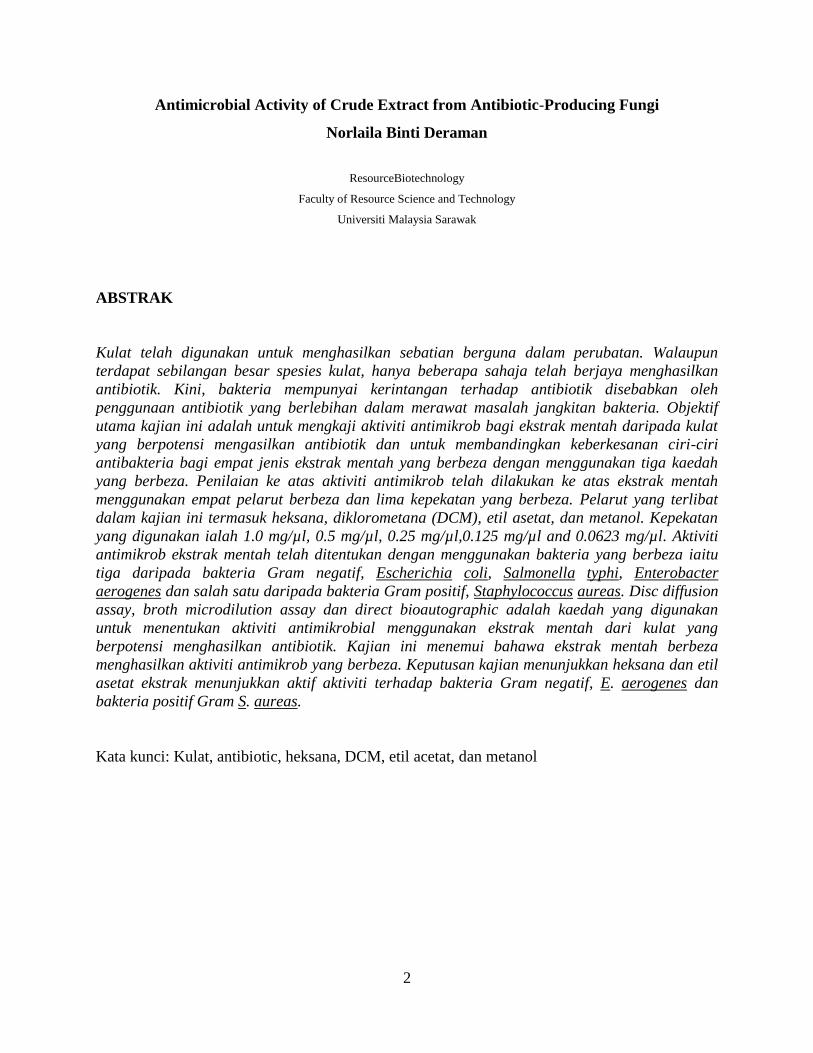
ABSTRACT
Fungi have been used for producing medically useful compound. Although there are a large
number of fungi species, only a relative few have been found to produce antibiotics. Nowadays,
the bacteria become resistance toward antibiotics because of overuse of the antibiotics in the
treatment of bacterial infection. The objectives of this study are to determine antimicrobial
activity of crude extracts from antibiotic producing fungi and to compare the effectiveness of
antibacterial properties among four different crude extracts using three different assays.
Evaluation on antimicrobial activity was done on crude extract using four different solvents and
five different concentrations. The solvents involved in this research including hexane,
dichloromethane (DCM), ethyl acetate, and methanol. The concentration used were 1.0 mg/µl,
0.5 mg/µl, 0.25 mg/µl,0.125 mg/µl and 0.0623 mg/µl. Antimicrobial activity of crude extracts
was determined using different bacteria which are three Gram-negative bacteria, Escherichia
coli, Salmonella typhi, Enterobacter aerogenes and one Gram-positive bacteria, Staphylococcus
aureas. The disc diffusion assay, broth microdilution assay and direct bioautographic assay was
the method used to determine antimicrobial activity using the crude extracts from the antibiotic
producing fungi. All of the crude extracts showed different antimicrobial activity toward
different bacteria. From the result hexane and ethyl acetate extracts showed active against Gram
negative bacteria, E. aerogenes and Gram positive bacteria S. aureas.
Key words: Fungi, antibiotic, hexane, DCM, ethyl acetate, and methanol
2
Antimicrobial Activity of Crude Extract from Antibiotic-Producing Fungi
Norlaila Binti Deraman
ResourceBiotechnology
Faculty of Resource Science and Technology
Universiti Malaysia Sarawak
ABSTRAK
Kulat telah digunakan untuk menghasilkan sebatian berguna dalam perubatan. Walaupun
terdapat sebilangan besar spesies kulat, hanya beberapa sahaja telah berjaya menghasilkan
antibiotik. Kini, bakteria mempunyai kerintangan terhadap antibiotik disebabkan oleh
penggunaan antibiotik yang berlebihan dalam merawat masalah jangkitan bakteria. Objektif
utama kajian ini adalah untuk mengkaji aktiviti antimikrob bagi ekstrak mentah daripada kulat
yang berpotensi mengasilkan antibiotik dan untuk membandingkan keberkesanan ciri-ciri
antibakteria bagi empat jenis ekstrak mentah yang berbeza dengan menggunakan tiga kaedah
yang berbeza. Penilaian ke atas aktiviti antimikrob telah dilakukan ke atas ekstrak mentah
menggunakan empat pelarut berbeza dan lima kepekatan yang berbeza. Pelarut yang terlibat
dalam kajian ini termasuk heksana, diklorometana (DCM), etil asetat, dan metanol. Kepekatan
yang digunakan ialah 1.0 mg/µl, 0.5 mg/µl, 0.25 mg/µl,0.125 mg/µl and 0.0623 mg/µl. Aktiviti
antimikrob ekstrak mentah telah ditentukan dengan menggunakan bakteria yang berbeza iaitu
tiga daripada bakteria Gram negatif, Escherichia coli, Salmonella typhi, Enterobacter
aerogenes dan salah satu daripada bakteria Gram positif, Staphylococcus aureas. Disc diffusion
assay, broth microdilution assay dan direct bioautographic adalah kaedah yang digunakan
untuk menentukan aktiviti antimikrobial menggunakan ekstrak mentah dari kulat yang
berpotensi menghasilkan antibiotik. Kajian ini menemui bahawa ekstrak mentah berbeza
menghasilkan aktiviti antimikrob yang berbeza. Keputusan kajian menunjukkan heksana dan etil
asetat ekstrak menunjukkan aktif aktiviti terhadap bakteria Gram negatif, E. aerogenes dan
bakteria positif Gram S. aureas.
Kata kunci: Kulat, antibiotic, heksana, DCM, etil acetat, dan metanol

3
1.0 INTRODUCTION
Nowadays, as a result of excessive use of antibiotic in treatment of infectious diseases,
microorganisms have developed resistance toward antibiotics (Melgarego et al., 2008). The
antimicrobial resistance particularly among bacteria has become an important issue which has
serious implications on the prevention and treatment of infectious diseases (Diarmaid and Dan,
2001). Besides, the emergence and spread of antimicrobial resistance remains a global public
health concern. Antibiotic resistance is commonly used to describe the situation when the
concentrations of antibiotic needed to kill the bacteria cannot be achieved at the site of the
infection (Berdy, 2005). This phenomenon has made the treatment of the bacterial infections
become increasingly difficult. Hence, more efforts and researches need to be carried out to
discover new antimicrobial drug in order to overcome the widespread of the bacteria resistances.
The antibiotics which have been produced by the fungi are increasing due to the
emergence of the bacteria that are resistant toward antibiotics (Bronzwaer, 2002). Antibiotics can
be produced by almost all types of living things. They are produced by prokaryotic and
eukaryotic organisms belonging to the plant and animal kingdom (Berdy, 2005). Antibiotic also
have been successfully used for extensive application in the treatment of infectious diseases of
man, animal, and plant (Cassell, 2001). Besides, antibiotics are affective against bacteria because
they attack the unique peptidoglycan cell wall or smaller ribosomal unit of the bacteria (Butler
and Cooper, 2011).

4
Penicillin is one of the examples of antibiotic that was discovered accidentally in 1928 by
Fleming, who showed its efficiency in laboratory cultures against many disease producing
bacteria. This discovery marked the beginning of the development of antibacterial compounds
produced by living organisms (Taylor et al., 2003). Since the discovery of penicillin, the first β-
lactam antibiotic, in modern medicine fungi turned out to be important antibiotic for curing life
treating infectious diseases. Penicillin was introduced as the first antibiotic, since then literally
thousands of metabolite which been produced mainly by fungi has been screened for
antimicrobial activities (Bronzwaer, 2002).
Crude extract as secondary metabolite was used to test the inhibition of bacterial and
fungal growth against human pathogenic bacteria and fungi (Hussain and Ananthan, 2009).
Antimicrobial activity can be determined using disc diffusion assay by inoculating the
suspension of bacteria through swabbing method on Mueller-Hinton Agar (MHA) plates
(Cleidson, 2007). This method described to determine the Minimum Inhibitory Concentration
(MIC) of extracts against the standard bacteria strains (Citron and Warren, 2005).
On the other hand, disc diffusion method was used to determine antibacterial and
antifungal activities (Al-Fatimi, 2010). Bioautography assay is a method for directly detection of
antibacterial compounds on nutrient agar plate. This assay is also practiced as a method to
localize antibacterial activity on chromatogram and has found widespread application in the
search for new antibiotic. Besides, the broth microdilution method described the modification
that was used to determine the MIC of extracts against the standard bacteria strains in liquid
culture (Cleidson, 2007).

5
Objectives of this study are to:
1. determine antimicrobial activity of crude extracts from antibiotic producing fungi.
2. compare the effectiveness of antibacterial properties among four different of crude
extracts using three different assays.

6
2.0 LITERATURE REVIEW
2.1 Fungi
Fungi are one of the organisms that used as a model for the study of various fields in biology
including biochemistry, genetic, molecular biology, interaction in environment and also used
widely in industry and biotechnology. Fungi also used in research study because they are easily
cultured, occupy little space, multiply rapidly and also have short life cycle (Feofilova, 2001).
The diversity of fungi in the organization of their life cycles, cellular structure, composition and
metabolism is important in mycology study (Wainwright, 1992).
Fungi are achlorophyllous, heterotrophic, eukaryotic and spore-bearing organism
surrounded by a well defined cell wall made up of chitin, with or without fungal cellulose, along
with many other complex organic molecules. Fungi usually obtain food by absorption, except a
few lower groups where they take in food by ingestion (Kornfeld, 2002). Fungi play such a
dominant role in human society that it could be readily argued that it is the most important
biotechnologically useful organisms. One of the most economically important uses of fungi is in
the industrial production of biochemicals such as organic acid that involve citric, fumaric, lactic
and gibberellins (Abraham, 2001).
The term ‘endophyte’ is used to define fungi or bacteria that occur inside asymptomatic
plant tissues. Fungal endophytes are ubiquitous and are dominated by Ascomycota (Ganley,
2005). People in the India subcontinent have a long history using medical plants to cure various
diseases. Medical plants of Western Ghats of India are reported to have a diverse community of
endophyte fungi (Raviraja, 2005). Few studies on the endophytic fungi of these plants have been

7
conducted. The present study was undertaken to investigate the diversity of endophytic fungi and
their seasonal colonization pattern in medical shrub species commonly used in Malnad region,
Western Ghats of Karnataka, Sourthern India. Besides that, a total of 6125 fungal endophytes
were isolated from 9000 leaf segments of 15 medicinal shrubs growing in Malnad region during
winter, monsoon and summer seasons. These fungal isolates belonged to Ascomycota,
Coelomycetes, Hyphomycetes, Mucoromycotina and sterile forms (Fernando, 2008). In addition,
fungal endophytes have been recognized as a respiratory on novel compounds of immense value
in agariculture, industry and medicine (Tan and Zou, 2001)
2.2 Test Bacteria
Staphylococcus aureas is a commonly bacteria that is easy to find on the skin and in the nose of
healthy persons. This bacterium has the ability to grow comparatively well under conditions of
high osmotic pressure and low moisture. Among the entire Staphylococcus group, S. aureus is
the one that cause most infection (Stoppler, 2009). The bacteria gained their pathogenicity via
production of many toxins that increases the ability to invade the body or damage the tissue
(Tortoro, 2004).
Escherichia coli is one of bacteria that are commonly found in the gut of warm blooded
organisms. There are several types of E. coli exist as part of the normal flora in human gut and
also have several beneficial functions, such as for production of vitamin K2. They also can
prevent harmful bacteria, known as pathogenic bacteria, because of establishing themselves in
the intestine (Sondi, 2004).

8
Enterobacter aerogenes is a Gram-negative bacteria and rod shaped microorganism from
Enterobacteriaceae famility. This microorganism is important nosocomial pathogen that
responsible for various infections, including lower respiratory tract infection, skin and soft-tissue
infection and wound infection. Enterobacters species possess inducible beta-lactamases, which
are undetectable in vitro but are responsible for resistance action during treatment. Physicians
treating patients with Enterobacter infections are adviced to avoid certain antibiotics, because
resistant mutants can quickly appeared (Sandra and Tenney, 2000).
Salmonella typhi is bacteria which can cause disease in human. This bacterium is the
causive agent of typhoid fever. Although typhoid fever is not widespread in the United States, it
very common in under developed countries and causes a serious often fatal disease. The main
source of S. typhi is from drinking infected water. Food may also be contaminated with S. typhi if
it was washed or irrigated with contaminated water (Kidgell, 2002).
2.3 Antibiotics
Originally, the term antibiotic referred only to organic compounds produced by bacteria
and fungi, which are toxic to other organisms. Antibiotics represent the single contribution of
drug therapy for the health care of increasing population of the world, and provide effective
control of many microbial pathogens that have been the cause of death of human and animals
(Makut and Owolewa, 2011). Antibiotics produced by fungi, are widely used in current
chemotherapy especially the penicillin, cephalosporin and fusidic acid, which have antibacterial
and antifungal activity (Romanowski, 2007).
9
Screening of antibiotic has been widely performed for about 50 years, and new antibiotics
are still being found. However, the possibility of discovering new antibiotics through random
screening of microorganisms such as actinomycetes and other bacteria producing antibiotics is
reduced nowadays. Because of that, new approaches are required for finding new antibiotics
efficiently. The term ‘antibiotic’ literally means ‘against life’. An antibiotic was originally
defined as a substance, produced by one microorganism (Berdy, 2005).
Many of the microbial products including antibiotics are considered to be secondary
metabolites. It is because they seem to have no direct role in those aspects of metabolism which
support necessary functions in the cell namely energy production, growth and reproduction.
There is a great structural variety among the secondary metabolites. Some antimicrobial active
and some are not. Antibiotic has a powerful action on a wide range of infectious bacteria include
in both, gram positive and gram negative (Weinstein, 2003). The streptomycin is example of
antibiotic that was isolated in 1944 by Waksman, a Microbiologist, from a species of soil
bacteria, called Streptomycesgriseus, particularly Tubercle bacilli, and has proved to be very
valuable against tuberculosis (Stewart and William, 2001). In 1947, another antibiotic,
chloromycetin was discovered by Burkholder (Cars, 2001).
Antibiotic had been used by human in three ways. Two of the ways are in human
medicine, which is the use of antibiotics to treat sick animals and the prophylactic use of
antibiotics to prevent diseases. The third one is on agriculture, which has no parallel to human
medicine and used as growth promoters. That is, the long-term administration of low levels

10
antibiotics to some animals increases the weight gain per unit of food and allows animals to
reach full weight more quickly (Darmaid and Dan,2001).
Various types of antibiotics work as bactericidal or bacteriostatic. Bactericidal antibiotic
kills the bacteria generally by either interfering with the formation of the bacterium’s cell wall or
its cell contents in the cytoplasm. Penicillin, daptomycin, fluoroquinolones and metronidazole
are some example of bactericidal antibiotics (Figure 2.1). Whereas, bacteriostatic antibiotic
inhibit the bacteria from multiplying by interfering with bacterial protein production, DNA
replication, or other aspects of bacterial cellular metabolism which can subsequently stop the
bacteria multiplication (Martyn, 2008).
In addition, although the concept of antibiotics has evolved over time to include plant and
animal products as well as synthetic and semisynthetic compounds that been used in theraphy,
the word ‘antibiotic’ has become entrenched as a descriptor for any molecule produced in the
laboratory with the ability of cidal (killing) or static (inhibitory) to inhibit specific group of
microorganisms (Grace, 2006). The range of bacteria or other microorganisms that is affected by
a certain antibiotic is expressed as its spectrum of action.
The spectrum have classified into two which are broad spectrum or narrow spectrum.
Broad spectrum is referring to the antibiotics effective against prokaryotes that kill or inhibit a
wide range of Gram-positive and Gram-negative bacteria. However, narrow spectrum is when
antibiotics effective mainly against Gram-positive or Gram-negative bacteria. If antibiotic
against a single organism or disease, they are referred to as limited spectrum (Todar, 2009).

11
Penicillin G
Figure 2.1: Molecular structure of an antibiotic Penicillin G.
2.4 Antibiotic Resistant
Antibiotic resistance is the natural consequence of the selective pressure imposed by antibiotic
drugs upon bacteria populations. Antibiotic resistance occurs when pathogenic microorganisms
are capable to inactive antibiotics or survive under the selective pressure of antibiotic. The rapid
emergence and spread of antibiotic resistance genes is due to the consumption of large amount of
antibiotics (Cleidson, 2007). Antimicrobial agents represent the greatest advance in modern
curative medicine.
Resistance to antimicrobial agents has existed since before they were introduced into
human probably because most of the classes of compounds used clinically are produced also by
microorganisms in the environment (Helen, 2001). The introduction and increasing clinical use
of each antimicrobial agent and also followed by an increasing isolation rate of resistant bacteria.
Resistance was shown in a short period of time by mutational target alteration against antibiotic
such as quinolones, rifampicin, fucidic acid and mupirocin after their introduction into clinical
use (Diarmaid and Dan, 2001).
12
Resistance can spread through species selection, mutation, gene epidemics, and strain
epidemics. Resistance can arise through mutations, which can be defined as random and
spontaneous genetic changes. Antibiotic do not cause mutations occur, but their use clearly
generates an intense pressure for the selection of resistance mutants that arise naturally at low
frequency. Bacteria can acquire genetic resistance determinants from other bacteria, either as
plasmid or chromosomal insert. Besides, plasmid and some chromosomally inserted transposons
are often freely transmissible and their epidemic spread allows resistance to extend to diverse
organisms (Helen, 2001).
The rapid emergence of mutational resistance can swiftly reduce the effectiveness of an
antibiotic. Methicillin resistant Staphyloccus aureus (MRSA) were first reported in the early
1960’s and are now regarded as a major hospital acquired pathogen worldwide. The term
methicillin resistant is historically used to describe resistance to any of this class of
antimicrobials. Fluoroquinolones were originally active against MRSA, but staphylococci have
an efflux pump and resistance arises cause by mutation of gene call norA. Because of that, most
MRSA are now resistant to antibiotics (Helen, 2010).
2.5 Antibiotic-Producing Microorganisms
The ability of certain fungi and bacteria to produce chemical substances which inhibit or destroy
pathogenic organisms (Waksman, 1952) has supported the fact that these unrelated groups are
involved in most of the natural antibiotics production (Todar, 2009). These substances are
hypothesized to confer a selective advantage to the producer when competition is significant

13
microbial fitness. A fungus such as Penicillium chrysogenum is an important industrial organism
due to its ability to produce several types of β-lactam antibiotic (Hogg, 2005).
Different antibacterial properties could be obtained through the substitution of R-group
subtituent of the penicillium nucleus (Frisvad and Andersen, 2008).The ability to produce
antibiotics has been found mainly in fungi of the group Aspergillales and also in a few bacteria.
The streptomycetes are remarkable for the chemical diversity of antibiotic that they produce.
Altogether about 2,000 antibiotics have been characterized so far, but only 50 are using
therapeutically. Antibiotic produced by fungi, are widely used in current chemotheraphy
especially the penicillin, cephalosporin and fucidic acid, which shows antibacterial and
antifungal activity (Grace, 2006).
A number of antibiotic drugs have been discovered from soil-inhabiting microorganisms
which include 20% from fungi, 70% from actinomycetes and 10% from eubacteria (Makut and
Owolewa, 2011). Antibiotics are produced by many microorganisms in various ecological
conditions. Producers of antibiotic can be found in rivers, lakes, decaying plants and animal
remains. However majority of microorganisms that produce antibiotic are soil inhabitants
(Chandra, 2010).

14
2.6 Crude Extract
Crude extract was used to test the inhibition of bacterial and fungal growth against human
pathogenic bacteria and fungi (Hussain and Ananthan, 2009). The fungal extract mostly contains
compounds from the secondary metabolism, but also some primary metabolites (Smedsgaard and
Nielsen, 2005). A profile of secondary metabolites is compiled by the mycologist and chemist is
based on fungal extracts. It consists of compounds produced on one or more media and includes
antibiotics (Frisvad and Andersen, 2008). Fungi, plants, lichen fungi, and actinomycetes are the
four groups of organisms are particularly good producers of secondary metabolites,
whereas yeasts, protozoa, and animals are less efficient producers. Therefore, secondary
metabolites have mostly been used in plant and fungal taxonomy (Frisvad and Larsen, 2007).
2.7 Antimicrobial Assay
Antimicrobial assay is a method to discover antibiotic. The current available screening methods
for the detection of antimicrobial activity of natural product are classified into three groups, disc
diffusion assay, broth microdilution assay and direct bioautography assay (Cleidson, 2007).
Antimicrobial activity of crude extract by using disc diffusion technique was carried out with
careful consideration of factors, such as inoculation size, incubation temperature ,as well as type
and depth of agar to ensure that the test are standardized, reproducible and reflected the
effectiveness of the antimicrobial agents (Rennie et al., 2007).
![TRINEXAPAC-ETHYL (271)€¦ · Results Test material purity and specification Reference Solubility in organic solvents [g/L at 25 °C] acetone > 500 dichloromethane > 500 ethyl acetate](https://static.fdocuments.net/doc/165x107/607eb36b396380183d26530e/trinexapac-ethyl-271-results-test-material-purity-and-specification-reference.jpg)