Kinetic and Structural Analysis for Potent Antifolate Inhibition of ...

Molecules 2013, 18, 8319-8341; doi:10.3390/molecules18078319
molecules ISSN 1420-3049
www.mdpi.com/journal/molecules
Article
Factors Influencing the Antifolate Activity of Synthetic Tea-Derived Catechins
Magalí Sáez-Ayala 1, María Piedad Fernández-Pérez 1, Soledad Chazarra 1, Nani Mchedlishvili 2,
Alberto Tárraga-Tomás 3 and José Neptuno Rodríguez-López 1,*
1 Department of Biochemistry and Molecular Biology A, School of Biology, Regional Campus of
International Excellence “Campus Mare Nostrum”, University of Murcia, 30100, Murcia, Spain;
E-Mails: [email protected] (M.S.-A.); [email protected] (M.P.F.-P.); [email protected] (S.C.) 2 Durmishidze Institute of Biochemistry and Biotechnology of Agrarian University of Georgia, 0131,
Tbilisi, Georgia; E-Mail: [email protected] 3 Department of Organic Chemistry, Faculty of Chemistry, Regional Campus of International
Excellence “Campus Mare Nostrum”, University of Murcia, 30100, Murcia, Spain;
E-Mail: [email protected]
* Author to whom correspondence should be addressed; E-Mail: [email protected];
Tel.: +34-868-888-284; Fax: +34-868-884-782.
Received: 7 June 2013; in revised form: 5 July 2013 / Accepted: 12 July 2013 /
Published: 16 July 2013
Abstract: Novel tea catechin derivatives have been synthesized, and a structure-activity
study, related to the capacity of these and other polyphenols to bind dihydrofolate
reductase (DHFR), has been performed. The data showed an effective binding between all
molecules and the free enzyme, and the dissociation constants of the synthetic compounds
and of the natural analogues were on the same order. Polyphenols with a catechin
configuration were better DHFR inhibitors than those with an epicatechin configuration.
Antiproliferative activity was also studied in cultured tumour cells, and the data showed
that the activity of the novel derivatives was higher in catechin isomers. Derivatives with a
hydroxyl group para on the ester-bonded gallate moiety presented a high in vitro binding
to DHFR, but exhibited transport problems in cell culture due to ionization at physiologic
pHs. The impact of the binding of catechins to serum albumin on their biological activity
was also evaluated. The information provided in this study could be important for the
design of novel medicinal active compounds derived from tea catechins. The data suggest
that changes in their structure to avoid serum albumin interactions and to facilitate
plasmatic membrane transport are essential for the intracellular functions of catechins.
OPEN ACCESS

Molecules 2013, 18 8320
Keywords: tea catechins; dihydrofolate reductase; antifolates; albumin; drug design
1. Introduction
Tea is the most popular beverage, consumed by two-thirds of the World’s population. Among all
teas, green tea is the most studied for its reputed health benefits [1]. Tea polyphenols are considered to
contribute to preventive effects against various pathological disorders, including cancer. The major
components of tea are catechins, which contain a benzopyran skeleton with a phenyl group substituted
at the 2-position and a hydroxyl (or ester) function at the 3-position. Variations in the catechin
structure include the stereochemistry of the 2,3-substituents and the number of hydroxyl groups in the
B-ring and D-ring (Figure 1). Belonging to the flavanol class of flavonoids, the most abundant
catechins found in tea leaves include epigallocatechin-3-gallate [EGCG], epigallocatechin [EGC],
epicatechin-3-gallate [ECG], epicatechin [EC], catechin-3-gallate [CG], and gallocatechin-3-gallate
[GCG] (Figure 1). Several in vivo experiments have shown the chemo-preventative effects of EGCG
against all stages of carcinogenesis in animal models of breast, lung, skin, prostate, and colon
cancers [2,3]. There has been extensive investigation into the mechanism by which EGCG might act in
cancer prevention, and recent attempts to define the molecular action mechanism of green tea
components have found that EGCG affects several molecular targets and carcinogenesis pathways. The
3-gallyl moiety of catechins is essential for the inhibition of several of their proposed enzymatic
targets, such as the proteasome [4], 5-cytosine DNA methyltransferase 1 (DNMT1) [5], glutamate
dehydrogenase [6], and dihydrofolate reductase (DHFR) [7]. Recently, we reported that the
ester-bound gallate catechins isolated from green tea are potent inhibitors of DHFR activity at
concentrations found in the serum and tissues of green tea drinkers [7]. Since this first report
describing the antifolate activity of tea polyphenols, several studies by us and by other research groups
have confirmed this activity [8–10] and have reported that EGCG inhibits DHFR from a variety of
biological sources [11–14].
Figure 1. Flavonoids present in tea with epicatechin (ECG, EGCG) and catechin configurations (CG).
OHO
OH
OH
OH
O O
HO
OH
OH
OHO
OH
OH
OH
O O
HO
OH
OH
OHO
OH
OH
OH
O O
HO
OH
OH
OH
CG ECG EGCG
A C
B
D
However, the excellent anticancer properties of tea catechins are significantly limited by their poor
bioavailability, which is related to their low stability in neutral or slightly alkaline solutions, and by
their inability to easily cross cellular membranes [15]. In an attempt to solve these bioavailability
problems, we synthesized a 3,4,5-trimethoxybenzoyl analogue of ECG (TMECG) that exhibits high

Molecules 2013, 18 8321
antiproliferative activity against several cancer cell lines, especially melanoma [16]. Methoxy
protection of the reactive hydroxyl groups in the D-ring of ECG yielded a derivative stable under cell
culture conditions that is more efficiently transported through the cell membranes than its natural tea
parent catechin [16]. Because the major polyphenols present in tea have epicatechin configurations,
many of the studies designed to elucidate the biological activities of these tea catechins have been
performed with epicatechin derivatives, but catechin gallate (CG) also inhibits the proliferation of
cancer cells derived from human oral cavity tissues [17]. As part of our on-going efforts to develop
new tea-derived compounds, we synthesized a trimethoxybenzoyl analogue of CG (TMCG) [18]. This
compound shared bioavailability advantages with its epimer TMECG, inhibited growth, and induced
apoptosis in several human cancer cell lines. TMCG exhibited characteristics of a slow-binding
inhibitor of DHFR; an explanation for the irreversibility of the slowly dissociating enzyme complex
was provided by molecular modelling [18].
In an attempt to explore the structure-activity relationship of natural and synthetic polyphenols as
inhibitors of DHFR, several new derivatives with different stereochemistry and D-ring substitutions
were designed and synthesized. This allowed us to compare the epimeric differences between
epicatechin and catechin configurations and the different substitutions of hydroxyl groups on the
D-ring with respect to DHFR binding and antiproliferative actions against cancer cells. We achieved
the synthesis of derivatives with methoxy groups in the meta position on the esterified D-ring in both
catechin and epicatechin configurations. We also synthesized the fully acetylated derivative of this
catechin isomer and its monoacetylated derivative with a hydroxyl group para on the D-ring. The
factors affecting the biological activities of these synthetic polyphenols, including their interaction
with serum albumin, were studied.
2. Results and Discussion
2.1. Synthesis
The syntheses of TMECG (13a) and TMCG (12a) was carried out starting from the commercially
available catechin, following a procedure previously described by our research group [16,18].
Consequently, this general route was also used to prepare the new compounds 12b–d, 13b. The
reaction sequence was designed to avoid problems associated with unspecific blockage of the
3-hydroxy group of epicatechin [19,20]. Therefore, all compounds (both catechin and epicatechin
configurations) were synthesized starting from catechin following the multi-step reaction sequence
shown in Scheme 1, which involved an inversion of configuration at C-3 through an
oxidation–reduction process to obtain compounds with an epicatechin configuration. After selectively
protecting the phenolic hydroxyl groups of catechin and epicatechin, the resulting compounds were
treated with the appropriate acid halides to give rise to the corresponding esters which, in a final step,
undergo deprotection of the phenolic groups by treatment with H2 in the presence of 10% Pd/C.
It is worth noting that for introducing two methoxy groups meta to the ester-bonded gallate moiety,
such as in SYCG (12b) and SYECG (13b), we used the 4-(benzyloxy)-3,5-dimethoxybenzoyl chloride
(7), which was prepared from the commercially available syringic acid (3,5-dimethoxy-4-
hydroxybenzoic acid) in a three step sequence: (i) treatment with benzyl bromide in the presence of

Molecules 2013, 18 8322
K2CO3 to give 5; (ii) hydrolysis of the benzyl ester group to the corresponding carboxylic acid
derivative 6; and iii) treatment of 6 with oxalyl chloride to yield 7 (Scheme 2). Thus, reaction of 7 with
the isomers 1 and 3 in the presence of DMAP (dimethylaminopyridine), yielded the corresponding
esters 10b and 11b which by hydrogenolysis underwent cleavage of the benzyl ether functionalities to
produce the final compounds 12b and 13b in high yield and purity.
Scheme 1. Synthesis of the tea catechin derivatives 12a–d and 13a–b.
O
OH
OBn
OBnBnO
OBn
O
O
OOR1
OR2
OR1
OH
OHHO
OH
O
OH
OH
OHHO
OH
O
O
OBn
OBnBnO
OBn
O
OH
OBn
OBnBnO
OBn
O
O
OOR1
OR2
OR1
OBn
OBnBnO
OBn
O
O
OOR1
OR2
OR1
OBn
OBnBnO
OBn
O
O
OOR1
OR2
OR1
OH
OHHO
OH
O
O
OOCH3
OAc
OCH3
OAc
OAcAcO
OAc
10a, R1 = CH3, R2 = CH310b, R1 = CH3, R2 = Bn10c, R1 = CH3, R2 = Ac
(i)
(ii)
(iii)
(iv)
(iv)
(v)
(v)
(vi)
1
2
3 11a, R1 = CH3, R2 = CH311b, R1 = CH3, R2 = Bn
TMCG, 12a, R1 = CH3, R2 = CH3SYCG, 12b, R1 = CH3, R2 = OHMASYCG, 12c, R1 = CH3, R2 = Ac
TMECG, 13a, R1 = CH3, R2 = CH3SYECG, 13b, R1 = CH3, R2 = OH
ASYCG, 12d
Catechin
Reagents and conditions: (i) BnBr, K2CO3, DMF, −10 °C to RT, overnight; (ii) Dess–Martin periodinane,
CH2Cl2, RT, 3 h; (iii) l-Selectride, nBu4NCl, THF, −78 °C, 3 h; (iv) 4, 7 or 9, CH2Cl2, DMAP, RT, 18 h;
(v) H2, 10% Pd/C, THF/MeOH (3:1), RT, 15 h.
To synthesize the fully acetylated derivative ASYCG (12d), compound 12b was peracetylated by
reaction with acetic anhydride in the presence of pyridine. However, the synthesis of MASYCG (12c)
bearing only one acetyl group in the para position to the ester-bonded gallate moiety, required the
previous acetylation of the syringic acid to give 8, which on treatment with thionyl chloride in CH2Cl2
gave rise to 4-acetoxy-3,5-dimethoxybenzoyl chloride (9, Scheme 2). Thus, treatment of 1 with the
acid chloride 9 in CH2Cl2 and in the presence of DMAP, yielded 10c, which undergoes hydrogenolysis
to give 12c in high yield and purity (Scheme 1).

Molecules 2013, 18 8323
All compounds were characterized by mono- and bi-dimensional 1H- and 13C-NMR and mass
spectrometry techniques (see Supplementary Materials).
Scheme 2. Synthesis of acid chlorides 4, 7 and 9.
H3CO
OCH3
OCH3
O OH
H3CO
OCH3
OCH3
O Cl
PCl5
Toluene
43,4,5-trimethoxybenzoic acid
H3CO
OBn
OCH3
O OBn
BnBr
Acetone5
K2CO3 H3CO
OBn
OCH3
O OH
6
NaOH
H3CO
OBn
OCH3
O Cl
7
C2O2Cl2
CH2Cl2
MeOH
H3CO
OH
OCH3
O OH
H3CO
O
OCH3
O OH
8
Ac2O
O
H3CO
O
OCH3
O Cl
9
SOCl2
O
Syringic acid
2.2. Biological Activities
2.2.1. Binding of polyphenols to DHFR
Natural and synthetic derivatives of tea catechins have shown to be good inhibitors of human
DHFR [7,11,16,18]. To determine the ability of natural and synthetic polyphenols to bind DHFR, we
performed a structure-activity study by analyzing the binding of these polyphenols to DHFR using
fluorescence quenching. Effective binding to free recombinant human DHFR (rHDHFR) was
determined by measuring the decrease in enzyme fluorescence that occurs upon the formation of the
enzyme-inhibitor complex. When DHFR fluorescence was excited at 290 nm, its emission spectra
showed a maximum at 340–350 nm. The binding of inhibitors quenched this fluorescence, and the data
showed an effective union between all molecules and the free enzyme (Table 1). The dissociation
constants of the synthetic compounds were on the same order as those of the natural analogues,
indicating the high DHFR binding capacities of these new derivatives.

Molecules 2013, 18 8324
Table 1. Dissociation constants of free rHDHFR for natural and synthetic catechins.
* p < 0.05 with respect to their respective catechin isomers.
Compound KD (μM)
CG 0.56 ± 0.07 ECG 0.81 ± 0.09 *
TMCG 0.9 ± 0.1 TMECG 2.1 ± 0.2 * SYCG 0.72 ± 0.10
SYECG 0.77 ± 0.07
In general, the catechin isomers bound to the enzyme with a lower dissociation constant than the
epicatechin isomers, indicating that the polyphenols with a catechin configuration (CG, TMCG, and
SYCG) had better binding capacity than those with an epicatechin configuration (ECG, TMECG, and
SYECG). This result also indicates that the cis disposition of the lateral esterified ring interferes with
binding compared to the trans disposition. Moreover, we studied the influence of different positions of
methoxy groups on the ester-bonded gallate moiety on the binding to DHFR. We analyzed natural
compounds (three hydroxyl groups), trimethoxy-derivatives (three methoxy groups in the meta and
para positions), and new derivatives of syringic acid (two methoxy groups meta and one hydroxyl
group para) (Table 2). These results showed that the presence of methoxy groups in ring D hindered
the binding of polyphenols to DHFR and that one hydroxyl group in the para position on lateral ring D
was important for the stabilization of the enzyme-inhibitor complex.
Table 2. Order of the binding strengths of catechins to rHDHFR depending on the
substitution in the D-ring. * p < 0.05 with respect to the binding of natural compounds
to DHFR.
HO
OH
OH
O O
H3CO
OH
OCH3
O O
H3CO
OCH3
OCH3
O O
≥ >
(KD, μM)
Catechin 0.56 0.72 0.90 * Epicatechin 0.81 0.77 2.10 *
2.2.2. Cancer Cell Antiproliferative Activity
Antifolate compounds are designed to inhibit DHFR and act specifically during DNA and RNA
synthesis, making them more toxic to rapidly dividing cells. Recent evidences indicate that tea-
catechins and derived compounds could inhibit DHFR and block the folate cycle in cancer cells [16,18].
Although inhibition of DHFR by these compounds could be a plausible explanation for their antifolate
activity, the possibility of these compounds targeting (folate)-enzymes other than DHFR could not be
excluded. To determine whether these newly synthetized compounds disrupt the folate cycle in cell
culture, we studied their antiproliferative activity in the melanoma cell line SK-MEL-28 (Table 3).

Molecules 2013, 18 8325
Concentration- and time-dependent effects were analyzed using the colorimetric 4,5-(3)-
dimethylthiazol-2-yl)-2,5-diphenyltetrazolium bromide (MTT) method [18].
Table 3. Antiproliferative effects of natural and synthetic catechins in melanoma cells.
Half-maximal inhibitory concentration (IC50) of compounds against the SK-MEL-28
melanoma cell line after 6 days of treatment. * p < 0.05 when compared with their
respective fully methylated polyphenols in ring D (TMCG and TMECG, respectively).
** p < 0.05 when compared with SYCG.
Compound IC50 (μM)
CG 82.2 ± 8.0 ECG > 100
TMCG 1.5 ± 0.4 TMECG 2.5 ± 0.6 SYCG 126.3 ± 10.0 *
SYECG 198.9 ± 12.0 * MASYCG 60.9 ± 6.0 ** ASYCG 37.4 ± 4.0 **
The results showed that all compounds caused a decrease in the number of viable cells depending
on the concentration and time. Previous results indicated that the reduced viability of cancer cells in
the presence of TMECG or TMCG was indeed due to apoptosis induction [16,18]. Compounds with a
catechin configuration presented higher antiproliferative activity than those with an epicatechin
configuration (p < 0.05), as observed in in vitro enzymatic assays with DHFR. However, the two
compounds with one hydroxyl group para on a lateral esterified ring D (SYCG and SYECG), that bind
tightly to DHFR in vitro, did not show the expected antiproliferative activity. To verify that this effect
was not due to a particular cell line, we carried out the same assays in other cell lines of melanoma,
colon, and breast cancer with SYCG (data not shown), which proved that this effect was a general
effect. Although SYCG and SYECG were stronger inhibitors of DHFR than the other compounds (as
observed by binding studies), they showed a reduced efficacy in inhibiting the growth of tumour cells.
This effect might be a result of a problem in cellular transport. Although they could be good inhibitors
of DHFR, these compounds cannot cross the plasma membrane to reach their cellular target.
2.2.3. pH-Related Effects on Catechin Membrane Transport
Importantly, recent studies have presented data suggesting that pH could be of relevance to tea
catechin transport and to their biological activity [11,21]. These results led us to study the effect of pH
on the protonation state of the inhibitors of DHFR. It has been shown that the protonation state of
methotrexate, trimethoprim, and pyrimethamine modulates the binding of these antifolates to DHFRs
from several sources [22]. The absorption spectra of SYCG and SYECG are strongly affected by the
pH (Figure 2). The absorbance maximum at 278 nm undergoes a bathochromic displacement to 328 nm,
which is greatest at alkaline pH (pH > 9.0). However, the absorption spectra of the trimethoxylated
derivatives, TMCG and TMECG, showed no significant changes in the pH range of 5.0–9.0 (Figure 2).

Molecules 2013, 18 8326
Figure 2. Effect of pH on the ionization state of SYCG, SYECG, TMCG, and TMECG.
UV-visible absorption spectra of the compounds at different pH values. The inset is a
representation of A328/270 vs. pH.
SYCG
λ (nm)
250 300 350 400 450
A
0.0
0.1
0.2
0.3
0.4
0.5
0.6
0.7
pH 5pH 7pH 7.5 pH 8pH 8.5pH 9
pH
5 6 7 8 9 10
A3
28n
m
0.0
0.2
0.4
0.6
0.8
TMCG
λ (nm)
250 300 350 400 450
A
0.0
0.1
0.2
0.3
0.4
0.5
0.6
0.7
pH 5pH 6pH 7pH 8pH 9
pH
4 5 6 7 8 9 10
A2
70n
m
0.0
0.2
0.4
0.6
0.8
1.0
SYECG
λ (nm)
250 300 350 400 450
A
0.0
0.1
0.2
0.3
0.4
0.5
pH5 pH7 pH7.5 pH8 pH8.5 pH9
pH
4 5 6 7 8 9 10
A32
8n
m
0.0
0.1
0.2
0.3
0.4
0.5
0.6
TMECG
λ (nm)
250 300 350 400 450
A
0.0
0.1
0.2
0.3
0.4
0.5
0.6
0.7
pH5 pH6 pH7 pH8 pH9
pH
4 5 6 7 8 9 10A
270
nm
0.0
0.2
0.4
0.6
0.8
1.0
To determine if the spectral changes in the SYCG and SYECG solutions with pH were due to an
irreversible oxidation reaction or to a reversible protonation step, a pH titration experiment was carried
out. An alkaline solution of SYCG and SYECG (pH 8.5) was acidified to pH 2.5 by the addition of
dilute HCl. A change in the spectra was observed, indicating a reversible protonation reaction. The
spectral changes with pH observed in SYCG and SYECG can be attributed to the hydroxyl group para
on the ester-bonded gallate moiety. At acidic pH, these derivatives are mainly present as protonated
species, whereas at basic pH or values near neutrality, these derivatives evolve toward the
deprotonated species, which are stabilized by resonance (Scheme 3). At physiologic pH, both SYCG
and SYECG are partially in their ionic form, which could be responsible for their limited traffic
through the plasma membrane and thereby account for their low antiproliferative activities. As
described, an important factor that decreases catechin bioavailability might be their inability to easily
cross cellular membranes by passive diffusion. The presence of several hydroxyl groups in these
molecules gives them a high hydrophilic character, which increases the difficulty of passing across
lipid membranes.

Molecules 2013, 18 8327
Scheme 3. Schematic representation of the effect of pH on SYCG and SYECG ionization
showing the proposed protonated and deprotonated species.
O
O
OOCH3
O
OCH3
OH
OHHO
OH
O
O
OOCH3
O
OCH3
OH
OHHO
OH
O
O
OOCH3
OH
OCH3
OH
OHHO
OH
H+
H+
For the TMCG and TMECG derivatives, the presence of three methoxy groups increases the
hydrophobic character, so they are easily transported [16], which favours their anti-tumour activities
with respect to natural catechins, despite being weaker inhibitors of DHFR. However, SYCG and
SYECG present an additional inconvenient related with the overall negative charges at physiological
pH, which hinder their ability to cross the cellular membrane. To test this hypothesis and to prevent the
ionization of this hydroxyl group to facilitate their transport into the cell, we synthesized two
derivatives from the structure of SYCG with fully or partially acetylated hydroxyl groups.
Esters are the most common pro-drugs used, and it is estimated that 49% of all marketed pro-drugs
are activated by enzymatic hydrolysis [23]. Ester pro-drugs are produced by masking the polar groups,
resulting in an enhanced lipophilicity and in the protection of the hydroxyl groups from ionization,
biotransformation or oxidative degradation. Therefore, we synthesized two derivatives of SYCG as
potential pro-drugs. After enzymatic hydrolysis of the ester bound inside cells, SYCG will be
recovered to perform its antifolate activity. We synthesized the fully acetylated derivative (ASYCG)
and the monoacetylated derivative of the hydroxyl group para on the ester-bonded gallate moiety
(MAYCG) and studied their antiproliferative activity in SK-MEL-28 by analyzing concentration- and
time-dependent effects (Figure 3). The presented data indicate that the anti-tumour activities of these
pro-drugs increased with respect to that of SYCG. The fully acetylated derivative was more active than
the partially acetylated derivative (ASYCG > MASYCG). This effect could be partially explained by
the fact that ASYCG lacks hydroxyl groups, facilitating its transport into the cell. Although the
antiproliferative activity of the monoacetylated derivative improved with respect to that of SYCG, the
numerous hydroxyl groups in the molecule may still complicate its transport across the plasma
membrane. All together, the results showed improved transport of these acetylated compounds into the
cells, but these effects were lower than expected because they did not reflect the strong in vitro binding
to DHFR by SYCG, which was higher than that of TMCG. This effect could be a result of an
incomplete enzymatic hydrolysis of acetyl groups, which hinder DHFR from binding to the free
hydroxyl groups of the molecule because of a low recovery of OH groups.

Molecules 2013, 18 8328
Figure 3. (a) Influence of the acetylation of hydroxyl groups of SYCG on the
antiproliferative activity in melanoma cells. (b) Half-maximal inhibitory concentration
(IC50) of compounds against the SK-MEL-28 melanoma cell line after six days of
treatment. * p < 0.05 when compared with SYCG.
2.2.4. Influence of Catechin-Serum Albumin Interactions on Polyphenol Activity
It is widely accepted that pharmacokinetic analyses in plasma do not accurately determine the
overall levels of catechins because polyphenols are known to partition into blood cells and plasma
proteins. This bound pool may be considered as a “reservoir”, partially protected from conjugative
enzymes and thus with a potentially extended lifetime, which can replace the molecules eliminated via
metabolism and excretion. Specifically, recent studies have suggested that tea catechins form
complexes with human serum albumin (HSA) for transport in human blood, and their binding affinity
for albumin is believed to modulate their bioavailability [24]. Recently, it has been demonstrated that
the affinity of tea catechins for albumin varies according to their chemical structure [24,25]. Thus,
galloylated catechins have higher binding affinities with HSA than non-galloylated catechins, and the
binding affinities of several methylated derivatives were lower than that of EGCG [24]. In agreement
with these previous data, we have observed that TMECG has an intermediate binding affinity to HSA
(KA = 4.2 × 10⁴ M−1) compared with that of EGCG (KA = 2.6 × 10⁵ M−1) or EC (KA = 3.2 × 10³ M−1).
However, it must be kept in mind that, although the intrinsic affinity of TMECG for albumin may be
weaker than that of EGCG, the physiologic concentration of serum albumin (~0.6 mmol/L) is most
likely large enough to allow the extensive binding of TMECG to this serum protein.
SYCG MASYCG ASYCG
0
20
40
60
80
100
120
140
Compound
IC50
(μ
M)
O
O
OOCH3
O
OCH3
OH
OHHO
OH
O
O
O
OOCH3
OH
OCH3
OH
OHHO
OH
< <
O
O
OOCH3
OCH3
O
OO
O
O
O
OO
O
O
a
b
*
*

Molecules 2013, 18 8329
Although the conventional view is that cellular uptake is proportional to the unbound concentration
of metabolites, the effect of albumin binding on the biological activity of polyphenols is unclear. On
the one hand, the incubation of quercetin in human whole blood or in suspensions of erythrocytes in
the absence of plasma proteins suggests that binding to albumin could considerably decrease the
association of quercetin with these cells [26]. On the other hand, the cancerous cells and tissues in
humans might constitute a different scenario. In this respect, albumin is believed to play an increasing
role as a drug carrier in the clinical setting. Due to pathophysiological conditions in neoplastic tissue,
high amounts of albumin accumulate in tumours and are metabolised by malignant cells. Primary
investigations have shown that tumours metabolize substantial amounts of albumin, using it as source
for nitrogen and energy [27]. Several studies with tumour-bearing animals have provided further evidence
that albumin accumulates in tumours because of their altered physiology and metabolism [28]. Due to
the leakiness of tumour vessels, the rate of albumin extravasation is markedly increased. Moreover, the
lack of a functional lymphatic system results in the accumulation of albumin in tumour tissue. The
uptake of albumin in tumour cells occurs by fluid phase endocytosis followed by lysosomal
breakdown. As a result, albumin-coupled drugs are liberated within the tumour cells [28].
To investigate the influence of catechin-serum albumin interactions on polyphenols activity and to
determine whether the degree of catechin binding to albumin may have consequences for the rate of
clearance of these drugs and/or for their delivery to cancerous cells and tissues, we analyzed and
compared the binding of EGCG and TMCG to bovine serum albumin (BSA). As shown in Figure 4,
the fluorescence emission spectrum of BSA has an emission maximum at 350 nm when excited at 280
nm. Upon progressive additions of various concentrations of EGCG and TMCG to BSA, the emission
intensity decreases, as shown in Figure 4(a). The quenching of the fluorescence intensity of BSA
implies that the catechins bind at close proximity to the tryptophan and tyrosine residues and that the
molecular arrangement of BSA is affected by the interactions. A progressive red shift of the fluorescence
emission of BSA was observed with increasing concentration of EGCG, while no such peak shift was
observed for TMCG, suggesting weaker interactions for this methylated catechin. The binding
constants (KA) for the BSA-EGCG and BSA-TMCG complexes are 1.54 × 10⁷ M−1 and 1.9 × 10⁵ M−1,
respectively (Table 4), and their plots are shown in Figure 4(b). The values of n are approximately 1
for both catechin-BSA complexes, indicating that there is only one binding site involved in each of the
cases [Figure 4(b)]. In light of these findings, it can be reasoned that the binding affinity to BSA is
higher for the galloylated catechin than for the methylated one, which emphasises that the three
phenolic groups can enhance hydrogen bonding between BSA and EGCG [25].

Molecules 2013, 18 8330
Figure 4. (a) The fluorescence emission spectra of the catechin-BSA system (T = 25 °C;
λex = 280 nm). For EGCG curves, (1–11) denote 0 to 6.5 µM, and for TMCG curves, (1–6)
denote 0 to 22 µM. (b) Double logarithmic plots of the catechin-BSA system calculated
from BSA fluorescence quenching as represented in panel a.
TMCG1
Wavelength (nm)
280 300 320 340 360 380 400 420 4400
200
400
600
6
EGCG1
Wavelength (nm)
280 300 320 340 360 380 400 420 4400
200
400
600
11
log [TMCG]
-5,5 -5,0 -4,5 -4,0-2,0
-1,5
-1,0
-0,5
0,0
0,5
log[EGCG]
-6,4 -6,2 -6,0 -5,8 -5,6 -5,4 -5,2 -5,0-2,0
-1,5
-1,0
-0,5
0,0
0,5
Flu
ore
scen
cein
tens
ity
Log
(Fo
-F)/
F
Next, the site selective binding of both EGCG and TMCG to BSA was analyzed. The capability of
serum albumins to bind to aromatic and heterocyclic compounds is largely dependent on the existence
of two major binding regions, namely site-I and site-II [25]. Warfarin and ibuprofen are fluorescence
probes whose primary binding sites to BSA are site-I and site-II, respectively. Competitive binding
experiments [Figure 5(a) and Table 4] reveal that both EGCG and TMCG have higher specificity for
site-I, at which they compete with warfarin.
Table 4. Binding constants of catechins (EGCG and TMCG) to BSA in the presence of the
site markers warfarin and ibuprofen at 25 °C.
Catechin Site Marker Binding constant (KA) (×106 M−1)
EGCG Blank 15.40 Warfarin 0.13 Ibuprofen 12.20
TMCG Blank 0.19 Warfarin 0.05 Ibuprofen 0.11
a b

Molecules 2013, 18 8331
Figure 5. (a) Effect of site markers on the catechin-BSA complex (T = 25 °C; λex = 280 nm).
Effect of warfarin () or ibuprofen (●) on the quenching of BSA with EGCG and TMCG.
(b) Effect of warfarin (10 µM) or ibuprofen (10 µM) on the pro-apoptotic activity of
TMCG. * p < 0.05 with respect to cells treated with a single TMCG treatment.
[EGCG] (µM)
0 2 4 6
Flu
ore
sce
nce
(%
)
50
60
70
80
90
100
[TMCG] (µM)
0 5 10 15 20 25
Flu
ore
sce
nce
(%
)
80
90
100
a
[EGCG] (µM)
0 2 4 6
Flu
ore
sce
nce
(%
)
50
60
70
80
90
100
[TMCG] (µM)
0 5 10 15 20 25
Flu
ore
sce
nce
(%
)
80
90
100
a
Serum is commonly used as a supplement in cell culture media. It supports cell growth and product
formation by providing a broad spectrum of macromolecules, attachment factors, nutrients, and
hormone and growth factors. The most commonly used animal serum supplement is foetal bovine
serum, FBS (alternatively called foetal calf serum—FCS). Because BSA is a major component of FBS,
the in vitro activity of catechins on tumour cells might be affected by their interaction with BSA. The
effect of albumin binding on the biological activity of polyphenols is unclear. Does the bound ligand
have some biological activity or does the polyphenol have to be in the free form to be active? To
answer this question and to determine whether BSA-catechin interactions increase or decrease catechin
anti-tumour activity, the proapoptotic activity of TMCG on SK-MEL-28 melanoma cells was probed
in the presence of warfarin or ibuprofen [Figure 5(b)]. As observed in this figure, warfarin or ibuprofen
alone did not induce apoptosis in melanoma cells; however, warfarin, but not ibuprofen, significantly
increased the proapoptotic activity of TMCG. These results indicate that serum albumins may have an
important role, not only in the transportation and deposition of catechin-derived drugs in the blood but
also on the impact on the distribution, free concentration, and metabolism of these drugs. Because the
b

Molecules 2013, 18 8332
binding of catechins to BSA seems to control drug availability, the lower affinity of TMCG and
TMECG for serum albumins, with respect to natural tea catechins, might contribute to the high in vivo
anti-tumour activity of these methylated synthetic catechins. Therefore, the polyphenols characterised
here might be useful to address this question in future in vivo studies.
3. Experimental
3.1. Synthesis
All reactions were carried out using solvents that were dried by routine procedures. 1H and 13C-NMR
spectra were recorded on Bruker Avance 200 MHz, Bruker Avance 300 MHz, and Bruker Avance
400 MHz instruments. The following abbreviations are used to represent the multiplicity of the signals:
s (singlet), bs (broad singlet), d (doublet), dd (double doublet), m (multiplet), and q (quaternary carbon
atom). Chemical shifts are given with reference to the signal of (CH3)4Si in 1H and 13C-NMR spectra.
Full assignments of 1H- and 13C NMR spectra were carried out by heteronuclear multiple quantum
coherence (HMQC) experiment. Electrospray (ESI) mass spectra were recorded on Agilent 6220 TOF
and Agilent VL spectrometers and FAB mass spectra were recorded on a Fisons AUTOSPEC 5000
VG spectrometer. Melting points were determined on a Kofler hot-plate melting point apparatus and are
uncorrected. Compounds 1–4 were obtained using experimental procedures described elsewhere [16,19].
Compound 13b was previously isolated by Gao et al. from the extract of the roots of Lysidice
rhodostega and the spectral data of the synthesized compound are in complete agreement with their
previously reported values [29].
3.1.1. Synthesis of Acid Chlorides 7 and 9
Benzyl 4-(benzyloxy)-3,5-dimethoxybenzoate (5). A suspension of syringic acid (4.0 g, 20.1 mmol) and
K2CO3 (27.9 g, 201 mmol) in acetone (150 mL) was refluxed under nitrogen for 20 min. Afterwards,
benzyl bromide (9.6 mL, 80.7 mmol) was added dropwise and the mixture was refluxed for 7 h under
nitrogen. On cooling, the reaction mixture was filtered and the solution was concentrated under
vacuum to give 5 as a clear oil which was pure enough to be used in the next step without further
purification. 1H-NMR (CDCl3, 400 MHz) δ 7.37–7.17 (m, 12H, Ph), 5.28 (s, 2H, CH2O), 5.00 (s, 2H,
CH2O), 3.77 (s, 6H, 2 × OCH3). 13C-NMR (CDCl3, 100 MHz) δ 166.1 (q, COO), 153.2 (2 × q, Ar-OCH3),
141.1 (q, Ar-OBn), 137.7 (q, Ph-CH2O), 137.3 (q, Ph-CH2O), 128.4 (2 × CH, Ph), 128.3 (2 × CH, Ph),
128.2 (CH, Ph), 128.1 (2 × CH, Ph), 128.1 (2 × CH, Ph), 127.9 (CH, Ph), 125.2 (q, Ar-CO), 106.9
(2 × CH, Ar-H), 74.9 (CH2O), 66.7 (CH2O), 56.2 (2 × OCH3).
4-(Benzyloxy)-3,5-dimethoxybenzoic Acid (6). To a solution of benzyl 4-(benzyloxy)-3,5-
dimethoxybenzoate 5 in methanol (100 mL) an aqueous solution of NaOH (5M, 25 mL) was added.
The mixture was refluxed for 5 h and the resulting solution was concentrated under vacuum to remove
the organic solvent. The residue was then dissolved in water and extracted with n-hexane (2 × 30 mL).
The aqueous phase was acidified with HCl to pH 2 to give a white precipitate which was filtered,
washed with chilled water and dried under reduced pressure (yield = 98%). MS (ESI) m/z (%) 287
(M+−1, 100). 1H-NMR (CDCl3, 200 MHz) δ 9.80 (bs, 1H, COOH), 7.42–7.17 (m, 7H, Ph), 5.04 (s, 2H,

Molecules 2013, 18 8333
CH2O), 3.80 (s, 6H, 2×OCH3). 13C-NMR (CDCl3, 50 MHz) δ 171.7 (q, COOH), 153.2 (2 × q,
Ar-OCH3), 141.7 (q, Ar-OBn), 137.2 (q, Ph-CH2O), 128.4 (2 × CH, Ph), 128.1 (2 × CH, Ph), 128.0
(CH, Ph), 124.2 (q, Ar-COO), 107.3 (2 × CH, Ar-H), 74.9 (CH2O), 56.1 (2 × OCH3).
4-(Benzyloxy)-3,5-dimethoxybenzoyl Chloride (7). To a solution of 6 (1 g, 3.4 mmol) in dry
dichloromethane (20 mL) and under nitrogen, oxalyl chloride (0.6 mL, 6.9 mmol) was added dropwise.
The reaction mixture was then stirred at room temperature for 8 h and then distilled under reduced
pressure to give 7 as a white solid (yield = 97%). 1H-NMR (CDCl3, 200 MHz) δ 7.42–7.17 (m, 7H,
Ph), 5.07 (s, 2H, CH2O), 3.82 (s, 6H, 2 × OCH3). 13C-NMR (CDCl3, 50 MHz) δ 167.5 (q, -COOCl),
153.3 (2 × q, Ar-OCH3), 143.2 (q, Ar-OBn), 136.9 (q, Ph-CH2O), 128.3 (2 × CH, Ph), 128.2 (2 × CH,
Ph), 128.1 (CH, Ph), 127.8 (q, Ar-COO), 108.8 (2 × CH, Ar-H), 75.0 (CH2O), 56.3 (2 × OCH3).
4-Acetoxy-3,5-dimethoxybenzoic Acid (8). Syringic acid (1 g, 5.04 mmol) was added to acetic
anhydride (7 mL) and the mixture was refluxed for 2 h. The hot reaction mixture was poured into
ice-cold water and was left stirring for 1 h to form a white precipitate. The resulting precipitate was
collected by filtration, washed with chilled water and dried under reduced pressure (yield = 83%). MS
(ESI) m/z (%) 239 (M+−1, 100). 1H-NMR (CDCl3, 200 MHz) δ 10.89 (bs, 1H, COOH), 7.32 (s, 2H,
Ar), 3.81 (s, 6H, 2 × OCH3), 2.28 (s, 3H, CH3COO). 13C-NMR (CDCl3, 50 MHz) δ 171.3 (q, -COOH),
168.1 (q, COO), 152.1 (2 × q, Ar-OCH3), 133.2 (q, Ar-COO), 127.1 (q, Ar-COOH), 106.7 (2 × CH,
Ar-H), 56.2 (2 × OCH3), 20.3 (CH3COO).
4-Acetoxy-3,5-dimethoxybenzoyl Chloride (9). A mixture of 4-acetoxy-3,5-dimethoxybenzoic acid 8
(600 mg, 2.49 mmol) and thionylchloride (10 mL) was refluxed under nitrogen atmosphere for 2 h. On
removing the excess of SOCl2 under vacuum a white solid was obtained in almost quantitative yield,
which was used without further purification. 1H-NMR (CDCl3, 200 MHz) δ 7.31 (s, 2H, Ar), 3.82
(s, 6H, 2 × OCH3), 2.28 (s, 3H, CH3COO). 13C-NMR (CDCl3, 50 MHz) δ 167.7 (q, COO), 167.6
(q, COO), 152.3 (2 × q, Ar-OCH3), 134.6 (q, Ar-COO), 130.8 (q, Ar-COO), 108.0 (2 × CH, Ar), 56.4
(2 × OCH3), 20.3 (CH3COO).
3.1.2. General Procedure for the Synthesis of Compounds 10b, 10c, and 11b
A solution of the appropriate acid chloride 7 or 9 (3.91 mmol) in dry CH2Cl2 (5 mL) was added
dropwise to a solution of the adequate derivative of catechin 1 or epicatechin 3 (1.3 g, 1.95 mmol) and
dimethylaminopyridine (DMAP) (0.59 g, 4.87 mmol) in the same solvent (30 mL) and under nitrogen.
The reaction mixture was stirred at room temperature for 18 h. Then, a solution of saturated sodium
bicarbonate (40 mL) was added and the mixture was extracted with ethyl acetate (2 × 30 mL). The
organic layers were washed with water (2 × 30 mL) and dried with anhydrous magnesium sulphate and
the solvent removed under vacuum. The resulting yellow oils were chromatographed on a silica gel
column using n-hexane/AcOEt/CH2Cl2 (6:6:2, v:v:v) as eluent. The solvent was removed under
reduced pressure and the solids obtained were recrystallized from Et2O/n-hexane.

Molecules 2013, 18 8334
5,7,3’,4’,4’’-penta-O-benzyl-3-O-(3’’,5’’-dimethoxybenzoyl)-catechin (10b). Pale yellow solid (yield =
94%). Mp: 90–93 °C. MS (ESI) m/z (%) 921 (M++1, 100). 1H-NMR (CDCl3, 400 MHz) δ 7.37–7.14
(m, 25H, Ph), 7.01 (s, 2H, H-2'' and H-6''), 6.96 (d, 1H, 4J = 1.6 Hz, H-2'), 6.87 (dd, 1H, 3J = 8.4 Hz, 4J = 1.6 Hz, H-6'), 6.79 (d, 1H, 3J = 8.4 Hz, H-5'), 6.19 (d, 1H, 4J = 2.4 Hz, H-6), 6.18 (d, 1H, 4J = 2.4 Hz,
H-8), 5.37 (m, 1H, H-2), 5.02 (m, 3H, CH2O, H-3), 4.97 (s, 2H, CH2O), 4.95 (s, 2H, CH2O), 4.92
(s, 4H, 2 × CH2O), 3.68 (s, 6H, 2 × OCH3), 3.05 (m, 1H, Hgem, H-4), 2.76 (m, 1H, Hgem, H-4). 13C-NMR (CDCl3, 100 MHz) δ 165.2 (q, COO), 158.8 (q, Ar-O), 157.6 (q, Ar-O), 155.0 (q, Ar-O),
153.1 (2 × q, Ar-O), 149.0 (q, Ar-O), 148.9 (q, Ar-O), 141.1 (q, Ar-O), 137.3 (q, PhCH2), 137.0 (q,
PhCH2), 136.9 (q, PhCH2), 136.8 (q, PhCH2), 136.7 (q, PhCH2), 131.0 (q, C-1’), 128.5 (CH, PhCH2),
128.4 (CH, PhCH2), 128.4 (CH, PhCH2), 128.3 (CH, PhCH2), 128.3 (CH, PhCH2), 128.1 (CH,
PhCH2), 127.9 (CH, PhCH2), 127.9 (CH, PhCH2), 127.8 (CH, PhCH2), 127.7 (2 × CH, PhCH2), 127.4
(CH, PhCH2), 127.3 (CH, PhCH2), 127.1 (2 × CH, PhCH2), 125.0 (q, C-1''), 120.1 (CH, C-6'), 114.8
(CH, C-5'), 113.5 (CH, C-2'), 106.8 (CH, C-2'' and C-6''), 101.4 (q, C-4a), 94.3 (CH, C-6), 93.7 (CH,
C-8), 78.5 (CH, C-2), 74.9 (CH2, CH2Ph), 71.3 (CH2, CH2Ph), 71.1 (CH2, CH2Ph), 70.1 (CH, C-3),
70.1 (CH2, CH2Ph), 69.9 (CH2, CH2Ph), 56.1 (OCH3), 24.6 (CH2, C-4).
4’’-O-acetyl-5,7,3’,4’-tetra-O-benzyl-3-O-(3’’,5’’-dimethoxybenzoyl)-catechin (10c). White solid
(yield = 75%). Mp: 92–96 °C. MS (FAB+) m/z (%) 873 (M++1, 10). 1H-NMR (CDCl3, 400 MHz) δ
7.33–7.19 (m, 25H, Ph), 7.04 (s, 2H, H-2'' and H-6''), 6.96 (d, 1H, 4J = 1.8 Hz, H-2'), 6.87 (dd, 1H, 3J = 8.2 Hz, 4J = 1.8 Hz, H-6’), 6.79 (d, 1H, 3J = 8.2 Hz, H-5'), 6.20 (d, 1H, 4J = 2.4 Hz, H-6), 6.18 (d,
1H, 4J = 2.4 Hz, H-8), 5.39 (m, 1H, H-3), 5.04 (s, 3H, CH2O and H-2), 4.98 (s, 2H, CH2O), 4.94 (s,
2H, CH2O), 4.93 (s, 2H, CH2O), 3.69 (s, 6H, 2 × OCH3), 3.04 (m, 1H, Hgem, H-4), 2.76 (m, 1H,
Hgem, H-4), 2.26 (s, 3H, CH3COO). 13C-NMR (CDCl3, 100 MHz) δ 168.1 (q, COOCH3), 164.9 (q,
COO), 158.8 (q, Ar-O), 157.6 (q, Ar-O), 154.9 (2 × q, Ar-O), 151.9 (q, Ar-O), 149.1 (q, Ar-O), 148.9
(q, Ar-O), 137.0 (q, PhCH2), 136.9 (q, PhCH2), 136.8 (q, PhCH2), 136.7 (q, PhCH2), 132.5 (q, Ar-O),
130.9 (q, C-1'), 128.6 (CH, PhCH2), 128.5 (CH, PhCH2), 128.4 (2 × CH, PhCH2), 128.0 (CH, PhCH2),
127.9 (2 × CH, PhCH2), 127.7 (CH, PhCH2), 127.5 (CH, PhCH2), 127.4 (CH, PhCH2), 127.1 (2 × CH
and q, PhCH2 and C-1''), 120.1 (CH, C-6'), 114.7 (CH, C-5'), 113.5 (CH, C-2'), 106.2 (CH, C-2'' and
C-6''), 101.3 (q, C-4a), 94.3 (CH, C-6), 93.7 (CH, C-8), 78.4 (CH, C-2), 71.4 (CH2, CH2Ph), 71.1
(CH2, CH2Ph), 70.3 (CH, C-3), 70.0 (CH2, CH2Ph), 69.9 (CH2, CH2Ph), 56.2 (OCH3), 24.5 (CH2, C-4),
20.3 (CH3COO).
5,7,3’,4’,4’’-penta-O-benzyl-3-O-(3’’,5’’-dimethoxybenzoyl)-epicatechin (11b). White solid (yield =
97%). MS (ESI) m/z (%) 920.9 (M++1, 100). 1H-NMR (CDCl3, 400 MHz) δ 7.32–7.20 (m, 25H, Ph),
7.07 (s, 2H, H-2'' and H-6''), 7.05 (d, 1H, 4J = 1.8 Hz, H-2'), 6.92 (dd, 1H, 3J = 8.2 Hz, 4J = 1.8 Hz,
H-6’), 6.80 (d, 1H, 3J = 8.2 Hz, H-5'), 6.22 (d, 1H, 4J = 2.2 Hz, H-6), 6.20 (d, 1H, 4J = 2.2 Hz, H-8),
5.01 (s, 4H, 2 × CH2O), 4.93 (s, 2H, CH2O), 4.91 (s, 2H, CH2O), 4.89 (s, 2H, CH2O), 4.90 (d, 1H, 3J = 11.7 Hz, H-2), 4.77 (d, 1H, 3J = 11.7 Hz, H-3), 3.66 (s, 6H, 2 × OCH3), 3.01 (m, 2H, Hgem, H-4). 13C-NMR (CDCl3, 100 MHz) δ 165.2 (q, COO), 158.7 (q, Ar-O), 157.8 (q, Ar-O), 155.5 (q, Ar-O),
153.0 (2 × q, Ar-O), 148.8 (2 × q, Ar-O), 141.0 (q, Ar-O), 137.2 (q, PhCH2), 137.0 (q, PhCH2), 136.9
(q, PhCH2), 136.7 (2 × q, PhCH2), 131.0 (q, C-1’), 128.5 (CH, PhCH2), 128.4 (CH, PhCH2), 128.3
(CH, PhCH2), 128.3 (CH, PhCH2), 128.2 (CH, PhCH2), 128.1 (CH, PhCH2), 127.9 (CH, PhCH2),

Molecules 2013, 18 8335
127.8 (CH, PhCH2), 127.8 (CH, PhCH2), 127.7 (CH, PhCH2), 127.7 (CH, PhCH2), 127.4 (CH,
PhCH2), 127.2 (CH, PhCH2), 127.1 (CH, PhCH2), 127.1 (CH, PhCH2), 125.1 (q, C-1''), 119.9 (CH,
C-6'), 114.4 (CH, C-5'), 113.8 (CH, C-2'), 106.9 (CH, C-2 and C-6''), 100.7 (q, C-4a), 94.4 (CH, C-6),
93.7 (CH, C-8), 77.3 (CH, C-2), 74.8 (CH2, CH2Ph), 71.3 (CH2, CH2Ph), 71.1 (CH2, CH2Ph), 70.0
(CH2, CH2Ph), 69.8 (CH2, CH2Ph), 68.7 (CH, C-3), 56.1 (OCH3), 25.8 (CH2, C-4).
3.1.3. General Procedure for the Synthesis of Compounds 12b, 12c, and 13b
Under normal pressure, a solution of the corresponding benzyl eter derivative 10b, 10c or 11b
(1.5 mmol) and 10% Pd/C (0.05 g of palladium, 0.47 mmol) in THF/MeOH (3:1) (40 mL) was treated
with molecular hydrogen. The solution was stirred for 15 h at room temperature and then filtered on a
cellite pad which, afterwards, was washed with CH2Cl2/MeOH (9:1) (250 mL). The solvent was
removed under vacuum and the resulting white solids were recrystallized from Et2O.
3-O-(4-hydroxy-3,5-dimethoxybenzoyl)-catechin (12b). White solid (yield = 80%). Mp: 110–114 °C.
MS (ESI) m/z (%) 469 (M+−1, 100). 1H-NMR (acetone-d6, 400 MHz) δ 8.40 (bs, 1H, OH), 8.17 (bs,
1H, OH), 8.05 (bs, 1H, OH), 7.99 (bs, 1H, OH), 7.97 (bs, 1H, OH), 7.15 (s, 2H, H-2'' and H-6''), 6.99
(d, 1H, 4J = 1.8 Hz, H-2'), 6.87 (dd, 1H, 3J = 8.2 Hz, 4J = 1.8 Hz, H-6'), 6.80 (d, 1H, 3J = 8.2 Hz, H-5’),
6.07 (d, 1H, 4J = 2.2 Hz, H-6), 5.96 (d, 1H, 4J = 2.2 Hz, H-8), 5.25 (m, 1H, H-3), 5.05 (d, 1H, J = 7.6 Hz,
H-2), 3.81 (s, 6H, 2 × OCH3), 3.12 (m, 1H, Hgem, H-4), 2.73 (m, 1H, Hgem, H-4). 13C-NMR
(acetone-d6, 100 MHz) δ 165.5 (q, COO), 157.7 (q, Ar-O), 156.9 (q, Ar-O), 156.2 (q, Ar-O), 147.9
(2 × q, Ar-O), 145.5 (2 × q, Ar-O), 141.3 (q, Ar-O), 130.8 (q, C-1'), 120.8 (q, C-1''), 119.1 (CH, C-6'),
115.5 (CH, C-5'), 114.4 (CH, C-2'), 107.5 (CH, C-2'' and C-6''), 99.1 (q, C-4a), 96.1 (CH, C-6), 95.1
(CH, C-8), 79.1 (CH, C-2), 71.1 (CH, C-3), 56.2 (CH3O), 25.6 (CH2, C-4).
3-O-(4-acetoxy-3,5-dimethoxybenzoyl)-catechin (12c). White solid (yield = 95%). Mp: 118–122 °C.
MS (ESI) m/z (%) 512 (M+−1, 100). 1H-NMR (acetone-d6, 400 MHz) δ 8.41 (bs, 1H, OH), 8.16 (bs,
1H, OH), 7.97 (bs, 1H, OH), 7.96 (bs, 1H, OH), 7.97 (bs, 1H, OH), 7.17 (s, 2H, H-2’’ and H-6’’), 7.00
(d, 1H, 4J = 2 Hz, H-2’), 6.88 (dd, 1H, 3J = 8 Hz, 4J = 2 Hz, H-6’), 6.81 (d, 1H, 3J = 8 Hz, H-5’), 6.07
(d, 1H, 4J = 2 Hz, H-6), 5.96 (d, 1H, 4J = 2 Hz, H-8), 5.29 (m, 1H, H-3), 5.05 (d, 1H, J = 7.6 Hz, H-2),
3.81 (s, 6H, 2×OCH3), 3.15 (m, 1H, Hgem, H-4), 2.76 (m, 1H, Hgem, H-4), 2.22 (s, 3H, CH3COO-). 13C-NMR (acetone-d6, 100 MHz) δ 167.9 (q, -COOCH3), 165.2 (q, COO), 158.0 (q, Ar-O), 157.0
(q, Ar-O), 156.4 (q, Ar-O), 153.0 (2 × q, Ar-O), 145.8 (q, Ar-O), 145.7 (q, Ar-O), 133.5 (q, Ar-O),
130.9 (q, C-1'), 128.9 (q, C-1''), 119.3 (CH, C-6'), 115.7 (CH, C-5'), 114.6 (CH, C-2'), 106.6 (CH, C-2''
and C-6''), 99.1 (q, C-4a), 96.2 (CH, C-6), 95.3 (CH, C-8), 79.1 (CH, C-2), 71.8 (CH, C-3), 56.4
(CH3O), 25.7 (CH2, C-4), 19.9 (CH3COO).
3-O-(4-hydroxy-3,5-dimethoxybenzoyl)-epicatechin (13b). White solid (yield = 46%). Mp: 120–123 °C.
MS (ESI) m/z (%) 469 (M+−1, 100). 1H-NMR (acetone-d6, 400 MHz) δ 8.32 (bs, 1H, OH), 8.12 (bs,
1H, OH), 8.01 (bs, 1H, OH), 7.96 (bs, 1H, OH), 7.82 (bs, 1H, OH), 7.17 (s, 2H, H-2'' and H-6''), 7.13
(d, 1H, 4J = 2 Hz, H-2'), 6.91 (dd, 1H, 3J = 8 Hz, 4J = 2 Hz, H-6’), 6.80 (d, 1H, 3J = 8 Hz, H-5’), 6.06
(d, 1H, 4J = 2.4 Hz, H-6), 6.04 (d, 1H, 4J = 2.4 Hz, H-8), 5.49 (m, 1H, H-3), 5.20 (s, 1H, H-2), 3.82 (s,
6H, 2 × OCH3), 3.08 (m, 1H, Hgem, H-4), 3.00 (m, 1H, Hgem, H-4). 13C-NMR (acetone-d6, 100 MHz)
Molecules 2013, 18 8336
δ 166.0 (q, COO), 157.8 (q, Ar-O), 157.7 (q, Ar-O), 156.9 (q, Ar-O), 148.2 (q, Ar-O), 145.5 (q, Ar-O),
145.4 (q, Ar-O), 141.5 (q, Ar-O), 131.4 (q, C-1'), 121.2 (q, C-1''), 118.8 (CH, C-6'), 115.6 (CH, C-5'),
114.7 (CH, C-2’), 107.6 (CH, C-2'' and C-6''), 98.7 (q, C-4a), 96.4 (CH, C-6), 95.5 (CH, C-8), 77.7
(CH, C-2), 70.2 (CH, C-3), 56.5 (CH3O), 26.0 (CH2, C-4).
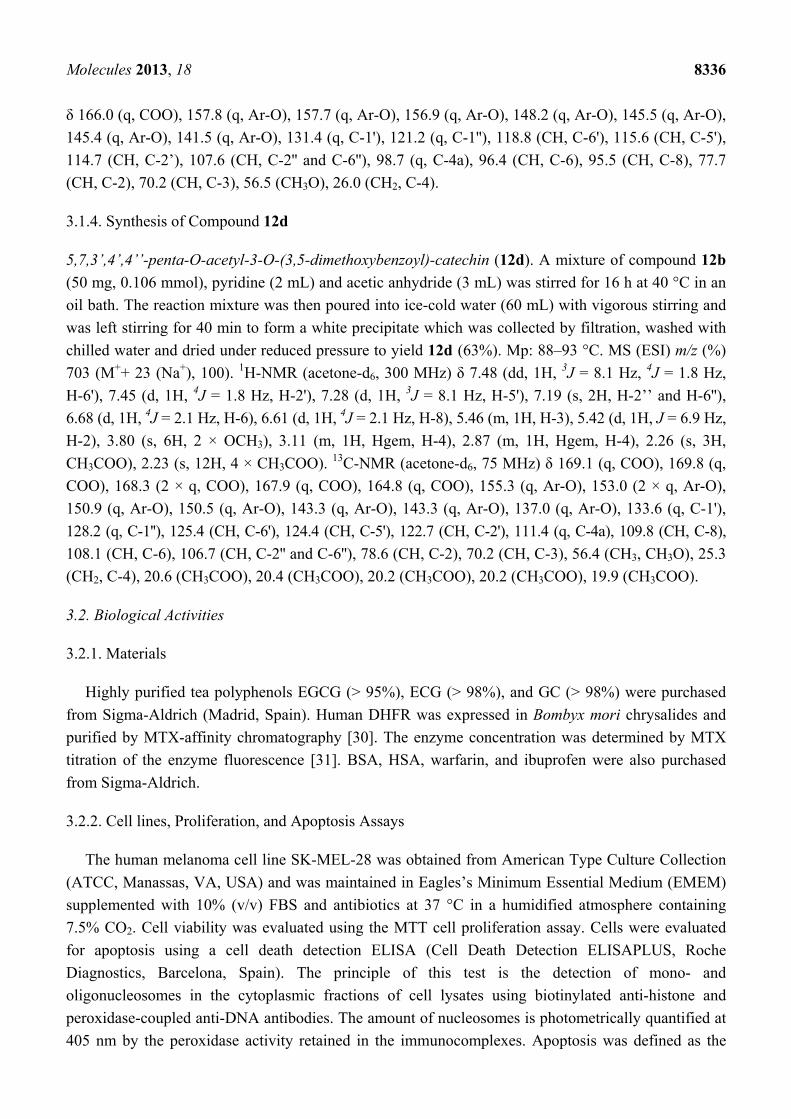
3.1.4. Synthesis of Compound 12d
5,7,3’,4’,4’’-penta-O-acetyl-3-O-(3,5-dimethoxybenzoyl)-catechin (12d). A mixture of compound 12b
(50 mg, 0.106 mmol), pyridine (2 mL) and acetic anhydride (3 mL) was stirred for 16 h at 40 °C in an
oil bath. The reaction mixture was then poured into ice-cold water (60 mL) with vigorous stirring and
was left stirring for 40 min to form a white precipitate which was collected by filtration, washed with
chilled water and dried under reduced pressure to yield 12d (63%). Mp: 88–93 °C. MS (ESI) m/z (%)
703 (M++ 23 (Na+), 100). 1H-NMR (acetone-d6, 300 MHz) δ 7.48 (dd, 1H, 3J = 8.1 Hz, 4J = 1.8 Hz,
H-6'), 7.45 (d, 1H, 4J = 1.8 Hz, H-2'), 7.28 (d, 1H, 3J = 8.1 Hz, H-5'), 7.19 (s, 2H, H-2’’ and H-6''),
6.68 (d, 1H, 4J = 2.1 Hz, H-6), 6.61 (d, 1H, 4J = 2.1 Hz, H-8), 5.46 (m, 1H, H-3), 5.42 (d, 1H, J = 6.9 Hz,
H-2), 3.80 (s, 6H, 2 × OCH3), 3.11 (m, 1H, Hgem, H-4), 2.87 (m, 1H, Hgem, H-4), 2.26 (s, 3H,
CH3COO), 2.23 (s, 12H, 4 × CH3COO). 13C-NMR (acetone-d6, 75 MHz) δ 169.1 (q, COO), 169.8 (q,
COO), 168.3 (2 × q, COO), 167.9 (q, COO), 164.8 (q, COO), 155.3 (q, Ar-O), 153.0 (2 × q, Ar-O),
150.9 (q, Ar-O), 150.5 (q, Ar-O), 143.3 (q, Ar-O), 143.3 (q, Ar-O), 137.0 (q, Ar-O), 133.6 (q, C-1'),
128.2 (q, C-1''), 125.4 (CH, C-6'), 124.4 (CH, C-5'), 122.7 (CH, C-2'), 111.4 (q, C-4a), 109.8 (CH, C-8),
108.1 (CH, C-6), 106.7 (CH, C-2'' and C-6''), 78.6 (CH, C-2), 70.2 (CH, C-3), 56.4 (CH3, CH3O), 25.3
(CH2, C-4), 20.6 (CH3COO), 20.4 (CH3COO), 20.2 (CH3COO), 20.2 (CH3COO), 19.9 (CH3COO).
3.2. Biological Activities
3.2.1. Materials
Highly purified tea polyphenols EGCG (> 95%), ECG (> 98%), and GC (> 98%) were purchased
from Sigma-Aldrich (Madrid, Spain). Human DHFR was expressed in Bombyx mori chrysalides and
purified by MTX-affinity chromatography [30]. The enzyme concentration was determined by MTX
titration of the enzyme fluorescence [31]. BSA, HSA, warfarin, and ibuprofen were also purchased
from Sigma-Aldrich.
3.2.2. Cell lines, Proliferation, and Apoptosis Assays
The human melanoma cell line SK-MEL-28 was obtained from American Type Culture Collection
(ATCC, Manassas, VA, USA) and was maintained in Eagles’s Minimum Essential Medium (EMEM)
supplemented with 10% (v/v) FBS and antibiotics at 37 °C in a humidified atmosphere containing
7.5% CO2. Cell viability was evaluated using the MTT cell proliferation assay. Cells were evaluated
for apoptosis using a cell death detection ELISA (Cell Death Detection ELISAPLUS, Roche
Diagnostics, Barcelona, Spain). The principle of this test is the detection of mono- and
oligonucleosomes in the cytoplasmic fractions of cell lysates using biotinylated anti-histone and
peroxidase-coupled anti-DNA antibodies. The amount of nucleosomes is photometrically quantified at
405 nm by the peroxidase activity retained in the immunocomplexes. Apoptosis was defined as the

Molecules 2013, 18 8337
specific enrichment of mono- and oligonucleosomes in the cytoplasm and was calculated by dividing
the absorbance of treated samples with the absorbance of untreated samples after correcting for the
number of cells. The induction of apoptosis in each melanoma cell line after a 7 h treatment with 2 μM
staurosporin (100% apoptotic cells) was used to calculate the number of apoptotic cells. For these
assays, cells were plated in a 96-well plate at a density of 1000–2000 cells/well. Compounds were
added once at the beginning of the experiments.
3.2.3. DHFR Binding Studies
The dissociation constants for the binding of polyphenols to free recombinant human DHFR
(rHDHFR) were determined by fluorescence titration in an automatic-scanning FluoroMax-3 (Jobin
Ybon, Horiba, Edison, NJ, USA) spectrofluorometer with 1.0 cm light path cells and a 150 W
mercury-xenon light source. The formation of a binary complex between the enzyme (2.5 × 10⁻⁷ M)
and the ligand (from 0 to 40 µM) was followed by measuring the quenching of the tryptophan
fluorescence of the enzyme upon the addition of microliter volumes of a concentrated stock solution of
ligand. Experiments were performed at pH 7.5 in a buffer containing 2-(N-morpholino)ethanesulfonic
acid (Mes, 0.025 M), sodium acetate (0.025 M), tris(hydroxymethyl)aminomethane (Tris 0.05 M), and
NaCl (0.1 M). The temperature was controlled at 25 °C using a Haake D1G circulating bath with a
heater and cooler. Fluorescence emission spectra were recorded when the human DHFR fluorescence
was excited at 290 nm, and titrations were performed as described elsewhere [11].
3.2.4. BSA Binding Studies
Fluorescence measurements were made on an automatic scanning Perkin-Elmer LS50B
spectrofluorometer with 1.0 cm light path cells equipped with a 150 W xenon (XBO) light source. In
all experiments, the fluorescence of BSA (5 × 10⁻⁶ M) in 50 mM phosphate buffer of pH 7.5 was
followed by excitation at 280 nm. The binding affinities of EGCG and TMCG with BSA in terms of
binding constants were determined using the following equation [25]:
log (F0−F)/F = log KA + n log [Q] (1)
where KA is the binding constant, n is the number of binding sites, F0 is the fluorescence intensity of
free BSA and F is the consequent fluorescence upon the addition of catechins (Q). A plot of log
(F0–F)/F against log [Q] was used to determine the values of KA and n from the intercept and the
slope, respectively.
To identify the binding sites of EGCG and TMCG in BSA, the displacements of warfarin and
ibuprofen (known to bind at site I and site II, respectively) were followed fluorometrically. Equimolar
solutions of BSA and warfarin (5 × 10⁻⁶ M each) were mixed for 1 h and titrated with EGCG and
TMCG in separate experiments. Similar solutions containing equimolar ibuprofen and BSA were
prepared and separately titrated with EGCG and TMCG to identify the binding sites of EGCG and
TMCG with BSA. Titration was followed fluorometrically using an excitation wavelength of 280 nm.

Molecules 2013, 18 8338
3.2.5. HSA Binding Studies
Binding of TMECG, EGCG, and EC to HSA was determined as specified for the BSA binding studies.
3.2.6. Spectrophotometric Assays
Ultraviolet-visible absorption spectra of catechins at different pHs were recorded on a UV-Vis
Perkin-Elmer Lambda-2 spectrophotometer with a spectral bandwidth of 1 nm at a scan speed of
60 nm s−1. The experiments were performed in the same buffer as described for DHFR binding studies,
and the ionic strength remained constant at an optimum value of 0.15 over the pH range that was used.
The pH of the reaction was measured before and after the experiment.
3.2.7. Statistical Analysis
In all experiments, the mean ± standard deviation (SD) values for three to five determinations in
triplicate were calculated. Statistically significant differences were evaluated using the Student’s t-test.
Differences were considered statistically significant at p < 0.05.
4. Conclusions
Recent studies have indicated that natural tea catechins or related synthetic catechins are good
inhibitors of DHFR. In this study, we show that polyphenols with catechin configuration are better
DHFR inhibitors than those with epicatechin configuration. However, in addition to their DHFR
inhibiting characteristics, other aspects should be considered for the design and syntheses of these
types of inhibitors. For example, although an OH group in para position of ring D seems to be an
important determinant that favours DHFR-catechin interaction, the pH-dependent ionization of this
hydroxyl group impedes drug transportation through the cell membrane, which highly reduces the
biological activity of these compounds. The interaction of catechins with albumin seems to be another
important factor that might modulate the in vivo activity of these drugs. Although natural galloylated
catechins, such as EGCG or ECG, are better inhibitors of DHFR, the high affinity of these catechins
for serum albumin might reduce their bioavailability. In summary, we found that two synthetic
methylated catechins, TMECG and TMCG, presented an ideal balance between DHFR inhibition and
other factors that improve their cell and body bioavailability, such as high hydrophobicity and low
interaction with serum albumins. These results demonstrate the high in vitro and in vivo anti-tumour
activity of these two catechins on different experimental models of cancer [32,33]. These observations,
together with their low toxicity on non-cancer cells [33,34], indicate that these drugs might have a
great potential for their application in cancer therapies. Finally, the observation that the displacement
of the equilibrium of the catechin-BSA complex using a competitive inhibitor of the albumin binding
site-I, such as warfarin, highly increases the anti-tumour activity of TMCG, may be useful for the
design and formulation of catechin-containing therapies. The binding of sulphonamide drugs to serum
albumin has long been considered important for their chemotherapeutic values, such as in vivo
antibacterial activity and the duration of action [35]. In this respect, the effect of these catechins on the
pharmacokinetics and pharmacodynamics of warfarin, a commonly used drug for preventing
Molecules 2013, 18 8339
intravascular coagulation, should be determined in order to evaluate the potential side-effects and
clinical consequence of these findings.
Supplementary Materials
Supplementary materials containing 1H and 13C NMR and mass spectrometry spectra of compounds
10b, 10c, 11b, 12b–d, and 13b can be accessed at: http://www.mdpi.com/1420-3049/18/7/8319/s1.
Acknowledgments
This work was supported by grants from the Fundación Séneca, Región de Murcia (FS-RM)
(15230/PI/10) and the European Commission (EU ERA293514). S.C was contracted by this EU
project. M.S.A. had a grant for research continuity from University of Murcia and M.P.F-P had a
fellowship from Ministerio de Ciencia e Innovación (MICINN).
Conflicts of Interest
The authors declare no conflicts of interest.
References
1. Mukhtar, H.; Ahmad, N. Cancer chemoprevention: future holds in multiple agents. Toxicol. Appl.
Pharmacol. 1999, 158, 207–210.
2. Yang, C.S.; Chung, J.Y.; Yang, G.Y.; Li, C.; Meng, X.; Lee, M.J. Mechanisms of inhibition of
carcinogenesis by tea. Biofactors 2000, 13, 73–79.
3. Shankar, S.; Ganapathy, S.; Srivastava, R.K. Green tea polyphenols: biology and therapeutic
implications in cancer. Front. Biosci. 2007, 12, 4881–4899.
4. Nam, S.; Smith, D.M.; Dou, Q.P. Ester bond-containing tea polyphenols potently inhibit
proteasome activity in vitro and in vivo. J. Biol. Chem. 2001, 276, 13322–13330.
5. Fang, M.Z.; Wang, Y.; Ai, N.; Hou, Z.; Sun, Y.; Lu, H.; Welsh, W.; Yang, C.S. Tea polyphenol
(−)-epigallocatechin-3-gallate inhibits DNA methyltransferase and reactivates methylation-silenced
genes in cancer cell lines. Cancer Res. 2003, 63, 7563–7570.
6. Li, C.; Allen, A.; Kwagh, J.; Doliba, N.M.; Qin, W.; Najafi, H.; Collins, H.W.; Matschinsky, F.M.;
Stanley, C.A.; Smith, T.J. Green tea polyphenols modulate insulin secretion by inhibiting
glutamate dehydrogenase. J. Biol. Chem. 2006, 281, 10214–10221.
7. Navarro-Peran, E.; Cabezas-Herrera, J.; Garcia-Canovas, F.; Durrant, M.C.; Thorneley, R.N.;
Rodriguez-Lopez, J.N. The antifolate activity of tea catechins. Cancer Res. 2005, 65, 2059–2064.
8. Alemdaroglu, N.C.; Wolffram, S.; Boissel, J.P.; Closs, E.; Spahn-Langguth, H.; Langguth, P.
Inhibition of folic acid uptake by catechins and tea extracts in Caco-2 cells. Planta Med. 2007, 73,
27–32.
9. Navarro-Peran, E.; Cabezas-Herrera, J.; Campo, L.S.; Rodriguez-Lopez, J.N. Effects of folate
cycle disruption by the green tea polyphenol epigallocatechin-3-gallate. Int. J. Biochem. Cell. Biol.
2007, 39, 2215–2225.

Molecules 2013, 18 8340
10. Navarro-Martinez, M.D.; Navarro-Peran, E.; Cabezas-Herrera, J.; Ruiz-Gomez, J.;
Garcia-Canovas, F.; Rodriguez-Lopez, J.N. Antifolate activity of epigallocatechin gallate against
Stenotrophomonas maltophilia. Antimicrob. Agents Chemother. 2005, 49, 2914–2920.
11. Navarro-Peran, E.; Cabezas-Herrera, J.; Hiner, A.N.; Sadunishvili, T.; Garcia-Canovas, F.;
Rodriguez-Lopez, J.N. Kinetics of the inhibition of bovine liver dihydrofolate reductase by tea
catechins: origin of slow-binding inhibition and pH studies. Biochemistry 2005, 44, 7512–7525.
12. Spina, M.; Cuccioloni, M.; Mozzicafreddo, M.; Montecchia, F.; Pucciarelli, S.; Eleuteri, A.M.;
Fioretti, E.; Angeletti, M. Mechanism of inhibition of wt-dihydrofolate reductase from E. coli by
tea epigallocatechin-gallate. Proteins 2008, 72, 240–251.
13. Kao, T.T.; Wang, K.C.; Chang, W.N.; Lin, C.Y.; Chen, B.H.; Wu, H.L.; Shi, G.Y.; Tsai, J.N.; Fu, T.F.
Characterization and comparative studies of zebrafish and human recombinant dihydrofolate
reductases--inhibition by folic acid and polyphenols. Drug Metab. Dispos. 2008, 36, 508–516.
14. Hannewald, P.; Maunit, B.; Muller, J.F. Screening of DHFR-binding drugs by MALDI-TOFMS.
Anal. Bioanal. Chem. 2008, 392, 1335–1344.
15. Hong, J.; Lu, H.; Meng, X.; Ryu, J.H.; Hara, Y.; Yang, C.S. Stability, cellular uptake,
biotransformation, and efflux of tea polyphenol (−)-epigallocatechin-3-gallate in HT-29 human
colon adenocarcinoma cells. Cancer Res. 2002, 62, 7241–7246.
16. Sanchez-del-Campo, L.; Oton, F.; Tarraga, A.; Cabezas-Herrera, J.; Chazarra, S.;
Rodriguez-Lopez, J.N. Synthesis and biological activity of a 3,4,5-trimethoxybenzoyl ester
analogue of epicatechin-3-gallate. J. Med. Chem. 2008, 51, 2018–2026.
17. Babich, H.; Zuckerbraun, H.L.; Weinerman, S.M. In vitro cytotoxicity of (-)-catechin gallate, a
minor polyphenol in green tea. Toxicol Lett 2007, 171, 171–180.
18. Saez-Ayala, M.; Sanchez-del-Campo, L.; Montenegro, M.F.; Chazarra, S.; Tarraga, A.;
Cabezas-Herrera, J.; Rodriguez-Lopez, J.N. Comparison of a pair of synthetic tea-catechin-derived
epimers: Synthesis, antifolate activity, and tyrosinase-mediated activation in melanoma.
ChemMedChem 2011, 6, 440–449.
19. Tückmantel, W.; Kozikowski, A.P.; Romanczyk, L.J. Studies in Polyphenol Chemistry and
Bioactivity. 1. Preparation of building blocks from (+)-Catechin. Procyanidin formation. synthesis
of the cancer cell growth inhibitor, 3-O-Galloyl-(2R,3R)-epicatechin-4β,8-[3-O-galloyl-(2R,3R)-
epicatechin]. J. Am. Chem. Soc. 1999, 121, 12073–12081.
20. Park, K.D.; Lee, S.G.; Kim, S.U.; Kim, S.H.; Sun, W.S.; Cho, S.J.; Jeong, D.H. Anticancer
activity of 3-O-acyl and alkyl-(-)-epicatechin derivatives. Bioorg. Med. Chem. Lett. 2004, 14,
5189–5192.
21. Hirasawa, M.; Takada, K. Multiple effects of green tea catechin on the antifungal activity of
antimycotics against Candida albicans. J. Antimicrob. Chemother. 2004, 53, 225–229.
22. Cocco, L.; Roth, B.; Temple, C., Jr.; Montgomery, J.A.; London, R.E.; Blakley, R.L. Protonated
state of methotrexate, trimethoprim, and pyrimethamine bound to dihydrofolate reductase.
Arch. Biochem. Biophys. 1983, 226, 567–577.
23. Vyas, S.; Manon, B.; Vir Singh, T.; Dev Sharma, P.; Sharma, M. Potential O-acyl-substituted
(−)-Epicatechin gallate prodrugs as inhibitors of DMBA/TPA-induced squamous cell carcinoma
of skin in Swiss albino mice. Chem. Biodivers. 2011, 8, 599–613.

Molecules 2013, 18 8341
24. Ishii, T.; Minoda, K.; Bae, M.J.; Mori, T.; Uekusa, Y.; Ichikawa, T.; Aihara, Y.; Furuta, T.;
Wakimoto, T.; Kan, T.; Nakayama, T. Binding affinity of tea catechins for HSA: characterization
by high-performance affinity chromatography with immobilized albumin column. Mol. Nutr.
Food Res. 2010, 54, 816–822.
25. Pal, S.; Saha, C.; Hossain, M.; Dey, S.K.; Kumar, G.S. Influence of galloyl moiety in interaction
of epicatechin with bovine serum albumin: A spectroscopic and thermodynamic characterization.
PLoS One 2012, 7, e43321.
26. Boulton, D.W.; Walle, U.K.; Walle, T. Extensive binding of the bioflavonoid quercetin to human
plasma proteins. J. Pharm. Pharmacol. 1998, 50, 243–249.
27. Kratz, F. Albumin as a drug carrier: Design of prodrugs, drug conjugates and nanoparticles.
J. Controlled Release 2008, 132, 171–183.
28. Wunder, A.; Muller-Ladner, U.; Stelzer, E.H.; Funk, J.; Neumann, E.; Stehle, G.; Pap, T.; Sinn, H.;
Gay, S.; Fiehn, C. Albumin-based drug delivery as novel therapeutic approach for rheumatoid
arthritis. J. Immunol. 2003, 170, 4793–4801.
29. Gao, S.; Feng, N.; Yu, S.; Yu, D.; Wang, X. Vasodilator constituents from the roots of Lysidice
rhodostega. Planta Med. 2004, 70, 1128–1134.
30. Chazarra, S.; Aznar-Cervantes, S.; Sanchez-del-Campo, L.; Cabezas-Herrera, J.; Xiaofeng, W.;
Cenis, J.L.; Rodriguez-Lopez, J.N. Purification and kinetic properties of human recombinant
dihydrofolate reductase produced in Bombyx mori chrysalides. Appl. Biochem. Biotechnol. 2010,
162, 1834–1846.
31. Smith, S.L.; Patrick, P.; Stone, D.; Phillips, A.W.; Burchall, J.J. Porcine liver dihydrofolate
reductase. Purification, properties, and amino acid sequence. J. Biol. Chem. 1979, 254, 11475–11484.
32. Montenegro, M.F.; Saez-Ayala, M.; Pinero-Madrona, A.; Cabezas-Herrera, J.;
Rodriguez-Lopez, J.N. Reactivation of the tumour suppressor RASSF1A in breast cancer by
simultaneous targeting of DNA and E2F1 methylation. PLoS One 2012, 7, e52231.
33. Sáez-Ayala, M.; Montenegro, M.F.; Sánchez-del-Campo, L.; Fernández-Pérez, M.P.;
Chazarra, S.; Freter, R.; Middleton, M.; Piñero-Madrona, A.; Cabezas-Herrera, J.; Goding, C.R.;
Rodríguez-López , J.N. Directed phenotype-switching as an effective anti-melanoma strategy.
Cancer Cell. 2013, 24, 105–119.
34. Sánchez-del-Campo, L.; Rodríguez-López, J.N. Targeting the methionine cycle for melanoma
therapy with 3-O-(3,4,5-trimethoxybenzoyl)-(-)-epicatechin. Int. J. Cancer 2008, 123, 2446–2455.
35. Fujita, T. Hydrophobic bonding of sulfonamide drugs with serum albumin. J. Med. Chem. 1972,
15, 1049–1056.
Sample Availability: Samples of the compounds 12b and 13b are available from the authors.
© 2013 by the authors; licensee MDPI, Basel, Switzerland. This article is an open access article
distributed under the terms and conditions of the Creative Commons Attribution license
(http://creativecommons.org/licenses/by/3.0/).