Facial and Orbital Fractures - Lieberman's...
49
Holly B. Hindman Gillian Lieberman, MD Holly B. Hindman, Harvard Medical School, Year III Gillian Lieberman, MD April 2002 Facial and Orbital Fractures
Transcript of Facial and Orbital Fractures - Lieberman's...
Holly B. Hindman
Gillian Lieberman, MD
Holly B. Hindman, Harvard Medical School, Year IIIGillian Lieberman, MD
April 2002
Facial and Orbital Fractures
2
Holly B. Hindman
Gillian Lieberman, MD
Outline of Discussion
• Introduction to our patient• Orbital anatomy• Recommended imaging studies• Presentation and radiological findings of
various facial and orbital fractures• Potential complications of orbital fractures• Revisiting our patient
3
Holly B. Hindman
Gillian Lieberman, MD
Patient Presentation – P.Q.
CC: Trauma patient, s/p fall from 70 feet.HPI: brought to E.R. s/p fall from 70 feet with
multiple injuries including facial and orbital fractures.
4
Holly B. Hindman
Gillian Lieberman, MD
Defining the Orbital Walls
• Medial Wall: ethmoid bone (paper thin), lacrimal bone, body of spenoid (posteriorly), frontal bone (superiorly), maxilla (inferiorly)
• Lateral Wall: zygomatic bone anteriorly, greater wing of sphenoid bone posteriorly.
• Roof: frontal bone, lesser wing of sphenoid bone containing optic canal
• Floor: maxilla and zygomatic bone anteriorly, palatine bone posteriorly
5
Holly B. Hindman
Gillian Lieberman, MD
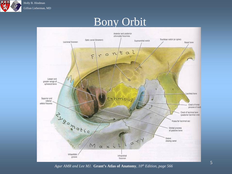
Bony Orbit
Agur AMR and Lee MJ. Grant’s Atlas of Anatomy, 10th Edition, page 566
6
Holly B. Hindman
Gillian Lieberman, MD
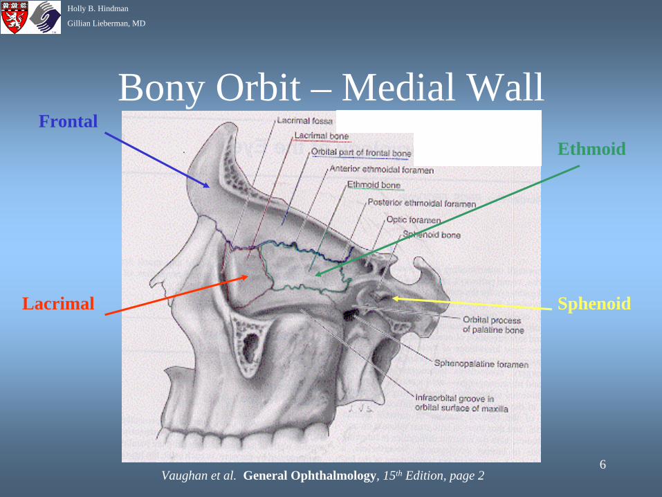
Bony Orbit – Medial Wall
Vaughan et al. General Ophthalmology, 15th Edition, page 2
Frontal
Lacrimal
Ethmoid
Sphenoid
7
Holly B. Hindman
Gillian Lieberman, MD
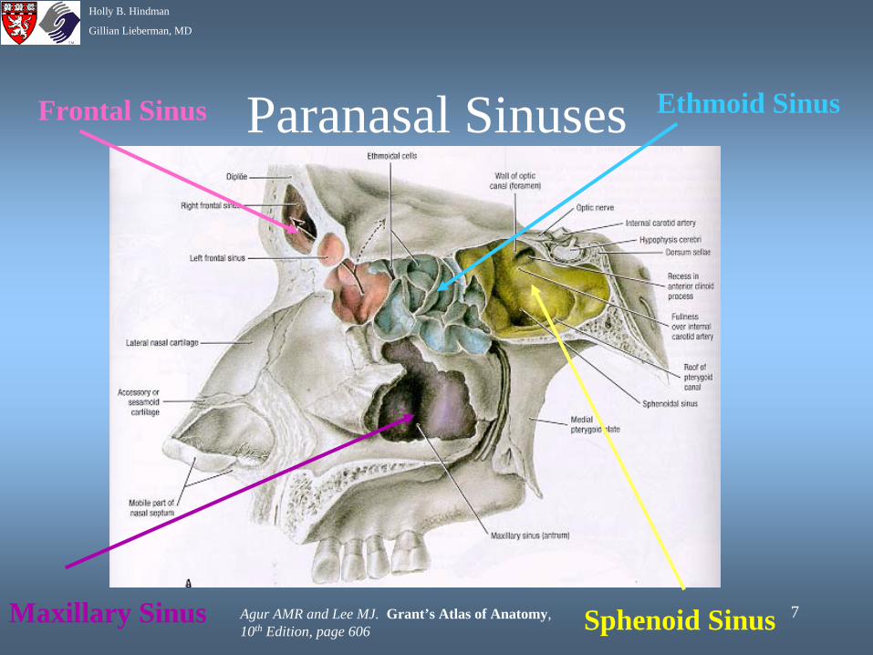
Paranasal SinusesFrontal Sinus Ethmoid Sinus
Sphenoid SinusMaxillary Sinus Agur AMR and Lee MJ. Grant’s Atlas of Anatomy, 10th Edition, page 606
8
Holly B. Hindman
Gillian Lieberman, MD
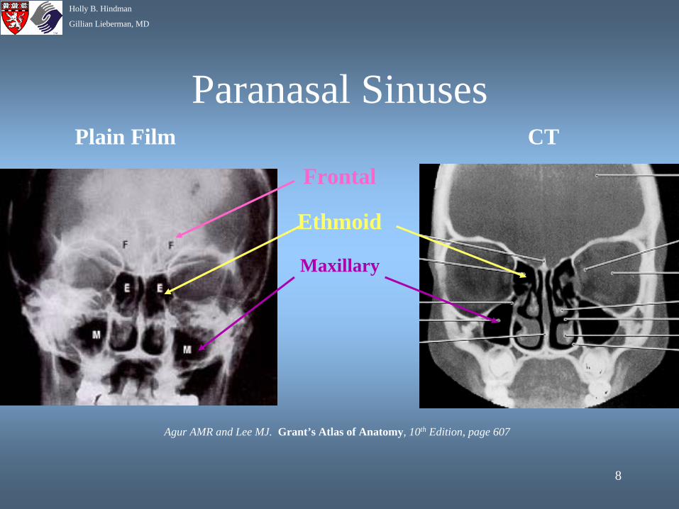
Paranasal SinusesPlain Film CT
Frontal
Ethmoid
Maxillary
Agur AMR and Lee MJ. Grant’s Atlas of Anatomy, 10th Edition, page 607
9
Holly B. Hindman
Gillian Lieberman, MD
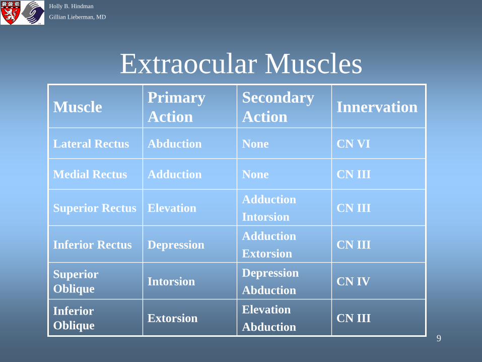
Extraocular MusclesMuscle Primary
ActionSecondary Action Innervation
Lateral Rectus Abduction None CN VI
Medial Rectus Adduction None CN III
Superior Rectus ElevationAdductionIntorsion
CN III
Inferior Rectus DepressionAdductionExtorsion
CN III
Superior Oblique Intorsion
DepressionAbduction
CN IV
Inferior Oblique Extorsion
ElevationAbduction
CN III
10
Holly B. Hindman
Gillian Lieberman, MD
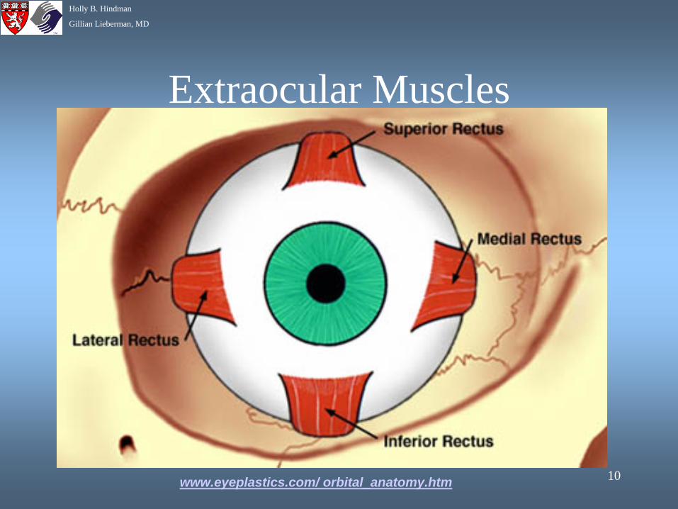
Extraocular Muscles
www.eyeplastics.com/ orbital_anatomy.htm
11
Holly B. Hindman
Gillian Lieberman, MD
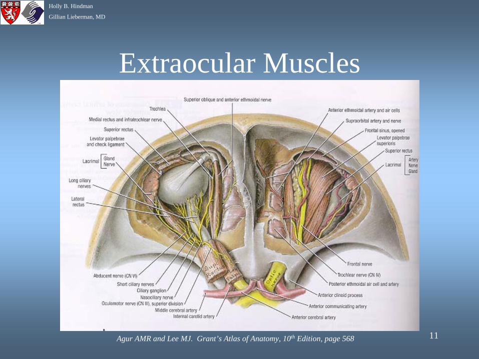
Extraocular Muscles
Agur AMR and Lee MJ. Grant’s Atlas of Anatomy, 10th Edition, page 568
12
Holly B. Hindman
Gillian Lieberman, MD
Orbital Arteries and VeinsArteries
Ophthalmic Artery is the first intracranial branch of internal carotid Artery. Accompanies optic nerve through optic canal and branches into:
1) Retinal Artery2) Lacrimal Artery3) Muscular Branches4) Long and Short Posterior Ciliary
Arteries5) Medial Palpebral Arteries6) Supraorbital and Supratrochlear
Arteries
VeinsVortex veins, anterior ciliary veins,
and central retinal vein drain into superior and inferior ophthalmic veins
Superior ophthalmic vein passes through superior orbital fissure and communicates with cavernous sinus
Inferior ophthalmic vein passes through inferior orbital fissure to communicate with pterygoid plexus
13
Holly B. Hindman
Gillian Lieberman, MD
Nerves of the Orbit
Oculomotor Nerve (CN III): enters via superior orbital fissure•Superior Division: levator palpebrae, superior rectus muscle•Inferior Division: medial and inferior recti, inferior oblique muscles, parasympathetic fibers to ciliary ganglion
Trochlear Nerve (CN IV): enters via superior orbital fissure • innervates superior oblique muscle
Abducens Nerve (CN VI): enters via the superior orbital fissure•Innervates lateral rectus muscle.
14
Holly B. Hindman
Gillian Lieberman, MD
Nerves of the Orbit
Trigeminal Nerve (CN V):•Ophthalmic Branch: enters via superior orbital fissure
1) lacrimal nerve: provides sensory innervation to lacrimal gland
2) frontal nerve: divides into supraorbital and supratrochlear nerves and provides sensation to brow and forehead
3) nasociliary nerve: sensation to cornea, iris, and ciliary body
•Maxillary Branch: enters via inferior orbital fissure becomes the infraorbital nerve and exits via the infraorbital foramen provide sensory innervation to lower lid and cheek
15
Holly B. Hindman
Gillian Lieberman, MD
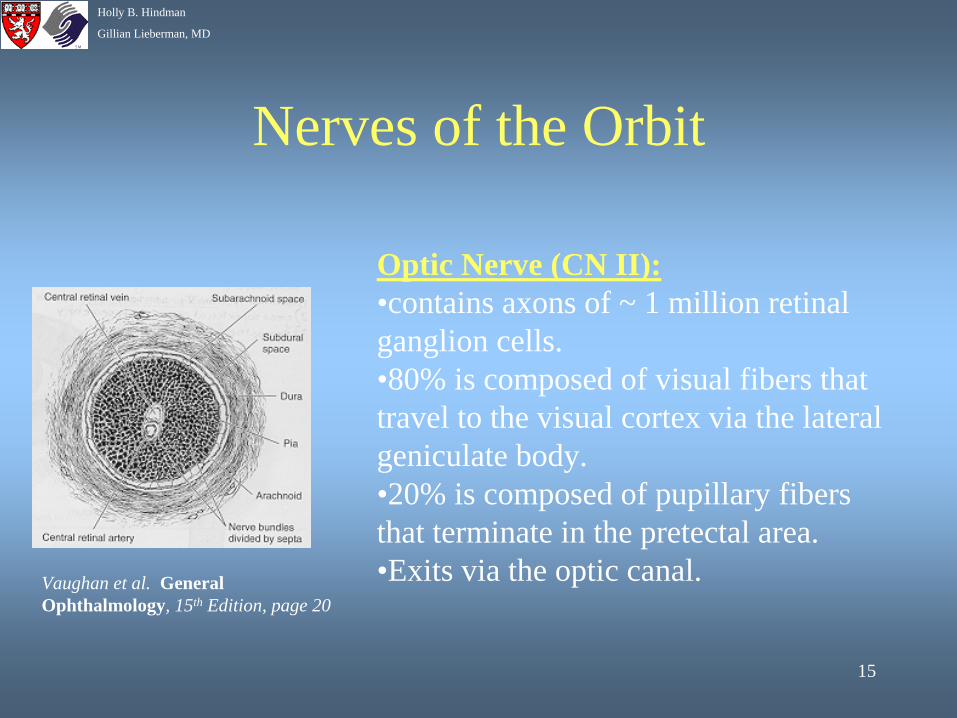
Nerves of the Orbit
Optic Nerve (CN II):•contains axons of ~ 1 million retinal ganglion cells.•80% is composed of visual fibers that travel to the visual cortex via the lateral geniculate body.•20% is composed of pupillary fibers that terminate in the pretectal area.•Exits via the optic canal.Vaughan et al. General
Ophthalmology, 15th Edition, page 20
16
Holly B. Hindman
Gillian Lieberman, MD
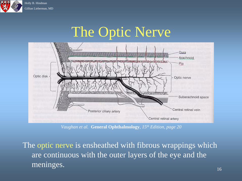
The Optic Nerve
The optic nerve is ensheathed with fibrous wrappings which are continuous with the outer layers of the eye and the meninges.
Vaughan et al. General Ophthalmology, 15th Edition, page 20
17
Holly B. Hindman
Gillian Lieberman, MD
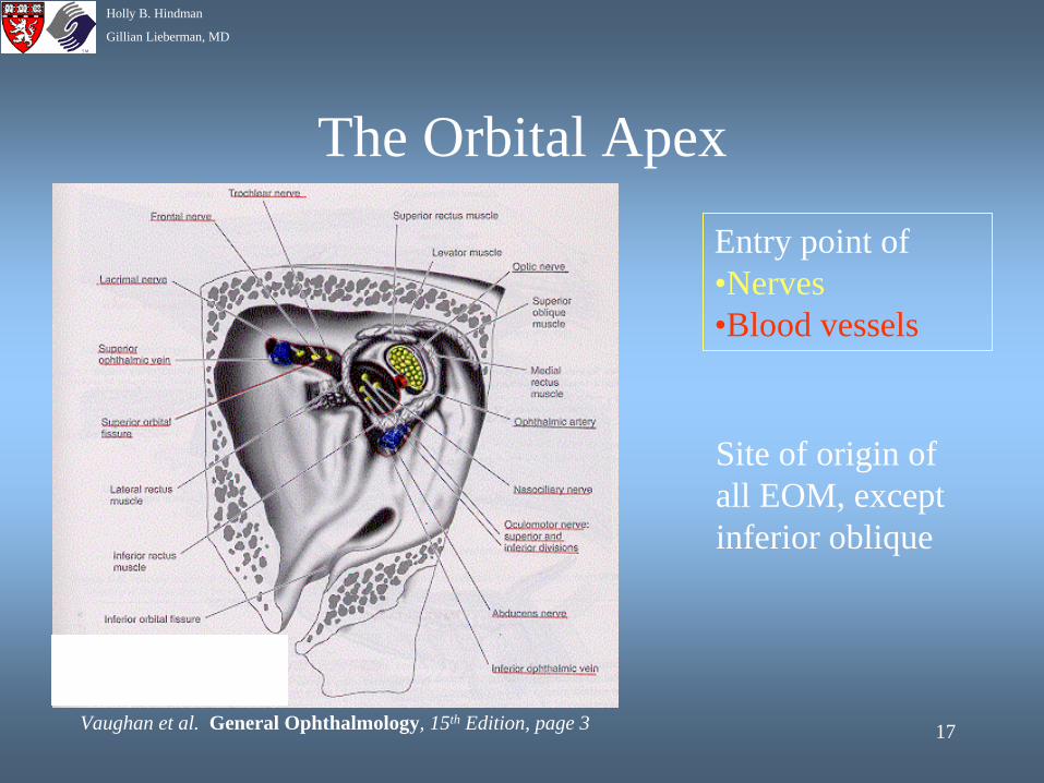
The Orbital Apex
Entry point of •Nerves•Blood vessels
Site of origin of all EOM, except inferior oblique
Vaughan et al. General Ophthalmology, 15th Edition, page 3
18
Holly B. Hindman
Gillian Lieberman, MD
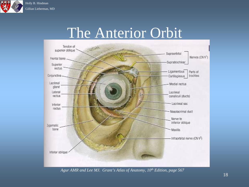
The Anterior Orbit
Agur AMR and Lee MJ. Grant’s Atlas of Anatomy, 10th Edition, page 567
19
Holly B. Hindman
Gillian Lieberman, MD
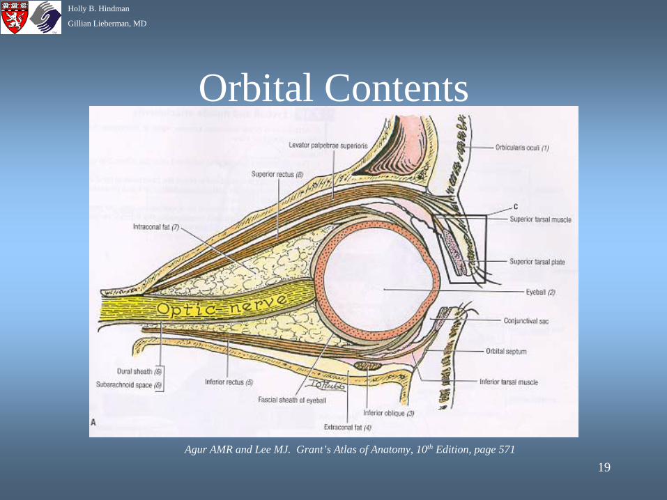
Orbital Contents
Agur AMR and Lee MJ. Grant’s Atlas of Anatomy, 10th Edition, page 571
20
Holly B. Hindman
Gillian Lieberman, MD
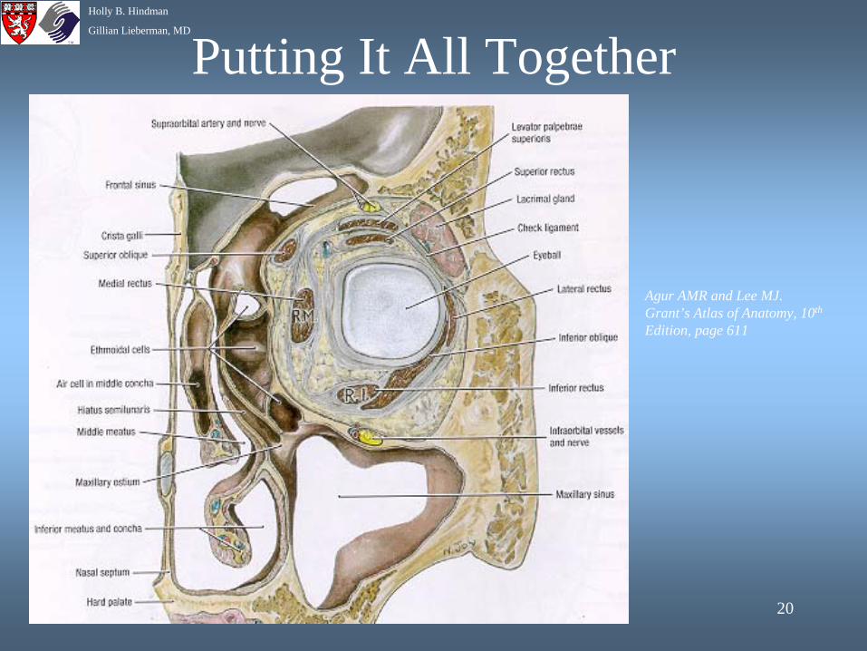
Putting It All Together
Agur AMR and Lee MJ. Grant’s Atlas of Anatomy, 10th
Edition, page 611
21
Holly B. Hindman
Gillian Lieberman, MD
Causes of Orbital Trauma
• Motor vehicle accidents• High acceleration injuries• Violent crime• Athletic accidents• Industrial accidents
22
Holly B. Hindman
Gillian Lieberman, MD
Imaging Studies• Plain Films in patients who show no neurological
abnormalities or in patients who have suspected foreign body. Use Caldwell and Waters views.
• High resolution axial CT is primary imaging modality using both axial and coronal views.
• CT angiogography if there is concern for vascular injury such as carotid cavernous fistula.
• MR useful for evaluating vascular injuries and psuedoaneurysms, lacrimal drainage injury, motility disorders, and for surgical planning. Contraindicated until metallic foreign body ruled out.
• US can detect intraocular foreign bodies, globe rupture, suprachoroidal hemorrhage, and retinal detachment.
23
Holly B. Hindman
Gillian Lieberman, MD
Types of Orbital FracturesOrbital fractures are often associated with optic nerve
injuries, paranasal sinus injuries, and/or intracranial injuries.
Types of orbital fractures include:• Le Fort Fractures• Medial Orbital Fractures• Orbital Floor Fractures• Orbital Roof Fractures• Lateral (Zygomatic, Tripod) Fractures• Naso-Ethmoidal Orbital Fractures• Orbital Apex Fractures
24
Holly B. Hindman
Gillian Lieberman, MD
Definitions• Blow-out Fracture:• outward fracture of involved orbital bones.• Usually involves medial wall and floor. • Results in increased intraorbital volume and
enophthalmos.
• Blow-in Fracture:• fracture of orbital bones inward into the orbital
space. • Results in decreased orbital volume and proptosis.
25
Holly B. Hindman
Gillian Lieberman, MD
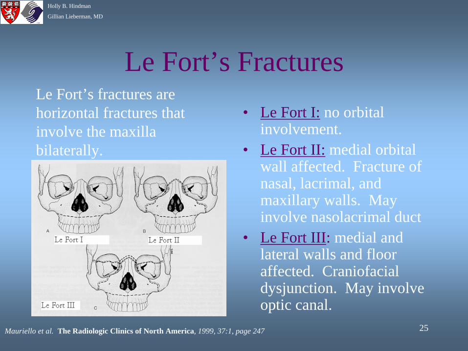
Le Fort’s Fractures• Le Fort I: no orbital
involvement.• Le Fort II: medial orbital
wall affected. Fracture of nasal, lacrimal, and maxillary walls. May involve nasolacrimal duct
• Le Fort III: medial and lateral walls and floor affected. Craniofacial dysjunction. May involve optic canal.
Le Fort’s fractures are horizontal fractures that involve the maxilla bilaterally.
Mauriello et al. The Radiologic Clinics of North America, 1999, 37:1, page 247
26
Holly B. Hindman
Gillian Lieberman, MD
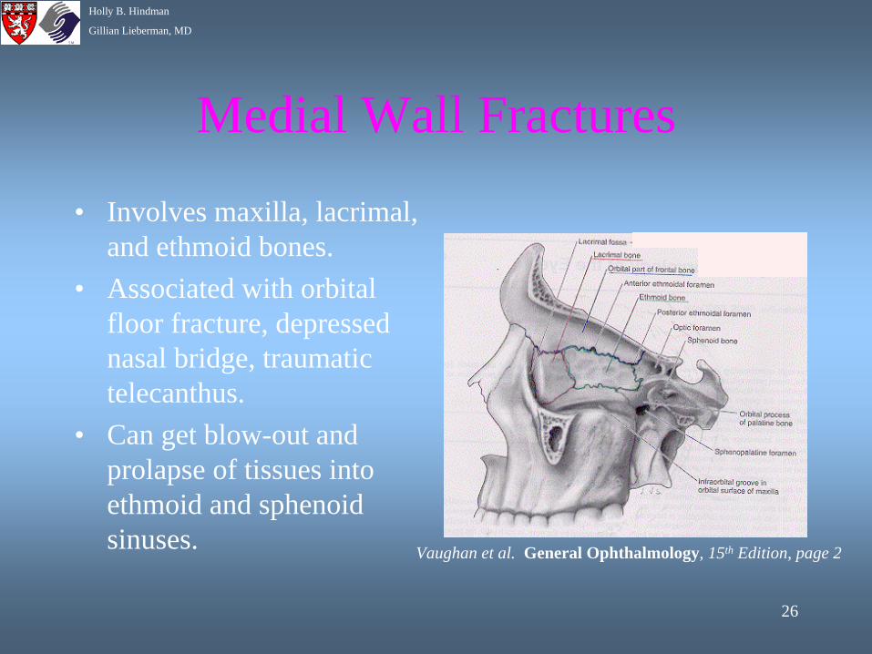
Medial Wall Fractures
• Involves maxilla, lacrimal, and ethmoid bones.
• Associated with orbital floor fracture, depressed nasal bridge, traumatic telecanthus.
• Can get blow-out and prolapse of tissues into ethmoid and sphenoid sinuses. Vaughan et al. General Ophthalmology, 15th Edition, page 2
27
Holly B. Hindman
Gillian Lieberman, MD
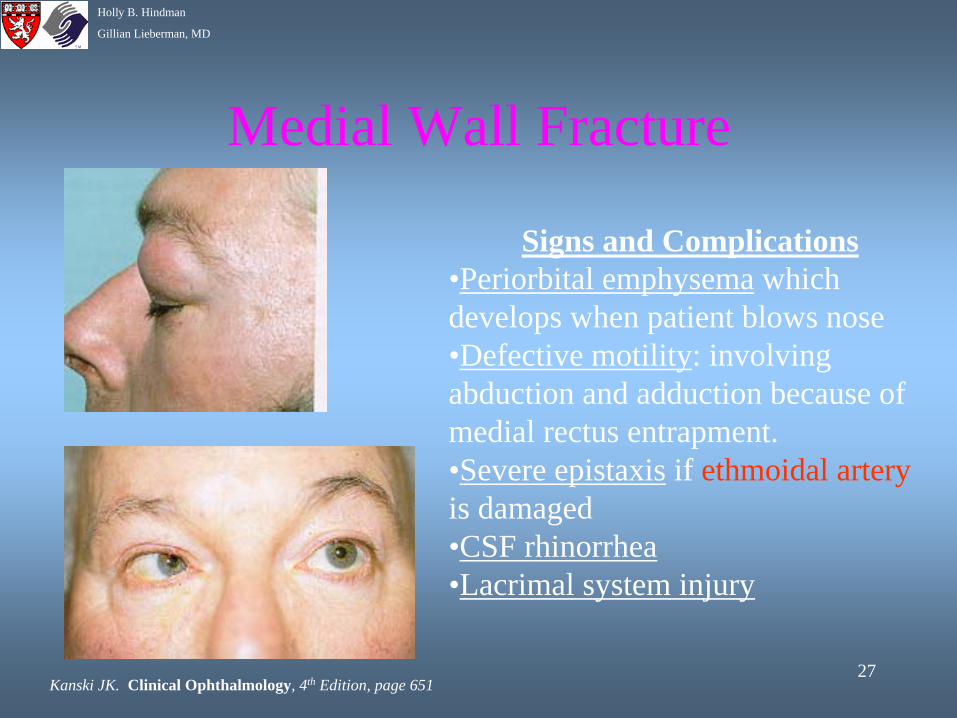
Medial Wall Fracture
Signs and Complications•Periorbital emphysema which develops when patient blows nose•Defective motility: involving abduction and adduction because of medial rectus entrapment.•Severe epistaxis if ethmoidal artery is damaged•CSF rhinorrhea•Lacrimal system injury
Kanski JK. Clinical Ophthalmology, 4th Edition, page 651
28
Holly B. Hindman
Gillian Lieberman, MD
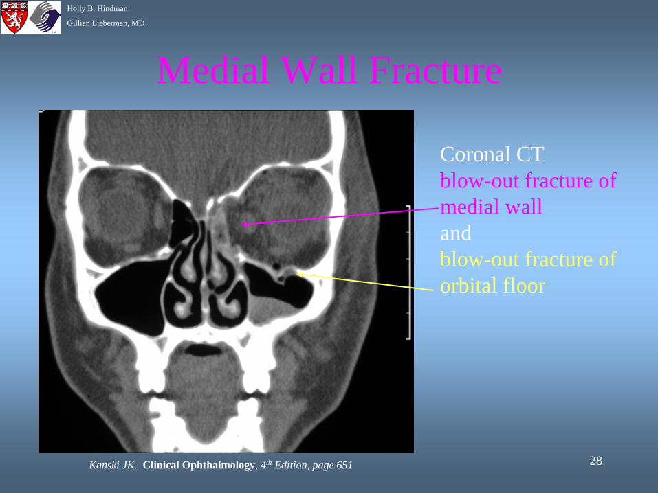
Medial Wall Fracture
Kanski JK. Clinical Ophthalmology, 4th Edition, page 651
Coronal CTblow-out fracture of medial wall andblow-out fracture of orbital floor
29
Holly B. Hindman
Gillian Lieberman, MD
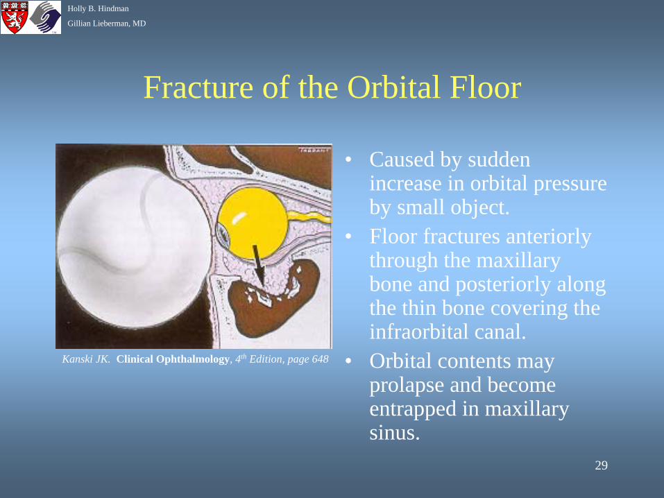
Fracture of the Orbital Floor
• Caused by sudden increase in orbital pressure by small object.
• Floor fractures anteriorly through the maxillary bone and posteriorly along the thin bone covering the infraorbital canal.
• Orbital contents may prolapse and become entrapped in maxillary sinus.
Kanski JK. Clinical Ophthalmology, 4th Edition, page 648
30
Holly B. Hindman
Gillian Lieberman, MD
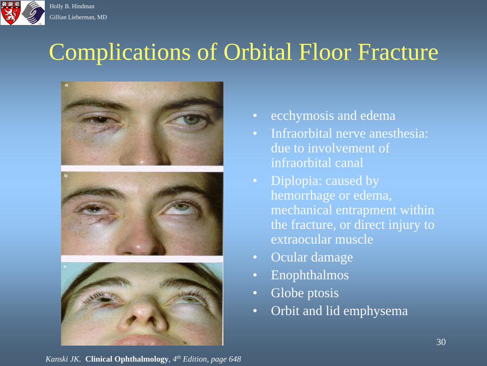
Complications of Orbital Floor Fracture
• ecchymosis and edema• Infraorbital nerve anesthesia:
due to involvement of infraorbital canal
• Diplopia: caused by hemorrhage or edema, mechanical entrapment within the fracture, or direct injury to extraocular muscle
• Ocular damage • Enophthalmos• Globe ptosis• Orbit and lid emphysema
Kanski JK. Clinical Ophthalmology, 4th Edition, page 648
31
Holly B. Hindman
Gillian Lieberman, MD
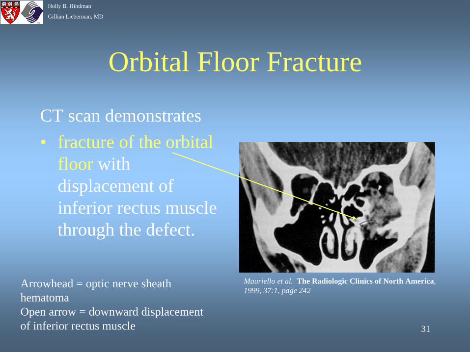
Orbital Floor Fracture
CT scan demonstrates• fracture of the orbital
floor with displacement of inferior rectus muscle through the defect.
Arrowhead = optic nerve sheath hematomaOpen arrow = downward displacement of inferior rectus muscle
Mauriello et al. The Radiologic Clinics of North America, 1999, 37:1, page 242
32
Holly B. Hindman
Gillian Lieberman, MD
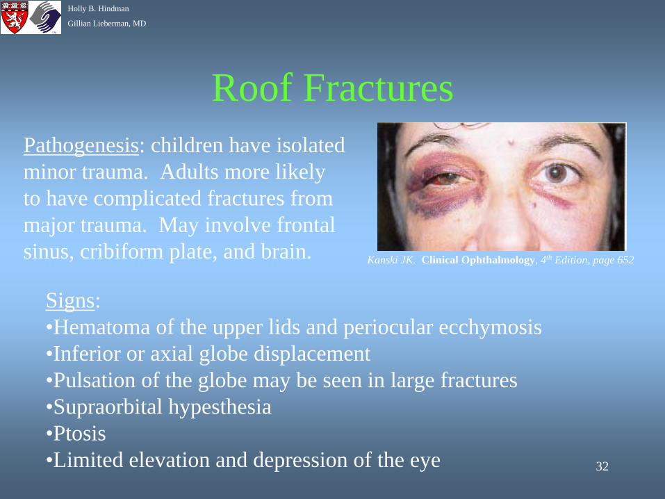
Roof Fractures
Signs:•Hematoma of the upper lids and periocular ecchymosis•Inferior or axial globe displacement•Pulsation of the globe may be seen in large fractures•Supraorbital hypesthesia•Ptosis•Limited elevation and depression of the eye
Kanski JK. Clinical Ophthalmology, 4th Edition, page 652
Pathogenesis: children have isolated minor trauma. Adults more likely to have complicated fractures from major trauma. May involve frontal sinus, cribiform plate, and brain.
33
Holly B. Hindman
Gillian Lieberman, MD
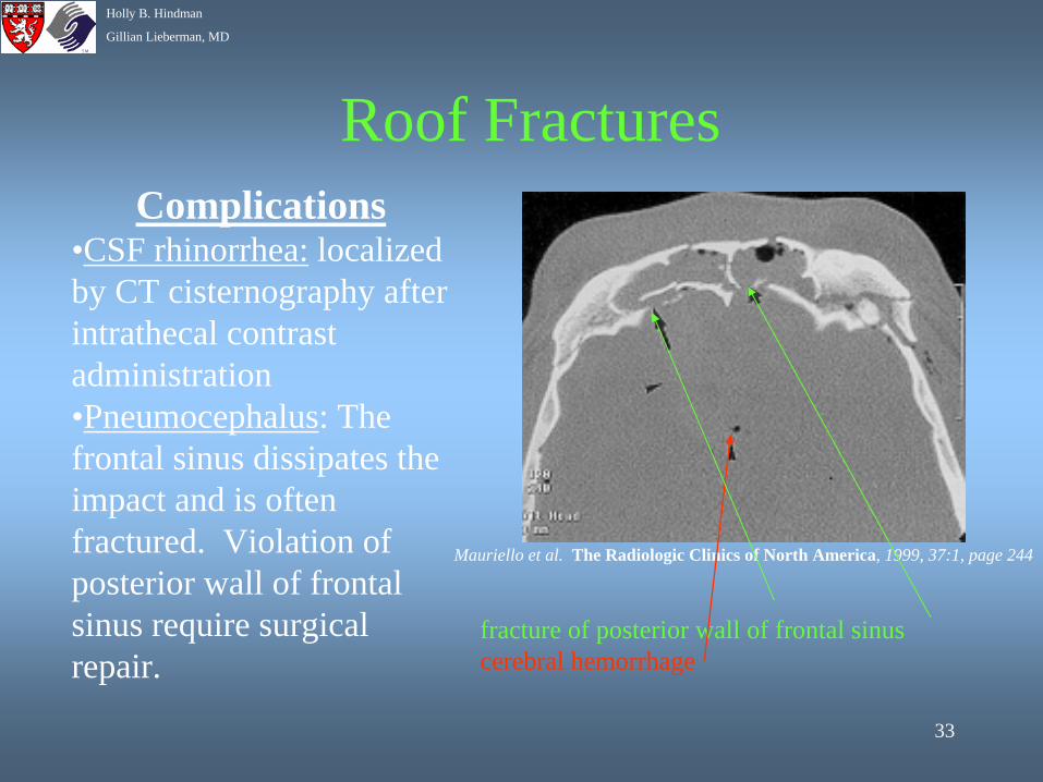
Roof FracturesComplications
•CSF rhinorrhea: localized by CT cisternography after intrathecal contrast administration•Pneumocephalus: The frontal sinus dissipates the impact and is often fractured. Violation of posterior wall of frontal sinus require surgical repair.
fracture of posterior wall of frontal sinuscerebral hemorrhage
Mauriello et al. The Radiologic Clinics of North America, 1999, 37:1, page 244
34
Holly B. Hindman
Gillian Lieberman, MD
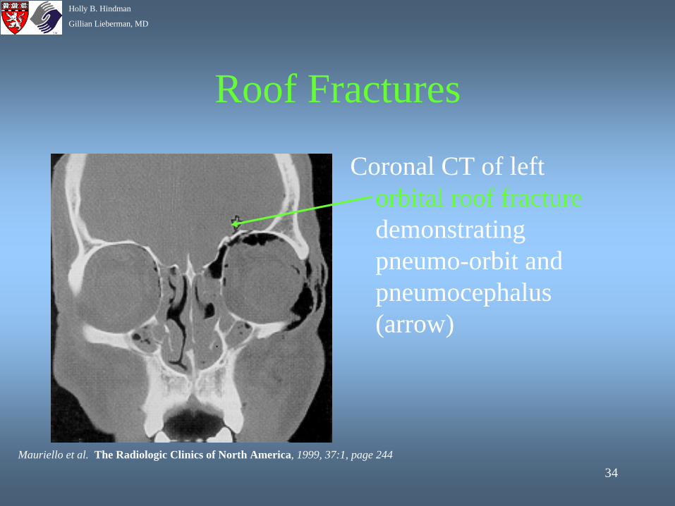
Roof Fractures
Coronal CT of left orbital roof fracture demonstrating pneumo-orbit and pneumocephalus (arrow)
Mauriello et al. The Radiologic Clinics of North America, 1999, 37:1, page 244
35
Holly B. Hindman
Gillian Lieberman, MD
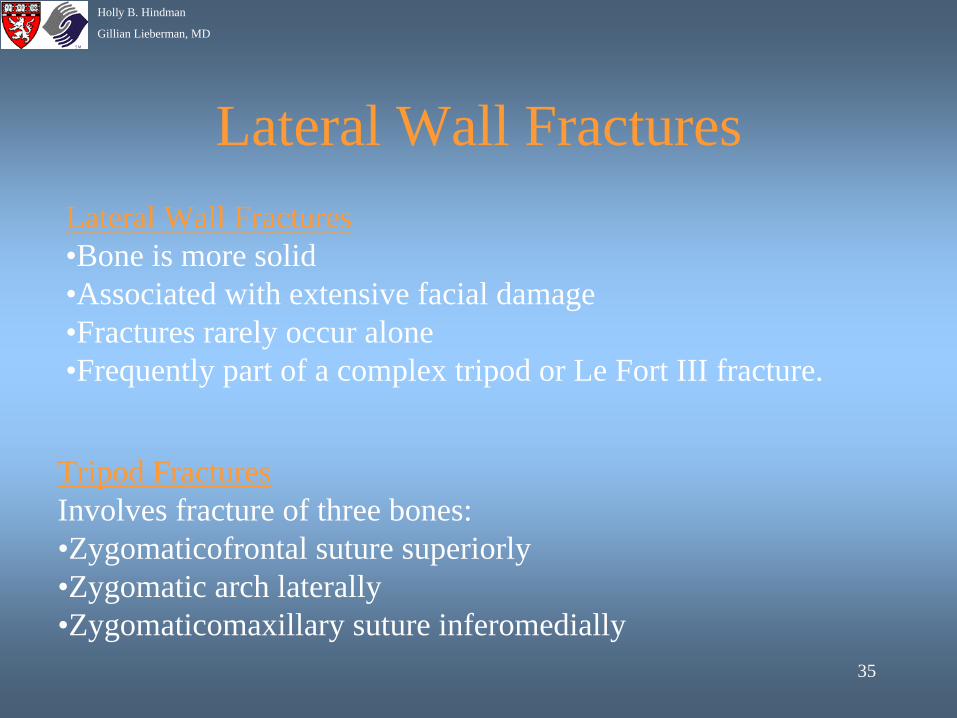
Lateral Wall FracturesLateral Wall Fractures•Bone is more solid•Associated with extensive facial damage •Fractures rarely occur alone •Frequently part of a complex tripod or Le Fort III fracture.
Tripod FracturesInvolves fracture of three bones:•Zygomaticofrontal suture superiorly•Zygomatic arch laterally•Zygomaticomaxillary suture inferomedially
36
Holly B. Hindman
Gillian Lieberman, MD
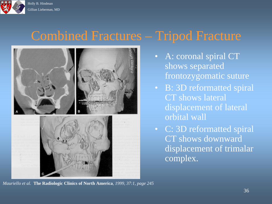
Combined Fractures – Tripod Fracture• A: coronal spiral CT
shows separated frontozygomatic suture
• B: 3D reformatted spiral CT shows lateral displacement of lateral orbital wall
• C: 3D reformatted spiral CT shows downward displacement of trimalar complex.
Mauriello et al. The Radiologic Clinics of North America, 1999, 37:1, page 245
37
Holly B. Hindman
Gillian Lieberman, MD
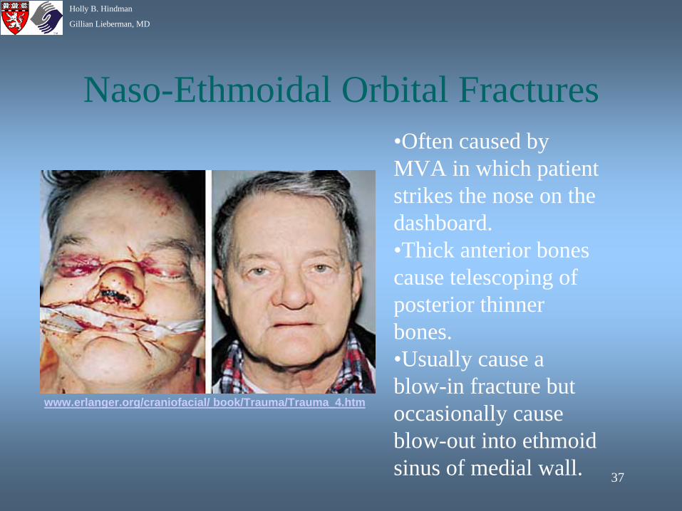
Naso-Ethmoidal Orbital Fractures•Often caused by MVA in which patient strikes the nose on the dashboard.•Thick anterior bones cause telescoping of posterior thinner bones.•Usually cause a blow-in fracture but occasionally cause blow-out into ethmoid sinus of medial wall.
www.erlanger.org/craniofacial/ book/Trauma/Trauma_4.htm
38
Holly B. Hindman
Gillian Lieberman, MD
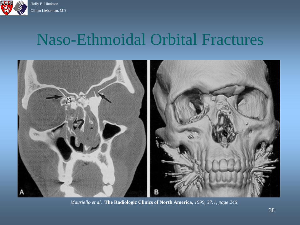
Naso-Ethmoidal Orbital Fractures
Mauriello et al. The Radiologic Clinics of North America, 1999, 37:1, page 246
39
Holly B. Hindman
Gillian Lieberman, MD
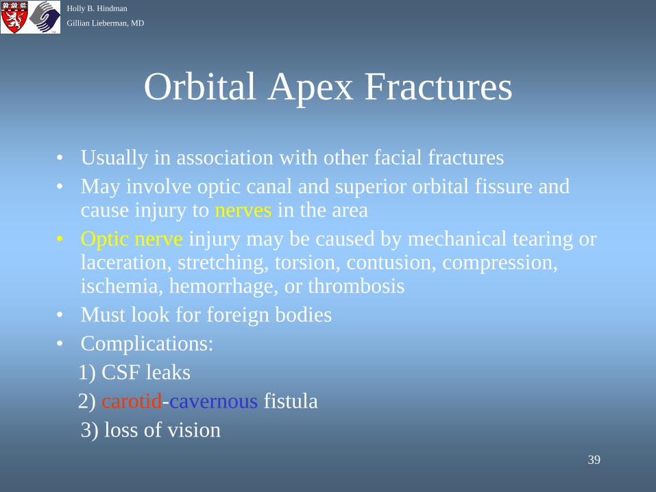
Orbital Apex Fractures• Usually in association with other facial fractures• May involve optic canal and superior orbital fissure and
cause injury to nerves in the area• Optic nerve injury may be caused by mechanical tearing or
laceration, stretching, torsion, contusion, compression, ischemia, hemorrhage, or thrombosis
• Must look for foreign bodies• Complications:
1) CSF leaks 2) carotid-cavernous fistula 3) loss of vision
40
Holly B. Hindman
Gillian Lieberman, MD
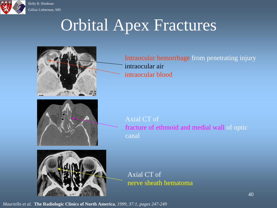
Orbital Apex Fractures
Intraocular hemorrhage from penetrating injury intraocular airintraocular blood
Axial CT of fracture of ethmoid and medial wall of optic canal
Axial CT of nerve sheath hematoma
Mauriello et al. The Radiologic Clinics of North America, 1999, 37:1, pages 247-249
41
Holly B. Hindman
Gillian Lieberman, MD
Complications of Orbital Trauma
• Foreign bodies (Radiographs, US, CT, NOT MRI)• Diplopia from muscle entrapment• Globe rupture• Suprachoroidal hemorrhage (US)• Retinal detachment (US)• Carotid cavernous fistula (CT, MRI,
arteriography)• Lens dislocation (US)• enophthalmos
42
Holly B. Hindman
Gillian Lieberman, MD
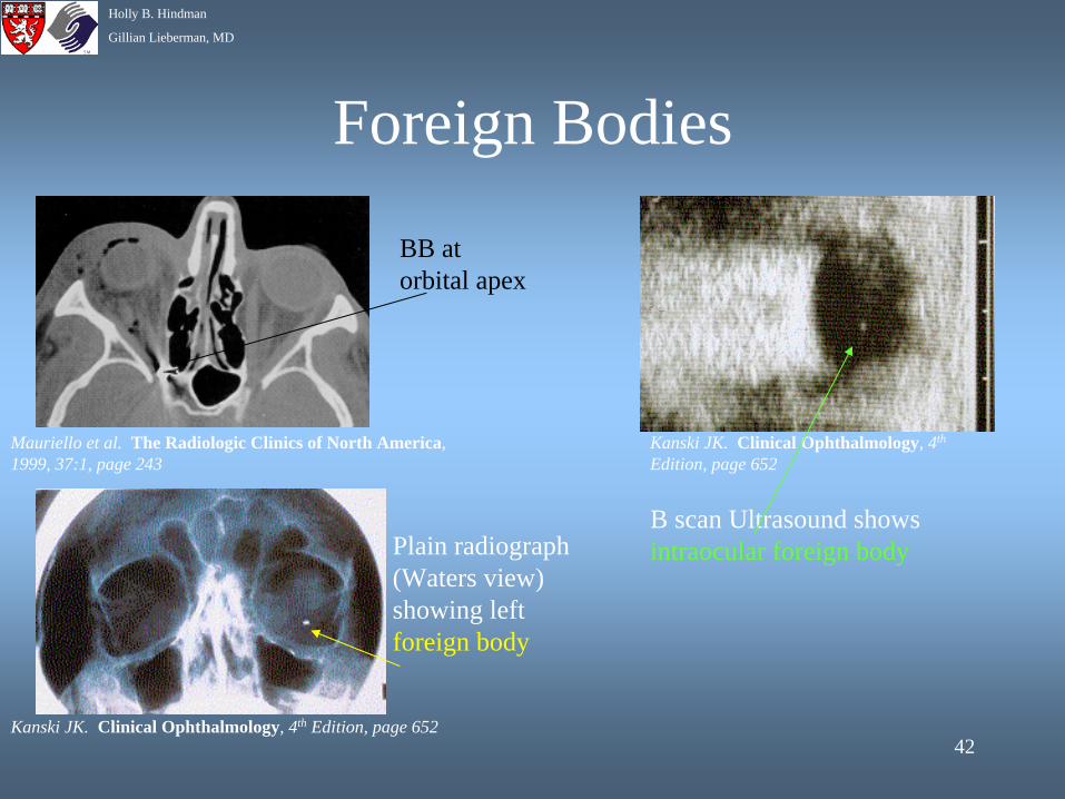
Foreign Bodies
Mauriello et al. The Radiologic Clinics of North America, 1999, 37:1, page 243
Kanski JK. Clinical Ophthalmology, 4th Edition, page 652
Plain radiograph (Waters view) showing left foreign body
BB at orbital apex
Kanski JK. Clinical Ophthalmology, 4th
Edition, page 652
B scan Ultrasound shows intraocular foreign body
43
Holly B. Hindman
Gillian Lieberman, MD
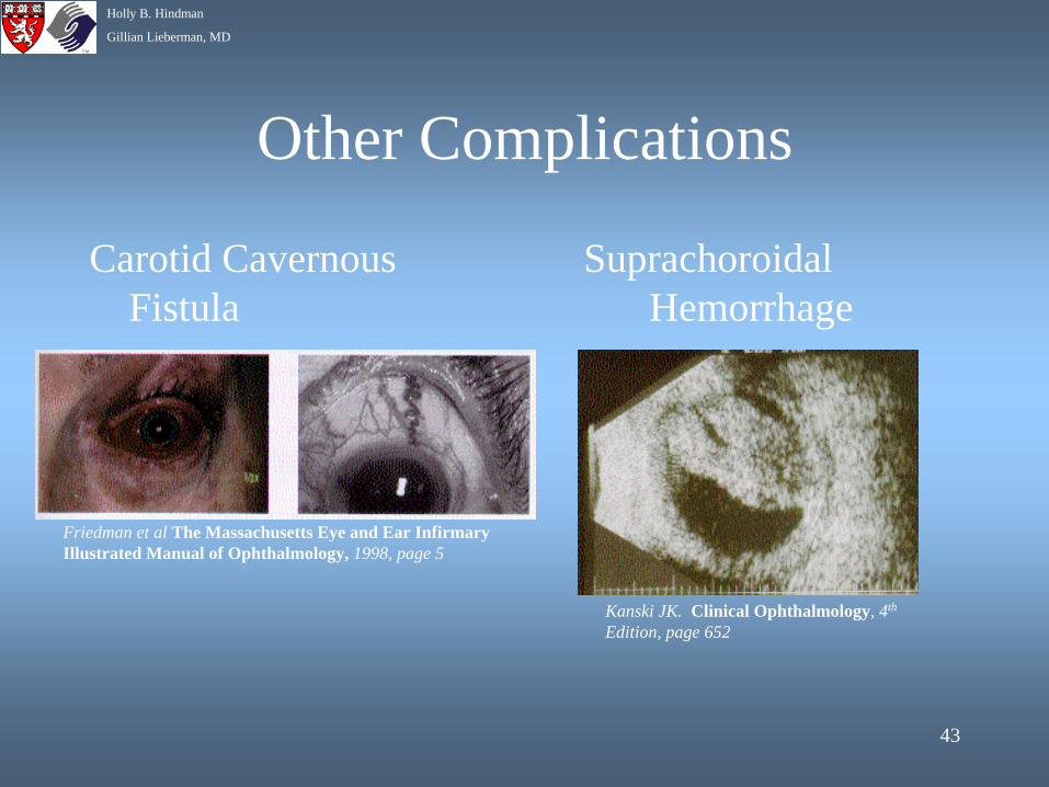
Other Complications
Carotid Cavernous Fistula
Suprachoroidal Hemorrhage
Friedman et al The Massachusetts Eye and Ear Infirmary Illustrated Manual of Ophthalmology, 1998, page 5
Kanski JK. Clinical Ophthalmology, 4th
Edition, page 652
44
Holly B. Hindman
Gillian Lieberman, MD
Patient D.W.
Findings on CT: contiguous axial images from the foramen magnum through the cranial vertex
• Multiple comminuted fractures involving the bilateral maxillary sinuses and ethmoid air cells
• Fracture of lamina papyracea bilaterally• Fracture of the left zygomatic arch• Extensive blood and soft tissue density within
maxillary, ethmoid, sphenoid, and frontal sinuses as well as mastoid air cells
• No gross abnormalities of the brain
45
Holly B. Hindman
Gillian Lieberman, MD
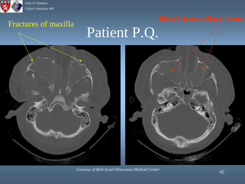
Patient P.Q.Fractures of maxilla Blood in maxillary sinus
Courtesy of Beth Israel Deaconess Medical Center
46
Holly B. Hindman
Gillian Lieberman, MD
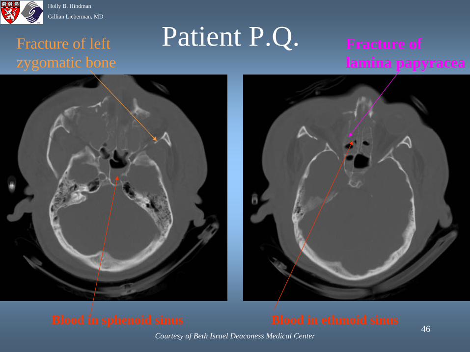
Fracture of left zygomatic bone
Blood in sphenoid sinus Blood in ethmoid sinus
Fracture of lamina papyracea
Courtesy of Beth Israel Deaconess Medical Center
Patient P.Q.
47
Holly B. Hindman
Gillian Lieberman, MD
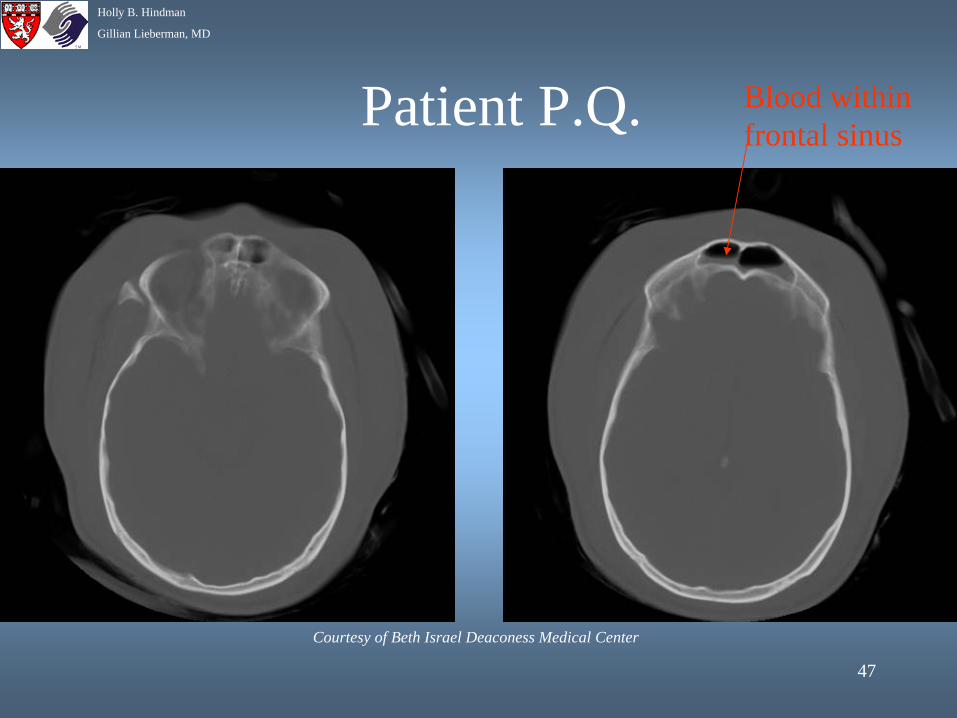
Blood within frontal sinus
Courtesy of Beth Israel Deaconess Medical Center
Patient P.Q.
48
Holly B. Hindman
Gillian Lieberman, MD
References• Agur AMR, Lee MJ. Grant’s Atlas of Anatomy, 10th Edition. Lippincott Williams and Wilkins, 1999.• Coleman DJ, Silverman RH, Daly SM, Rondeau MJ. Advances in Ophthalmic Ultrasound. Radiologic
Clinics of North America, 1998; 36:6, 1073-1082.• Ettl A, Salmonowitz E, Koornneef L, Zonnefeld FW. High Resolution MR Imaging – Anatomy of the
Orbit. The Radiologic Clinics of North America, 1998; 36:6, 1021-1045.• Friedman NJ, Pineda R, Kaiser PK. The Massachusetts Eye and Ear Infirmary Illustrated Manual of
Ophthalmology. W.B. Saunders Company, 1998.• Kanski JK. Clinical Ophthalmology. 4th Edition. Butterworth-Heinemann, 2000.• Koornneef L, Zonneveld FW. The Role of Direct Multiplanar High-Resolution CT in the Assessment
and Management of Orbital Trauma. Radiologic Clinics of North America, 1987; 25:4, 753-766.• Mauriello JA, Lee HJ, Nguyen L. CT of Soft Tissue Injury and Orbital Fractures. Radiologic Clinics
of North America 1999; 37:1, 241-252.• Novelline, RA. Squire’s Fundamentals of Radiology, 5th Edition. Butterworth-Heinemann, 2000.• Vaughan D, Asbury T, Riordan-Eva P. General Ophthalmology, 15th Edition. McGraw-Hill, 1999.• www.eyeplastics.com/ orbital_anatomy.htm• www.erlanger.org/craniofacial/ book/Trauma/Trauma_4.htm
49
Holly B. Hindman
Gillian Lieberman, MD
Acknowledgements
• Webmasters Larry Barbaras and Cara Lyn D’amour
• Gillian Lieberman, MD • Pamela Lepkowski• Nicole Thobe, MD