Extranodal lymphoma arising within the maxillary … percent of lymphomas arise in tissues outside...
14
Vol.:(0123456789) 1 3 Oral Radiology (2018) 34:113–126 https://doi.org/10.1007/s11282-017-0309-5 ORIGINAL ARTICLE Extranodal lymphoma arising within the maxillary alveolus: a systematic review David MacDonald 1 · Sean Lim 1 Received: 6 September 2017 / Accepted: 19 October 2017 / Published online: 8 December 2017 © Japanese Society for Oral and Maxillofacial Radiology and Springer Japan KK, part of Springer Nature 2017 Abstract Objectives Extranodal lymphomas affecting the head and neck infrequently arise within the jaw bones. Although clinical examination and conventional radiography may initially suffice for such lesions arising within the mandible, those arising within the maxillary alveolus generally require cross-sectional imaging because of the complex anatomy of this region. This study was performed to determine the prevalence, demographic characteristics, and clinical presentations of these lesions and the imaging modalities used for their diagnosis. Study design A systematic review (SR) on case series and another SR on case reports were performed to investigate the demographic, clinical, and radiological features of extranodal lymphomas arising within the maxillary alveolus. Results Most case series were derived from just four nations, whereas the case reports were derived from a wider range of ethnicities. The more detailed case reports significantly reported at least one imaging modality. Most patients were aware of their lesions for nearly 2 months before presentation. The most frequent symptom was swelling. Most case reports included a provisional diagnosis, the most frequent of which was dental infection followed by squamous cell carcinoma. Discussion Extranodal lymphomas arising within the maxillary alveolus were sufficiently frequent in four communities to be reported in two or more case series, and the occasional single case report indicated that such lesions are more widespread globally. Although the SR on case series revealed differences in the relative period prevalence and maxillary/mandibular ratio, the SR on case reports revealed details of the clinical presentation and imaging modalities used. Keywords Lymphoma · Systematic review · Radiology Introduction Lymphoma accounts for about 5% of head and neck malig- nancies [1]. During the last 50 years, a new classification of lymphoma has appeared almost every decade; the 2016 revision of the World Health Organization classification [2] is the most recent. Lymphoma is divided into Hodgkin lym- phoma (HL) and non-Hodgkin lymphoma (NHL) based on its histopathological and clinical features. NHL is further divided into B- and T-cell lymphoma. Thirty percent of lymphomas arise in tissues outside the lymph nodes. These are called extranodal lymphomas [1] and can be either HL or NHL [3]. Extranodal lymphoma arising within the jawbones is so rare that no cases were pre- sent in a case series of 361 oral and paraoral lymphomas [4]. Since the case series reported by Eisenbud et al. [5] in 1984, increasingly, more cases of extranodal lymphomas arising within the jawbones have been reported. A possible cause of the initial accumulation of lymphoid tissue (from which the lymphoma arises) within the jawbones is chronic dental disease, which may arise from an untreated or inadequately treated nonvital tooth or periodontal disease. Although such lesions affecting the mandibular alveolus are adequately dis- played by the conventional radiography, those in the max- illary alveolus are not readily displayed. Cross-sectional imaging modalities, including cone-beam computed tomog- raphy (CBCT), are necessary for lesions arising within the The original version of this article was revised: Modifications have been made in the result and discussion section. Full information regarding corrections made can be found in the correction article for this article. * David MacDonald [email protected] 1 Division of Oral and Maxillofacial Radiology, Faculty of Dentistry, UBC, 2199 Wesbrook Mall, Vancouver, BC V6T 1Z3, Canada
Transcript of Extranodal lymphoma arising within the maxillary … percent of lymphomas arise in tissues outside...

Vol.:(0123456789)1 3
Oral Radiology (2018) 34:113–126 https://doi.org/10.1007/s11282-017-0309-5
ORIGINAL ARTICLE
Extranodal lymphoma arising within the maxillary alveolus: a systematic review
David MacDonald1 · Sean Lim1
Received: 6 September 2017 / Accepted: 19 October 2017 / Published online: 8 December 2017 © Japanese Society for Oral and Maxillofacial Radiology and Springer Japan KK, part of Springer Nature 2017
AbstractObjectives Extranodal lymphomas affecting the head and neck infrequently arise within the jaw bones. Although clinical examination and conventional radiography may initially suffice for such lesions arising within the mandible, those arising within the maxillary alveolus generally require cross-sectional imaging because of the complex anatomy of this region. This study was performed to determine the prevalence, demographic characteristics, and clinical presentations of these lesions and the imaging modalities used for their diagnosis.Study design A systematic review (SR) on case series and another SR on case reports were performed to investigate the demographic, clinical, and radiological features of extranodal lymphomas arising within the maxillary alveolus.Results Most case series were derived from just four nations, whereas the case reports were derived from a wider range of ethnicities. The more detailed case reports significantly reported at least one imaging modality. Most patients were aware of their lesions for nearly 2 months before presentation. The most frequent symptom was swelling. Most case reports included a provisional diagnosis, the most frequent of which was dental infection followed by squamous cell carcinoma.Discussion Extranodal lymphomas arising within the maxillary alveolus were sufficiently frequent in four communities to be reported in two or more case series, and the occasional single case report indicated that such lesions are more widespread globally. Although the SR on case series revealed differences in the relative period prevalence and maxillary/mandibular ratio, the SR on case reports revealed details of the clinical presentation and imaging modalities used.
Keywords Lymphoma · Systematic review · Radiology
Introduction
Lymphoma accounts for about 5% of head and neck malig-nancies [1]. During the last 50 years, a new classification of lymphoma has appeared almost every decade; the 2016 revision of the World Health Organization classification [2] is the most recent. Lymphoma is divided into Hodgkin lym-phoma (HL) and non-Hodgkin lymphoma (NHL) based on
its histopathological and clinical features. NHL is further divided into B- and T-cell lymphoma.
Thirty percent of lymphomas arise in tissues outside the lymph nodes. These are called extranodal lymphomas [1] and can be either HL or NHL [3]. Extranodal lymphoma arising within the jawbones is so rare that no cases were pre-sent in a case series of 361 oral and paraoral lymphomas [4]. Since the case series reported by Eisenbud et al. [5] in 1984, increasingly, more cases of extranodal lymphomas arising within the jawbones have been reported. A possible cause of the initial accumulation of lymphoid tissue (from which the lymphoma arises) within the jawbones is chronic dental disease, which may arise from an untreated or inadequately treated nonvital tooth or periodontal disease. Although such lesions affecting the mandibular alveolus are adequately dis-played by the conventional radiography, those in the max-illary alveolus are not readily displayed. Cross-sectional imaging modalities, including cone-beam computed tomog-raphy (CBCT), are necessary for lesions arising within the
The original version of this article was revised: Modifications have been made in the result and discussion section. Full information regarding corrections made can be found in the correction article for this article.
* David MacDonald [email protected]
1 Division of Oral and Maxillofacial Radiology, Faculty of Dentistry, UBC, 2199 Wesbrook Mall, Vancouver, BC V6T 1Z3, Canada

114 Oral Radiology (2018) 34:113–126
1 3
anatomically complex maxilla [6–8]. A recent case report appeared to be the first to compare the merits of the conven-tional radiography, CBCT, computed tomography (CT), and magnetic resonance imaging (MRI) in the diagnosis of NHL arising within the maxillary alveolus [9].
We performed two systematic reviews (SRs) to examine the demographic and the clinical features at the first presen-tation and the radiological modalities used to investigate this rare but important phenomenon. The particular demographic and clinical features and radiological modalities considered by both SRs were prompted by the aforementioned recent case report [9]. The overall aim of these SRs was broadly similar to those of earlier SRs of other oral and maxillofacial lesions [10–21]: to determine whether the demographics, clinical presentation, and imaging modalities differ between case series and case reports of European, East Asian, and sub-Saharan origin. The following more specific questions were asked to both address the above aim and extract further information pertinent to clinical practice.
a. What is the relative period prevalence of NHLs aris-ing within the maxillary alveolus within an SR of case series?
b. Which global communities have a greater prevalence of NHLs arising within the maxillary alveolus?
c. Which demographic and clinical details are most likely to be associated with the first presentation of NHLs arising within the maxillary alveolus? This includes the patient’s prior awareness of the lesion before the first presentation.
d. What advanced imaging modalities were prescribed in the course of diagnosis of NHLs arising within the max-illary alveolus?
Methods
The approach to these SRs followed the procedure set out in earlier SRs for other oral and maxillofacial lesions [10–21]. This approach was based on the format established for this purpose [22]. An essential element of an SR is a meta-anal-ysis [22]. A meta-analysis, as defined by the dictionary of epidemiology, is “the process of using statistical methods to combine the results of different studies” [23]. Fisher’s exact test (FET) was applied to categorical data, because the total sample sizes were small [24]. Significance was defined by a two-tailed P value of < 0.05. All calculations were per-formed using the VassarStats software [25]. Although the literature search performed for the case reports revealed that extranodal lymphomas affecting the jaws were NHLs, a search strategy was developed that would enhance capture of HLs.
The Medline PubMed database was searched. The PubMed Medical Subject Heading (MeSH) “Lymphoma” is defined as “a general term for various neoplastic diseases of the lymphoid tissue.” It includes HLs and NHLs and was used in conjunction with “jaw” in the present study. Text word searches [22] of “lymphoma AND jaw” and “non-Hodgkin lymphoma maxilla” were also performed. These searches were last conducted on 30 August 2017.
Emphasis was placed on recall rather than precision to include as many reports as possible [22]. This strategy was further augmented by examination of the reference lists of the reports identified by the MeSH and text word searches. Major journals were also hand-searched.
SR of consecutive case series of extranodal lymphomas arising within the maxillary alveolus (Table 1)
The selection criteria for the case series were as follows:
1. The article revealed that the case was a lymphoma.2. The article intended to report the entire case series of
lymphomas and not a selection of cases. Case series that were restricted to a particular age group or type of lymphoma (e.g., B-cell type or Burkitt lymphoma) or clinicopathological group (e.g., patients with acquired immunodeficiency syndrome) were excluded. The only exception was case series of NHL.
3. The article intended to include cases that primarily arose from the alveolus of both jaws. Although such a case series may not include a maxillary case, it was included to allow the SR to determine the ratio of maxillary to mandibular cases (maxilla/mandible ratio). Neverthe-less, case series that were expressly concerned only with mandibular cases were excluded, whereas those expressly concerned with maxillary cases were included (although the latter were unable to contribute to the maxilla/mandible ratio).
4. The case series revealed that the lymphoma(s) arose from within the alveolus of the jaws. In the absence of radiological evidence (or, failing that, clinical evidence) to the contrary, an unequivocal statement that the lym-phoma arose within the alveolus of the jaws would be sufficient for inclusion of the report. Cases indicating secondary involvement of the maxillary alveolus from a lymphoma arising from the palate or from within the maxillary sinus were excluded.

115Oral Radiology (2018) 34:113–126
1 3
Tabl
e 1
Sys
tem
atic
revi
ew o
f con
secu
tive
case
serie
s of e
xtra
noda
l lym
phom
as a
risin
g w
ithin
the
alve
olus
of t
he m
axill
a
Firs
t aut
hor,
year
of
publ
icat
ion,
an
d se
arch
te
rm
M =
MeS
H
W =
Tex
t w
ord
O =
Oth
er
Ethn
icity
Year
s of
study
Rela
tive
perio
d pr
eval
ence
(m
axill
as
per y
ear o
f stu
dy)
Type
and
nu
mbe
r of
cas
es
In th
em
axill
a
Max
illa/
man
dibl
e ra
tio
Sex
Age
(y
ear)
Prio
r aw
aren
ess
(yea
r)
Clin
ical
feat
ures
at fi
rst p
rese
ntat
ion
Rad
iolo
gica
l mod
aliti
es
MF
Swel
ling
Pain
Num
bD
isch
.M
obile
te
eth
Sext
ant
CR
CT
MR
IC
BC
T
Ant
Post
Eise
nbud
19
84b M
US
Cau
-ca
sian
ING
ING
8N8/
5II
GII
GII
GII
G4
12
ING
ING
26
IIG
ING
ING
ING
Sloo
tweg
19
85 W
Dut
chU
a34
0.29
10N
10/2
55
56 ±
210.
08 ±
0.13
ING
ING
ING
ING
ING
ING
IIG
ING
ING
ING
ING
Fuku
da
1987
OJa
pane
se12
Nil
Nil
0/3
Nil
Nil
Nil
Nil
Nil
Nil
Nil
Nil
Nil
Nil
Nil
Nil
Nil
Nil
Nil
How
ell
1987
WU
S C
au-
casi
anIN
GIN
G3B
3/6
21
68 ±
9IN
GIN
GIN
GN
oIN
GIN
GIN
G1
IIG
ING
ING
ING
Söde
rhol
m
1990
WFi
nnis
ha11
ING
3N3/
113
048
± 20
ING
8IN
G2
ING
ING
ING
ING
Yes
Yes
ING
ING
Wol
vius
19
95 W
Dut
chA
a20
0.25
5B5/
5II
GII
GII
GIN
GII
GII
GII
GIN
GIN
GIN
GIN
GII
GII
GIN
GIN
G
Pazo
ki
2003
WU
SaIN
GIN
G1B
1/3
10
580.
081
0N
ilN
ilN
il1
0 l
IIG
Nil
Nil
Nil
Ugb
oko
2004
WN
iger
ian
50.
603N
3/0
21
32 ±
160.
16 ±
0.08
30
1IN
GIN
G2
1Ye
sIN
GIN
GIN
G
Kol
oko-
troni
s 20
05 W
Gre
eka
3.5
Nil
Nil
0/18
Nil
Nil
Nil
Nil
Nil
Nil
Nil
Nil
Nil
Nil
Nil
Nil
Nil
Nil
Nil
van
der
Waa
l 200
5 O
Dut
chA
a5
Nil
Nil
0/2
Nil
Nil
Nil
Nil
Nil
Nil
Nil
Nil
ING
ING
ING
Nil
Nil
Nil
Nil
Dja
van-
mar
di
2008
W
Fren
cha
ING
ING
5N5/
11II
GII
GII
GII
GII
GII
GII
GIN
GIN
GIN
G4
ING
ING
ING
ING
Kes
zler
20
08 W
Arg
en-
tinia
na20
0.30
6N6/
23
3II
GII
GII
GII
GIN
GIN
GIN
GIN
GIN
GIN
GIN
GIN
GIN
G
Etem
ad-M
20
10 O
Iran
ianM
200.
204N
4/9
ING
ING
IIG
ING
ING
ING
ING
ING
ING
ING
ING
ING
ING
ING
ING
Moh
tash
am
2011
WIr
ania
nT11
0.73
7B, 1
T8/
3IN
GIN
GIN
GIN
GIN
GIN
GIN
GIN
GIN
GIN
GIN
GIN
GIN
GIN
GIN
G

116 Oral Radiology (2018) 34:113–126
1 3
A A
sian
; Ant
ant
erio
r (se
xtan
t); B
B c
ell;
Bk B
lack
; C C
auca
sian
; CBC
T co
ne-b
eam
com
pute
d to
mog
raph
y; C
R co
nven
tiona
l rad
iogr
aphy
; CT
med
ical
com
pute
d to
mog
raph
y; D
isch
. dis
char
ge;
Endo
. end
odon
tics (
root
can
al) t
reat
men
t; D
utch
A A
mste
rdam
, Hol
land
; Dut
chU
Utre
cht,
Hol
land
; IIG
inad
equa
te in
form
atio
n gi
ven;
Inci
d. in
cide
ntal
find
ing;
ING
info
rmat
ion
not g
iven
; Ira
ni-
anM
a c
ase
serie
s fro
m M
asha
d in
the
north
east
of Ir
an w
ith a
subs
tant
ial m
inor
ity o
f Eas
t Asi
an o
rigin
; Ira
nina
nT a
cas
e se
ries f
rom
Teh
eran
, Ira
nian
nat
iona
l cap
ital;
MRI
mag
netic
reso
nanc
e im
agin
g; N
non
-Hod
gkin
lym
phom
a; N
il no
cas
es o
f non
-Hod
gkin
lym
phom
a ar
isin
g w
ithin
the
max
illar
y al
veol
us fo
r tha
t stu
dy; P
.Ext
pos
t-ext
ract
ion;
pos
t pos
terio
r (se
xtan
t); r
pt re
port(
s); T
T
cell:
US,
Uni
ted
Stat
esa A
lthou
gh th
e pa
tient
’s e
thni
city
was
not
giv
en, i
t may
be
pres
umed
to b
e C
auca
sian
(Ira
nian
s wer
e N
OT
incl
uded
; see
“D
iscu
ssio
n”)
b Alth
ough
8 o
f the
14
case
s wer
e ex
pres
sly d
escr
ibed
as C
auca
sian
, 2 o
f the
orig
inal
31
wer
e de
scrib
ed a
s Bla
ck
Tabl
e 1
(con
tinue
d)
Firs
t aut
hor,
year
of
publ
icat
ion,
an
d se
arch
te
rm
M =
MeS
H
W =
Tex
t w
ord
O =
Oth
er
Ethn
icity
Year
s of
study
Rela
tive
perio
d pr
eval
ence
(m
axill
as
per y
ear o
f stu
dy)
Type
and
nu
mbe
r of
cas
es
In th
em
axill
a
Max
illa/
man
dibl
e ra
tio
Sex
Age
(y
ear)
Prio
r aw
aren
ess
(yea
r)
Clin
ical
feat
ures
at fi
rst p
rese
ntat
ion
Rad
iolo
gica
l mod
aliti
es
MF
Swel
ling
Pain
Num
bD
isch
.M
obile
te
eth
Sext
ant
CR
CT
MR
IC
BC
T
Ant
Post
Tria
ntafi
l-lid
ou
2012
M
Gre
eka
60.
332B
2/1
11
48 ±
1IN
GIN
GIN
GIN
GIN
GIN
GIN
GIN
GIN
GIN
GIN
GIN
G
Ram
anat
han
2014
MM
alay
-si
an22
0.32
2B, 3
T,
2N7/
95
238
± 29
ING
ING
ING
ING
ING
ING
ING
ING
ING
ING
ING
ING
Tota
l = 65
ca
ses
43Ca ,
7A,
3Bk,
12
Ira-
nian
s
14 ±
90.
38 ±
0.18
20B
, 4T,
41
N65
/90
2213
49.7
13
repo
rts16
15
00
512
2 rp
t1
rpt
0 rp
t0
rpt

117Oral Radiology (2018) 34:113–126
1 3
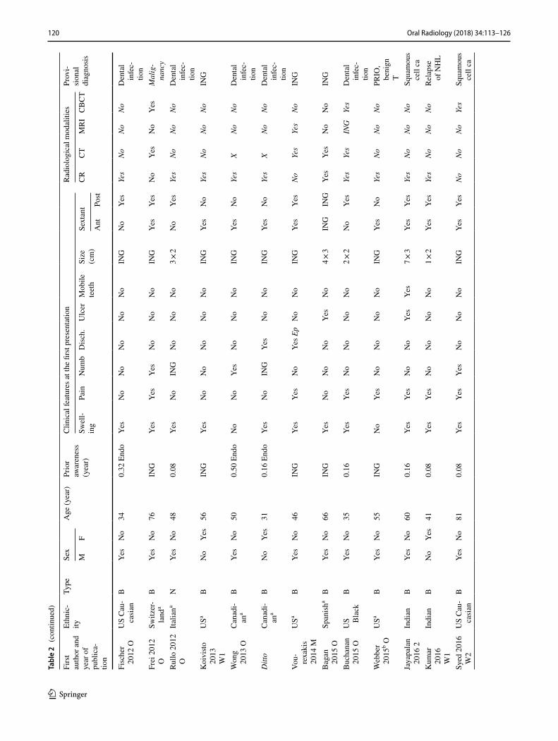
SR of reported cases (case reports) of extranodal lymphomas arising within the maxillary alveolus (Table 2 [26])
A case report is used to describe the medical history of a sin-gle patient in the form of a story or anecdote. The case report is frequently used to alert colleagues to cases that deviate from the norm in important ways with regard to presenta-tion, treatment, or outcome. The case report is also invalu-able for detailed descriptions of rare lesions [27, 28], such as extranodal lymphomas arising within the alveolus of the maxilla. Individual case reports are “often run together to form a case series, in which the demographic, clinical, and other presentations of more than one patient with a particular condition are described to illustrate an aspect of the condi-tion” [27]. However, the amount of detail available for each individual case in that case series may be lost. This will be further explored in the “Discussion”. For this reason, the present study included an SR of case reports in addition to an SR of case series.
The SR of case reports focused solely on cases of extran-odal lymphomas of any kind arising within the alveolus of the maxilla. Included in this SR of case reports were indi-vidual cases reported with adequate detail in the case series (Table 1). Such cases provided details on at least the demo-graphics, clinical features, and imaging modalities used at the first presentation.
To assist in answering the above-mentioned research questions, the case reports included in the SR were divided into four global groups broadly reflecting their ethnic origin: European, sub-Saharan African, East Asian, and Indian.
Results
Forty-six reports satisfied the selection criteria: 4 of the 146 hits in the search of the MeSH terms “Lymphoma AND Jaw,” 24 of the 739 hits in the search of the text words “lym-phoma AND jaw,” 4 of the 105 hits in the search of the text word “non-Hodgkin lymphoma maxilla,” and 14 reports obtained from the reference lists and hand-searching.
The 16 case series [5, 29–43] included in the SR are shown in Table 1. Two case series were excluded. Velez and Hogge [44] reported only B-cell NHLs, and Kemp et al. [45] grouped NHLs arising in the maxillary alveolus together with those arising in the hard palate. For 3 of the 16 case series [30, 36, 37] that did not include a case aris-ing within the maxillary alveolus, “Nil” is entered in the “relative period prevalence” column in Table 1. The relative period prevalence indicates the likelihood of a particular lesion presenting within a particular community each year. This is an average for that particular report depending on the total number of cases reported and the number of years
covered by that report. The relative period prevalence was determinable in 8 of the 16 case series. Among all studies, a mean of 0.38 ± 0.18 extranodal lymphomas arose within the maxillary alveolus per year, suggesting that globally, an NHL may arise in the maxillary alveolus in such a case series every third year.
Of the 16 case series in Table 1, 10 were derived from 4 countries (3 from the United States, 3 from Holland, 2 from Greece, and 2 from Iran). Most of these reports were derived from different cities in the United States, Holland (Amsterdam and Utrecht), and Iran (Teheran and Mashhad). Two of the three Dutch reports were from Amsterdam and published a decade apart, and the two Greek reports were from different hospitals in the same city (Thessaloniki) and published about the same time. One of these hospitals in Thessaloniki had a higher relative period prevalence than the other. Teheran had a higher relative period prevalence than Mashhad, which had almost the lowest relative period prevalence overall. The maxilla/mandible ratio declined over time in both the US and Dutch reports. Patients of European origin predominated in both the SRs of case series (76%) and case reports (73%) (Tables 1, 2, respectively).
Of the cases shown in Table 2, 29 [9, 46–72] were reported as single case reports. A case series of three B-cell NHLs [44] and a double case report [73] are included in the SR in Table 2. To these were added cases for which the demographic and clinical details were reported in the case series. Four case series provided these details for at least one of their cases [5, 29, 34, 35]. Details about the imag-ing modalities used for diagnosis were added for some of these cases. Ten more nations are represented in Table 2 than Table 1. Although many of those case reports were European (UK, Poland, Croatia, Italy, Spain, and Switzerland), China, India, Brazil, and Canada were also represented.
Table 1 includes four cases of T-cell lymphoma, whereas Table 2 shows only one [53]. Table 2 includes one case of HL, whereas almost every other case was identified as an NHL, many of which were identified as B-cell lymphomas.
Although most NHLs arising within the maxillary alveo-lus first presented in the posterior sextant in both Tables 1 and 2, seven cases in Table 2 first presented in both the ante-rior and posterior sextants. Five were reported in the last 5 years, all with a provisional diagnosis of a malignancy.
In Tables 1 and 2, NHLs arising within the maxillary alveolus presented more frequently in males. This predilec-tion for males (23 males to 6 females) was more significant for patients of European origin than for those of sub-Saharan African origin (3 males to 5 females; FET = 0.035).
The vast majority of extranodal lymphomas aris-ing within the alveolus of the maxilla presented in the patient’s fifth decade of life. Table 2 shows that the mean age for all 43 patients at the first presenta-tion was 48.26 ± 18.89 years. For the 29 patients of

118 Oral Radiology (2018) 34:113–126
1 3
Tabl
e 2
Sys
tem
atic
revi
ew o
f cas
e re
ports
of e
xtra
noda
l lym
phom
as a
risin
g w
ithin
the
alve
olus
of t
he m
axill
a
Firs
t au
thor
and
ye
ar o
f pu
blic
a-tio
n
Ethn
ic-
ityTy
peSe
xA
ge (y
ear)
Prio
r aw
aren
ess
(yea
r)
Clin
ical
feat
ures
at t
he fi
rst p
rese
ntat
ion
Rad
iolo
gica
l mod
aliti
esPr
ovi-
sion
al
diag
nosi
sM
FSw
ell-
ing
Pain
Num
bD
isch
.U
lcer
Mob
ile
teet
hSi
ze
(cm
)Se
xtan
tC
RC
TM
RI
CB
CT
Ant
Post
Eise
nbud
19
84 M
US
Cau
-ca
sian
NN
oYe
s44
0.00
Inci
d.Ye
sN
oYe
sN
oN
oN
oIN
GYe
sN
oYe
sN
oN
oN
RIN
G
Ditt
oU
S C
au-
casi
anN
Yes
No
42En
doYe
sYe
sN
oN
oN
oN
oIN
GYe
sN
oYe
sN
oN
oN
RIN
G
Ditt
oU
S C
au-
casi
anN
Yes
No
590.
06Ye
sN
oYe
sN
oN
oN
oIN
GN
oYe
sYe
sN
oN
oN
RIN
G
Ditt
oU
S C
au-
casi
anN
No
Yes
500.
4Ye
sN
oIN
GN
oN
oN
oIN
GN
oYe
sYe
sN
oN
oN
RIN
G
How
ell
1987
W
1
US
Cau
-ca
sian
BYe
sN
o78
ING
Yes
No
No
No
No
No
ING
ING
ING
Yes
No
No
NR
ING
Ditt
oU
S C
au-
casi
anB
No
Yes
63IN
GYe
sN
oN
oN
oN
oN
oIN
GN
oYe
sYe
sN
oN
oN
RIN
G
Ditt
oU
S C
au-
casi
anB
Yes
No
620.
00 In
cid.
No
No
No
No
No
No
ING
ING
ING
Yes
No
No
NR
ING
Key
es
1988
W
1
USa
LYe
sN
o24
0.12
Yes
Yes
No
No
No
No
ING
Yes
No
Yes
No
No
NR
ING
Ronc
hi
1988
W
1
Italia
naH
No
Yes
780.
08Ye
sN
oIN
GN
oN
oN
o1
dia
Yes
No
Yes
No
No
NR
ING
Li 1
991
W1
Chi
nese
NN
oYe
s39
0.08
Yes
No
No
No
No
No
ING
No
Yes
Yes
Yes
No
NR
Squa
mou
s ce
ll ca
Rog
1991
O
US
Cau
-ca
sian
NYe
sN
o19
0.08
End
oYe
sYe
sN
oN
oN
oN
oIN
GN
oYe
sYe
sIN
GN
oN
RD
enta
l in
fec-
tion
Thom
as
1991
W
1
Brit
isha
LYe
sN
o75
0.33
Ext
Yes
Yes
ING
No
Yes
No
ING
No
Yes
Yes
Yes
No
NR
Squa
mou
s ce
ll ca
Hok
ett
2000
OU
SaB
Yes
No
64Pe
riodo
ntT
Yes
Yes
ING
No
No
No
4 × 5
Yes
Yes
Yes
Yes
No
No
ING
Koz
ak-
iew
icz
2003
W1
Polis
haB
Yes
No
540.
00 In
cid.
No
Yes
ING
No
No
No
ING
No
Yes
Yes
No
No
No
ING
Pazo
ki
2003
W
1
USa
BYe
sN
o58
0.08
Yes
No
ING
No
No
No
ING
Yes
No
Yes
ING
ING
ING
ING

119Oral Radiology (2018) 34:113–126
1 3
Tabl
e 2
(con
tinue
d)
Firs
t au
thor
and
ye
ar o
f pu
blic
a-tio
n
Ethn
ic-
ityTy
peSe
xA
ge (y
ear)
Prio
r aw
aren
ess
(yea
r)
Clin
ical
feat
ures
at t
he fi
rst p
rese
ntat
ion
Rad
iolo
gica
l mod
aliti
esPr
ovi-
sion
al
diag
nosi
sM
FSw
ell-
ing
Pain
Num
bD
isch
.U
lcer
Mob
ile
teet
hSi
ze
(cm
)Se
xtan
tC
RC
TM
RI
CB
CT
Ant
Post
Kob
ler
2005
OC
roat
aB
Yes
No
630.
00 In
cid.
No
No
No
No
No
No
ING
ING
ING
Yes
No
No
No
ING
Ugb
oko
2004
W
1
Nig
eria
nN
Yes
No
180.
12Ye
sN
oN
oN
oN
oYe
sIN
GIN
GIN
GYe
sIN
GIN
GIN
GIN
G
Ditt
oN
iger
ian
NYe
sN
o50
0.40
Yes
No
No
No
Yes
No
ING
ING
ING
Yes
ING
ING
ING
ING
Ditt
oN
iger
ian
NN
oYe
s29
0.40
Yes
No
Yes
No
No
No
ING
ING
ING
Yes
ING
ING
ING
ING
Mac
Don
-al
d 20
05
O
Can
adi-
ana
NIN
GIN
GIN
GIN
GIN
GIN
GIN
GIN
GIN
GIN
GIN
GN
oYe
sYe
sIN
GIN
GYe
sIN
G
Yepe
s 20
05 O
US
Cau
-ca
sian
BYe
sN
o24
ING
vita
lYe
sYe
sN
oN
oN
oYe
sIN
GN
oYe
sYe
sYe
sN
oN
oM
alig
-na
ncy
Cav
alca
nte
2009
OB
razi
l B
lack
BN
oYe
s6
0.08
Yes
Yes
No
No
No
No
4 × 3
× 2
No
Yes
Yes
Yes
No
No
Den
tal
infe
c-tio
nSa
und
2010
W
1
Brit
ish
Bla
ckN
No
Yes
380.
50N
oYe
sN
oN
oN
oN
oIN
GYe
sN
oYe
sYe
sN
oN
oD
enta
l in
fec-
tion
Vale
n-zu
ela-
S 20
10 O
Span
ish
CB
Yes
No
50.
08Ye
sYe
sN
oN
oN
oN
o5 ×
5N
oYe
sYe
sYe
sN
oN
oD
enta
l in
fec-
tion
Yam
ada
2010
W
1
Japa
nese
TYe
sN
o44
0.08
Yes
No
Yes
No
No
No
ING
Yes
No
Yes
Yes
No
No
ING
Agr
awal
20
11
W1
Indi
anB
No
Yes
300.
04 E
xtYe
sN
oN
oN
oN
oN
oIN
GN
oYe
sYe
sYe
sN
oN
oSq
uam
ous
cell
ca
Vele
z 20
11
W1
US B
lack
BN
oYe
s53
ING
Ext
Yes
No
No
No
No
Yes
ING
No
Yes
Yes
No
No
No
Odo
n-to
gen.
N
eoD
itto
US B
lack
BN
oYe
s48
ING
Yes
No
No
No
Yes
Yes
6 × 3
No
Yes
Yes
No
No
No
Squa
mou
s ce
ll ca
Ditt
oU
SaB
Yes
No
48IN
G E
ndo
Yes
Yes
No
No
No
No
ING
No
Yes
Yes
Yes
No
No
Fibr
oos-
seou
s le
sion
Mat
suza
ki
2011
OJa
pane
seB
No
Yes
68IN
GYe
sN
oIN
GN
oN
oN
o5 ×
4 ×
3N
oYe
sYe
sN
oYe
sN
oM
alig
-na
ncy
120 Oral Radiology (2018) 34:113–126
1 3
Tabl
e 2
(con
tinue
d)
Firs
t au
thor
and
ye
ar o
f pu
blic
a-tio
n
Ethn
ic-
ityTy
peSe
xA
ge (y
ear)
Prio
r aw
aren
ess
(yea
r)
Clin
ical
feat
ures
at t
he fi
rst p
rese
ntat
ion
Rad
iolo
gica
l mod
aliti
esPr
ovi-
sion
al
diag
nosi
sM
FSw
ell-
ing
Pain
Num
bD
isch
.U
lcer
Mob
ile
teet
hSi
ze
(cm
)Se
xtan
tC
RC
TM
RI
CB
CT
Ant
Post
Fisc
her
2012
OU
S C
au-
casi
anB
Yes
No
340.
32 E
ndo
Yes
No
No
No
No
No
ING
No
Yes
Yes
No
No
No
Den
tal
infe
c-tio
nFr
ei 2
012
OSw
itzer
-la
nda
BYe
sN
o76
ING
Yes
Yes
Yes
No
No
No
ING
Yes
Yes
No
Yes
No
Yes
Mal
ig-
nanc
yRu
llo 2
012
OIta
liana
NYe
sN
o48
0.08
Yes
No
ING
No
No
No
3 × 2
No
Yes
Yes
No
No
No
Den
tal
infe
c-tio
nK
oivi
sto
2013
W
1
USa
BN
oYe
s56
ING
Yes
No
No
No
No
No
ING
Yes
No
Yes
No
No
No
ING
Won
g 20
13 O
Can
adi-
ana
BYe
sN
o50
0.50
End
oN
oN
oYe
sN
oN
oN
oIN
GYe
sN
oYe
sX
No
No
Den
tal
infe
c-tio
nD
itto
Can
adi-
ana
BN
oYe
s31
0.16
End
oYe
sN
oIN
GYe
sN
oN
oIN
GYe
sN
oYe
sX
No
No
Den
tal
infe
c-tio
nVo
u- rexa
kis
2014
M
USa
BYe
sN
o46
ING
Yes
Yes
No
Yes E
pN
oN
oIN
GYe
sYe
sN
oYe
sYe
sN
oIN
G
Bag
an
2015
OSp
anis
haB
Yes
No
66IN
GYe
sN
oN
oN
oYe
sN
o4 ×
3IN
GIN
GYe
sYe
sN
oN
oIN
G
Buc
hana
n 20
15 O
US B
lack
BYe
sN
o35
0.16
Yes
Yes
No
No
No
No
2 × 2
No
Yes
Yes
Yes
ING
Yes
Den
tal
infe
c-tio
nW
ebbe
r 20
15b O
USa
BYe
sN
o55
ING
No
Yes
No
No
No
No
ING
Yes
No
Yes
No
No
No
PRIO
, be
nign
T
Jaya
pala
n 20
16 2
Indi
anB
Yes
No
600.
16Ye
sYe
sN
oN
oYe
sYe
s7 ×
3Ye
sYe
sYe
sN
oN
oN
oSq
uam
ous
cell
caK
umar
20
16
W1
Indi
anB
No
Yes
410.
08Ye
sYe
sN
oN
oN
oN
o1 ×
2Ye
sYe
sYe
sN
oN
oN
oRe
laps
e of
NH
L
Syed
201
6 W
2U
S C
au-
casi
anB
Yes
No
810.
08Ye
sYe
sYe
sN
oN
oN
oIN
GYe
sYe
sN
oN
oN
oYe
sSq
uam
ous
cell
ca

121Oral Radiology (2018) 34:113–126
1 3
Tabl
e 2
(con
tinue
d)
Firs
t au
thor
and
ye
ar o
f pu
blic
a-tio
n
Ethn
ic-
ityTy
peSe
xA
ge (y
ear)
Prio
r aw
aren
ess
(yea
r)
Clin
ical
feat
ures
at t
he fi
rst p
rese
ntat
ion
Rad
iolo
gica
l mod
aliti
esPr
ovi-
sion
al
diag
nosi
sM
FSw
ell-
ing
Pain
Num
bD
isch
.U
lcer
Mob
ile
teet
hSi
ze
(cm
)Se
xtan
tC
RC
TM
RI
CB
CT
Ant
Post
Dol
an
2017
OU
S C
au-
casi
anB
Yes
No
680.
33Ye
sN
oYe
sN
oN
oN
o3
dia
No
Yes
Yes
Yes
No
No
Non
-od
onto
-ge
n le
sM
ac-
Don
ald
2017
W1
Chi
nese
BN
oYe
s59
0.00
End
oN
oN
oN
oYe
sN
oN
o4 ×
2 ×
3Ye
sYe
sYe
sYe
sYe
sYe
sSq
uam
ous
cell
ca
Tota
l : 4
5 ca
ses
30C
, 4A
, 8B
k, 3
In
di-
ans
29B
, 1H
, 1T
, 12
N,
2L
2816
48.6
± 18
.80.
16 ±
0.16
37/4
418
/44
8/35
3/44
5/44
5/44
13 c
ases
18/3
827
/38
42/4
516
/37
3/39
5/29
7 Sq
ua-
mou
s ce
ll ca
3
Oth
er
mal
ig-
nanc
ies
9 D
enta
l in
fec-
tion
1 re
laps
ed
NH
L
X, C
ompu
ted
tom
ogra
phy
exam
inat
ions
in th
e stu
dy b
y W
ong
et a
l. ar
e ex
clud
ed b
ecau
se th
ey w
ere
perfo
rmed
pos
tope
rativ
ely
and
wer
e no
t par
t of t
he in
itial
dia
gnos
is a
nd tr
eatm
ent p
lann
ing
All
entri
es u
nder
“R
adio
logi
cal m
odal
ities
” w
ere
deriv
ed fr
om c
ase
repo
rts, a
nd th
eref
ore,
NO
T fro
m c
ase
serie
s (se
e Ta
ble
1) a
re in
ital
ics.
Onl
y th
ese
case
repo
rts w
ere
used
to d
eter
min
e si
g-ni
fican
ce w
ith T
able
1 in
term
s of i
mag
ing
mod
aliti
esA
Asi
an; A
nt a
nter
ior (
sext
ant);
B B
cel
l; C
BCT
cone
-bea
m c
ompu
ted
tom
ogra
phy;
ben
ign
T be
nign
tum
or; B
k B
lack
/Sub
Saha
ran
Afr
ican
orig
in; C
Cau
casi
an; C
R co
nven
tiona
l rad
iogr
aphy
; CT
med
ical
com
pute
d to
mog
raph
y; d
ia. d
iam
eter
; Dis
ch. d
isch
arge
; End
o af
ter e
ndod
ontic
trea
tmen
t; EC
S Ew
ing’
s sar
com
a; E
xt p
ost-e
xtra
ctio
n; E
p ep
istax
is; G
C g
iant
cel
l gra
nulo
ma;
H H
odgk
in
lym
phom
a; II
G in
adeq
uate
info
rmat
ion
give
n; In
cid.
inci
dent
al fi
ndin
g; IN
G in
form
atio
n no
t giv
en; L
lym
phom
a; L
CH
Lan
gerh
ans
cell
histi
ocyt
osis
; M M
eSH
; MRI
mag
netic
reso
nanc
e im
ag-
ing;
N n
on-H
odgk
in ly
mph
oma;
Non
-odo
ntog
en le
s non
-odo
ntog
enic
lesi
on; N
R no
t rel
evan
t (C
BC
T w
as n
ot a
vaila
ble
then
); O
oth
er (s
earc
h str
ateg
y); O
dont
ogen
. Neo
. odo
ntog
enic
neo
plas
m;
Peri
odon
tT p
erio
dont
al tr
eatm
ent;
PRIO
per
iapi
cal r
adio
luce
ncy
of in
flam
mat
ory
orig
in (c
yst a
nd g
ranu
lom
a); p
ost p
oste
rior (
sext
ant);
Squ
amou
s cel
l ca
squa
mou
s cel
l car
cino
ma;
T T
cel
l: U
S,
Uni
ted
Stat
es; W
1 fir
st te
xt w
ord
(sea
rch
term
); W
2 se
cond
text
wor
d (s
earc
h te
rm)
a Alth
ough
the
patie
nt’s
eth
nici
ty w
as n
ot g
iven
, it m
ay b
e pr
esum
ed to
be
whi
teb W
ebbe
r et a
l. in
clud
ed a
pos
itron
em
issi
on to
mog
raph
ic st
udy

122 Oral Radiology (2018) 34:113–126
1 3
European origin, the mean age at presentation was 52.18 ± 19.67 years; that for the 4 patients of East Asian origin was 52.50 ± 13.38 years; that for the 8 patients of sub-Saharan African origin was 36.11 ± 16.01 years; and that for the 3 Indians was 43.67 ± 15.18 years. The differ-ence in the age at the first presentation between patients of European versus sub-Saharan African origin was signifi-cant (t = 2.46; 34 degrees of freedom P > 0.01).
As shown in Table 1, two case series reported a period of prior awareness. This period was a mean of 1 month in a European case series [29] and 2 months in a sub-Saharan African case series [35]. As shown in Table 2, 30 case reports reported a mean period of prior awareness of nearly 2 months.
Almost all patients in Table 2 presented with symptoms; < 10% were discovered incidentally. The most prevalent symptom in almost all cases was swelling (84%), whereas only 41% presented with pain. Eight patients (23%) pre-sented with numbness. Nine patients in Table 2 had a pre-existing infection (the lymphoma was detected secondary to extractions in three reports and endodontic treatment in the other six).
Unlike Table 1, Table 2 recorded non-comments with regards important symptoms such as swelling, pain, dis-charge, ulcers and tooth mobility at presentation as “No.” The reasoning for this was that if the report included at least one symptom, then it is reasonable to assume that the clinicians also assessed the patients for other symptoms. The exception was numbness, which is addressed later in the “Discussion”. Non-comments with regard to imaging modalities were recorded as “No” simply because case reports are likely to be more detailed than case series. Furthermore, it is also unlikely that a case report would deliberately exclude mentioning the use of advanced imag-ing modalities.
With the exception of the three Nigerian case reports, all case reports in Table 2 described at least one imaging modality. Although all case reports in Table 2 reported at least one imaging modality, only 3 of the 16 case series in Table 1 did so. Eighteen cases that were derived from case reports (not included in Table 1) reported at least one advanced imaging modality (CT, MRI, or CBCT) com-pared with 0 of the 7 cases listed in Table 1; this difference was significant (FET = 0.011). All three advanced imag-ing modalities were reported more recently in accordance with the time periods of their general clinical availabil-ity globally and nationally. The earliest reported use of CBCT [54], which was performed to investigate a lym-phoma, was reported within a few years of its initial clini-cal availability.
Eight of the 13 cases that reported the size of the lesion in Table 2 were derived for measurements made by advanced imaging modalities.
The radiological features observed in the conventional radiography and advanced imaging modalities in Table 2 were previously addressed in a recent case report [9].
A differential diagnosis was available for 23 of the 44 cases in Table 2. Twenty of the 23 cases were from case reports. Nine were provisionally diagnosed as dental infec-tions, seven as squamous cell carcinoma (SCC), and three as malignancies. One case was suspected to be a recurrent case of NHL based on the patient’s medical history.
Discussion
Although consecutive case series allow for better determi-nation of the relative period prevalence of a lesion affect-ing a particular community during a specified period [10], they are frequently lacking in clinical details other than the patients’ age, sex, and affected jaw. Earlier SRs [10–21] are often even less forthcoming with radiological details. Conversely, case reports are more detailed with regard to clinical and radiological presentations and a differential or provisional diagnosis. Although the value of the case report is generally somewhat restricted to a particular lesion with an unusual presentation, be it clinical, radiological, and/or histopathological, an extranodal lymphoma arising within the alveolus of the jaws is already unusual as revealed earlier by the absence of any such lesions appearing in a very large case series of oral lymphomas [4]. Therefore, when assessed in an SR, these more detailed reports of individual cases give the best current overall picture to the clinician until even more detailed case series are published.
Although the markedly higher prevalence of males in Table 1 is consistent with the overall higher prevalence of males with NHL across all age groups and ethnicities, recent reports have indicated that this prevalence is particularly higher in Africans, Middle Easterners, and East and South Asians than in North Americans [74–77].
Although extranodal lymphomas arising within the jaws are relatively frequent, with at least two case series reported in the United States, Holland, Greece, and Iran (Table 1), occasional single case reports appear in a wider range of ethnicities from every inhabited continent (Table 2). This should prompt global vigilance regarding the presence of this lesion in all populations.
The decline in the maxilla/mandible ratio in the US and Dutch case series over time suggests that lymphomas aris-ing within the maxillary alveolus in these two nations are decreasing in prevalence. The absence of such a pattern in the Iranian pair of case series reported 1 year apart could reflect ethnic differences between the two Iranian cities in these case series. They were from the two largest Iranian cities, Teheran and Mashhad. Mashhad has a substantial Turkmen community, which is East Asian in contrast to the

123Oral Radiology (2018) 34:113–126
1 3
mainly Indo-European community in Teheran. The relative period prevalence in Teheran was 0.74 [41], the highest of all case series in Table 1, whereas that of Mashhad was 0.20 [40], almost the lowest of all case series. The difference in the maxilla/mandible ratio between these two case series tended to significant (FET = 0.080). Ethnicity may also play a role in some of the other outliers observed in Tables 1 and 2. After the Teheran Iranians [41], the Nigerians [35] had the second highest relative period prevalence of 0.60.
The significantly earlier first presentation of NHLs aris-ing within the maxillary alveolus in patients of sub-Saharan African origin is noteworthy and should prompt suspicion of NHL or at least another malignancy whenever such a lesion fails to respond to treatment for dental inflammation or its presentation suggests a malignant lesion.
Although Indians, certainly North Indians, are geneti-cally similar to Middle Easterners and Europeans [78], sig-nificant differences in the presentation of nevoid basal cell carcinoma between these patients and North Europeans have been observed [79]. Although the age of two [69, 70] of the three Indian patients at the first presentation was well within the first standard deviation of the mean age for Euro-peans, the third Indian patient was younger and outside the first standard deviation. This third Indian was a 30-year-old woman in otherwise good health. Her initial complaint was related to pain and swelling at an extraction site. The swell-ing increased to a substantial size during a 2-week period, at which time she was referred. A substantial increase in size during a > 1-week period was also observed in a recent report of a Chinese woman [9]. Two other outliers were observed in patients of European origin: a 5-year-old Span-ish boy [57] and a 31-year-old Canadian woman [73]. The boy had Burkett lymphoma, which is generally endemic in sub-Saharan Africa but occurs sporadically elsewhere [57]. Unlike the Indian woman, the Canadian woman reported recurring swelling which recurred despite endodontic treat-ment [73]. Therefore, as mentioned above for patients of sub-Saharan African origin, failure to respond to the usual treatment for dental inflammation should prompt an appro-priate referral.
Environmental and health care factors (e.g., vaccina-tions) can influence Epstein–Barr virus positivity [80]. In one report, the rate of Epstein–Barr virus positivity in T- and B-cell lymphomas was 36 and 7%, respectively [81]. The sole case of T-cell lymphoma in Table 2 involved a Japanese patient from Kyushu [58], where T-cell lymphomas account for 74% of NHLs in contrast to 25% for most of East Asia and about 5% for communities largely of European origin (see Table 5 in the report by Peh [81]). Fukuda et al. [30] reported that 2 of their 20 cases of mandibular lymphomas in Japanese patients were T-cell lymphomas.
One-third of patients in Table 2 were clearly aware of their lesions 1 month prior to their first presentation, which
is generally a shorter time period than that for most benign neoplasms and cysts arising within the jaws [10–21]. There-fore, the brevity of this period of prior awareness may serve as an additional prompter to consider a more serious dis-ease if a lesion, provisionally diagnosed as an inflamma-tory lesion, does not respond promptly to appropriate treat-ment. Nevertheless, the period of awareness of a lesion that is eventually diagnosed as an extranodal lymphoma aris-ing within the alveolus of the jaws may vary among differ-ent ethnicities (e.g., 2 months for Nigerians [32] but only 1 month for Dutch patients [29]).
Swelling is the predominant symptom at the first pres-entation, whereas pain and numbness are less frequent. Numbness, an important indicator of malignancy within the mandible [82], is a frequently reported symptom of mandibular lymphoma [34]. Nevertheless, numbness has been infrequently reported for lymphomas arising within the alveolus of the maxilla. This may reflect the fact that the inferior orbital nerve, the supplier of innervation to the upper lip, is separated from an extranodal lymphoma arising with the alveolus by the frequently voluminous lumen of the maxillary sinus. Conversely, the inferior alveolar nerve, the supplier of innervation to the lower lip, courses through the mandibular canal and is, therefore, in closer proximity to an extranodal lymphoma arising within the mandibular alveolus. More than half of the 13 lymphomas in Table 2 measured by advanced imaging modalities were of substan-tial dimensions, and although some can be expected to have reached the inferior orbital nerve, this may not be enough to cause numbness. Evidence for this comes from a case series of NHLs and SCCs arising within the maxillary sinus itself. Although it is reasonable to expect that NHLs arising within the maxillary sinus are in closer proximity to the inferior orbital nerve and are, therefore, more likely to be associated with a numb upper lip, Kato et al. [83] made no mention of this. Instead, they reported that the most frequent symptoms of NHL arising within the maxillary sinus are facial swell-ing, epistaxis, nasal obstruction, and headaches [83].
Although the relative paucity of clinical detail in the case series of our SR (Table 1) is not dissimilar to that in SRs performed for other lesions [10–21], the complete exclusion of the conventional radiography is remarkable (although understandable). Most the case series included in the SR (Table 1) focused on the prevalence and out-comes of treatment or how the latter may be related to the histopathology. Because the histopathological diagnosis is generally obtained after the clinical and radiological examinations have been completed, the clinical and radio-logical features at the first presentation may not have been considered particularly relevant to the subsequent man-agement and patient outcomes. Nevertheless, it is reason-able to assume that conventional radiography had been completed in every case at the appropriate time. Likewise,

124 Oral Radiology (2018) 34:113–126
1 3
this assumption may be extended to medical CT for all cases (Table 2), except for the earliest case series when this technology was not yet available. This last remark is even truer for MRI and CBCT, which only really became widely available 25 and 10 years ago, respectively.
Most of the cases in Table 2, particularly the case reports [9, 46–73], were associated with a provisional diagnosis of a dental infection or SCC. Only the provi-sional diagnosis of a single maxillary lesion indicated NHL [70], simply because this had already been diag-nosed and treated and had been in remission for 2 years. That maxillary lesion was considered to reflect recurrent disease, which was found elsewhere in the patient and led to her death shortly thereafter. The importance of oral and maxillofacial clinicians’ awareness of lymphomas is that lymphoid cancers, which in addition to NHL and HL include myeloma and lymphocytic leukemia, vary among global communities. Specifically, NHL and HL are lowest in East Asians, particularly the Hong Kong Chinese, and highest in Western Europe, the United States, and Aus-tralia [75–77].
Our overall conclusions are as follows:
1. Although extranodal lymphomas arising within the alveolus of the jaws were sufficiently frequent in four communities to be reported in two or more case series, the occasional single case report indicates that such lym-phomas are more widespread globally.
2. Although HL can occasionally arise within the alveolus, the overwhelming majority are NHLs.
3. Although the SR of case series displayed differences between HL and NHL with regard to the relative period prevalence and maxillary/mandibular ratio, the SR of case reports revealed details regarding the clinical pres-entation and imaging modalities used.
4. The most frequent clinical feature at the first presenta-tion of an extranodal lymphoma arising in the maxillary alveolus was a swelling followed by pain or discomfort. Numbness is an important feature of extranodal lym-phoma arising with the mandibular alveolus; neverthe-less, numbness of the upper lip occurred in nearly one-quarter of cases of NHL arising within the maxillary alveolus.
5. Many patients were aware of their lesions 1–2 months prior to their first presentation. This brevity of the period of prior awareness should prompt consideration of more serious disease, particularly if the lesion does not respond promptly to appropriate treatment for a dental infection.
6. Most of the case reports included a provisional diagno-sis. The most frequent was a dental infection, and the next most frequent was SCC.
Compliance with ethical standards
Conflict of interest David MacDonald and Sean Lim declare that they have no conflicts of interest.
Human rights statement All procedures followed were in accordance with the ethical standards of the responsible committee on human experimentation (institutional and national) and with the Helsinki Declaration of 1964 and later versions.
Informed consent Informed consent was obtained from all patients for being included in the study.
References
1. Lubek JE, Shihabi A, Murphy LA, Berman JN. Hematopoi-etic neck lesions. Atlas Oral Maxillofac Surg Clin N Am. 2015;23:31–7.
2. Swerdlow SH, Campo E, Pileri SA, Harris NL, Stein H, Siebert R, et al. The 2016 revision of the World Health Organization clas-sification of lymphoid neoplasms. Blood. 2016;127:2375–90.
3. Paes FM, Kalkanis DG, Sideras PA, Serafini AN. FDG PET/CT of extranodal involvement in non-Hodgkin lymphoma and Hodgkin disease. Radiographics. 2010;30:269–91.
4. Epstein JB, Epstein JD, Le ND, Gorsky M. Characteristics of oral and paraoral malignant lymphoma: a population-based review of 361 cases. Oral Surg Oral Med Oral Pathol Oral Radiol Endod. 2001;92:519–25.
5. Eisenbud L, Sciubba J, Mir R, Sachs SA. Oral presentations in non-Hodgkin’s lymphoma: a review of thirty-one cases. Part II. Fourteen cases arising in bone. Oral Surg Oral Med Oral Pathol. 1984;57:272–80.
6. MacDonald DS, editor. Maxillary antrum. In: Oral and maxil-lofacial radiology: a diagnostic approach. Ames: Wiley; 2011. pp. 197–9.
7. MacDonald DS. Maxillofacial fibro-osseous lesions. Clin Radiol. 2015;70:25–36. https://doi.org/10.1016/j.crad.2014.06.022.
8. MacDonald DS. Lesions of the jaws presenting as radiolucencies on cone-beam computed tomography. Clin Radiol. 2016;71:972–85. https://doi.org/10.1016/j.crad.2016.05.018.
9. MacDonald DS, Li T, Leung SF, Curtin J, Leung A, Martin MA. Extranodal lymphoma arising within the maxillary alveolus: a case report. Oral Surg Oral Med Oral Pathol Oral Radiol Endod. 2017;124:e233–e8. https://doi.org/10.1016/j.oooo.2017.04.015.
10. MacDonald-Jankowski DS, Yeung R, Lee KM, Li TK. Odonto-genic myxomas in the Hong Kong Chinese: clinico-radiological presentation and systematic review. Dentomaxillofac Radiol. 2002;31:71–83.
11. MacDonald-Jankowski DS, Yeung R, Lee KM, Li TK. Ameloblas-toma in the Hong Kong Chinese. Part 1: systematic review and clinical presentation. Dentomaxillofac Radiol. 2004;33:71–82.
12. MacDonald-Jankowski DS, Yeung R, Lee KM, Li TK. Amelo-blastoma in the Hong Kong Chinese. Part 2: systematic review and radiological presentation. Dentomaxillofac Radiol. 2004;33:141–51.
13. MacDonald-Jankowski DS. Florid cemento-osseous dysplasia: a systematic review. Dentomaxillofac Radiol. 2003;32:141–9.
14. MacDonald-Jankowski D, Chan KC. Clinical presentation of den-tigerous cysts: systematic review. Asian J Oral Maxillofac Surg. 2005;15:109–20.
15. MacDonald-Jankowski DS. Focal cemento-osseous dysplasia: a systematic review. Dentomaxillofac Radiol. 2008;37:350–60. https://doi.org/10.1259/dmfr/31641295.
125Oral Radiology (2018) 34:113–126
1 3
16. MacDonald-Jankowski D. Fibrous dysplasia: a systematic review. Dentomaxillofac Radiol. 2009;38:196–215. https://doi.org/10.1259/dmfr/16645318.
17. MacDonald-Jankowski DS. Ossifying fibroma: a systematic review. Dentomaxillofac Radiol. 2009;38:495–513. https://doi.org/10.1259/dmfr/70933621.
18. MacDonald-Jankowski DS. Orthokeratinized odontogenic cyst: a systematic review. Dentomaxillofac Radiol. 2010;39:455–67. https://doi.org/10.1259/dmfr/19728573.
19. MacDonald-Jankowski DS. Glandular odontogenic cyst: system-atic review. Dentomaxillofac Radiol. 2010;39:127–39. https://doi.org/10.1259/dmfr/30943934.
20. MacDonald-Jankowski DS. Keratocystic odontogenic tumour: sys-tematic review. Dentomaxillofac Radiol. 2011;40:1–23. https://doi.org/10.1259/dmfr/29949053.
21. MacDonald DS. A systematic review of the literature of nevoid basal cell carcinoma syndrome affecting East Asians and North Europeans. Oral Surg Oral Med Oral Pathol Oral Radiol. 2015;120:396–407.
22. MacDonald-Jankowski DS, Dozier MF. Systematic review in diag-nostic radiology. Dentomaxillofac Radiol. 2001;30:78–83.
23. Clarkson JE, Bonner BC, Deery C, Grimshaw J. Evidence-based dentistry for effective practice. London: Martin Dunitz; 2003. pp. 68, 75.
24. Brunette DM. Critical thinking: understanding and evaluating dental research. 2nd ed. Hanover Park: Quintessence Publishing; 2007. pp. 127–8.
25. VassarStats. 2 × 2 contingency table. http://vassarstats.net/tab2x2.html. Accessed 30 Aug 2017.
26. Yepes JF, Alawi F, Stanton DC, Stoopler ET. Extranodal marginal zone lymphoma: a case report and review of the literature. Gen Dent. 2005;53:335–8.
27. Greenhalgh T. How to read a paper. Getting your bearings (decid-ing what the paper is about). BMJ. 1997;315:243–6.
28. Vandenbroucke JP. In defense of case reports and case series. Ann Intern Med. 2001;134:330–4.
29. Slootweg PJ, Wittkampf AR, Kluin PM, de Wilde PC, van Unnik JA. Extranodal non-Hodgkin’s lymphoma of the oral tissues. An analysis of 20 cases. J Maxillofac Surg. 1985;13:85–92.
30. Fukuda Y, Ishida T, Fujimoto M, Ueda T, Aozasa K. Malignant lymphoma of the oral cavity: clinicopathologic analysis of 20 cases. J Oral Pathol. 1987;16:8–12.
31. Howell RE, Handlers JP, Abrams AM, Melrose RJ. Extranodal oral lymphoma. Part II. Relationships between clinical features and the Lukes-Collins classification of 34 cases. Oral Surg Oral Med Oral Pathol. 1987;64:597–602.
32. Söderholm AL, Lindqvist C, Heikinheimo K, Forssell K, Hap-ponen RP. Non-Hodgkin’s lymphomas presenting through oral symptoms. Int J Oral Maxillofac Surg. 1990;19:131–4.
33. Wolvius EB, van der Valk P, van der Wal JE, van Diest PJ, Huij-gens PC, van der Waal I, et al. Primary extranodal non-Hodgkin lymphoma of the oral cavity. An analysis of 34 cases. Eur J Cancer B Oral Oncol. 1994;30B:121–5.
34. Pazoki A, Jansisyanont P, Ord RA. Primary non-Hodgkin’s lym-phoma of the jaws: report of 4 cases and review of the literature. J Oral Maxillofac Surg. 2003;61:112–7.
35. Ugboko VI, Oginni FO, Adelusola KA, Durosinmi MA. Orofacial non-Hodgkins lymphoma in Nigerians. J Oral Maxillofac Surg. 2004;62:1347–50.
36. Kolokotronis A, Konstantinou N, Christakis I, Papadimitriou P, Matiakis A, Zaraboukas T, et al. Localized B-cell non-Hodg-kin’s lymphoma of oral cavity and maxillofacial region: a clini-cal study. Oral Surg Oral Med Oral Pathol Oral Radiol Endod. 2005;99:303–10.
37. van der Waal RI, Huijgens PC, van der Valk P, van der Waal I. Characteristics of 40 primary extranodal non-Hodgkin
lymphomas of the oral cavity in perspective of the new WHO classification and the International Prognostic Index. Int J Oral Maxillofac Surg. 2005;34:391–5.
38. Djavanmardi L, Oprean N, Alantar A, Bousetta K, Princ G. Malignant non-Hodgkin’s lymphoma (NHL) of the jaws: a review of 16 cases. J Craniomaxillofac Surg. 2008;36:410–4.
39. Keszler A, Piloni MJ, Paparella ML, Soler Mde D, Ron PC, Narbaitz M. Extranodal oral non-Hodgkin’s lymphomas. A ret-rospective study of 40 cases in Argentina. Acta Odontol Lati-noam. 2008;21:43–8.
40. Etemad-Moghadam S, Tirgary F, Keshavarz S, Alaeddini M. Head and neck non-Hodgkin’s lymphoma: a 20-year demographic study of 381 cases. Int J Oral Maxillofac Surg. 2010;39:869–72.
41. Mohtasham N, Babakoohi S, Sarraf-Yazdy M, Sadr B, Ghaffar-zadegan K, Shiva A, et al. Oral and jaw lymphoma in an Iranian population. J Craniofac Surg. 2011;22:868–70.
42. Triantafillidou K, Dimitrakopoulos J, Iordanidis F, Gkagkalis A. Extranodal non-Hodgkin lymphomas of the oral cavity and max-illofacial region: a clinical study of 58 cases and review of the literature. J Oral Maxillofac Surg. 2012;70:2776–85.
43. Ramanathan A, Mahmoud HA, Hui LP, Mei NY, Valliappan V, Zain RB. Oral extranodal non-Hodgkin’s lymphoma: series of forty two cases in Malaysia. Asian Pac J Cancer Prev. 2014;15:1633–7.
44. Velez I, Hogge M. Primary maxillofacial large B-cell lymphoma in immunocompetent patients: report of 5 cases. Case Rep Radiol. 2011;2011:108023.
45. Kemp S, Gallagher G, Kabani S, Noonan V, O’Hara C. Oral non-Hodgkin’s lymphoma: review of the literature and World Health Organization classification with reference to 40 cases. Oral Surg Oral Med Oral Pathol Oral Radiol Endod. 2008;105:194–201.
46. Keyes GG, Balaban FS, Lattanzi DA. Periradicular lymphoma: differentiation from inflammation. Oral Surg Oral Med Oral Pathol. 1988;66:230–5.
47. Ronchi P, Epifani C, Lunetta PH. Hodgkin’s disease of the jaw: report of a case. J Oral Maxillofac Surg. 1988;46:155–8.
48. Li TK, MacDonald-Jankowski DS. An unusual presentation of a high-grade, non Hodgkin’s lymphoma in the maxilla. Dentomaxil-lofac Radiol. 1991;20:224–6.
49. Rog RP. Beware of malignant lymphoma masquerading as facial inflammatory processes. Oral Surg Oral Med Oral Pathol. 1991;71:415–9.
50. Thomas DW, Gray W, Tate RJ. Non-Hodgkin’s lymphoma pre-senting at the site of a recent dental extraction: a report of two cases. Br J Oral Maxillofac Surg. 1991;29:34–7.
51. Hokett SD, Cuenin MF, Peacock ME, Thompson SH, Van Dyke TE. Non-Hodgkin’s lymphoma and periodontitis. A case report. J Periodontol. 2000;71:504–9.
52. Kozakiewicz M, Karolewski M, Kobos JW, Stołecka Z. Malignant lymphoma of the jaw bone. Med Sci Monit. 2003;9:CS110–C4.
53. Kobler P, Borcic J, Filipovic Zore I, Nola M, Sertic D. Primary non-Hodgkin’s lymphoma of the oral cavity. Oral Oncol Extra. 2005;41:12–4.
54. MacDonald-Jankowski DS, Orpe E. Computed tomography for oral and maxillofacial surgeons. Part 2: cone-beam computed tomography. Asian J Oral Maxillofac Surg. 2006;18:85–92.
55. Cavalcante AS, Anbinder AL, Pontes EM, Carvalho YR. B-cell lymphoblastic lymphoma in the maxilla of a child: a rare case report. Int J Oral Maxillofac Surg. 2009;38:1326–30.
56. Saund D, Kotecha S, Rout J, Dietrich T. Non-resolving periapical inflammation: malignant deception. Int Endod J. 2010;43:84–90.
57. Valenzuela-Salas B, Dean-Ferrer A, Alamillos-Granados FJ. Burkitt’s lymphoma: a child’s case presenting in the maxilla. Clinical and radiological aspects. Med Oral Patol Oral Cir Bucal. 2010;15:e479–82.
126 Oral Radiology (2018) 34:113–126
1 3
58. Yamada T, Mishima K, Ota A, Moritani N, Matsumura T, Katase N, et al. A case of ATLL (adult T-cell leukemia/lymphoma) mim-icking odontogenic infection. Oral Surg Oral Med Oral Pathol Oral Radiol Endod. 2010;109:e51–5.
59. Agrawal MG, Agrawal SM, Kambalimath DH. Non-Hodgkins lymphoma of maxilla. A rare entity. Natl J Maxillofac Surg. 2011;2:210–3.
60. Matsuzaki H, Katase N, Hara M, Asaumi J, Yanagi Y, Unetsubo T. Primary extranodal lymphoma of the maxilla: a case report with imaging features and dynamic data analysis of magnetic resonance imaging. Oral Surg Oral Med Oral Pathol Oral Radiol Endod. 2011;112:e59–69.
61. Fischer DJ, Klasser GD, Kaufmann R. Intraoral swelling and peri-apical radiolucency. J Am Dent Assoc. 2012;143:985–8.
62. Frei M, Dubach P, Reichart P, Schmitt A, Mueller-Garamvolgyi E, Bornstein M. Diffuse swelling of the buccal mucosa and pal-ate as first and only manifestation of an extranodal non-Hodgkin ‘double-hit’ lymphoma: report of a case. Oral Maxillofac Surg. 2012;16:69–74.
63. Rullo R, Addabbo F, Festa VM. Painless, rapidly increasing max-illary swelling and erythematous mucosa: differential diagnosis and therapy. J Can Dent Assoc. 2012;78:c50.
64. Koivisto T, Bowles WR, Magajna WA, Rohrer M. Malignant lym-phoma in maxilla with cystic involvement: a case report. J Endod. 2013;39:935–8.
65. Vourexakis Z, Dulguerov P. Extensive maxillofacial plasma-blastic lymphoma in an immunocompetent patient. BMJ Case Rep. 2014;2014:bcr2014204042. https://doi.org/10.1136/bcr-2014-204042.
66. Bagan JV, Carbonell F, Gómez MJ, Sánchez M, Navarro A, Leo-poldo M, et al. Extra-nodal B-cell non-Hodgkin’s lymphomas of the head and neck: a study of 68 cases. Am J Otolaryngol. 2015;36:57–62.
67. Buchanan A, Kalathingal S, Capes J, Kurago Z. Unusual pres-entation of extranodal diffuse large B-cell lymphoma in the head and neck: description of a case with emphasis on radiographic features and review of the literature. Dentomaxillofac Radiol. 2015;44:20140288. https://doi.org/10.1259/dmfr.20140288.
68. Webber B, Webber M, Keinan D. Extranodal large B cell lym-phoma of the anterior maxilla. Case report and review of litera-ture. N Y State Dent J. 2015;81:34–8.
69. Jayapalan CS, Pynadath MK, Mangalath U, George A, Aslam S, Hafiz A. Clinical diagnostic dilemma in an uncharacteristic rapidly enlarging swelling of the anterior maxilla: extranodal dif-fuse large B cell lymphoma. BMJ Case Rep. 2016. https://doi.org/10.1136/bcr-2015-213141.
70. Kumar MS, Gannepalli A, Chandragiri A, Amarnath K. Diffuse large B-cell lymphoma of maxilla: a case report of late relapse. J Clin Diagn Res. 2016;10:ZD12–Z14. https://doi.org/10.7860/JCDR/2016/16139.7695.
71. Syed A, Singer S, Mupparapu M. Non-Hodgkin’s lymphoma in the oral cavity. J Mich Dent Assoc. 2016;98:40–3.
72. Dolan J, DeGraft-Johnson A, McDonald N, Ward B, Phillips T, Munz S. Maxillary and mandibular non-Hodgkin lymphoma with concurrent periapical endodontic disease: diagnosis and manage-ment. J Endod. 2017;43:1744–9.
73. Wong GB, Spadafora S, Barbon N, Caputo M. Primary extranodal B-cell non-Hodgkin lymphoma mimicking an endodontic lesion: report of 2 cases. J Can Dent Assoc. 2013;79:d93.
74. Bassig BA, Au WY, Mang O, Ngan R, Morton LM, Ip DK, et al. Subtype-specific incidence rates of lymphoid malignancies in Hong Kong compared to the United States, 2001–2010. Cancer Epidemiol. 2016;42:15–23.
75. Perry AM, Perner Y, Diebold J, Nathwani BN, MacLennan KA, Müller-Hermelink HK, et al. Non-Hodgkin lymphoma in Southern Africa: review of 487 cases from the international non-Hodgkin lymphoma classification project. Br J Haematol. 2016;172:716–23.
76. Perry AM, Diebold J, Nathwani BN, MacLennan KA, Müller-Hermelink HK, Bast M, et al. Relative frequency of non-Hodg-kin lymphoma subtypes in selected centres in North Africa, the middle east and India: a review of 971 cases. Br J Haematol. 2016;172:699–708.
77. Perry AM, Diebold J, Nathwani BN, MacLennan KA, Müller-Hermelink HK, Bast M, et al. Non-Hodgkin lymphoma in the far east: review of 730 cases from the international non-Hodgkin lymphoma classification project. Ann Hematol. 2016;95:245–51.
78. Moorjani P, Thangaraj K, Patterson N, Lipson M, Loh PR, Govin-daraj P, et al. Genetic evidence for recent population mixture in India. Am J Hum Genet. 2013;93:422–38.
79. MacDonald DS, Li T, Goto TK. A consecutive case series of nevoid basal cell carcinoma syndrome affecting the Hong Kong Chinese. Oral Surg Oral Med Oral Path Oral Radiol. 2015;120:396–407.
80. Condon LM, Cederberg LE, Rabinovitch MD, Liebo RV, Go JC, Delaney AS, et al. Age-specific prevalence of Epstein-Barr virus infection among Minnesota children: effects of race/ethnicity and family environment. Clin Infect Dis. 2014;59:501–8.
81. Peh SC. Host ethnicity influences non-Hodgkin’s lymphoma subtype frequency and Epstein-Barr virus association rate: the experience of a multi-ethnic patient population in Malaysia. His-topathology. 2001;38:458–65.
82. Tejani N, Cooper A, Rezo A, Pranavan G, Yip D. Numb chin syndrome: a case series of a clinical syndrome associated with malignancy. J Med Imaging Radiat Oncol. 2014;58:700–5.
83. Kato H, Kanematsu M, Watanabe H, Kawaguchi S, Mizuta K, Aoki M. Differentiation of extranodal non-Hodgkins lymphoma from squamous cell carcinoma of the maxillary sinus: a multimo-dality imaging approach. Springerplus. 2015;4:228. https://doi.org/10.1186/s40064-015-0974-y.


![Primary extranodal marginal zone Bcell lymphoma … palatal soft tissues [5]. Extranodal marginal zone lymphomas (ENMZL) constitute a heterogeneous group ... Characterization of oral](https://static.fdocuments.net/doc/165x107/5af0b8a07f8b9ac62b8f041e/primary-extranodal-marginal-zone-bcell-lymphoma-palatal-soft-tissues-5-extranodal.jpg)