
Extradural Hemangioma of Thoracic Spine - …file.scirp.org/pdf/OJMN_2014102216035031.pdfTitle...
4
Open Journal of Modern Neurosurgery, 2014, 4, 190-192 Published Online October 2014 in SciRes. http://www.scirp.org/journal/ojmn http://dx.doi.org/10.4236/ojmn.2014.44033 How to cite this paper: Aggouri, M., Berete, I., Himmich, M., Chakour, K. and Chaoui, M.F. (2014) Extradural Hemangioma of Thoracic Spine. Open Journal of Modern Neurosurgery, 4, 190-192. http://dx.doi.org/10.4236/ojmn.2014.44033 Extradural Hemangioma of Thoracic Spine Mohamed Aggouri, Ibrahima Berete, Meryem Himmich, Khalid Chakour, Chaoui Mohamed El Faiz Chaoui Department of Neurosurgery, University Hospital Hassan II-Fez, Fez, Morocco Email: [email protected] Received 24 June 2014; revised 24 July 2014; accepted 24 August 2014 Copyright © 2014 by authors and Scientific Research Publishing Inc. This work is licensed under the Creative Commons Attribution International License (CC BY). http://creativecommons.org/licenses/by/4.0/ Abstract A 50-year-old woman presented with mid-thoracic backache and progressive, spastic, paraparesis for 3 months. Magnetic resonance imaging revealed an epidural mass without bone lesion at the level of thoracic vertebrae (T7, T8 and T9). The mass had a paraspinal invasion. The spinal cord was compressed ventrally. The lesion was totally excised through an anterolateral transthoracic approach. Histopathological examination revealed a cavernous hemangioma. The patient im- proved dramatically after the excision of the lesion. We report this case for its rarity. Keywords Cavernous Hemangioma, Epidural, Magnetic Resonance Imaging, Myelopathy, Thoracic Spine 1. Introduction Cavernous hemangioma of the central nervous system is a vascular malformation which is a developmental ha- martoma, also known as cavernoma or cavernous malformation or venous angioma. Cavernomas may affect any segment of the neuraxis. Most of these malformations are intracranial. Pure spinal epidural cavernomas are very rare. We report on a case of thoracic extradural cavernous hemangioma, with emphasis on the clinical, radiolog- ical aspects. 2. Case Report A 50-year-old woman was admitted with back pain and progressive paraparesis of 3 months duration. The gen- eral examination revealed no significant abnormalities. A detailed Central Nervous System examination showed no neurological deficit in the upper limbs. The lower limbs were spastics, with a bilateral sustained clonus, power of grade 3/5 and gait disturbance. A sensory deficit was detectable up to the level of T6. The routine haematological investigations showed no abnormality. Magnetic resonance imaging (MRI) with a 1.5-T unit re- vealed an extradural mass at T7 to T9 level that filled the whole canal, compressed and displaced spinal cord
Transcript of Extradural Hemangioma of Thoracic Spine - …file.scirp.org/pdf/OJMN_2014102216035031.pdfTitle...
Open Journal of Modern Neurosurgery, 2014, 4, 190-192 Published Online October 2014 in SciRes. http://www.scirp.org/journal/ojmn http://dx.doi.org/10.4236/ojmn.2014.44033
How to cite this paper: Aggouri, M., Berete, I., Himmich, M., Chakour, K. and Chaoui, M.F. (2014) Extradural Hemangioma of Thoracic Spine. Open Journal of Modern Neurosurgery, 4, 190-192. http://dx.doi.org/10.4236/ojmn.2014.44033
Extradural Hemangioma of Thoracic Spine Mohamed Aggouri, Ibrahima Berete, Meryem Himmich, Khalid Chakour, Chaoui Mohamed El Faiz Chaoui Department of Neurosurgery, University Hospital Hassan II-Fez, Fez, Morocco Email: [email protected] Received 24 June 2014; revised 24 July 2014; accepted 24 August 2014
Copyright © 2014 by authors and Scientific Research Publishing Inc. This work is licensed under the Creative Commons Attribution International License (CC BY). http://creativecommons.org/licenses/by/4.0/
Abstract A 50-year-old woman presented with mid-thoracic backache and progressive, spastic, paraparesis for 3 months. Magnetic resonance imaging revealed an epidural mass without bone lesion at the level of thoracic vertebrae (T7, T8 and T9). The mass had a paraspinal invasion. The spinal cord was compressed ventrally. The lesion was totally excised through an anterolateral transthoracic approach. Histopathological examination revealed a cavernous hemangioma. The patient im-proved dramatically after the excision of the lesion. We report this case for its rarity.
Keywords Cavernous Hemangioma, Epidural, Magnetic Resonance Imaging, Myelopathy, Thoracic Spine
1. Introduction Cavernous hemangioma of the central nervous system is a vascular malformation which is a developmental ha-martoma, also known as cavernoma or cavernous malformation or venous angioma. Cavernomas may affect any segment of the neuraxis. Most of these malformations are intracranial. Pure spinal epidural cavernomas are very rare. We report on a case of thoracic extradural cavernous hemangioma, with emphasis on the clinical, radiolog-ical aspects.
2. Case Report A 50-year-old woman was admitted with back pain and progressive paraparesis of 3 months duration. The gen-eral examination revealed no significant abnormalities. A detailed Central Nervous System examination showed no neurological deficit in the upper limbs. The lower limbs were spastics, with a bilateral sustained clonus, power of grade 3/5 and gait disturbance. A sensory deficit was detectable up to the level of T6. The routine haematological investigations showed no abnormality. Magnetic resonance imaging (MRI) with a 1.5-T unit re-vealed an extradural mass at T7 to T9 level that filled the whole canal, compressed and displaced spinal cord
M. Aggouri et al.
191
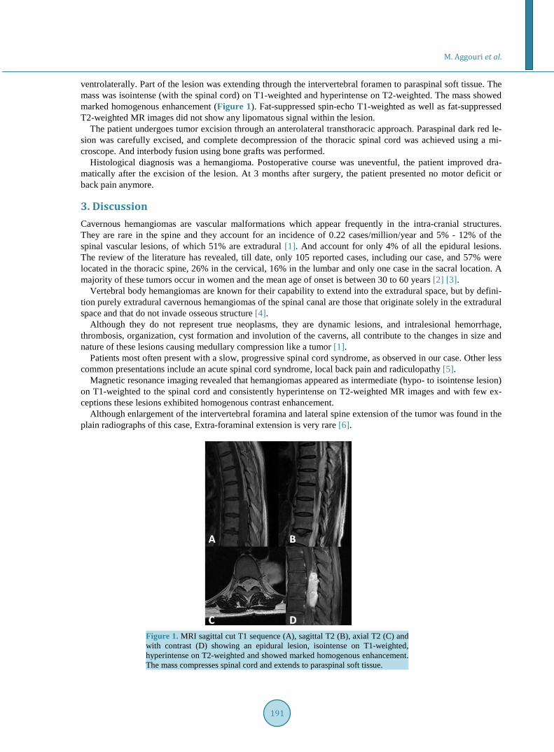
ventrolaterally. Part of the lesion was extending through the intervertebral foramen to paraspinal soft tissue. The mass was isointense (with the spinal cord) on T1-weighted and hyperintense on T2-weighted. The mass showed marked homogenous enhancement (Figure 1). Fat-suppressed spin-echo T1-weighted as well as fat-suppressed T2-weighted MR images did not show any lipomatous signal within the lesion.
The patient undergoes tumor excision through an anterolateral transthoracic approach. Paraspinal dark red le-sion was carefully excised, and complete decompression of the thoracic spinal cord was achieved using a mi-croscope. And interbody fusion using bone grafts was performed.
Histological diagnosis was a hemangioma. Postoperative course was uneventful, the patient improved dra-matically after the excision of the lesion. At 3 months after surgery, the patient presented no motor deficit or back pain anymore.
3. Discussion Cavernous hemangiomas are vascular malformations which appear frequently in the intra-cranial structures. They are rare in the spine and they account for an incidence of 0.22 cases/million/year and 5% - 12% of the spinal vascular lesions, of which 51% are extradural [1]. And account for only 4% of all the epidural lesions. The review of the literature has revealed, till date, only 105 reported cases, including our case, and 57% were located in the thoracic spine, 26% in the cervical, 16% in the lumbar and only one case in the sacral location. A majority of these tumors occur in women and the mean age of onset is between 30 to 60 years [2] [3].
Vertebral body hemangiomas are known for their capability to extend into the extradural space, but by defini-tion purely extradural cavernous hemangiomas of the spinal canal are those that originate solely in the extradural space and that do not invade osseous structure [4].
Although they do not represent true neoplasms, they are dynamic lesions, and intralesional hemorrhage, thrombosis, organization, cyst formation and involution of the caverns, all contribute to the changes in size and nature of these lesions causing medullary compression like a tumor [1].
Patients most often present with a slow, progressive spinal cord syndrome, as observed in our case. Other less common presentations include an acute spinal cord syndrome, local back pain and radiculopathy [5].
Magnetic resonance imaging revealed that hemangiomas appeared as intermediate (hypo- to isointense lesion) on T1-weighted to the spinal cord and consistently hyperintense on T2-weighted MR images and with few ex-ceptions these lesions exhibited homogenous contrast enhancement.
Although enlargement of the intervertebral foramina and lateral spine extension of the tumor was found in the plain radiographs of this case, Extra-foraminal extension is very rare [6].
Figure 1. MRI sagittal cut T1 sequence (A), sagittal T2 (B), axial T2 (C) and with contrast (D) showing an epidural lesion, isointense on T1-weighted, hyperintense on T2-weighted and showed marked homogenous enhancement. The mass compresses spinal cord and extends to paraspinal soft tissue.
M. Aggouri et al.
192
Because hemangiomas are rare in the spine, and have no radiological characteristic signs, histological diagno-sis cannot be predicted with accuracy before surgery. Differential diagnosis include schwannoma, neurofibroma, angiolipoma, lymphoma, metastasis.
Complete removal of extradural hemangioma is the procedure of choice, and because hemangiomas rarely exhibit profuse intraoperative bleeding, radical extirpation is often possible [4].
As in this case, Most of the patients achieve a good recovery with the improvement of the neurological defi-cits and a complete resolution of the tumour, but the patients with acute and paraplegic conditions have poor outcomes [7].
4. Conclusion Even if entirely extradural Hemangioma with no bone involvement is rare, this lesion should be considered in the differential diagnosis of epidural tumors.
References [1] Khalatbari, M.R., Abbassioun, K. and Amirjmshidi, A. (2013) Solitary Spinal Epidural Cavernous Angioma: Report of
Nine Surgically Treated Cases and Review of The literature. European Spine Journal, 22, 542-547. http://dx.doi.org/10.1007/s00586-012-2526-2
[2] Cosgrove, G.R., Bertrand, G., Fontaine, S., Robitaille, Y. and Melanson, D. (1988) Cavernous Angiomas of the Spinal Cord. Journal of Neurosurgery, 68, 31-36. http://dx.doi.org/10.3171/jns.1988.68.1.0031
[3] Hatiboglu, M.A., Iplikcioglu, A.C. and Ozcan, D. (2006) Epidural Spinal Cavernous Hemangioma: Case Report. Neurologia Medico-Chirurgica (Tokyo), 46, 455-458. http://dx.doi.org/10.2176/nmc.46.455
[4] Hemalatha, A.L., et al. (2013) Pure Epidural Spinal Cavernous Hemangioma—With an Innocuous Face but a Perilous Behaviour. Journal of Clinical and Diagnostic Research, 7, 1434-1435.
[5] Cheng, L.T.E. (2005) Spinal Epidural Aemangioma Associated with Extensive Gastrointestinal Eaemangiomas. Inter-ventional Neuroradiology, 11, 161-166.
[6] Harrington Jr., J.F., Khan, A. and Grunnet, M. (1995) Spinal Epidural Cavernous Angioma Presenting as a Lumbar Radiculopathy with Analysis of Magnetic Resonance Imaging Characteristics: Case Report. Neurosurgery, 36, 581- 584. http://dx.doi.org/10.1227/00006123-199503000-00018
[7] Jo, B.J., Lee, S.H., Chung, S.E., Paeng, S.S., Kim, H.S., Yoon, S.W., et al. (2006) Pure Epidural Cavernous Heman-gioma of the Cervical Spine that Presented with an Acute Sensory Deficit Caused by Hemorrhage. Yonsei Medical Journal, 47, 877-880. http://dx.doi.org/10.3349/ymj.2006.47.6.877


![A Giant Extradural Infantile Hemangioma of the Middle ... · intracranial infantile hemangioma has been reported in the literature [2-5]. The most prevalent location for IH is posterior](https://static.fdocuments.net/doc/165x107/5d65e6a688c993aa7e8bba4a/a-giant-extradural-infantile-hemangioma-of-the-middle-intracranial-infantile.jpg)