EXTENDED REPORT Proof of concept: enthesitis and...
9
EXTENDED REPORT Proof of concept: enthesitis and new bone formation in spondyloarthritis are driven by mechanical strain and stromal cells Peggy Jacques, 1 Stijn Lambrecht, 1 Eveline Verheugen, 1 Elin Pauwels, 2 George Kollias, 3 Maria Armaka, 3 Marleen Verhoye, 4 Annemie Van der Linden, 4 Rik Achten, 5 Rik J Lories, 6 Dirk Elewaut 1 Handling editor Tore K Kvien ▸ Additional material is published online only. To view please visit the journal online (http://dx.doi.org/10.1136/ annrheumdis-2013-203643). For numbered affiliations see end of article. Correspondence to Dr Peggy Jacques or Dr Dirk Elewaut, Laboratory for Molecular Immunology and Inflammation, Ghent University Hospital, Department of Rheumatology, De Pintelaan 185, Ghent 9000, Belgium; [email protected] or [email protected] Received 18 March 2013 Revised 20 June 2013 Accepted 14 July 2013 Published Online First 6 August 2013 ▸ http://dx.doi.org/10.1136/ annrheumdis-2013-203924 To cite: Jacques P, Lambrecht S, Verheugen E, et al. Ann Rheum Dis 2014;73:437–445. ABSTRACT Objectives Spondyloarthritides (SpA) are characterised by both peripheral and axial arthritis. The hallmarks of peripheral SpA are the development of enthesitis, most typically of the Achilles tendon and plantar fascia, and new bone formation. This study was undertaken to unravel the mechanisms leading towards enthesitis and new bone formation in preclinical models of SpA. Results First, we demonstrated that TNF ΔARE mice show typical inflammatory features highly reminiscent of SpA. The first signs of inflammation were found at the entheses. Importantly, enthesitis occurred equally in the presence or absence of mature T and B cells, underscoring the importance of stromal cells. Hind limb unloading in TNF ΔARE mice significantly suppressed inflammation of the Achilles tendon compared with weight bearing controls. Erk1/2 signalling plays a crucial role in mechanotransduction-associated inflammation. Furthermore, new bone formation is strongly promoted at entheseal sites by biomechanical stress and correlates with the degree of inflammation. Conclusions These findings provide a formal proof of the concept that mechanical strain drives both entheseal inflammation and new bone formation in SpA. INTRODUCTION Spondyloarthritides (SpA) are a group of chronic inflammatory disorders characterised by asymmet- rical peripheral arthritis predominantly of lower limbs, and axial inflammation (sacroiliitis and spon- dylitis). The disease is typically accompanied by a variety of extra-articular manifestations, such as intestinal and ocular inflammation. In European countries, the overall incidence is estimated at 0.5%–2% of the Caucasian population, with onset frequently in the early adulthood. 12 Inflammation of attachment sites of ligaments and tendons to bones, enthesitis, is a hallmark of SpA which distin- guishes it from other inflammatory rheumatic dis- orders. 3 In addition, SpA is also characterised by new bone formation evolving into ankylosis, or into the formation of enthesophytes that also appear to originate from these insertion sites. 4 Radiographic progression of disease reflecting structural damage is characterised by new bone for- mation leading to sacroiliac and spinal ankylosis. Both inflammation and progressive structural damage contribute to the burden of disease. 5 The entheses are subjected to repetitive biomech- anical stressing forces that are applied during the course of normal muscle, ligament and tendon action; this suggests a link between biomechanical stress and SpA. McGonagle et al 6 proposed an enthesitis-based model for the pathogenesis of SpA where interactions between biomechanical factors and the innate immune response (eg, to bacterial products) may lead to disease. Nevertheless, the enthesitis concept has remained controversial as synovitis and bone marrow inflammation are also typical signs of active SpA. The anatomical proxim- ity and the ample molecular and cellular communi- cations between these tissues suggest their functional relationship in the pathogenesis of syno- vitis in SpA with the disease processes defined in the context of the synovio–entheseal complex. 7 Within this microenvironment, it has become clear that in SpA patients, new bone formation often occurs in close relationship with the entheses. Spinal syndesmophytes develop along the anterior intervertebral ligaments, and bony spurs are formed at the Achilles tendon and plantar fascia. Interestingly, the process of inflammation and subsequent bone erosion appeared in anatomically distinct sites as compared with new bone formation at the Achilles’ enthesis. 8 Erosions preferentially developed in regions undergoing compression, whereas spur formation occurred in regions prone to tensile forces. These anatomical data further cor- roborate current paradigms that strongly suggest a molecular uncoupling of inflammation and new bone formation in SpA. Despite a proven efficacy on inflammatory signs and symptoms, treatment with tumour necrosis factor (TNF) blocking agents in mouse models and in patients does not substan- tially affect progression of ankylosis. 9–11 Currently, it remains elusive whether inflamma- tion and new bone formation are closely linked or rather uncoupled, and why these processes coloca- lise at entheseal sites. In the current study, we hypothesised that bio- mechanical factors drive inflammation and new bone formation at entheseal sites. This hypothesis was studied in the TNF ΔARE mouse model in which chronic and deregulated TNF production leads to arthritis and a Crohn’s-like ileitis. 12–14 We previ- ously argued that this model, depending on endogenous TNF, involving peripheral and axial Editor’s choice Scan to access more free content Jacques P, et al. Ann Rheum Dis 2014;73:437–445. doi:10.1136/annrheumdis-2013-203643 437 Basic and translational research on 28 July 2018 by guest. Protected by copyright. http://ard.bmj.com/ Ann Rheum Dis: first published as 10.1136/annrheumdis-2013-203643 on 6 August 2013. Downloaded from
Transcript of EXTENDED REPORT Proof of concept: enthesitis and...

EXTENDED REPORT
Proof of concept: enthesitis and new boneformation in spondyloarthritis are drivenby mechanical strain and stromal cellsPeggy Jacques,1 Stijn Lambrecht,1 Eveline Verheugen,1 Elin Pauwels,2 George Kollias,3
Maria Armaka,3 Marleen Verhoye,4 Annemie Van der Linden,4 Rik Achten,5
Rik J Lories,6 Dirk Elewaut1
Handling editor Tore K Kvien
▸ Additional material ispublished online only. To viewplease visit the journal online(http://dx.doi.org/10.1136/annrheumdis-2013-203643).
For numbered affiliations seeend of article.
Correspondence toDr Peggy Jacques or Dr DirkElewaut, Laboratory forMolecular Immunology andInflammation, Ghent UniversityHospital, Department ofRheumatology, De Pintelaan185, Ghent 9000, Belgium;[email protected] [email protected]
Received 18 March 2013Revised 20 June 2013Accepted 14 July 2013Published Online First6 August 2013
▸ http://dx.doi.org/10.1136/annrheumdis-2013-203924
To cite: Jacques P,Lambrecht S, Verheugen E,et al. Ann Rheum Dis2014;73:437–445.
ABSTRACTObjectives Spondyloarthritides (SpA) are characterisedby both peripheral and axial arthritis. The hallmarks ofperipheral SpA are the development of enthesitis, mosttypically of the Achilles tendon and plantar fascia, andnew bone formation. This study was undertaken tounravel the mechanisms leading towards enthesitis andnew bone formation in preclinical models of SpA.Results First, we demonstrated that TNFΔARE miceshow typical inflammatory features highly reminiscent ofSpA. The first signs of inflammation were found at theentheses. Importantly, enthesitis occurred equally in thepresence or absence of mature T and B cells,underscoring the importance of stromal cells. Hind limbunloading in TNFΔARE mice significantly suppressedinflammation of the Achilles tendon compared withweight bearing controls. Erk1/2 signalling plays a crucialrole in mechanotransduction-associated inflammation.Furthermore, new bone formation is strongly promotedat entheseal sites by biomechanical stress and correlateswith the degree of inflammation.Conclusions These findings provide a formal proof ofthe concept that mechanical strain drives both enthesealinflammation and new bone formation in SpA.
INTRODUCTIONSpondyloarthritides (SpA) are a group of chronicinflammatory disorders characterised by asymmet-rical peripheral arthritis predominantly of lowerlimbs, and axial inflammation (sacroiliitis and spon-dylitis). The disease is typically accompanied by avariety of extra-articular manifestations, such asintestinal and ocular inflammation. In Europeancountries, the overall incidence is estimated at0.5%–2% of the Caucasian population, with onsetfrequently in the early adulthood.1 2 Inflammationof attachment sites of ligaments and tendons tobones, enthesitis, is a hallmark of SpA which distin-guishes it from other inflammatory rheumatic dis-orders.3 In addition, SpA is also characterised bynew bone formation evolving into ankylosis, orinto the formation of enthesophytes that alsoappear to originate from these insertion sites.4
Radiographic progression of disease reflectingstructural damage is characterised by new bone for-mation leading to sacroiliac and spinal ankylosis.Both inflammation and progressive structuraldamage contribute to the burden of disease.5
The entheses are subjected to repetitive biomech-anical stressing forces that are applied during thecourse of normal muscle, ligament and tendonaction; this suggests a link between biomechanicalstress and SpA. McGonagle et al6 proposed anenthesitis-based model for the pathogenesis of SpAwhere interactions between biomechanical factorsand the innate immune response (eg, to bacterialproducts) may lead to disease. Nevertheless, theenthesitis concept has remained controversial assynovitis and bone marrow inflammation are alsotypical signs of active SpA. The anatomical proxim-ity and the ample molecular and cellular communi-cations between these tissues suggest theirfunctional relationship in the pathogenesis of syno-vitis in SpA with the disease processes defined inthe context of the synovio–entheseal complex.7
Within this microenvironment, it has becomeclear that in SpA patients, new bone formationoften occurs in close relationship with the entheses.Spinal syndesmophytes develop along the anteriorintervertebral ligaments, and bony spurs areformed at the Achilles tendon and plantar fascia.Interestingly, the process of inflammation and
subsequent bone erosion appeared in anatomicallydistinct sites as compared with new bone formationat the Achilles’ enthesis.8 Erosions preferentiallydeveloped in regions undergoing compression,whereas spur formation occurred in regions proneto tensile forces. These anatomical data further cor-roborate current paradigms that strongly suggest amolecular uncoupling of inflammation and newbone formation in SpA. Despite a proven efficacyon inflammatory signs and symptoms, treatmentwith tumour necrosis factor (TNF) blocking agentsin mouse models and in patients does not substan-tially affect progression of ankylosis.9–11
Currently, it remains elusive whether inflamma-tion and new bone formation are closely linked orrather uncoupled, and why these processes coloca-lise at entheseal sites.In the current study, we hypothesised that bio-
mechanical factors drive inflammation and newbone formation at entheseal sites. This hypothesiswas studied in the TNFΔARE mouse model in whichchronic and deregulated TNF production leads toarthritis and a Crohn’s-like ileitis.12–14 We previ-ously argued that this model, depending onendogenous TNF, involving peripheral and axial
Editor’s choiceScan to access more
free content
Jacques P, et al. Ann Rheum Dis 2014;73:437–445. doi:10.1136/annrheumdis-2013-203643 437
Basic and translational research
on 28 July 2018 by guest. Protected by copyright.
http://ard.bmj.com
/A
nn Rheum
Dis: first published as 10.1136/annrheum
dis-2013-203643 on 6 August 2013. D
ownloaded from

joints and presenting extra-articular manifestations such asbowel inflammation, is a true translational model of SpA.13
Here, we observed that enthesitis is an early feature of thismurine model, and that its development is dependent upon bio-mechanical strain. Bone new formation at entheseal sites,another prominent feature of SpA, was also associated withweight bearing and biomechanical strain. Altogether, these datasubstantiate the hypothesis that entheseal inflammation and newbone formation are driven by biomechanical strain.
MATERIALS AND METHODSAnimals and housingTNFΔARE mice were provided by Kollias G.12 CAIA was inducedwith ArthritoMab from MD Biosciences according to the manu-facturer’s instructions. All mice were bred and housed in specificpathogen-free conditions in accordance with the general recom-mendations for animal breeding and housing, and all experi-ments were conducted in conditions according to the EthicalCommittee of Animal Welfare. For treatment with MAPK inhi-bitors, mice were injected daily with 50 μg/g of PD98059, aselective Erk1/2/MEK1 inhibitor to inhibit Erk1/2 signalling, orSB203580 for p38 blockade (both LC Laboratories) intraperito-neally, or with DMSO as a negative control.
HistologyMurine paws and spine were dissected, fixed in 4% formalde-hyde, decalcified in 0.5 M ethylenediamine tetraacetic acid(Sigma-Aldrich, St Louis, Missouri, USA) at 4° or in 5% formicacid until bones were pliable. Paraffin sections were stained withH&E for evaluation of inflammation and bone erosions, or withsafranin-O for cartilage appreciation. Enthesitis of Achillestendon was evaluated by two blinded assessors, and scored onthree parameters, infiltrate in the Achilles tendon, calcanealerosion and exudate at the synovio–entheseal complex, eachranging from 0 (normal) to 3. A composite score was built fromthese parameters. In addition, digital image analysis was per-formed by AxioVision software (Carl Zeiss MicroImagingGmbH, Göttingen, Germany). Extent of inflammation withinthe synovio–entheseal complex was normalised to the size ofthe growth plate. In a subgroup of mice from tail suspensionexperiments, inflammation in front paws was also evaluated asfollows: absence (0) or presence (1) of dactylitis and enthesitis.
Western blottingAchilles tendons were dissected on ice, snap frozen and lysed inlaemli buffer by a bead-beating procedure to liberate proteins.Immunoblotting using chemiluminescent detection forphospho-Erk1/2 and total Erk1/2 was conducted as previouslydescribed. Briefly, equal amounts were loaded on precast 10%SDS-PAGE Criterion gels (Bio-Rad Laboratories, Hercules,California, USA). Equal loading was verified by Ponceau S stain-ing (data not shown). MagicMark (Invitrogen, Paisley, UK)protein standards were run as Mw markers. Strips were focusedfor 35 kVh and SDS-PAGE was performed at 200 V forapproximately 1 h. Proteins were transferred to nitrocellulosemembranes (Bio-Rad Laboratories). The resulting membraneswere immunoblotted with antiphospho-Erk1/2 or antitotalErk1/2 (Cell Signaling Technology, Danvers, Massachusetts,USA), followed by antirabbit horseradish peroxidase-conjugatedAb and enhanced chemiluminescence (Pierce, Rockford, Illinois,USA). Chemiluminescence images were recorded using theVersaDoc-imaging system (Bio-Rad Laboratories). Densitometricanalysis of the images was performed by Quantity One Softwarev 4.4.0 (Bio-Rad Laboratories).
Magnetic resonance imagingWild type and littermate TNFΔARE mice were anaesthetised withisoflurane (IsoFlo, Abbott, Illinois, USA) administered in amixture of 30% O2 and 70% N2. During immobilisation, iso-flurane levels were kept at 3%. The dose was gradually loweredto 1.5%. Mice were submitted to a longitudinal MRI studyat age of 1, 2, 3 and 5 months. MRI was performed on a 9.4Tesla MR system (BRUKER, Ettlingen, Germany). CoronalT1-weighted 2D images of the sacroiliac joints were acquiredwith a FLASH sequence: TE=3.4 ms, TR=200 ms, FA=40°,FOV=15 mm, image matrix (256×256), 16 slices, slice thick-ness =0.5 mm. T2-weighted images were acquired with a RAREsequence: TE=36 ms, TR=3000 ms, ETL=8, FOV=19.2 mm,image matrix (256×192), 8 slices, slice thickness =1 mm. Tocompare the T2-weighted signal intensities of the iliac boneamong animals and age, relative signal intensities of the iliacbone with respect to the mean signal intensity of the muscletissue close to the ilium bone were calculated.
Micro-CT acquisitionsSamples (n=36) were scanned in a GE Healthcare eXploreLocus SP preclinical micro-CT specimen scanner (GE MedicalSystems, London, Ontario) with the following acquisition para-meters: 80 kV tube voltage, 80 microamperes tube current, 1×1detector binning, three times magnification, 500 projectionsacquired over 200°s using a 0.4° step size, 3000 ms exposuretime and frame averaging of 4. The acquired projections werereconstructed into a 500×500×940 matrix, with 8-mm voxelsize, by proprietary software (eXplore Reconstruction Utility,GE Medical Systems, London, Ontario) using a Feldkamp-typealgorithm with Parker’s weighting function.15 Each recon-structed micro-CT image was visually and quantitatively ana-lysed by MicroView software (GE Medical Systems).Quantitative analysis was done by drawing a volume of interest.Bone mineral density was calculated inside this region aftermeasuring the threshold for non-bone versus bone tissue, withthe threshold value automatically determined.
A second set of micro-CT scans were performed at the Centrefor X-ray Tomography of the Ghent University.16 To improvesoft tissue contrast, the mice legs were immersed in a 4% HgCl2aqueous solution for 24 h before scanning.31 The mice legswere scanned using the transmission head of a dual head x-raytube from Feinfocus (FXE 160.51), operated at 120 kV, and aVarian 2520 V Paxscan flat panel detector. A beam filtration of1 mm aluminium was applied. For each scan, 1441 projectionswere recorded, covering 360°, with an exposure time of 2 s perprojection. The resulting voxel size was 6.5 mm. Reconstructionof the projection data was done using Octopus, a reconstructionsoftware package developed at UGCT.17
StatisticsThe non-parametric Kruskal–Wallis test or parametric ANOVAwas used for comparison of three groups. The Mann–Whitneytest was used for comparison of two groups. Results were con-sidered statistically significant with two-sided p values <0.05.Regression analysis was performed to determine the correlationbetween osteophyte size and clinical score, irrespective of tailsuspension, with R² the adjusted correlation coefficient.
RESULTSFeatures of spondyloarthritis in TNFΔARE miceAs the precise sites where inflammation originates in SpAremain a matter of debate, a detailed clinical and histological
438 Jacques P, et al. Ann Rheum Dis 2014;73:437–445. doi:10.1136/annrheumdis-2013-203643
Basic and translational research
on 28 July 2018 by guest. Protected by copyright.
http://ard.bmj.com
/A
nn Rheum
Dis: first published as 10.1136/annrheum
dis-2013-203643 on 6 August 2013. D
ownloaded from

study was undertaken in TNFΔARE mice. Therefore, mice werescored every other day for arthritis development, and weresacrificed at different ages (4, 8, 12, 24 weeks) for histologicalassessment. In hind paws of TNFΔARE mice, inflammationinitiated at approximately 4 weeks of age within the collateralligaments of interphalangeal joints (figure 1B), within thesynovio–entheseal complex of the Achilles tendon (figure 1E,F)and the greater trochanter ligament of the hip (figure 1H,I). Bythe age of 6–8 weeks, inflammation spreads out into the syno-vium with pannus formation, and finally involves the entirejoint, with bone erosions (figure 1C). Thus, articular inflamma-tion initiates within the entheseal regions in TNFΔARE mice.
Another interesting hallmark of the disease is the developmentof spinal inflammation with sacroiliitis (figure 1J–L).13 The jointsbetween the sacrum and the pelvic ilium bone are joined byseveral ligaments and consist of a lower fibrocartilagenous partand an upper ligamentous part. As such, this region is also proneto mechanical strain. As sacroiliac joints are difficult to assess clin-ically, we performed a longitudinal in vivo MRI study, in whichTNFΔARE mice and controls were scanned monthly, starting atthe age of 4 weeks, and each month both T1 and T2 sequenceswere run (figure 2A). On T1 sequences, progressive narrowing ofjoint space and irregular articular surfaces reflected progressivestructural damage. On T2 sequences, the relative signal intensity
Figure 1 Features of spondyloarthritis in TNFΔARE mice: entheseal inflammation. (A) Hind paw distal interphalangeal joint of healthy controlmouse. (B) Early signs of enthesitis in hind paw of TNFΔARE mouse. (C) Advanced erosive stage. (D) Achilles tendon of healthy control mouse.(E and F) Achilles enthesitis in TNFΔARE mouse. (G) Hip joint of healthy control mouse. (H and I) Hip joint of TNFΔARE mouse, arrow head points atinflammation around the greater trochanter. ( J) Sacroiliac joint of a healthy control mouse. (K) Sacroiliac joint of young TNFΔARE mouse. (L)Advanced stage sacroiliitis in TNFΔARE mouse, white arrows point at erosions. Staining with H&E. Original magnification×100, except D, E, G and Horiginal magnification×40. Access the article online to view this figure in colour.
Jacques P, et al. Ann Rheum Dis 2014;73:437–445. doi:10.1136/annrheumdis-2013-203643 439
Basic and translational research
on 28 July 2018 by guest. Protected by copyright.
http://ard.bmj.com
/A
nn Rheum
Dis: first published as 10.1136/annrheum
dis-2013-203643 on 6 August 2013. D
ownloaded from

of the iliac bone was calculated for each mouse (figure 2B). Thesignal intensity in control mice clearly decreased with agebecause of an increase in mineral content (63% decrease at5 months compared with 1 month). The signal intensity inTNFΔARE mice only decreased by 29% between 1 and 5 monthsof age. This may reflect an increased water content (or bonemarrow oedema) and a smaller mineral content in TNFΔARE
mice. Alternatively, the differences in signal may also reflect thepersistence of red marrow (ie, increased cellularity) seen in TNFoverexpression models.18 The two-way analysis of variance(ANOVA) of relative signal intensity of the iliac bone valuesdemonstrated both a significant main group effect (TNFΔARE
mice vs controls) and age effect. At the age of 2, 3 and 5 months,the relative signal intensities of the TNFΔARE mice were signifi-cantly higher compared with the controls (figure 2B). Detailedimages obtained by this in vivo imaging approach provide a valu-able tool to study sacroiliitis development and allow the monitor-ing of therapeutic interventions in animal models for SpA.Collectively, these data highlight that inflammation occurring inTNFΔARE mice is highly reminiscent of human SpA.
Enthesitis occurs independent of mature T and B cellsIt was recently shown that interleukin (IL)-23 responsiveCD3+CD4–CD8– entheseal resident T cells are essential toinduce enthesitis in an IL-23 overexpression model.19 To assessthe role of adaptive immunity, we backcrossed TNFΔARE miceon to RAG-1 deficient animals that have no mature T cells, andassessed severity of enthesitis. In RAG-1 deficient TNFΔARE
mice, incidence of enthesitis was similar to that in T cell suffi-cient TNFΔARE mice and occurred in all mice examined(figure 3B,C). This finding highlights that both T cell dependentand independent mechanisms, most likely involving stromalcells, can contribute to enthesitis. Our data corroborate the pre-vious role for stromal cells in TNF driven joint inflammation.13
Hind limb unloading inhibits development of enthesitisin TNFΔARE miceWe hypothesised that continuous mechanical strain caused bynormal animal activity could be at the origin of enthesitis in thismodel. The Achilles tendon was chosen as the focus of interestfor this study. To address this we used an established model for
Figure 2 Consecutive monthly in vivo MRI of sacroiliac joints of healthy control and littermate TNFΔARE mice. (A) T1-weighted images (left panel)illustrate the development of sacroiliitis in TNFΔARE mice: joint space narrowing, irregular surfaces. T2-weighted images (right panel) illustrate thepersistence of red marrow and general osteopenia in TNFΔARE mice. (B) Graph gives the relative T2-weighted signal intensities of the ilium withrespect to a neighbouring muscle control region for control and TNFΔARE mice and for different ages. A two-way ANOVA demonstrates differencesin relative signal intensities between the group of control mice and TNFΔARE mice, and for the different ages of the mice (*=p<0.05).
440 Jacques P, et al. Ann Rheum Dis 2014;73:437–445. doi:10.1136/annrheumdis-2013-203643
Basic and translational research
on 28 July 2018 by guest. Protected by copyright.
http://ard.bmj.com
/A
nn Rheum
Dis: first published as 10.1136/annrheum
dis-2013-203643 on 6 August 2013. D
ownloaded from

hind limb unloading, tail suspension.20 21 In specially designedcages, TNFΔARE mice and healthy controls were tail suspended,and clinical signs of arthritis were assessed daily. Hind limbunloading was initiated before the known onset of illness,before 4 weeks of age, in a preventive setting. Following a14-day period of unloading, none of the mice subjected to thisprocedure displayed clinical signs of peripheral arthritis ofankles and hind paws, whereas all control mice that were nottail suspended, did. Furthermore, by detailed histological ana-lysis, we detected only minor inflammatory changes in tail sus-pended TNFΔARE mice, but significantly more inflammation intheir weight bearing controls (figure 3D–G). In a subgroup ofmice, arthritis was also evaluated in front paws. In general,severity of inflammation did not differ between weight carrying
front paws of tail-suspended and control mice (figure 3H–J).These data strongly suggest that the onset of enthesitis in thisanimal model is driven by mechanical load on hind paws.
Mechanical stress triggers Erk1/2 signalling in TNFΔARE miceActivation of mitogen-activated protein kinase (MAPK) signallingpathways was previously demonstrated as a response to stretch ina variety of stromal cell types.22–25 Therefore, we performed atail suspension study, whereby TNFΔARE mice were subjected totail suspension for 7 days, again in a preventive setting, beforethe onset of enthesitis. Tail suspended mice did not show signs ofclinical arthritis, as compared with weight bearing controls.Seven days later, half of the group of mice were allowed to walkaround for 10 min before they were sacrificed. Induction of
Figure 3 Inflammation at the synovio–entheseal complex is driven by biomechanical factors in TNFΔARE mice. (A) Healthy control mouse: overviewof ankle. (B) Achilles tendon from a RAG-1 deficient TNFΔARE mouse. (C) Achilles tendon from RAG-1 sufficient (heterozygous) littermate TNFΔARE
mouse. (D) Achilles tendon of 4-week-old healthy control mouse. (E) Non-tail suspended TNFΔARE mouse. (F) Littermate TNFΔARE mouse subjected totail suspension for 14 days continuously. (G) Composite histology score demonstrates reduced severity of enthesitis upon tail suspension, *=p<0.05.(H) Distal interphalangeal joint in front paw of a non-tail suspended TNFΔARE and (I) tail suspended TNFΔARE mouse. All original magnification ×40.( J) Graph demonstrates inflammation score in front paws from a subgroup of mice from tail suspension experiments, N=4 in each group, values aremean and SEM. Access the article online to view this figure in colour.
Jacques P, et al. Ann Rheum Dis 2014;73:437–445. doi:10.1136/annrheumdis-2013-203643 441
Basic and translational research
on 28 July 2018 by guest. Protected by copyright.
http://ard.bmj.com
/A
nn Rheum
Dis: first published as 10.1136/annrheum
dis-2013-203643 on 6 August 2013. D
ownloaded from
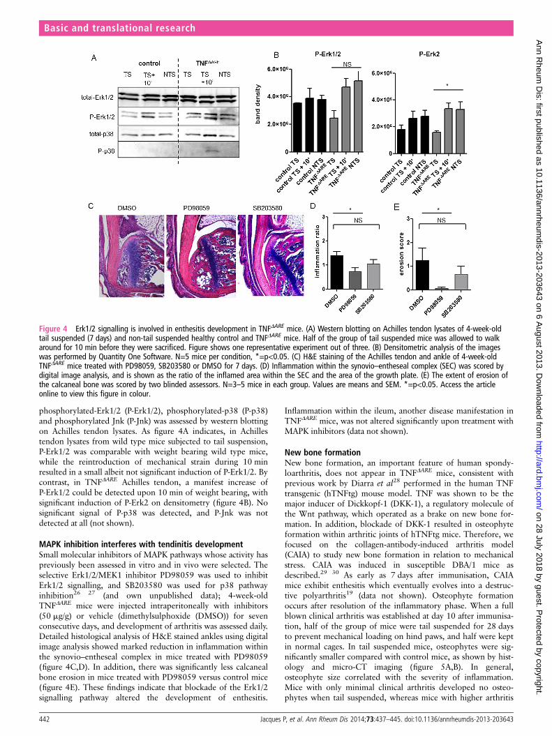
phosphorylated-Erk1/2 (P-Erk1/2), phosphorylated-p38 (P-p38)and phosphorylated Jnk (P-Jnk) was assessed by western blottingon Achilles tendon lysates. As figure 4A indicates, in Achillestendon lysates from wild type mice subjected to tail suspension,P-Erk1/2 was comparable with weight bearing wild type mice,while the reintroduction of mechanical strain during 10 minresulted in a small albeit not significant induction of P-Erk1/2. Bycontrast, in TNFΔARE Achilles tendon, a manifest increase ofP-Erk1/2 could be detected upon 10 min of weight bearing, withsignificant induction of P-Erk2 on densitometry (figure 4B). Nosignificant signal of P-p38 was detected, and P-Jnk was notdetected at all (not shown).
MAPK inhibition interferes with tendinitis developmentSmall molecular inhibitors of MAPK pathways whose activity haspreviously been assessed in vitro and in vivo were selected. Theselective Erk1/2/MEK1 inhibitor PD98059 was used to inhibitErk1/2 signalling, and SB203580 was used for p38 pathwayinhibition26 27 (and own unpublished data); 4-week-oldTNFΔARE mice were injected intraperitoneally with inhibitors(50 mg/g) or vehicle (dimethylsulphoxide (DMSO)) for sevenconsecutive days, and development of arthritis was assessed daily.Detailed histological analysis of H&E stained ankles using digitalimage analysis showed marked reduction in inflammation withinthe synovio–entheseal complex in mice treated with PD98059(figure 4C,D). In addition, there was significantly less calcanealbone erosion in mice treated with PD98059 versus control mice(figure 4E). These findings indicate that blockade of the Erk1/2signalling pathway altered the development of enthesitis.
Inflammation within the ileum, another disease manifestation inTNFΔARE mice, was not altered significantly upon treatment withMAPK inhibitors (data not shown).
New bone formationNew bone formation, an important feature of human spondy-loarthritis, does not appear in TNFΔARE mice, consistent withprevious work by Diarra et al28 performed in the human TNFtransgenic (hTNFtg) mouse model. TNF was shown to be themajor inducer of Dickkopf-1 (DKK-1), a regulatory molecule ofthe Wnt pathway, which operated as a brake on new bone for-mation. In addition, blockade of DKK-1 resulted in osteophyteformation within arthritic joints of hTNFtg mice. Therefore, wefocused on the collagen-antibody-induced arthritis model(CAIA) to study new bone formation in relation to mechanicalstress. CAIA was induced in susceptible DBA/1 mice asdescribed.29 30 As early as 7 days after immunisation, CAIAmice exhibit enthesitis which eventually evolves into a destruc-tive polyarthritis19 (data not shown). Osteophyte formationoccurs after resolution of the inflammatory phase. When a fullblown clinical arthritis was established at day 10 after immunisa-tion, half of the group of mice were tail suspended for 28 daysto prevent mechanical loading on hind paws, and half were keptin normal cages. In tail suspended mice, osteophytes were sig-nificantly smaller compared with control mice, as shown by hist-ology and micro-CT imaging (figure 5A,B). In general,osteophyte size correlated with the severity of inflammation.Mice with only minimal clinical arthritis developed no osteo-phytes when tail suspended, whereas mice with higher arthritis
Figure 4 Erk1/2 signalling is involved in enthesitis development in TNFΔARE mice. (A) Western blotting on Achilles tendon lysates of 4-week-oldtail suspended (7 days) and non-tail suspended healthy control and TNFΔARE mice. Half of the group of tail suspended mice was allowed to walkaround for 10 min before they were sacrificed. Figure shows one representative experiment out of three. (B) Densitometric analysis of the imageswas performed by Quantity One Software. N=5 mice per condition, *=p<0.05. (C) H&E staining of the Achilles tendon and ankle of 4-week-oldTNFΔARE mice treated with PD98059, SB203580 or DMSO for 7 days. (D) Inflammation within the synovio–entheseal complex (SEC) was scored bydigital image analysis, and is shown as the ratio of the inflamed area within the SEC and the area of the growth plate. (E) The extent of erosion ofthe calcaneal bone was scored by two blinded assessors. N=3–5 mice in each group. Values are means and SEM. *=p<0.05. Access the articleonline to view this figure in colour.
442 Jacques P, et al. Ann Rheum Dis 2014;73:437–445. doi:10.1136/annrheumdis-2013-203643
Basic and translational research
on 28 July 2018 by guest. Protected by copyright.
http://ard.bmj.com
/A
nn Rheum
Dis: first published as 10.1136/annrheum
dis-2013-203643 on 6 August 2013. D
ownloaded from

scores developed significantly smaller osteophytes when tail sus-pended compared with non-tail suspended mice (figure 5C,D).
New bone formation occurred at distance from the jointarticular surfaces. Moreover, immersion of the entire paw inmercury chloride (HgCl2), which allows uptake in soft tissuessuch as tendons,31 followed by high resolution CT, revealedthat these osteophytes are formed mainly at entheseal sites(see online supplementary figure S1 and S2). Collectively, thesefindings underscore a role for mechanical strain in new boneformation at entheseal sites.
DISCUSSIONIt has been a great matter of debate where disease originates inSpA patients since, during the course of disease, synovium,enthesis as well as bone marrow can become involved. Due totheir location at the interface of ligaments and bone, entheses areespecially prone to mechanical forces. It is generally acceptedalthough not formally proven that both mechanical and inflam-matory factors can trigger enthesitis, with the enthesitis-basedorigin of SpA as a formal hypothesis.6 According to an occupa-tional study by Ward et al,32 physical activities that requiredrepetitive strain such as stretching, bending or twisting, as well asexposure to whole body vibration, were associated with moreradiographic damage in ankylosing spondylitis. Yet, experimentalevidence supporting these theories was lacking. We provide evi-dence for a mechanotransduction-associated origin of enthesitisand new bone formation at entheseal sites.
First, enthesitis is an early disease feature in TNFΔARE mice.Hind limb unloading prevented the onset of Achilles enthesitis
in TNFΔARE mice. Front paws of tail suspended TNFΔARE miceshowed similar arthritis severity compared with weight bearingcontrols, although the body weight of these mice was loaded onthe front paws. However, careful observation revealed that tailsuspended mice tended to move less than the control group,which provides an explanation for the contra-intuitive findingthat severity of inflammation in front paws does not increaseafter tail suspension and hind limb unloading. We hypothesisethat minimal biomechanical stress is sufficient to induce a sus-tained inflammatory response. Thus, biomechanical stress ratherfunctions as an on/off switch and there is not necessarily alinear relationship between the amount of stress and the degreeof inflammation, at least not above a given level of biomechan-ical stress. The resulting minor increase, if any, may not bedetected by the scoring system, which has limited power.
Therefore, mechanical strain may be considered the trigger ofan inflammatory process, and provides an explanation for theunsolved paradigm of enthesitis development in SpA.
It was previously demonstrated that in the presence ofchronic TNF overexposure, signalling through TNFRI in syn-ovial fibroblasts and intestinal myofibroblasts appears to be suffi-cient to develop arthritis and Crohn’s-like ileitis in TNFΔARE
mice.13 Therefore, crosstalk between the myeloid cell compart-ment and stromal cells such as fibroblasts is critically involved inthe development of both the articular and the intestinal pheno-type. Importantly, mature T cells are not involved in enthesitisdevelopment in TNFΔARE mice, as demonstrated in RAG-1 defi-cient mice. This highlights that several pathways exist to induceenthesitis, some of which rely on IL-23R+ innate-like entheseal
Figure 5 New bone formation is driven by mechanical strain. (A and B) Micro-CT images and corresponding H&E staining of the region within thesquare from healthy control, non-tail suspended and tail suspended collagen-antibody-induced arthritis mice. (C) Osteophyte size was scored by twoblinded assessors. Graph shows osteophyte size for mice with minor (0–1) or major (2–3) clinical arthritis score (one paw). N=10 mice in eachgroup, *=p<0.05. (D) Correlation of osteophyte size with clinical score. R2=0.28, p=0.03. Access the article online to view this figure in colour.
Jacques P, et al. Ann Rheum Dis 2014;73:437–445. doi:10.1136/annrheumdis-2013-203643 443
Basic and translational research
on 28 July 2018 by guest. Protected by copyright.
http://ard.bmj.com
/A
nn Rheum
Dis: first published as 10.1136/annrheum
dis-2013-203643 on 6 August 2013. D
ownloaded from

resident cells,19 while others are strictly dependent uponstromal cell function. In more complex situations such as in SpApatients, both T cell dependent and independent mechanismsmay exist and could have additive effects.
Mechanical stress is sensed by cells via mechanoreceptorssuch as integrins, and transmitted through MAPK signallingpathways.33 In this study, activation of Erk1/2 signalling path-ways in Achilles tendon lysates could be efficiently prohibited byhind limb unloading, while reintroduction of mechanical strainresulted in immediate phosphorylation of Erk1/2 (within10 min), both in wild type and TNFΔARE tendons. Smallmolecular MAPK inhibitors were proven effective in animalmodels for arthritis, and may be promising therapeutic targetsin treatment of inflammatory diseases.34 35 However, limiteddata are available on treatment options of enthesitis in particu-lar. In the present study, the effects of an Erk1/2 inhibitor and ap38 inhibitor on enthesitis development were evaluated. WithErk1/2 inhibition, we detected a clear decrease in inflammationwithin the synovio–entheseal complex; in addition, there was asignificant reduction in calcaneal bone erosion.
The MAPK pathways have previously been shown to modulateTNF biosynthesis, and TNF signalling is involved both upstreamand downstream of MAPK activation.36 In particular, the adeno-sine uracil (AU) rich elements within the TNF transcript areknown targets for p38-mediated activation of TNF translationupon lipopolysaccharide stimulation.12 In addition, the Erk1/2pathway requires the presence of ARE for post-transcriptionalregulation, that is, nuclear export of the TNF message.37 In wildtype mice, MAPK blockade in general results in a dose dependentinhibition of TNF translation. As these targets are deleted inTNFΔARE mice, MAPK blockade would be unable to inhibit TNFtranslation via this pathway. Therefore, the observed biologicaleffects of Erk1/2 blockade in TNFΔARE mice may even be under-estimated and partly TNF independent. However, data fromother models suggest that inhibition of MAPK has complexeffects. In the ankylosing enthesitis model in DBA/1 mice, inhib-ition of p38 accelerated ankylosis rather than inhibiting arthritis,although in vitro data suggested an inhibitory effect of the com-pound used on transforming growth factor and bone morpho-genetic protein induced chondro- and osteogenesis.38
A second hallmark of spondyloarthritis is the occurrence ofnew bone formation leading to ankylosis. However, in TNFΔARE
mice, new bone formation could never be demonstrated, pre-sumably due to continuous and deregulated TNF productionthat functions as a brake on this process through for exampleDKK1, which is a potent inhibitor of Wnt signalling. In add-ition, new bone formation is much less pronounced in theCB57BL/6 background than on other backgrounds such asDBA/1. Successful inhibition of inflammation in mouse modelsand in patients, for instance with TNF blocking agents, does notaffect ankylosis or radiographic progression.9–11 However,recent data suggest that long term treatment may be effective onstructural changes.39 40 The sequence of inflammation followedby new bone formation is not formally proven, although theremay be a preference for syndesmophytes to develop at inflamedvertebral edges in human SpA.41 42 In addition, recent MRIstudies suggested that new bone formation more likely developsin advanced inflammatory spinal lesions. In these lesions, reso-lution of inflammation results in fat metaplasia and ultimately innew bone formation.43 44 Here, we demonstrated that in CAIAmice, osteophytes are significantly smaller when weight bearingis prohibited, supporting a role for mechanical strain in theirdevelopment. In addition, tail suspension was able to preventthe development of osteophytes when only minimal signs of
inflammation were evident, and could minimise osteophyte sizein more advanced arthritis. In general, osteophyte size corre-lated with the severity of inflammation. Although this particularaspect was not the focus of this study, it indirectly supports therelationship between inflammation and new bone formation.
In summary, these findings comprise the first formal proof ofa new paradigm that mechanical forces are the underlying causeof enthesitis and new bone formation in SpA. This opensavenues for a new field of mechanotransduction-associated path-ways in SpA.
Author affiliations1Laboratory for Molecular Immunology and Inflammation, Department ofRheumatology, University Hospital, Ghent, Belgium2UGCT, Department of Physics and Astronomy, University of Ghent, Ghent, Belgium3Alexander Fleming Biomedical Sciences Research Center, Vari, Greece4Bio-Imaging Lab, University of Antwerp, Antwerp, Belgium5Department of Radiology, University Hospital, Ghent, Belgium6Laboratory of Tissue Homeostasis and Disease, Skeletal Biology and EngineeringCenter, Department of Development and Regeneration, KU Leuven, Belgium
Acknowledgements The authors wish to thank C Van Hove at INFINITY forperforming and analysing micro-CT images and L Van Praet for statistical analysis.
Contributors All authors contributed to the conception and design of the study,acquisition, analysis or interpretation of data, and drafting or revising the articlecritically for intellectual content. All authors approved the final version forpublication.
Funding This work is supported by the Fund for Scientific Research—Flanders, by aconcerted action grant from Ghent University, and by the Special Research Fund ofthe Ghent University (BOF, GOA 01G01008). DE is part of a multidisciplinaryresearch platform (MRP) of Ghent University and is supported by an InteruniversityAttraction Pole (Project P7/07).
Competing interests None.
Provenance and peer review Not commissioned; externally peer reviewed.
Data sharing statement Data not shown are available upon email request to thecorresponding author.
REFERENCES1 Braun J, Bollow M, Remlinger G, et al. Prevalence of spondylarthropathies in
HLA-B27 positive and negative blood donors. Arthritis Rheum 1998;41:58–67.2 Sieper J, Rudwaleit M, Khan MA, et al. Concepts and epidemiology of
spondyloarthritis. Best Pract Res Clin Rheumatol 2006;20:401–17.3 McGonagle D, Gibbon W, Emery P. Classification of inflammatory arthritis by
enthesitis. Lancet 1998;352:1137–40.4 Benjamin M, McGonagle D. The anatomical basis for disease localisation in
seronegative spondyloarthropathy at entheses and related sites. J Anat 2001;199(Pt 5):503–26.
5 Machado P, Landewe R, Braun J, et al. Both structural damage and inflammation ofthe spine contribute to impairment of spinal mobility in patients with ankylosingspondylitis. Ann Rheum Dis 2010;69:1465–70.
6 McGonagle D, Stockwin L, Isaacs J, et al. An enthesitis based model for thepathogenesis of spondyloarthropathy additive effects of microbial adjuvant andbiomechanical factors at disease sites. J Rheumatol 2001;28:2155–9.
7 McGonagle D, Lories RJ, Tan AL, et al. The concept of a "synovio-enthesealcomplex" and its implications for understanding joint inflammation and damage inpsoriatic arthritis and beyond. Arthritis Rheum 2007;56:2482–91.
8 McGonagle D, Wakefield RJ, Tan AL, et al. Distinct topography of erosion and newbone formation in achilles tendon enthesitis: implications for understanding the linkbetween inflammation and bone formation in spondylarthritis. Arthritis Rheum2008;58:2694–9.
9 van der Heijde D, Salonen D, Weissman BN, et al. Assessment of radiographicprogression in the spines of patients with ankylosing spondylitis treated withadalimumab for up to 2 years. Arthritis Res Ther 2009;11:R127.
10 van der Heijde D, Landewe R, Baraliakos X, et al. Radiographic findings followingtwo years of infliximab therapy in patients with ankylosing spondylitis. ArthritisRheum 2008;58:3063–70.
11 van der Heijde D, Landewe R, Einstein S, et al. Radiographic progression ofankylosing spondylitis after up to two years of treatment with etanercept. ArthritisRheum 2008;58:1324–31.
12 Kontoyiannis D, Pasparakis M, Pizarro TT, et al. Impaired on/off regulation of TNFbiosynthesis in mice lacking TNF AU-rich elements: implications for joint andgut-associated immunopathologies. Immunity 1999;10:387–98.
444 Jacques P, et al. Ann Rheum Dis 2014;73:437–445. doi:10.1136/annrheumdis-2013-203643
Basic and translational research
on 28 July 2018 by guest. Protected by copyright.
http://ard.bmj.com
/A
nn Rheum
Dis: first published as 10.1136/annrheum
dis-2013-203643 on 6 August 2013. D
ownloaded from

13 Armaka M, Apostolaki M, Jacques P, et al. Mesenchymal cell targeting by TNF as acommon pathogenic principle in chronic inflammatory joint and intestinal diseases.J Exp Med 2008;205:331–7.
14 Jacques P, Venken K, Van Beneden K, et al. Invariant natural killer T cellsare natural regulators of murine spondylarthritis. Arthritis Rheum 2010;62:988–99.
15 Parker DL. Optimal short scan convolution reconstruction for Fanbeam Ct. Med Phys1982;9:254–7.
16 Masschaele BC, Cnudde V, Dierick M, et al. UGCT: new x-ray radiography andtomography facility. Nucl Instrum Meth A 2007;580:266–9.
17 Vlassenbroeck J, Dierick M, Masschaele B, et al. Software tools for quantificationof X-ray microtomography. Nucl Instrum Meth A 2007;580:442–5.
18 Proulx ST, Kwok E, You Z, et al. Elucidating bone marrow edema and myelopoiesisin murine arthritis using contrast-enhanced magnetic resonance imaging. ArthritisRheum 2008;58:2019–29.
19 Sherlock JP, Joyce-Shaikh B, Turner SP, et al. IL-23 induces spondyloarthropathy byacting on ROR-gammat+ CD3+CD4-CD8- entheseal resident T cells. Nat Med2012;18:1069–76.
20 Morey-Holton ER, Globus RK. Hindlimb unloading of growing rats: a model forpredicting skeletal changes during space flight. Bone 1998;22(5 Suppl):83S–8S.
21 Morey-Holton ER, Globus RK. Hindlimb unloading rodent model: technical aspects.J Appl Physiol 2002;92:1367–77.
22 Cornelissen J, Armstrong J, Holt CM. Mechanical stretch induces phosphorylation ofp38-MAPK and apoptosis in human saphenous vein. Arterioscler Thromb Vasc Biol2004;24:451–6.
23 Correa-Meyer E, Pesce L, Guerrero C, et al. Cyclic stretch activates ERK1/2 via Gproteins and EGFR in alveolar epithelial cells. Am J Physiol Lung Cell Mol Physiol2002;282:L883–91.
24 Lal H, Verma SK, Smith M, et al. Stretch-induced MAP kinase activation in cardiacmyocytes: differential regulation through beta1-integrin and focal adhesion kinase.J Mol Cell Cardiol 2007;43:137–47.
25 Papakrivopoulou J, Lindahl GE, Bishop JE, et al. Differential roles of extracellularsignal-regulated kinase 1/2 and p38MAPK in mechanical load-inducedprocollagen alpha1(I) gene expression in cardiac fibroblasts. Cardiovasc Res2004;61:736–44.
26 Alessi DR, Cuenda A, Cohen P, et al. PD 098059 is a specific inhibitor of theactivation of mitogen-activated protein kinase kinase in vitro and in vivo. J BiolChem 1995;270:27489–94.
27 Cuenda A, Rouse J, Doza YN, et al. SB 203580 is a specific inhibitor of a MAPkinase homologue which is stimulated by cellular stresses and interleukin-1. FEBSletters 1995;364:229–33.
28 Diarra D, Stolina M, Polzer K, et al. Dickkopf-1 is a master regulator of jointremodeling. Nat Med 2007;13:156–63.
29 Nandakumar KS, Holmdahl R. Efficient promotion of collagen antibody inducedarthritis (CAIA) using four monoclonal antibodies specific for the major epitopesrecognized in both collagen induced arthritis and rheumatoid arthritis. J ImmunolMethods 2005;304:126–36.
30 Nandakumar KS, Andren M, Martinsson P, et al. Induction of arthritis by singlemonoclonal IgG anti-collagen type II antibodies and enhancement of arthritis inmice lacking inhibitory FcgammaRIIB. Eur J Immunol 2003;33:2269–77.
31 Pauwels E, Van Loo D, Cornillie P, et al. An exploratory study of contrast agents forsoft tissue visualization by means of high resolution X-ray computed tomographyimaging. J Microsc 2013;250:21–31.
32 Ward MM, Reveille JD, Learch TJ, et al. Occupational physical activities and long-term functional and radiographic outcomes in patients with ankylosing spondylitis.Arthritis Rheum 2008;59:822–32.
33 Knies Y, Bernd A, Kaufmann R, et al. Mechanical stretch induces clustering ofbeta1-integrins and facilitates adhesion. Exp Dermatol 2006;15:347–55.
34 Thiel MJ, Schaefer CJ, Lesch ME, et al. Central role of the MEK/ERK MAP kinasepathway in a mouse model of rheumatoid arthritis: potential proinflammatorymechanisms. Arthritis Rheum 2007;56:3347–57.
35 Mihara K, Almansa C, Smeets RL, et al. A potent and selective p38 inhibitorprotects against bone damage in murine collagen-induced arthritis: a comparisonwith neutralization of mouse TNFalpha. Br J Pharmacol 2008;154:153–64.
36 Schett G, Tohidast-Akrad M, Smolen JS, et al. Activation, differential localization, andregulation of the stress-activated protein kinases, extracellular signal-regulated kinase,c-JUN N-terminal kinase, and p38 mitogen-activated protein kinase, in synovial tissueand cells in rheumatoid arthritis. Arthritis Rheum 2000;43:2501–12.
37 Skinner SJ, Deleault KM, Fecteau R, et al. Extracellular signal-regulated kinaseregulation of tumor necrosis factor-alpha mRNA nucleocytoplasmic transport requiresTAP-NxT1 binding and the AU-rich element. J Biol Chem 2008;283:3191–9.
38 Braem K, Luyten FP, Lories RJ. Blocking p38 signalling inhibits chondrogenesis invitro but not ankylosis in a model of ankylosing spondylitis in vivo. Ann Rheum Dis2012;71:722–8.
39 Baraliakos X, Haibel H, Listing J, et al. Continuous long-term anti-TNF therapy doesnot lead to an increase in the rate of new bone formation over 8 years in patientswith ankylosing spondylitis. Ann Rheum Dis Published Online First: 27 Mar 2013.doi:10.1136/annrheumdis-2012-202698.
40 Haroon N, Inman RD, Learch TJ, et al. The impact of TNF-inhibitors on radiographicprogression in ankylosing spondylitis. Arthritis Rheum Published Online First: 1 Jul2013. doi:10.1002/art.38070. ACR abstract 782. 2012.
41 van der Heijde D, Machado P, Braun J, et al. MRI inflammation at the vertebral unitonly marginally predicts new syndesmophyte formation: a multilevel analysis inpatients with ankylosing spondylitis. Ann Rheum Dis 2012;71:369–73.
42 Maksymowych WP, Chiowchanwisawakit P, Clare T, et al. Inflammatory lesionsof the spine on magnetic resonance imaging predict the development of newsyndesmophytes in ankylosing spondylitis: evidence of a relationship betweeninflammation and new bone formation. Arthritis Rheum 2009;60:93–102.
43 Maksymowych WP, Morency N, Conner-Spady B, et al. Suppression of inflammationand effects on new bone formation in ankylosing spondylitis: evidence for awindow of opportunity in disease modification. Ann Rheum Dis 2013;72:23–8.
44 Chiowchanwisawakit P, Lambert RG, Conner-Spady B, et al. Focal fat lesions atvertebral corners on magnetic resonance imaging predict the development of newsyndesmophytes in ankylosing spondylitis. Arthritis Rheum 2011;63:2215–25.
Jacques P, et al. Ann Rheum Dis 2014;73:437–445. doi:10.1136/annrheumdis-2013-203643 445
Basic and translational research
on 28 July 2018 by guest. Protected by copyright.
http://ard.bmj.com
/A
nn Rheum
Dis: first published as 10.1136/annrheum
dis-2013-203643 on 6 August 2013. D
ownloaded from


![Types of Fireballs (Extended Version)giuliog/typesfire.pdf · 2018-10-29 · Types of Fireballs (Extended Version) 3 proof assistants, see Gr egoire and Leroy’s [29]. Typically,](https://static.fdocuments.net/doc/165x107/5f044daf7e708231d40d4fa1/types-of-fireballs-extended-version-giuliog-2018-10-29-types-of-fireballs.jpg)