
Extended hysterectomy (С1 nerve-sparing dissection) … randomized trials comparing radical...
27
www.clinicaloncology.com.ua 1 Extended hysterectomy (С 1 nerve-sparing dissection) in patients with invasive cancer of cervix V.S. Svintsitsky, L.I. Vorobjova, E.A. Stahovsky, T.V. Dermenzhy, N.F. Legerda, O.I. Iatsyna National Cancer Institute, Kyiv Surgical operations for cervical cancer are often accompanied by disruption of the normal functioning of urinary and reproductive systems. The main cause of these disorders is the surgical trauma of the pelvic autonomic nervous system innervating the urinary and reproductive systems’ organs and that located in the immediate proximity from the area of surgical intervention. The aim of the study was to justify the use of extended hysterectomy (C1 nerve- sparing dissection) in the clinical practice for treatment of patients with infiltrative cervical cancer and to evaluate the results of such treatment. 20 patients (mean age 32,7 ± 4,9 years) with infiltrative cervical cancer underwent extended hysterectomy (C1 nerve-sparing dissection) in gynecological oncology department of the National Cancer Institute in 2012. The first experience of extended hysterectomy for treatment of patients with infiltrative cervical cancer shows promising results in reducing the number of early postoperative complications in the urinary system. But for assessment the long-term results of treatment, the frequency of complications and quality of life further set of clinical data, randomized trials in specialized centers are required. Key words: cervical cancer, extended hysterectomy (С1 nerve-sparing dissection), cystomanometry, hypogastric nerve. Introduction Cancer of the cervix (CC) is the second most common cancer among women. According to WHO, about 500 000 new cases and 250 000 deaths are detected each year. In Ukraine in 2011 standardized incidence rate of CC equaled to 15.4 per 100 000 of female population, standardized mortality rate - 5.5. Current estimates indicate that 5344 women were diagnosed with CC and 2194 died from the disease in 2011 [1]. Recently incidence of CC among women before 40 years of age has been dramatically increasing [1,4]. Surgical treatment of invasive CC has more than 100 year’s history. Currently, radical hysterectomy for stage IB-IIB invasive CC, widely known as Wertheim operation, is the most common and frequently used approach worldwide. According to Piver, Rutledge and Smith, there are five classes of extended hysterectomy used in treating women with invasive CC (Piver M., Ratledge F., Smith J., 1974) [2, 8, 23]. Class I - extrafascial hysterectomy. Class II –
Transcript of Extended hysterectomy (С1 nerve-sparing dissection) … randomized trials comparing radical...
www.clinicaloncology.com.ua 1
Extended hysterectomy (С1 nerve-sparing dissection) in patients with invasive cancer
of cervix
V.S. Svintsitsky, L.I. Vorobjova, E.A. Stahovsky, T.V. Dermenzhy, N.F. Legerda, O.I. Iatsyna
National Cancer Institute, Kyiv
Surgical operations for cervical cancer are often accompanied by disruption of the normal
functioning of urinary and reproductive systems. The main cause of these disorders is the
surgical trauma of the pelvic autonomic nervous system innervating the urinary and reproductive
systems’ organs and that located in the immediate proximity from the area of surgical
intervention. The aim of the study was to justify the use of extended hysterectomy (C1 nerve-
sparing dissection) in the clinical practice for treatment of patients with infiltrative cervical
cancer and to evaluate the results of such treatment. 20 patients (mean age 32,7 ± 4,9 years) with
infiltrative cervical cancer underwent extended hysterectomy (C1 nerve-sparing dissection) in
gynecological oncology department of the National Cancer Institute in 2012.
The first experience of extended hysterectomy for treatment of patients with infiltrative cervical
cancer shows promising results in reducing the number of early postoperative complications in
the urinary system. But for assessment the long-term results of treatment, the frequency of
complications and quality of life further set of clinical data, randomized trials in specialized
centers are required.
Key words: cervical cancer, extended hysterectomy (С1 nerve-sparing dissection),
cystomanometry, hypogastric nerve.
Introduction
Cancer of the cervix (CC) is the second most common cancer among women. According
to WHO, about 500 000 new cases and 250 000 deaths are detected each year. In Ukraine in
2011 standardized incidence rate of CC equaled to 15.4 per 100 000 of female population,
standardized mortality rate - 5.5. Current estimates indicate that 5344 women were diagnosed
with CC and 2194 died from the disease in 2011 [1]. Recently incidence of CC among women
before 40 years of age has been dramatically increasing [1,4].
Surgical treatment of invasive CC has more than 100 year’s history. Currently, radical
hysterectomy for stage IB-IIB invasive CC, widely known as Wertheim operation, is the most
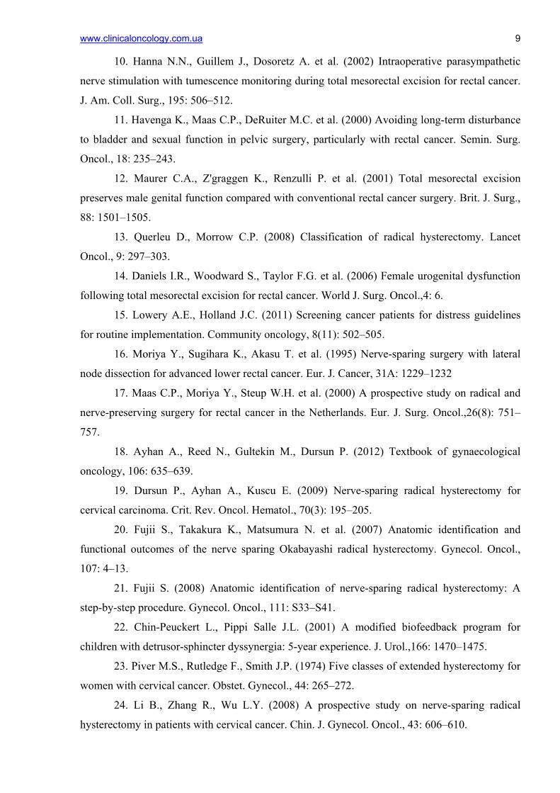
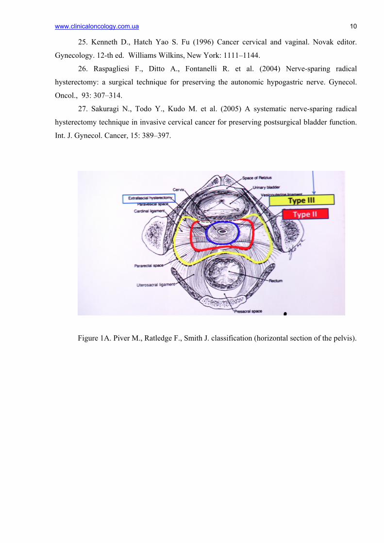
common and frequently used approach worldwide. According to Piver, Rutledge and Smith,
there are five classes of extended hysterectomy used in treating women with invasive CC (Piver
M., Ratledge F., Smith J., 1974) [2, 8, 23]. Class I - extrafascial hysterectomy. Class II –
www.clinicaloncology.com.ua 2 modified radical hysterectomy with removal of medial half of the cardinal and utero-sacral
ligaments, class III - classical Meigs’ radical hysterectomy [2, 8] with resection of utero-sacral,
cardinal ligaments and upper half of the vagina and pelvic lymphadenectomy. This approach has
become the standard technique for invasive cervical cancer in USA (Kenneth D.,
Hatch, Yao S. Fu, 1996) [8, 25]. Class IV – complete dissection of the ureter from the pubo-
vesical ligament, the superior vesicle artery is sacrificed and upper three-quarters of the vagina
removed. Class V include excision of distal ureter or bladder and reimplantation of ureter into
the bladder (Fig. 1A,B).
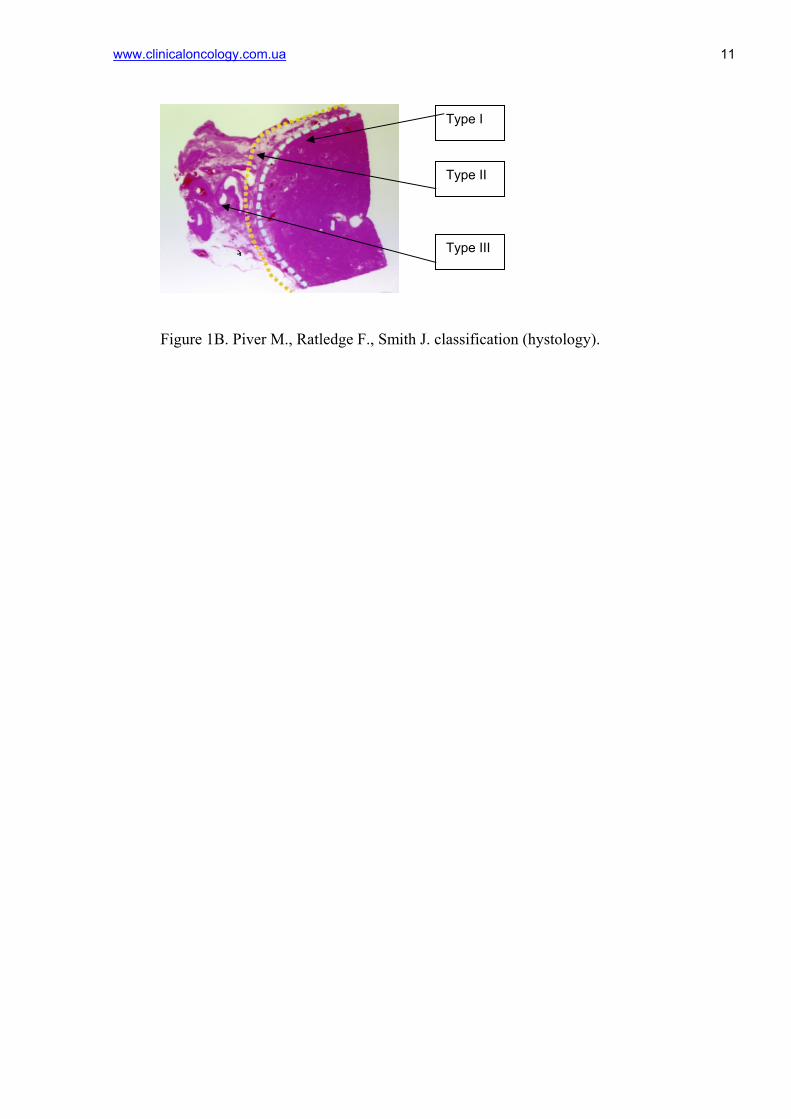
An updated and a new, simple and anatomy based classification of hysterectomy for
invasive CC was published by Querleu and Morrow in 2008 (Fig. 2) [13]:
Type A. Extrafascial hysterectomy:
Lateral parametrium removed to the ureter;
Ureter not tunneled;
Anterior and posterior parametrium not removed;
Vessels removed maximally close to the uterus;
Vaginal resection is minimal without removal of the paracolpos.
Тype В. :
Ureter tunneled;
Partial resection of utero-sacral and vesico-uterine ligaments;
Resection of para-cervical ligament at the level of ureteral tunnel.
Тype С. :
Transection of lateral parametrium to the iliac vessels;
Transection of vesico-uterine ligament at the bladder;
Transection of utero-sacral ligament at the rectum;
Ureter completely mobilized.
Тype С1 (with autonomic nerve sparing/preservation):
Lateral parametrium – preservation of splanchnic nerves;
Ventral parametrium – preservation of vesical branch of pelvis plexus;
Dorsal parametrium – preservation of hypogastric nerve (Fig. 3).
In Fig.3 the levels of dissection of cardinal ligament in different types of hysterectomies are
shown.
Тype С2 (without autonomic nerve sparing/preservation):
Lateral parametrium - intersection of the splanchnic nerve;
Ventral parametrium – intersection of vesical branch of pelvic plexus;
www.clinicaloncology.com.ua 3
Dorsal parametrium – all branches of hypogastric nerve are dissected.
In Fig.4 removed uterus after extended hysterectomy type C2 is shown.
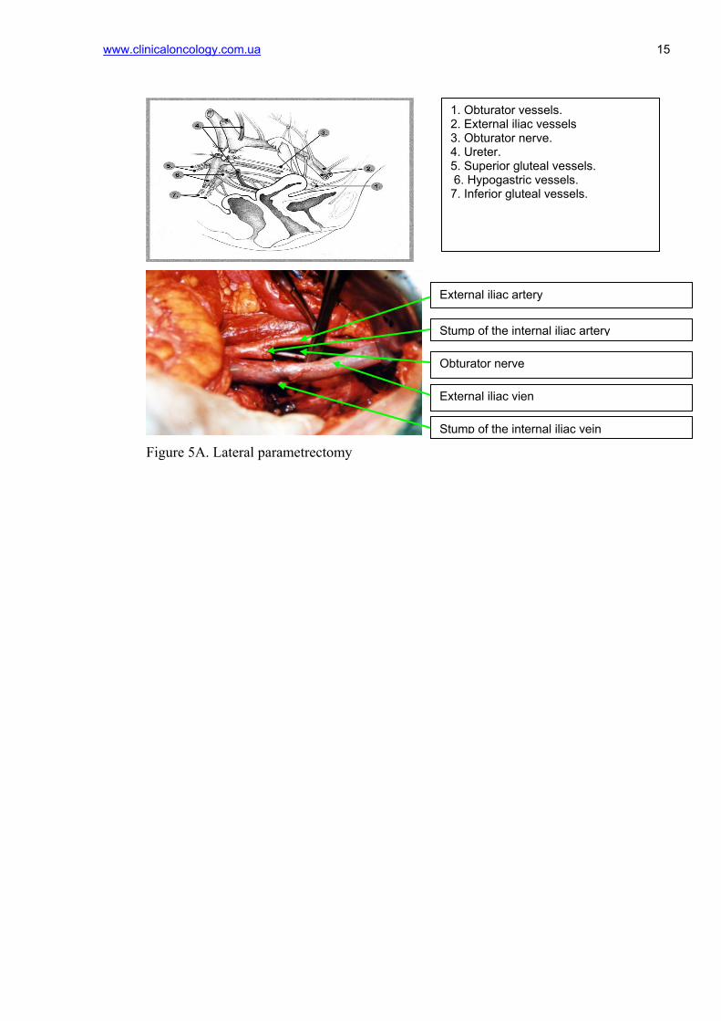
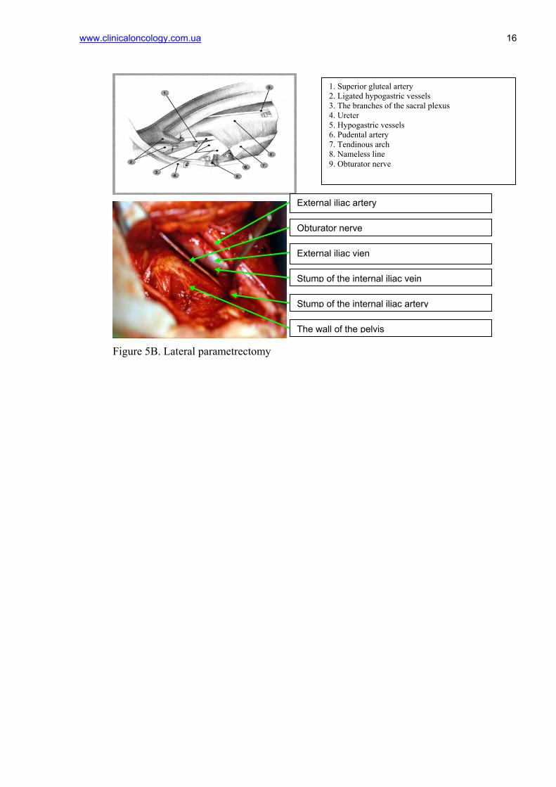
Тype D. Lateral parametrectomy:
The line of resection runs between internal obdurator internus muscle and
lumbosacral plexus;
Resection of internal iliac arteries and veins (Fig. 5А, B).
Surgical operations for CC are often accompanied by disorders of the normal functioning
of the urinary and reproductive systems’ organs. Frequency of urination disorders after surgery
of the cervix is 78%, the frequency of sexual disorders - 90%. The main cause of these disorders
is the surgical trauma of the pelvic autonomic nervous system innervating the urinary and
reproductive systems' organs and are located in the immediate proximity from the area of
surgical intervention (Levickis J., 1995; Hanna N.N., 2002) [3,9,10]. There are five main
elements of the pelvic autonomic nervous system: the unpaired upper hypogastric plexus, the
paired hypogastric nerves and the lower hypogastric plexus. The main symptoms of dysfunction
of the urinary system are urinary retention or enuresis (Havenga K., 2000) [3, 11]. Dysfunctions
of the woman’s reproductive system are manifested by vaginal secretions and rhythmic
contractions of vagina (Maurer C.A., 2001; Daniels I.R., 2006) [3, 12, 14].
Emerging advances in surgical techniques and other treatment modalities have led to
development of the nerve-sparing operations - complex of surgical techniques preserving nerve
structures of the pelvis (Moriya Y., 1995; Maas C.P., 2000) [5,6,16,17]. Numerous studies have
confirmed that preservation of the pelvic autonomic nerves allows significantly reduce the
number of postoperative urinary disorders. The high frequency of urinary functional disorders
and urological complications after radical hysterectomy gave impetus to the development of
nerve-sparing dissection technique. Functional preservation of pelvic innervation (primarily
vascular) is possible in oncological patients by transvaginal hysterectomy with microsurgical
dissection using laparoscopic technique. It is important to note that most of the researches
regarding nerve-sparing operations in surgery of the cervix conducted by foreign authors [24, 26,
27]. In Ukraine, nerve-sparing technique has been recently developed in specialized medical
centers. Despite advances in nerve-sparing techniques there is no adequate information regarding
the indications depending on the stage of the tumor process, tumor localization and type of
surgery.
Materials and methods
In order to study the effectiveness of nerve-sparing hysterectomy in the treatment of patients
with infiltrative CC in gynecological oncology department of the National Cancer Institute the
www.clinicaloncology.com.ua 4 indications, techniques, operation data, postoperative complications were evaluated.
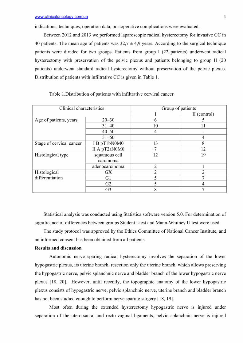
Between 2012 and 2013 we performed laparoscopic radical hysterectomy for invasive CC in
40 patients. The mean age of patients was 32,7 ± 4,9 years. According to the surgical technique
patients were divided for two groups. Patients from group I (22 patients) underwent radical
hysterectomy with preservation of the pelvic plexus and patients belonging to group II (20
patients) underwent standard radical hysterectomy without preservation of the pelvic plexus.
Distribution of patients with infiltrative CC is given in Table 1.
Таble 1.Distribution of patients with infiltrative cervical cancer
Group of patients Clinical characteristics
I II (control) 20–30 6 5 31–40 10 11 40–50 4 -
Age of patients, years
51–60 4 I B pT1bN0M0 13 8 Stage of cervical cancer II A pT2aN0M0 7 12 squamous cell
carcinoma 12 19 Histological type
adenocarcinoma 2 1 GX 2 2 G1 5 7 G2 5 4
Histological differentiation
G3 8 7
Statistical analysis was conducted using Statistica software version 5.0. For determination of
significance of differences between groups Student t-test and Mann-Whitney U test were used.
The study protocol was approved by the Ethics Committee of National Cancer Institute, and
an informed consent has been obtained from all patients.
Results and discussion
Autonomic nerve sparing radical hysterectomy involves the separation of the lower
hypogastric plexus, its uterine branch, resection only the uterine branch, which allows preserving
the hypogastric nerve, pelvic splanchnic nerve and bladder branch of the lower hypogastric nerve
plexus [18, 20]. However, until recently, the topographic anatomy of the lower hypogastric
plexus consists of hypogastric nerve, pelvic splanchnic nerve, uterine branch and bladder branch
has not been studied enough to perform nerve sparing surgery [18, 19].
Most often during the extended hysterectomy hypogastric nerve is injured under
separation of the utero-sacral and recto-vaginal ligaments, pelvic splanchnic nerve is injured
www.clinicaloncology.com.ua 5 under separation of the deep uterine vein of the cardinal ligament, and the bladder branch of the
upper hypogastric plexus is injured under bandaging and separation of paracolpium [18,20]. Less
radical surgery in which the incidence of such traumas is less compared to standard
hysterectomy was developed by Japanese oncogynecologists in 1950 [18, 19]. Okabayashi
proposed a method for extended hysterectomy, including the separation of the posterior leaf of
the vesico-uterine ligament, which makes possible to separate only the uterine branch of lower
hypogastric plexus, preserve innervation of the bladder, and avoid dysfunction of its contractile
function in the postoperative period [18, 20]. Without stratification of the posterior leaf of
vesico-uterine ligament it is difficult to visualize the lower hypogastric plexus, especially during
standard Wertheim’s operation. There are many published data regarding nerve sparing surgery
using various surgical techniques. However, randomized trials comparing radical hysterectomy
of C1 and C2 types were not carried out, which makes difficult to analyze published data on the
dysfunction of the urinary and reproductive systems after surgical treatment [18, 20].
The uterus is innervated by part of the plexus hypogastricus inferior, which forms a so-
called plexus of the Rhine or plexus uterovaginalis, s. uterinus magnus (the front part of the
bottom of the plexus hypogastricus inferior).
Rhine plexus forms branches: 1) from the anterior roots of II, III and IV sacral nerves
from which branch out 4-6 chains nn. erigentes (nn. erigentes referred to nn. pelvici). These
chains connected with branches of the sacral ganglions of the sympathetic border trunk; 2) from
the sacral and coccygeal part of the sympathetic border trunk; 3) through plexus hypogastric
inferior, from the lumbar sympathetic trunks, as well as X, XI, XII thoracic nerves, 4) from the
plexus haemorrhoidales inferior, extending from the plexus mesogasrica inferior, by which
functional relationship is established between genital apparatus and the rectum (Fig. 6).
Pelvic plexus contains mainly sympathetic chain, and chain nn. pelvici and therefore is a
mixed plexus. Thus, the uterus, vagina, bladder and rectum are innervated by the motor and
sensory nerves of sympathetic and parasympathetic autonomic nervous system. Sympathetic
chain extending froms Th11-L2 forms the upper hypogastric plexus. Parasympathetic chain
extending from S2-S4 to the walls of the pelvis forms the pelvic splanchnic nerve. These chains
merge and form inferior hypogastric plexus, the branches of which innervate the uterus and
bladder.
Topographic anatomy of the inferior hypogastric plexus, including the hypogastric nerve,
pelvic splanchnic nerve, bladder branch, uterine branch of this plexus, is quite complex and it is
not easy to be visualized during the radical hysterectomy. In order to preserve the pelvic
splanchnic nerve, it is necessary to visualize the inferior hypogastric plexus, this requires a
thorough knowledge of the anatomy of the pelvis and the parametrial area (topography of the
www.clinicaloncology.com.ua 6 deep veins of the uterus) [18, 20, 21]. Moreover it is also very important to have knowledge of
the anatomy of the vesico-uterine ligament, especially its posterior leaf.
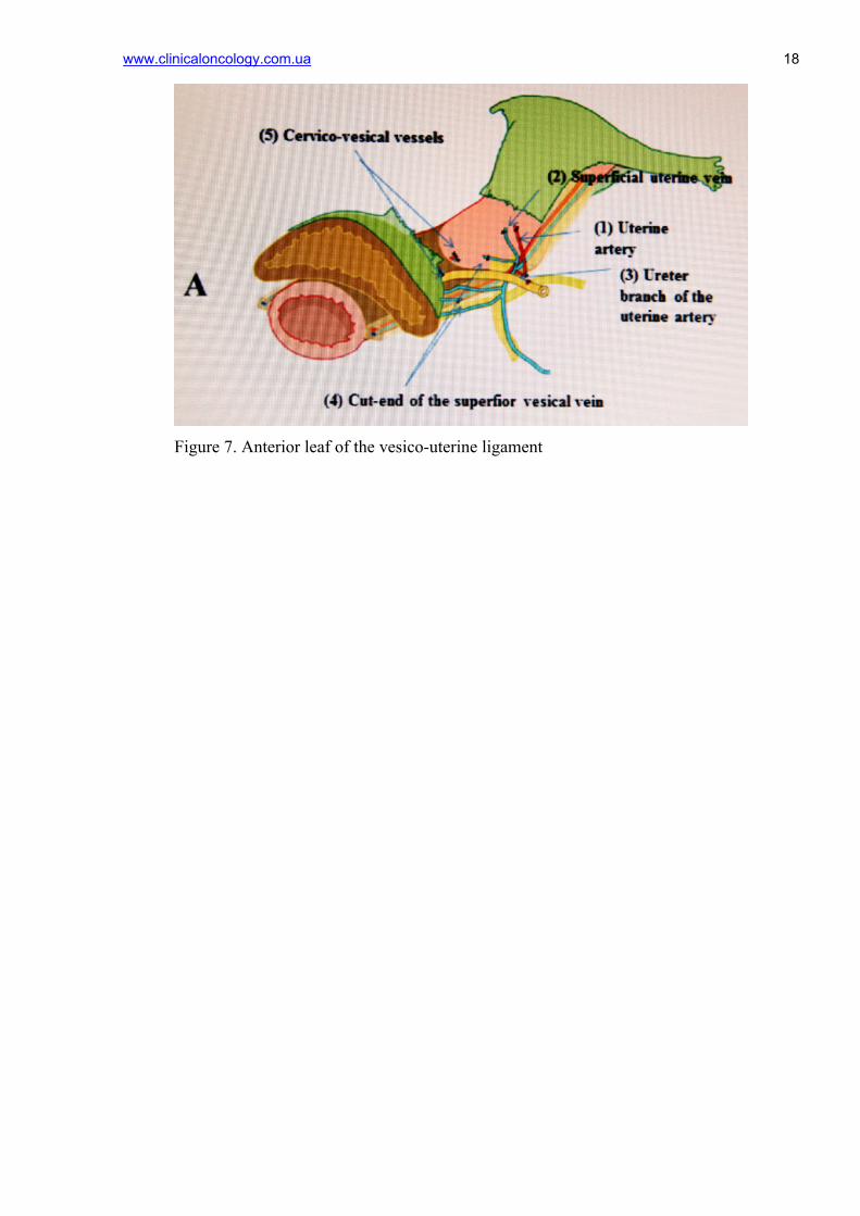
The anatomy of the vesico-uterine ligament (anterior/posterior leaf) is given on Fig. 7,8.
In the anterior leaf of vesico-uterine ligament are visuzlized: uterine artery, superficial
veins of the uterus, ureter branch of the uterine artery, superficial bladder veins flowing into
superficial uterine veins, cervico-vesical vessels. Division of blood vessels from the connective
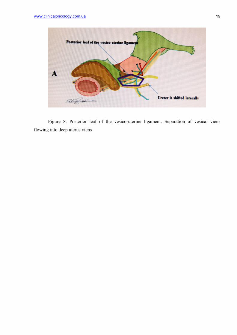
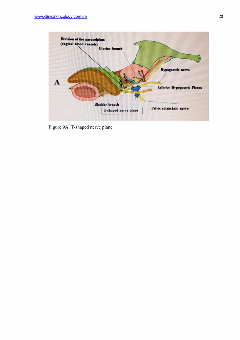
tissue of anterior leaf of the vesico-uterine ligament is presented. In the posterior leaf of vesico-
uterine ligaments two major bladder veins flowing into the deep veins of the uterus are usually
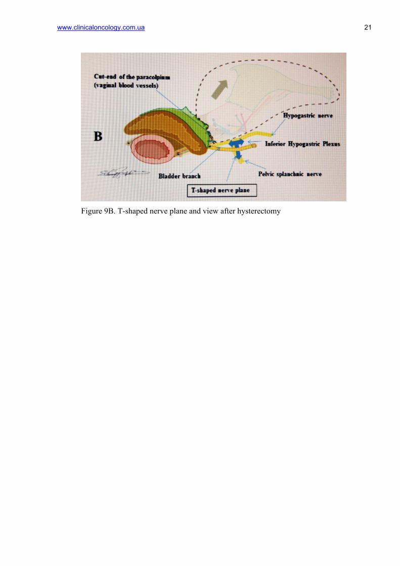
localized (Fig. 8). Dissection of these veins gives access to the inferior hypogastric plexus, which
allows separating only uterine branch. After isolation of paracolpium T-shaped hypogastric
nerve plane is visualized and only the uterine branch is cut-ended, vaginal cuff is released, the
uterus is removed (Fig. 9А, B).
The schematic drawings present extended hysterectomy (C1 nerve-sparing dissection) in
patients with invasive CC, from Textbook of gynaecological oncology, 2012 (Ayhan A., Reed
N., Gultekin M., Dursun P.).
Stages of nerve-sparing hysterectomy, which was held in gynecological oncology
department of the National Cancer Institute, are presented below:
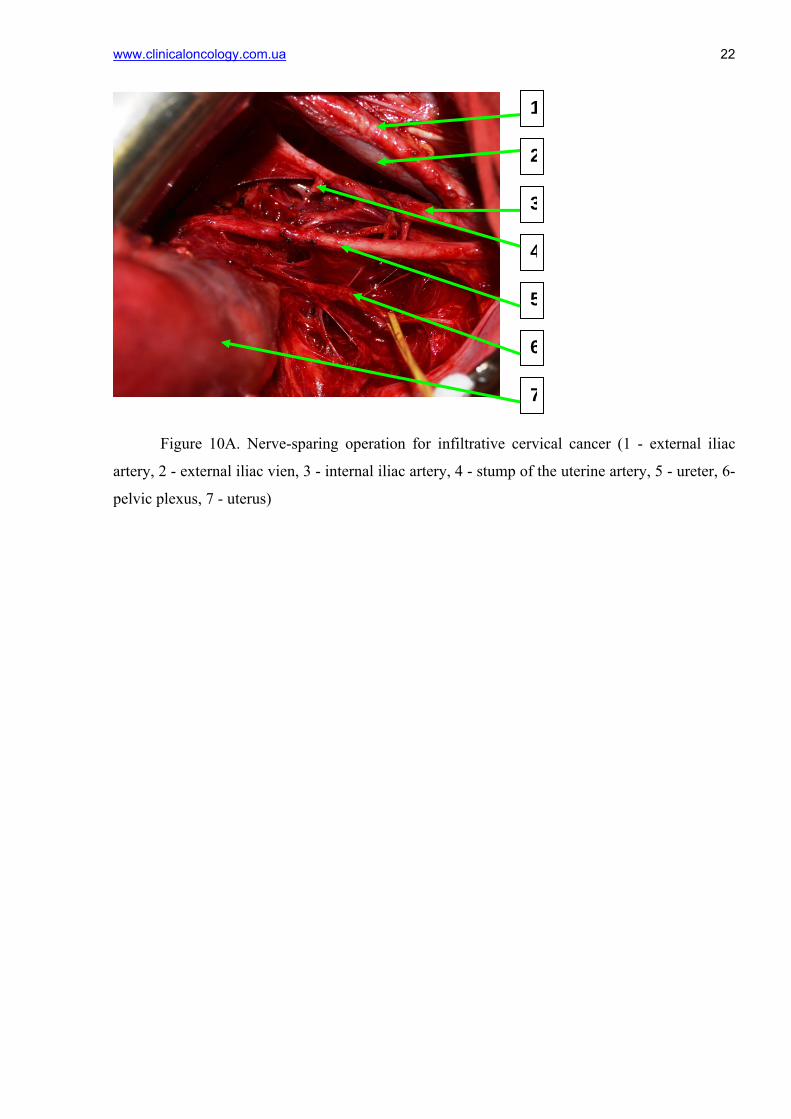
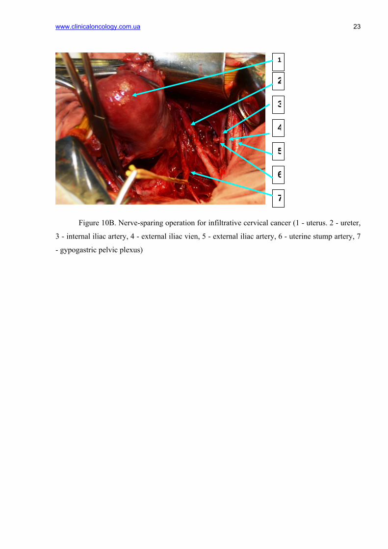
Division of uterine branches from the inferior hypogastric plexus. Below hypogastric
nerve, branch of the inferior hypogastric plexus is separated from the blood vessels of the
paracolpium (Figure 10 A, B). Division is between bladder branch and blood vessels of the
paracolpium in the connective tissue between the pelvic nerve plane and the cervix/upper part of
the vagina at the hypogastric nerve on the ventral side and deep uterine veins on the dorsal side.
From the side of the uterus, uterine branch of the inferior hypogastric plexus clamped, cut off
after a sensation resembling a gap stretched string. Dissection of the recto-vaginal ligament
separates the blood vessels of paracolpium from the T-shaped nerve plane. After division of the
recto-vaginal ligament of the upper part of the vagina, bladder branch of the inferior hypogastric
plexus, forming a T-shaped nerve plane, dissection and ligating of blood vessels of paracolpium
distally along the vagina below the level of cervix leision is performed (Fig. 11,12).
Formation of protective wall for juxtavesical portion of the ureter by the walls of the
rectum and bladder.
The inflection of the ureter is one of the most serious post-operative problems leading to
hydroureter and hydronephrosis with possible dysfunction of the kidneys. To prevent the
immersion of juxtavesical part of ureter into the pelvis and its bend two seams are applied
laterally from the ureter between the outer wall of the rectum and the bladder.
www.clinicaloncology.com.ua 7
The function of the lower urinary tract after radical hysterectomy with preservation of the
pelvic plexus in patients with infiltrative CC has been studied using cystomanometry on
urodynamic rack URO-PRO by the standard method.
In the early postoperative period, in all patients contractile function of the bladder has
been assessed.
Cystomanometry – registration of changes in intravesical pressure in the process of its
filling and urination. Cystomanometry reflects: 1) censor characteristics of bladder; 2) adaptive
properties of the detrusor (its ability to maintain a low intravesical pressure during filling and
lack of unbraked contractions; 3) dynamic of the intravesical pressure during urination [7, 15,
22].
Extensibility of the bladder was considered as a change in detrusor pressure at a certain
change in the filling volume and was calculated according to the formula: К=∆V/∆Р, where К –
extensibility of the bladder wall (in ml / cm wat.column), ∆V — volume change, ∆Р – detrusor
pressure change at the time of change the volume. Under preservation of the pelvic nerve plexus
extensibility of the bladder wall was more than 10 mL / cm of water column with up to 100 mL
volume and more than 25 mL / cm of water column with up to 500 ml volume (Fig. 13,14).
After nerve-sparing hysterectomy in 80% of patients of group I contractile function of the
bladder recovered completely in 2-3 days after surgery, while in the control group - only in 20%
of patients. In 7 days after the radical hysterectomy in all patients of group I normal function of
the lower urinary tract was completely restored while in patients of group II recovery period
lasted from 7 to 21 days (Table 2).
Conducted study of extended hysterectomy (C1 nerve-sparing dissection) for infiltrative
CC shows promising results in reducing the number of early postoperative complications in the
urinary system. However to assess the long-term results of treatment, the frequency of
complications and quality of life of patients further set of clinical data, randomized trials in
specialized centers are required.
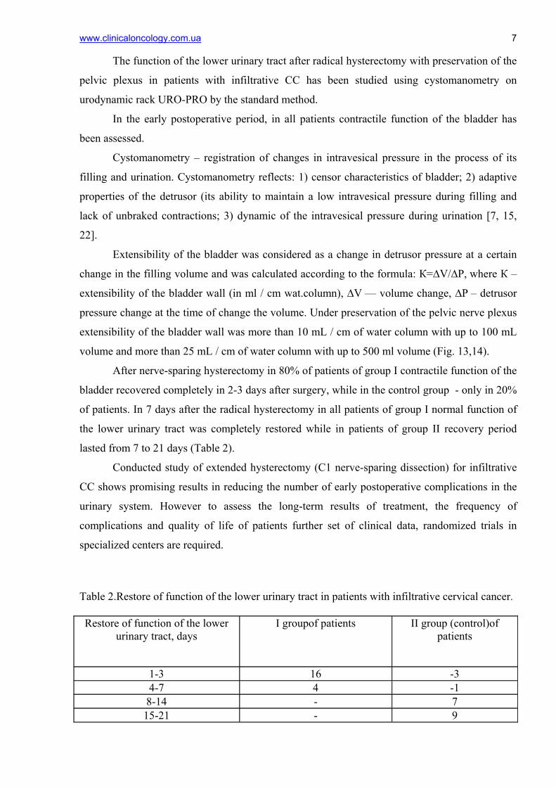
Table 2.Restore of function of the lower urinary tract in patients with infiltrative cervical cancer.
Restore of function of the lower
urinary tract, days I groupof patients II group (control)of
patients
1-3 16 -3 4-7 4 -1 8-14 - 7 15-21 - 9
www.clinicaloncology.com.ua 8
Conclusion
1. Radical nerve-sparing hysterectomy in patients with infiltrative cervical cancer allows
preserving the function of the lower urinary tract in the postoperative period: the
indicators of cystomanometry in these patients 60% higher than in patients of the control
group.
2. Radical nerve-sparing hysterectomy should not be performed in patients with the IIB
FIGO stage of the cervical cancer, since the location of the lower hypogastric plexus is
very close to the site of the tumor lesion.
References
1. Cancer in Ukraine, 2010–2011. Morbidity, mortality, indicators of cancer care service
(2012) Bulletin of National Cancer Registry of Ukraine, 13: 124 (in Ukrainian).
2. Werner P., Zederl Y. (1960) Wertheim’s radical surgery for cancer of cervix.
Medicine, Moskow: 74p. (in Russian).
3. Belyaev A.M., Manikhas G.M., Domanski A.A., Bratov O.Z. (2010) The ability to
perform nerve-sparing surgery in different categories of patients with rectal cancer. Biomedical
Journal. Medline.ru, 11: 597-610 (in Russian).
4. Vorobjova L.І. (2010) Current approaches to diagnostic, treatment and prevention of
cervical cancer. Female doctor 5: 18–21 (in Ukrainian).
6. Belyaev A.M., Domanski A.A., Zakharenko А.А. et all. (2011) Results of nerve-
sparing operation in surgery of complicated rectal cancer. Biomedical Journal. Medline.ru, 12:
495-510 (in Russian).
7. Yatsyna O.I., Stakhovsky E.O., Vitruk Y.V., Chernienko Y.L. Patent № 58393,
Ukraine, MPK6 A61V5/00. Method for determining the storage function of orthotopic ileal
bladder. Number 201011467, appl. 27.09.2010, publ. 11.04.11, Bull. Number 7: P. 27 (in
Ukrainian).
8. Kuznetsov V.V., Lebedev A.I., Morkhov K.Y., Gritsay A.N. (2002) Surgery of
invasive cancer of the cervix. Practical Oncology, 3 (3): 178-182 (in Russian).
9. Levickis J. et al. (1995) Bladder and erectile disfunction before and after rectal surgery
for cancer. Br. J. Urol., 76: 752–756.
www.clinicaloncology.com.ua 9
10. Hanna N.N., Guillem J., Dosoretz A. et al. (2002) Intraoperative parasympathetic
nerve stimulation with tumescence monitoring during total mesorectal excision for rectal cancer.
J. Am. Coll. Surg., 195: 506–512.
11. Havenga K., Maas C.P., DeRuiter M.C. et al. (2000) Avoiding long-term disturbance
to bladder and sexual function in pelvic surgery, particularly with rectal cancer. Semin. Surg.
Oncol., 18: 235–243.
12. Maurer C.A., Z'graggen K., Renzulli P. et al. (2001) Total mesorectal excision
preserves male genital function compared with conventional rectal cancer surgery. Brit. J. Surg.,
88: 1501–1505.
13. Querleu D., Morrow C.P. (2008) Classification of radical hysterectomy. Lancet
Oncol., 9: 297–303.
14. Daniels I.R., Woodward S., Taylor F.G. et al. (2006) Female urogenital dysfunction
following total mesorectal excision for rectal cancer. World J. Surg. Oncol.,4: 6.
15. Lowery A.E., Holland J.C. (2011) Screening cancer patients for distress guidelines
for routine implementation. Community oncology, 8(11): 502–505.
16. Moriya Y., Sugihara K., Akasu T. et al. (1995) Nerve-sparing surgery with lateral
node dissection for advanced lower rectal сancer. Eur. J. Cancer, 31A: 1229–1232
17. Maas C.P., Moriya Y., Steup W.H. et al. (2000) A prospective study on radical and
nerve-preserving surgery for rectal cancer in the Netherlands. Eur. J. Surg. Oncol.,26(8): 751–
757.
18. Ayhan A., Reed N., Gultekin M., Dursun P. (2012) Textbook of gynaecological
oncology, 106: 635–639.
19. Dursun P., Ayhan A., Kuscu E. (2009) Nerve-sparing radical hysterectomy for
cervical carcinoma. Crit. Rev. Oncol. Hematol., 70(3): 195–205.
20. Fujii S., Takakura K., Matsumura N. et al. (2007) Anatomic identification and
functional outcomes of the nerve sparing Okabayashi radical hysterectomy. Gynecol. Oncol.,
107: 4–13.
21. Fujii S. (2008) Anatomic identification of nerve-sparing radical hysterectomy: A
step-by-step procedure. Gynecol. Oncol., 111: S33–S41.
22. Chin-Peuckert L., Pippi Salle J.L. (2001) A modified biofeedback program for
children with detrusor-sphincter dyssynergia: 5-year experience. J. Urol.,166: 1470–1475.
23. Piver M.S., Rutledge F., Smith J.P. (1974) Five classes of extended hysterectomy for
women with cervical cancer. Obstet. Gynecol., 44: 265–272.
24. Li B., Zhang R., Wu L.Y. (2008) A prospective study on nerve-sparing radical
hysterectomy in patients with cervical cancer. Chin. J. Gynecol. Oncol., 43: 606–610.
www.clinicaloncology.com.ua 10
25. Kenneth D., Hatch Yao S. Fu (1996) Cancer cervical and vaginal. Novak editor.
Gynecology. 12-th ed. Williams Wilkins, New York: 1111–1144.
26. Raspagliesi F., Ditto A., Fontanelli R. et al. (2004) Nerve-sparing radical
hysterectomy: a surgical technique for preserving the autonomic hypogastric nerve. Gynecol.
Oncol., 93: 307–314.
27. Sakuragi N., Todo Y., Kudo M. et al. (2005) A systematic nerve-sparing radical
hysterectomy technique in invasive cervical cancer for preserving postsurgical bladder function.
Int. J. Gynecol. Cancer, 15: 389–397.
Figure 1А. Piver M., Ratledge F., Smith J. classification (horizontal section of the pelvis).
www.clinicaloncology.com.ua 11
Тype І
Тype ІІ
Тype ІІІ
Figure 1B. Piver M., Ratledge F., Smith J. classification (hystology).
www.clinicaloncology.com.ua
12Vagina
paracolpium
Ischium
Rectum
Рис 2. D. Querleu, C.P. Morrow classification (horizontal section of the pelvis).

www.clinicaloncology.com.ua 13
Figure 3. Dissection levels of cardinal ligament in different types of hysterectomy.

www.clinicaloncology.com.ua 14
Corporis uteri
Figure 4. Removed uterus after C2 extended hysterectomy.
Параметральная клетчатка
Vaginal cuff
www.clinicaloncology.com.ua
15
1. Obturator vessels. 2. External iliac vessels 3. Obturator nerve. 4. Ureter. 5. Superior gluteal vessels. 6. Hypogastric vessels. 7. Inferior gluteal vessels.
External iliac artery
Figure 5А. Lateral parametrectomy
Stump of the internal iliac artery
Stump of the internal iliac vein
Obturator nerve
External iliac vien
www.clinicaloncology.com.ua 16
1. Superior gluteal artery 2. Ligated hypogastric vessels 3. The branches of the sacral plexus 4. Ureter 5. Hypogastric vessels 6. Pudental artery 7. Tendinous arch 8. Nameless line 9. Obturator nerve
External iliac artery
Figure 5B. Lateral parametrectomy
Stump of the internal iliac artery
The wall of the pelvis
Obturator nerve
External iliac vien
Stump of the internal iliac vein

www.clinicaloncology.com.ua 17
Figure 6. Schematic representation of the anatomy of the lower hypogastric plexus
www.clinicaloncology.com.ua 18
Figure 7. Anterior leaf of the vesico-uterine ligament
www.clinicaloncology.com.ua 19
Figure 8. Posterior leaf of the vesico-uterine ligament. Separation of vesical viens
flowing into deep uterus viens
www.clinicaloncology.com.ua 20
Figure 9А. Т-shaped nerve plane
www.clinicaloncology.com.ua 21
Figure 9B. Т-shaped nerve plane and view after hysterectomy
www.clinicaloncology.com.ua
22
1
2
Figure 10А. Nerve-sparing operation for infiltrative cervical cancer (1 - external iliac
artery, 2 - external iliac vien, 3 - internal iliac artery, 4 - stump of the uterine artery, 5 - ureter, 6-
pelvic plexus, 7 - uterus)
7
6
5
4
3
www.clinicaloncology.com.ua
23
1
Figure 10B. Nerve-sparing operation for infiltrative cervical cancer (1 - uterus. 2 - ureter,
3 - internal iliac artery, 4 - external iliac vien, 5 - external iliac artery, 6 - uterine stump artery, 7
- gypogastric pelvic plexus)
7
6
5
4
2
3
www.clinicaloncology.com.ua
24
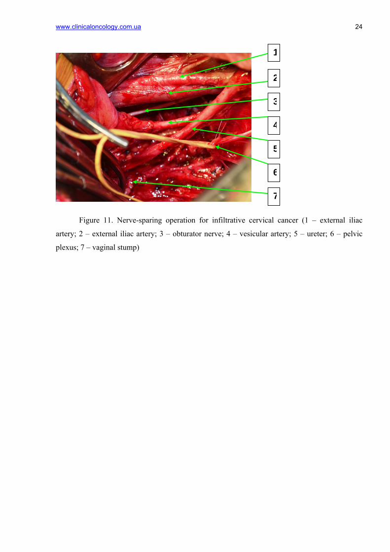
1
Figure 11. Nerve-sparing operation for infiltrative cervical cancer (1 – external iliac
artery; 2 – external iliac artery; 3 – obturator nerve; 4 – vesicular artery; 5 – ureter; 6 – pelvic
plexus; 7 – vaginal stump)
2
3
4
5
6
7
www.clinicaloncology.com.ua
25
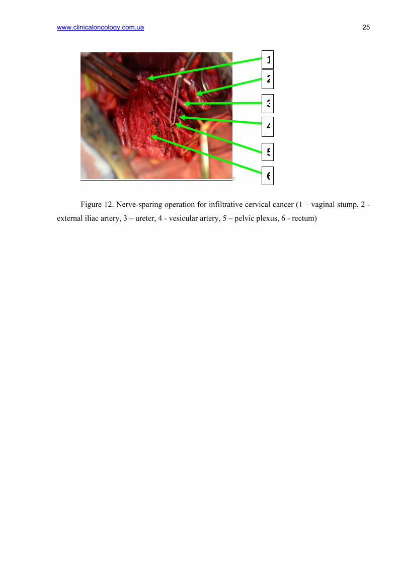
1
Figure 12. Nerve-sparing operation for infiltrative cervical cancer (1 – vaginal stump, 2 -
external iliac artery, 3 – ureter, 4 - vesicular artery, 5 – pelvic plexus, 6 - rectum)
3
4
5
2
6
1
www.clinicaloncology.com.ua 26
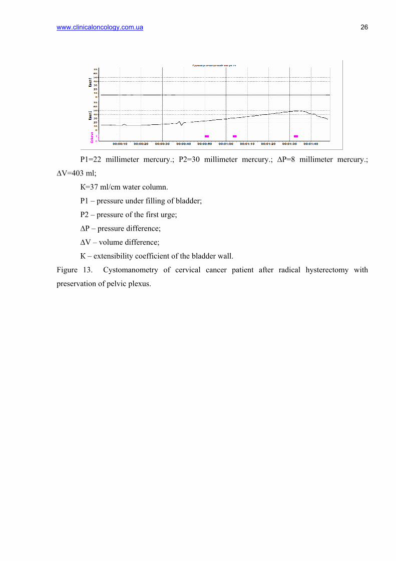
Р1=22 millimeter mercury.; Р2=30 millimeter mercury.; ∆Р=8 millimeter mercury.;
∆V=403 ml;
К=37 ml/cm water column.
Р1 – pressure under filling of bladder;
Р2 – pressure of the first urge;
∆Р – pressure difference;
∆V – volume difference;
К – extensibility coefficient of the bladder wall.
Figure 13. Cystomanometry of cervical cancer patient after radical hysterectomy with
preservation of pelvic plexus.
www.clinicaloncology.com.ua
27
.
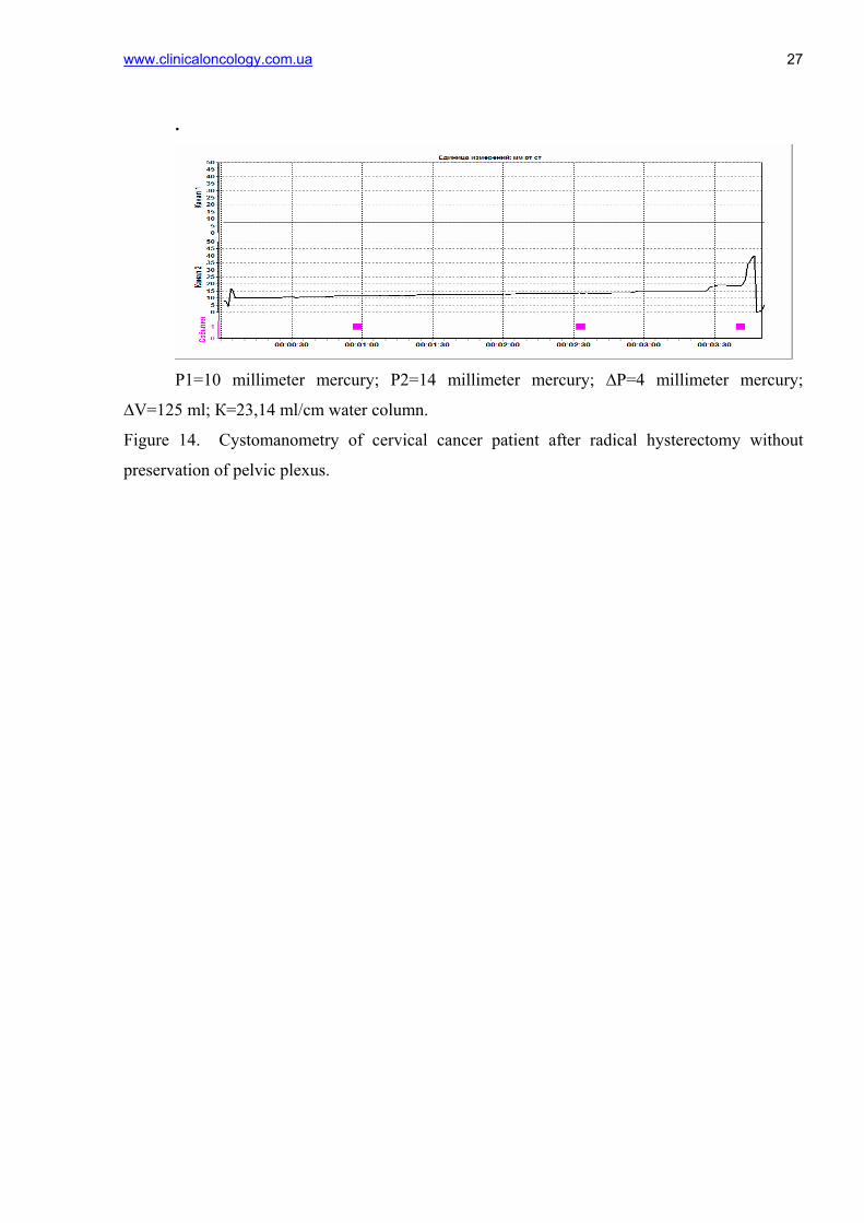
Р1=10 millimeter mercury; Р2=14 millimeter mercury; ∆Р=4 millimeter mercury;
∆V=125 ml; К=23,14 ml/cm water column.
Figure 14. Cystomanometry of cervical cancer patient after radical hysterectomy without
preservation of pelvic plexus.