EXPRESSION PATTERNS OF FLOWERING GENES DURING FLOWER ...
125
EXPRESSION PATTERNS OF FLOWERING GENES DURING FLOWER INDUCTION AND DETERMINATION IN SWEET ORANGE (Citrus sinensis L. OSBECK) By EDUARDO J. CHICA A DISSERTATION PRESENTED TO THE GRADUATE SCHOOL OF THE UNIVERSITY OF FLORIDA IN PARTIAL FULFILLMENT OF THE REQUIREMENTS FOR THE DEGREE OF DOCTOR OF PHILOSOPHY UNIVERSITY OF FLORIDA 2011
Transcript of EXPRESSION PATTERNS OF FLOWERING GENES DURING FLOWER ...

EXPRESSION PATTERNS OF FLOWERING GENES DURING FLOWER INDUCTIONAND DETERMINATION IN SWEET ORANGE (Citrus sinensis L. OSBECK)
By
EDUARDO J. CHICA
A DISSERTATION PRESENTED TO THE GRADUATE SCHOOLOF THE UNIVERSITY OF FLORIDA IN PARTIAL FULFILLMENT
OF THE REQUIREMENTS FOR THE DEGREE OFDOCTOR OF PHILOSOPHY
UNIVERSITY OF FLORIDA
2011

© 2011 Eduardo J. Chica
2

ACKNOWLEDGMENTS
I thank my advisors, Dr. Gene Albrigo and Dr. Christine Chase, for their help,
advice, support and all the things I learned from them during my graduate studies. I
consider myself fortunate for having had the opportunity to work under their guidance.
To Dr. Albrigo, thanks also for having taken the challenge of having me twice as his
student and for his friendship throughout these years. I also thank Drs. Kevin Folta,
James Syvertsen and Nian Wang, members of my supervisory committee, from whom
I learned many things and whose works inspired me to pursue ever-higher goals in
science.
I am very grateful also to Dr. Jacqueline Burns for having allowed me to conduct the
majority of my analyses in her lab. To Dr. Burns and the members of her lab, thanks also
for receiving me as another member of their team and for being a second lab-home for
me. I am very grateful too to Dr. Karen Koch for sparking in me new interests in science,
for her dedication to her students, and for key exchanges in developing ideas that served
as seed for this study.
I thank also my friends who became family during my years in graduate school,
they are too many to name but they know who they are. From each of these friends and
colleagues I have learned much, both scientifically and personally. I thank also my and
Lis’s family in Ecuador for their continuous support during these years abroad. Finally
and most especially, I thank Lis for giving sense to everything and for always signing Lis
:-)
3

TABLE OF CONTENTS
page
ACKNOWLEDGMENTS . . . . . . . . . . . . . . . . . . . . . . . . . . . . . . . . . . 3
LIST OF TABLES . . . . . . . . . . . . . . . . . . . . . . . . . . . . . . . . . . . . . . 6
LIST OF FIGURES . . . . . . . . . . . . . . . . . . . . . . . . . . . . . . . . . . . . . 7
ABSTRACT . . . . . . . . . . . . . . . . . . . . . . . . . . . . . . . . . . . . . . . . . 9
CHAPTER
1 INTRODUCTION AND LITERATURE REVIEW . . . . . . . . . . . . . . . . . . 11
1.1 Shoot Meristem Developmental Programs . . . . . . . . . . . . . . . . . . 111.1.1 Vegetative Growth: Maintaining Vegetative Indeterminacy . . . . . 121.1.2 Phase Change: Re-programing the Meristem to Produce Flowers . 151.1.3 Inflorescences: a Hybrid Program . . . . . . . . . . . . . . . . . . . 17
1.2 Floral Induction: a General Overview . . . . . . . . . . . . . . . . . . . . . 191.2.1 Acquisition of Competence . . . . . . . . . . . . . . . . . . . . . . 191.2.2 Floral Induction . . . . . . . . . . . . . . . . . . . . . . . . . . . . . 21
1.3 Floral Induction in citrus . . . . . . . . . . . . . . . . . . . . . . . . . . . . 241.3.1 Factors Regulating Floral Induction in Citrus . . . . . . . . . . . . . 251.3.2 Citrus Orthologs of Arabidopsis Flowering Genes . . . . . . . . . . 26
1.4 Hypothetical Model for the Transcriptional Regulation of Floral Inductionin citrus . . . . . . . . . . . . . . . . . . . . . . . . . . . . . . . . . . . . . 27
2 EXPRESSION PATTERNS OF FLOWERING GENES IN SWEET ORANGEIN RESPONSE TO FLORAL-INDUCTIVE WATER DEFICITS . . . . . . . . . . 32
2.1 Materials and Methods . . . . . . . . . . . . . . . . . . . . . . . . . . . . . 342.1.1 Plant Material . . . . . . . . . . . . . . . . . . . . . . . . . . . . . . 342.1.2 Experimental Conditions . . . . . . . . . . . . . . . . . . . . . . . . 342.1.3 qRT-PCR . . . . . . . . . . . . . . . . . . . . . . . . . . . . . . . . 372.1.4 Data Analysis . . . . . . . . . . . . . . . . . . . . . . . . . . . . . . 38
2.2 Results and Discussion . . . . . . . . . . . . . . . . . . . . . . . . . . . . 382.2.1 Floral-inductive Water Deficit Up-regulates CsFT but not CsSL1. . 382.2.2 CsFT Transcript Accumulation also Increases in Trees under Water
Deficit at Floral-inductive Temperatures . . . . . . . . . . . . . . . . 402.2.3 Water Deficit Reduce the Transcript Accumulation of Floral Identity
Genes in Buds during Floral Induction . . . . . . . . . . . . . . . . 412.2.4 Other Factors Modify the Response of Flowering Genes to
Floral-inductive Treatments in Field Trees . . . . . . . . . . . . . . 43
3 RELATIONSHIP BETWEEN EXPRESSION PATTERNS OF FLOWERINGGENES, FLOWERING INTENSITY GRADIENTS AND FLOWERING COHORTSIN SWEET ORANGE SHOOTS . . . . . . . . . . . . . . . . . . . . . . . . . . . 57
4

3.1 Materials and Methods . . . . . . . . . . . . . . . . . . . . . . . . . . . . . 593.1.1 Plant Material . . . . . . . . . . . . . . . . . . . . . . . . . . . . . . 593.1.2 Experimental Conditions . . . . . . . . . . . . . . . . . . . . . . . . 593.1.3 qRT-PCR . . . . . . . . . . . . . . . . . . . . . . . . . . . . . . . . 623.1.4 Data Analysis . . . . . . . . . . . . . . . . . . . . . . . . . . . . . . 62
3.2 Results and Discussion . . . . . . . . . . . . . . . . . . . . . . . . . . . . 633.2.1 CsFT Transcripts Accumulate at Equal Levels in Leaves Regardless
of Their Position in the Shoot. . . . . . . . . . . . . . . . . . . . . . 633.2.2 Accumulation of CsAP1 and CsLFY transcripts is Higher at Nodes
Closer to the Apex. . . . . . . . . . . . . . . . . . . . . . . . . . . . 633.2.3 TIBA Disrupts the Establishment of Flowering Gradients. . . . . . . 643.2.4 Transcript Accumulation of Floral Identity Genes after Intermittent
Induction is Related to Initiation of Flowering Cohorts. . . . . . . . 66
4 OTHER FACTORS ALTERING THE EXPRESSION OF SWEET ORANGEFLOWERING GENES DURING FLORAL INDUCTION . . . . . . . . . . . . . . 75
4.1 Materials and Methods . . . . . . . . . . . . . . . . . . . . . . . . . . . . . 764.1.1 Plant Material . . . . . . . . . . . . . . . . . . . . . . . . . . . . . . 764.1.2 Experimental Conditions . . . . . . . . . . . . . . . . . . . . . . . . 764.1.3 qRT-PCR . . . . . . . . . . . . . . . . . . . . . . . . . . . . . . . . 794.1.4 Data Analysis . . . . . . . . . . . . . . . . . . . . . . . . . . . . . . 80
4.2 Results and Discussion . . . . . . . . . . . . . . . . . . . . . . . . . . . . 804.2.1 Gibberellins Down-regulate the Accumulation of Putative Flowering
Signals and Floral Identity Genes Transcripts . . . . . . . . . . . . 804.2.2 Fruit Proximity . . . . . . . . . . . . . . . . . . . . . . . . . . . . . . 824.2.3 Effect of Light/Dark Cycles on CsFT Transcript Accumulation . . . 834.2.4 Accumulation of CsFT Transcripts Changes Throughout the Day . 844.2.5 Early Changes in Transcript Levels of CsFT in Response to Floral
Inductive Temperatures . . . . . . . . . . . . . . . . . . . . . . . . 85
5 CONCLUSIONS . . . . . . . . . . . . . . . . . . . . . . . . . . . . . . . . . . . 98
APPENDIX: DESIGN, VALIDATION AND OPTIMIZATION OF qPCR ASSAYS . . . . 102
REFERENCES . . . . . . . . . . . . . . . . . . . . . . . . . . . . . . . . . . . . . . . 107
BIOGRAPHICAL SKETCH . . . . . . . . . . . . . . . . . . . . . . . . . . . . . . . . 125
5
LIST OF TABLES
Table page
1-1 Citrus and Poncirus trifoliata orthologs of Arabidopsis flowering genes . . . . . 30
2-1 Flowering characteristics of ‘Washington Navel’ citrus trees exposed to waterdeficit. . . . . . . . . . . . . . . . . . . . . . . . . . . . . . . . . . . . . . . . . . 49
2-2 Flowering characteristics of ‘Washington Navel’ citrus trees exposed to waterdeficit at floral inductive temperatures. . . . . . . . . . . . . . . . . . . . . . . . 51
2-3 Flowering characteristics of field grown ‘Valencia’ trees under water deficitduring Winter. . . . . . . . . . . . . . . . . . . . . . . . . . . . . . . . . . . . . . 56
A-1 Primer pairs used for transcript quantification assays by qPCR. . . . . . . . . . 104
6

LIST OF FIGURES
Figure page
1-1 Model for the transcriptional regulation of floral induction in citrus . . . . . . . . 31
2-1 Expression of CsFT and CsSL1 in ‘Navel’ trees under water deficit. . . . . . . 48
2-2 Expression of CsFT and CsSL1 in ‘Navel’ trees under water deficit at floralinductive temperatures. . . . . . . . . . . . . . . . . . . . . . . . . . . . . . . . 50
2-3 Expression of CsAP1 and CsLFY in ‘Navel’ trees under water deficit. . . . . . . 52
2-4 Expression of CsAP1 and CsLFY in ‘Navel’ trees under water deficit at floralinductive temperatures. . . . . . . . . . . . . . . . . . . . . . . . . . . . . . . . 53
2-5 Expression of flowering-related genes in field grown ‘Valencia’ trees underwater deficit during Winter. . . . . . . . . . . . . . . . . . . . . . . . . . . . . . 54
2-6 Expression of flowering-related genes in field grown ‘Valencia’ trees underwater deficit during Summer. . . . . . . . . . . . . . . . . . . . . . . . . . . . . 55
3-1 Inflorescence gradient, types of new growth and inflorescence cohorts in C.sinensis spring flush. . . . . . . . . . . . . . . . . . . . . . . . . . . . . . . . . . 68
3-2 Number of inflorescences formed by node position in the spring. . . . . . . . . 69
3-3 Expression of CsFT in leaves at different positions. . . . . . . . . . . . . . . . . 70
3-4 Expression of floral identity genes in buds at different positions. . . . . . . . . . 71
3-5 Number of inflorescences formed by position in the TIBA-treated shoots. . . . . 72
3-6 Expression of floral identity genes in buds under intermittent floral induction. . 73
3-7 Number of inflorescences formed by position in potted trees under intermittentinduction. . . . . . . . . . . . . . . . . . . . . . . . . . . . . . . . . . . . . . . . 74
4-1 Expression of flowering-related genes in buds treated with gibberellic acid. . . 90
4-2 Expression of CsFT in leaves treated with gibberellic acid. . . . . . . . . . . . . 91
4-3 Expression of CsFT in leaves located at different distances from single fruits. . 92
4-4 Expression of CsFT under extreme photoperiods. . . . . . . . . . . . . . . . . 93
4-5 Expression of CsFT at different times of the day. . . . . . . . . . . . . . . . . . 94
4-6 Expression of CsFT after transfer to floral-inductive temperatures. . . . . . . . 95
4-7 Expression of CsFT in field trees after the pass of a cold front. . . . . . . . . . 96
4-8 Changes in expression of CsFT after transfer to different temperatures. . . . . 97
7

5-1 Graphical summary of conclusions . . . . . . . . . . . . . . . . . . . . . . . . . 101
A-2 Algorithm for the design, validation and optimization of qPCR assays . . . . . . 103
A-3 Amplification specificity of qPCR assays. . . . . . . . . . . . . . . . . . . . . . 105
A-4 Linear dynamic range and amplification efficiency of qPCR assays . . . . . . . 106
8

Abstract of Dissertation Presented to the Graduate Schoolof the University of Florida in Partial Fulfillment of theRequirements for the Degree of Doctor of Philosophy
EXPRESSION PATTERNS OF FLOWERING GENES DURING FLOWER INDUCTIONAND DETERMINATION IN SWEET ORANGE (Citrus sinensis L. OSBECK)
By
Eduardo J. Chica
August 2011
Chair: Gene AlbrigoCochair: Christine ChaseMajor: Horticultural Science
In recent years, several genes putatively involved in the regulation of floral
induction have been identified in C. sinensis. However, the expression patterns of
these genes in response to different treatments known to alter floral induction have
not been investigated. As the first level of regulation for the expression of a given
phenotype, characterizing transcript levels of C. sinensis flowering genes will be useful
for developing models that could enable discovery of mechanisms regulating floral
induction in C. sinensis and other species of subtropical origin. This study investigated
patterns of transcript accumulation of putative flowering signal genes CsFT and CsSL1,
and the floral identity genes CsAP1 and CsLFY in response to several drought, low
temperature and gibberellin treatments known to alter floral induction in C. sinensis.
Results supported a role for CsFT as an integrator of flowering signals initiated by low
temperatures and water deficit whereas CsSL1 was responsive only to signals initiated
by low temperatures. Accumulation of CsFT transcripts was proportional to the duration
of floral-inductive water deficit and to levels of floral-inductive temperatures. Water
deficit reduced CsAP1 and CsLFY transcript accumulation while trees were under water
deficit but induced higher levels of CsAP1 and CsLFY transcripts after irrigation was
resumed than in well-irrigated trees. The patterns of transcript accumulation of CsAP1
and CsLFY supported a role of these two genes as markers of floral initiation. Based on
9
the patterns of accumulation CsAP1 and CsLFY transcripts, floral determination occurs
right after floral induction and initiation of growth is required for their up-regulation.
Accumulation patterns of CsAP1 and CsLFY transcripts corresponded to the basipetal
gradient of flowering observed in C. sinensis shoots and the initiation of multiple
flowering cohorts. Gibberellins and the presence of fruit both had a negative effect
on the accumulation of CsFT transcripts and exogenous gibberellins also reduce
the accumulation of CsAP1 and CsLFY transcripts in buds. Accumulation of CsFT
transcripts changed diurnally, responded quickly to environmental stimuli, and required
alternation of light and dark cycles in order to sustain increasing levels of CsFT
transcripts accumulation. Results provide initial information about the regulation of
flowering in C. sinensis at the transcript level and could be helpful to design models of
how flowering is induced and regulated in C. sinensis, other citrus cultivars and other
subtropical species.
10

CHAPTER 1INTRODUCTION AND LITERATURE REVIEW
Floral induction marks the beginning of a major shift in the developmental program
of flowering plants. Through floral induction, shoot meristems of flowering plants
stop producing indeterminate vegetative structures and start generating determinate
reproductive structures in the form of flowers and inflorescences. Understanding this
shift in the developmental program of flowering plants is important for both, biological
and horticultural reasons. Biologically, the timely generation of flowers is a major factor
determining evolutionary fitness and the balance of the ecosystem to which a species
belongs. Horticulturally, flowering is a key factor determining fruit set, development and
crop load.
I studied changes in the expression of a set of flowering-related genes in response
to factors that affect flowering intensity in sweet orange trees (Citrus sinensis Osbeck).
The objective was to determine whether bloom characteristics in C. sinensis could be
modeled from the patterns of transcript accumulation of these flowering-related genes.
The starting point of my study was a hypothetical model (section 1.4) describing
how flowering is induced in C. sinensis shoot meristems. This model was built
mostly by merging information about the molecular mechanisms controlling flowering
in model species (A. thaliana, Populus sp. and Antirrhinum sp.) with information
about environmental regulation of flowering in C. sinensis. From this model, a set of
hypotheses was selected and tested experimentally. The following three sections review
the foundations for the proposed hypothetical model.
1.1 Shoot Meristem Developmental Programs
All aerial plant structures originate from shoot meristems. The type of structure
formed in every growth cycle is determined by developmental programs executed
in the active shoot meristem and developing primordia. In seed plants, two major
developmental programs determine whether new growth will go onto forming indeterminate
11
vegetative structures or determinate reproductive structures. Both major developmental
programs rely heavily on the establishment of spatial domains of expression and activity
of several genes, proteins and metabolites within the shoot meristem. Assuming that
the generation of indeterminate vegetative structures is the default program executed
in shoot meristems, shifting to the determinate reproductive program will imply two
major developmental changes: first, the shoot meristem must lose its capacity to
self-replicate (transition to determinacy), and, second, the morphology of vegetative
structures should be modified in order to form reproductive structures (generation of
reproductive structures). The following subsections review how indeterminacy and
vegetative character is maintained in non-flowering shoot meristems, what changes in
spatial domains of expression and activity of genes, proteins and metabolites have been
associated with changing developmental programs in shoot meristems and what are the
differences between flower and inflorescence development. Unless noted otherwise,
the information presented in the following subsections is derived from literature on
Arabidopsis thaliana because these topics are better understood in this species.
1.1.1 Vegetative Growth: Maintaining Vegetative Indeterminacy
During vegetative growth, leaves, internodes and axillary meristems are produced
in a modular, reiterative fashion from the flanks of an active shoot meristem (Sussex,
1989). In order to sustain indeterminate vegetative growth, shoot meristems must (1)
maintain a pool of undifferentiated cells to perpetuate the process and (2) actively
generate new vegetative structures through cell differentiation (Bowman and Eshed,
2000). The proper execution of these activities depends on the integration of positional
information that determines the fate of each cell in the shoot meristem (Laux and Mayer,
1998).
Based on cyto-histological data, the shoot meristem is organized in three zones:
(1) a central zone located at the tip of the meristem where cells divide sparingly, (2)
a peripheral zone on the flanks of the central zone where cells divide more often and
12

(3) a medullary zone or pith meristem beneath the central zone and flanked by the
peripheral zone with divisions as in the peripheral zone (Gifford and Corson, 1971).
The cells from the central zone remain undifferentiated whereas cells in the peripheral
zone and pith meristem start differentiating into specific cell types (Laux and Mayer,
1998). Hence, maintenance of the undifferentiated population of cells occurs in the
central zone whereas early differentiation/organ initiation occurs in the peripheral zone
and pith meristem. In addition to the central, peripheral and medullary zones, the shoot
meristem can also be organized in three concentric layers (L1, L2 and L3, the outermost
is L1) of cells clonally related to each other and originating from a minimal number of
mother cells (Stewart and Dermen, 1970). The boundaries of these zones and layers
are apparently defined by activity domains of several proteins and metabolites.
Genetic and molecular evidence indicates that the undifferentiated nature of the
cells in the central zone is maintained by the interaction of the proteins encoded by
the SHOOTMERISTEMLESS (STM), WUSCHEL (WUS), CLAVATA1 (CLV1), and
CLAVATA3 (CLV3) genes1 . The expression of STM and WUS in the cells of the central
zone promotes cell division and keep these cells undifferentiated (Gallois et al., 2002;
Lenhard et al., 2002; Long et al., 1996; Mayer et al., 1998). Shoot meristems of mutants
lacking either STM or WUS are either lost or disorganized and dysfunctional (Barton
and Poethig, 1993; Laux et al., 1996). The activity of STM and WUS is antagonized by
the activity of CLV1 and CLV3 (Clark et al., 1996, 1995, 1997; Reddy and Meyerowitz,
2005). CLV1 and CLV3 are components of a signaling pathway that maintains meristem
size (Brand et al., 2000; Clark et al., 1997; Fletcher et al., 1999; Stone et al., 1998) by
1 To refer to genes, mutants and proteins I will be following the formats in the GeneticNomenclature Guide for Arabidopsis thaliana published in TRENDS in Genetics (Meinkeet al., 1998). Briefly, wild-type gene names will be written in using italic uppercase letters(e.g. ABC), mutant alleles using italic lowercase letters (e.g. abc) and proteins usingnon-italic uppercase letters (e.g ABC).
13

promoting differentiation during organ formation (Laufs et al., 1998; Lenhard and Laux,
1999). Disruption of this signaling pathway in mutants lacking CLV1 results in over-sized
shoot meristems composed of masses of undifferentiated cells (Clark et al., 1993,
1995; Leyser and Furner, 1992). Thus, indeterminacy, enabled by self-regeneration of
a functional meristem, is maintained at the genetic level by the interactions between
the cell division and stem cell identity promoters STM/WUS and the signals from the
CLV1/CLV3 pathway.
The generation of new vegetative structures starts with the initiation of primordia
pre-founder cells (Carraro et al., 2006). The pre-founder cells originate from the central
zone of the shoot meristem and show upregulation of primordia initiation gene markers
such as ZWILLE (Moussian et al., 1998), PIN1 (Vernoux et al., 2000) and REVOLUTA
(Otsuga et al., 2001). The pre-founder cells then transition to primordia founder cells
(4-10 cells) located in the peripheral zone of the meristem (Reddy et al., 2004). Founder
cells show down regulation of KNOX genes (that are involved in the maintenance of
undifferentiated meristematic cells), and expression of primordia initiation markers such
as AINTEGUMENTA (ANT ) (Elliott et al., 1996) or LEAFY (LFY ) (Weigel et al., 1992).
ANT and LFY are also involved in organ identity (Krizek et al., 2000; Weigel et al.,
1992). At this stage, a boundary domain for the emerging primordia is established and
is defined by the expression of CUP-SHAPED COTYLEDONS genes (Aida et al., 1997;
Vroemen et al., 2003). The last stage of primordia formation is the establishment of
dorso-ventrality, followed by cell differentiation, proliferation and expansion (Carraro
et al., 2006). The processes in this last stage are controlled by genetic programs
specific to each type of organ formed (Blazquez et al., 2006). Thus, during early organ
morphogenesis, meristematic identity is first lost in a group of founder cells in the central
zone of the meristem and then organ identity is determined. These two processes are
under control of several genetic programs integrating signals from within the plant and
the environment.
14

1.1.2 Phase Change: Re-programing the Meristem to Produce Flowers
Floral initiation induces a major change in the shoot meristem’s organization and
physiology. After the activation of flowering signal integrators APETALA1 (AP1) and
LFY, the meristem stops producing leaf primordia and initiate floral organ primordia
instead (Mandel and Yanofsky, 1995; Weigel and Nilsson, 1995). Floral organ primordia
originate in the meristem as a series of concentric whorls with sepal primordia being
initiated first in the outermost whorl, followed by the petal primordia, then the stamen
primordia and finally the carpel primordia in the innermost whorl (Coen and Meyerowitz,
1991; Smyth et al., 1990). Flowers and shoots show structural homology, and thus
flowers can be imagined as shoot systems with minimal internodes, altered phyllotaxy
and modified leaves (Coen and Carpenter, 1993; Esau, 1977). An important distinction
between floral and shoot meristems is that whereas shoot meristems maintain a
population of undifferentiated cells and thus are capable of indeterminate growth, floral
meristems eventually lose this population of undifferentiated cells and become incapable
of undergoing further growth. Thus, during the phase change from vegetative to
reproductive growth, shoot meristems: (1) are re-programed to activate developmental
programs that modify the basic morphology of leaves to produce floral organs in the
emerging primordia, and (2) lose indeterminacy by failing to maintain a population of
undifferentiated cells in the central zone.
AP1 and LFY are the targets for flowering signals initiated by different internal and
environmental flowering promoter stimuli (Blazquez et al., 1998; Ruiz-Garcia et al.,
1997; Wagner et al., 1999). Upregulation of AP1 and LFY establish floral meristem
identity in shoot meristems (Mandel and Yanofsky, 1995; Weigel and Nilsson, 1995).
Once activated, AP1 and LFY reinforce each other’s expression and initiate floral
morphogenesis (Liljegren et al., 1999; Parcy et al., 1998; Wagner et al., 1999). Floral
morphogenesis in most angiosperms can be explained by the so called ABC+SEP
model (Jack, 2001). The ABC+SEP model considers that genes involved in floral
15

organ morphogenesis can be classified in four activity classes (Coen and Meyerowitz,
1991; Jack, 2001). Genes belonging to each of these activity classes are expressed
at specific times in specific whorls of the emerging primordia in the floral meristem
and the interaction of their products define the type of floral organ to be formed in
each whorl (Bowman et al., 1991; Coen and Meyerowitz, 1991; Jack, 2001). AP1 and
APETALA2 (AP2) are class A genes and are expressed in the two outermost whorls of
the floral meristem (i.e. whorls 1 and 22 ), APETALA3 (AP3) and PISTILLATA are class
B genes and are expressed in the two middle whorls (i.e. 2 and 3), and AGAMOUS
(AG) is a class C gene expressed in the two innermost whorls (i.e. 3 and 4) (Weigel and
Meyerowitz, 1994). The SEPALLATA genes (SEP1/2/3) are expressed in all four whorls
(except SEP3 that is expressed only in whorls 2-4) (Flanagan and Ma, 1994; Mandel
and Yanofsky, 1998; Savidge et al., 1995). Then, according to the model, expression
of class A in the first whorl initiates sepal primordia, joint expression of class A and
class B genes in the second whorl initiates petal primordia, joint expression of class B
and class C genes in the third whorl initiates stamen primordia and finally expression
of class C gene AG in the four whorl initiates carpel primordia and terminates growth
by inactivating WUS (Mizukami and Ma, 1995; Weigel and Meyerowitz, 1994). The
expression of SEP genes is required by class B and class C genes activity (Pelaz et al.,
2000).
Determinacy in the floral meristem is achieved primarily by inactivation of WUS
in the floral meristem (Lenhard et al., 2001; Lohmann et al., 2001; Prunet et al.,
2008). Inactivation of WUS occurs primarily through a positive-negative feedback loop
between AG and WUS (Lenhard et al., 2001). In early stages of flower development,
AG expression is activated by LFY and WUS (Lohmann et al., 2001). Later, expression
of AG inactivates WUS through the action of KNUCKLES, that provides temporal
2 A. thaliana floral meristems consists of four whorls
16

integration for the process. Genetic evidence indicates that other pathways might also
be involved in the inactivation of WUS (Ming and Ma, 2009). For instance, SUPER-
MAN terminates WUS expression independently of AG through a pathway mediated
by APETALA3 and PISTILLATA (Bowman et al., 1992; Schultz et al., 1991). Other
genes involved in floral meristem determinacy include CRAB CLAW, possibly acting
downstream of AG, APETALA3 and PISTILLATA (Bowman and Smyth, 1999; Lee
et al., 2005), and the group of REBELOTE, SQUINT and ULTRAPETALA possibly
acting upstream of both SUPERMAN and AG (Carles et al., 2004; Prunet et al., 2008).
Regardless of the variety of potential pathways and mechanism, inactivation of WUS
seems to be a necessary condition for determinacy in floral meristem.
1.1.3 Inflorescences: a Hybrid Program
Flowers can occur singly or in clusters forming an inflorescence. Inflorescences
can be determinate or indeterminate depending on whether additional growth is
possible through the maintenance of an active meristem. Regardless of the type of
inflorescence formed, inflorescence meristems are different from floral meristems in
that a population of undifferentiated cells in the central zone is maintained at least until
the topology of the inflorescence is established; therefore, certain vegetative character
is still conserved in inflorescence meristems. Determining whether meristems in an
emerging inflorescence will develop into shoot-like or flower structures seems to be
regulated (at the molecular level) by the interactions between AP1, LFY and TERMINAL
FLOWER 1 (TFL1)(Shannon and Meeks-Wagner, 1991).
Inflorescences of wild type Arabidopsis are indeterminate, and thus maintain a
population of undifferentiated cells in their apical meristems (Smyth et al., 1990). In
contrast, Arabidopsis mutants lacking TFL1 produce determinate inflorescences in
which the apical meristem produces a single flower (Alvarez et al., 1992; Schultz and
Haughn, 1993; Shannon and Meeks-Wagner, 1991), suggesting that TFL1 is involved
in maintaining undifferentiated apical meristems (Bradley et al., 1997). TFL1 is also
17

expressed during the vegetative phase, and its mutant shows delayed phase transitions
during development (Ratcliffe et al., 1998), suggesting that TFL1 is a broader regulator
of plant development.
In the inflorescence meristem, TFL1 is expressed primarily below the central
zone (Alvarez et al., 1992; Shannon and Meeks-Wagner, 1991). In the central zone,
TFL1 represses the expression of floral identity genes AP1 and LFY by delaying the
upregulation of AP1 and LFY and making the meristem less responsive to the activity
of AP1 and LFY (Ratcliffe et al., 1999). Thus, TFL1 keeps the meristem from acquiring
floral identity (Shannon and Meeks-Wagner, 1993). In turn, in the peripheral zone,
AP1 and LFY inhibit the expression of TFL1 (Liljegren et al., 1999; Parcy et al., 2002;
Shannon and Meeks-Wagner, 1991) and promote floral identity in the axillary meristems
(Mandel and Yanofsky, 1995; Weigel and Nilsson, 1995). TFL1 expression in the
central zone of the inflorescence meristem occurs before the upregulation of AP1 and
LFY during flower development and restricts AP1 and LFY to the peripheral zone of
the meristem where axillary meristems are forming (Ratcliffe et al., 1999). As these
axillary meristems develop, the expression of AP1 and LFY restrict the upregulation
of TFL1 in lateral positions and establish floral identity in these meristems (Ratcliffe
et al., 1999). If the axillary meristems form before AP1 and LFY are activated, TFL1
will be activated first and the axillary meristem will develop into an axillary inflorescence
(Ratcliffe et al., 1999). Hence, meristem fate in the inflorescence meristem seems to be
determined by the relative timing of upregulation of floral identity genes (AP1 and LFY )
and TFL1 and their mutual inhibition. Besides the TFL1 – (AP1+LFY ) regulatory loop
in A. thaliana, other mechanisms controlling the fate of inflorescence meristems have
recently been reported in other species but are not as extensively documented as the
TFL1 – (AP1+LFY ) loop (Bull-Herenu and Claßen-Bockhoff, 2011).
18

1.2 Floral Induction: a General Overview
Floral development requires the execution of 3 developmental processes in shoot
meristems. First, juvenile meristems unable to respond to floral inductive stimuli
become competent to flower as the plant ages. Then, floral competent meristems
become determined to flower by being exposed to floral inductive stimuli. Finally, floral
determined meristems initiate grow and form either flowers or inflorescences (McDaniel
et al., 1992)(reviewed in section 1.1). The specifics of each of these developmental
processes varies greatly across species and the environment in which each species
develops. In this section I review the specifics of the process of floral competence
acquisition and floral induction in the model A. thaliana and other species in response to
different flowering stimuli.
1.2.1 Acquisition of Competence
The acquisition of floral competence is the first developmental transition required
to initiate flowering (McDaniel et al., 1992). Most species, either annual or perennial,
go through a juvenile phase during their development in which meristems produce
only vegetative structures (usually with distinctive characteristics such as thorns,
trichome distribution, unique phyllotaxy and leaf shape) and are florally incompetent
(Poethig, 1990). The juvenile phase may last from days or weeks in most herbaceous
species to several years in most woody species (Poethig, 1990). The principal
factor associated to the juvenile-to-adult transition is the developmental age of the
plant (Lawson and Poethig, 1995). The specifics of the mechanisms regulating the
juvenile-to-adult transition have not been studied as extensively as other phase
transitions (Albani and Coupland, 2010; Poethig, 2003). However, the mechanism
regulating the juvenile-to-adult transition, and thus the acquisition of floral competence,
seems to contain 2 sub-processes: (1) a check process for the developmental age of
the plan that initiates or holds the transition to the adult phase, and (2) a developmental
program that induces changes in the morphology and physiology of new organs formed
19
in the adult phase; the latter program includes the acquisition of floral competence in
shoot meristems.
The check process for the developmental age of the plant could be controlled by
spatial and temporal signals (Brunner and Nilsson, 2004; Day et al., 2002; Lawson and
Poethig, 1995). Support for the involvement of a spatial signal comes from works in
which plant size rather than age has been correlated with the juvenile-to-adult transition
(Greenwood et al., 2010; Longman and Wareing, 1959; Olivera and Browning, 1993).
According to this hypothesis, juvenility is maintained by a signal produced in the roots
(Greenwood et al., 2010; McDaniel, 1980; Olivera and Browning, 1993; Schwabe and
Al-Doori, 1973), then, as the plant grows, the distance between the root and shoot
tips increases and the activity of the juvenility signal from roots decreases in distal
meristems promoting the transition to the adult phase (Brunner and Nilsson, 2004;
Day et al., 2002; Greenwood et al., 2010). This hypotheses, however, is challenged
by other works in which the juvenile-to-adult transition is not affected by plant size
but by plant age (Lawson and Poethig, 1995; Telfer et al., 1997). Two obstacles for
determining the exact mechanism for keeping track of developmental age are (1)
confounding effects among processes affected by both plant size and age (Lawson and
Poethig, 1995) and (2) the lack of a reliable juvenile-to-adult transition marker other
than reproductive competence (Jones, 1999). Still, evidence supports the hypothesis
of single central mechanism that keeps track of the developmental age of the plant
(Martınez-Zapater et al., 1995; Ratcliffe et al., 1998). For instance, genetic manipulation
of genes regulating flowering time in Arabidopsis also alter other phase transitions
(Ratcliffe et al., 1998; Steynen et al., 2001; Willmann and Poethig, 2011). In woody
species, the effect of manipulating flowering gene expression on adult phase transition
is more obvious since lengthy juvenile phases of 7-15 years are shortened to 1-2
years when flowering genes such as AP1, LFY or FLOWERING LOCUS T (FT ) are
over-expressed (Endo et al., 2005; Hsu et al., 2006; Pena et al., 2001). Other genes
20

also involved in timing the transition to the adult phase in Arabidopsis are EARLY
FLOWERING1, HASTY, ZIPPY, and SQUINT (Berardini et al., 2001; Hunter et al.,
2003; Scott et al., 1999; Telfer and Poethig, 1998); mutants lacking these genes develop
with a shorter juvenile phase compare to wild-type plants. However, even though
the juvenile phase in mutants lacking ZIPPY is shortened and adult vegetative traits
are expressed, floral competence is not immediately acquired, indicating that both
processes could be independent (Hunter et al., 2003).
On the other hand, several factors affecting the actual onset of the adult phase have
been identified. In A. thaliana and maize, transition to the adult phase is controlled by
the expression of microRNAs (miRNAs) (Chuck et al., 2007; Peragine et al., 2004; Wu
and Poethig, 2006). The signal triggering the transition to the adult phase in A. thaliana
and maize is the inactivation of miRNA miR156 (Chuck et al., 2007; Wu et al., 2009;
Wu and Poethig, 2006). Expression of miR156 occurs in leaf primordia and maintains
juvenile traits in the developing leaf (Yang et al., 2011). Maintenance of juvenile traits
by miR156 occurs by repression of members of the SBP/SBL family of transcription
factors (Chuck et al., 2007; Gandikota et al., 2007; Schwab et al., 2005; Schwarz et al.,
2008; Wu and Poethig, 2006). Some members of the SBP/SPL family of transcription
factors regulate the expression of several flowering genes such as AP1 and LFY (Wang
et al., 2009; Yamaguchi et al., 2009); thus, floral competence could be regulated by this
mechanism.
1.2.2 Floral Induction
Floral induction is the process by which florally competent meristems become
determined to flower (McDaniel et al., 1992). Floral induction occurs when competent
meristems are exposed to stimuli that initiate the development of inflorescences and
flowers (Araki, 2001). The specific stimuli inducing flowering vary depending on the
species, and are usually signals of developmental and environmental conditions
favoring reproductive success (Putterill et al., 2004). In model plants, the most
21

extensively studied floral promoting stimuli are changes in photoperiod, vernalization,
phytohormones and developmental age (Amasino, 2010; Komeda, 2004). Some
components of the molecular mechanisms sensing and transmitting flowering signals
in Arabidopsis seem also to be conserved, at least partially, in other species (Benlloch
et al., 2007; Sablowski, 2007).
Photoperiodic induction of flowering occurs either by extending or shortening
day-lengths. Long-day plants (also called short-night plants) flower as the day-length
increases whereas short-day plants flower as the night length increases (Amasino,
2010). The mechanisms coupling day-length sensing and floral initiation seem to be
similar in both long-day and short-day species (Hayama and Coupland, 2004; Turck
et al., 2008). In long-day Arabidopsis, increasing day-length is sensed by CONSTANS
(CO) whose expression is controlled by the circadian clock and peaks between 16h
and dusk (Suarez-Lopez et al., 2001). CO protein is targeted for degradation by the
proteasome under dark conditions (Valverde et al., 2004), so CO is only stable when
the day-length is long enough for light to stabilize CO (Hayama and Coupland, 2004;
Yanovsky and Kay, 2002). CO acts as a transcription factor for four other genes. Two
of these genes, SUPRESSOR OF OVEREXPRESSION OF CO1 (SOC1) and FT are
major integrators of flowering signals (Samach et al., 2000). CO triggers the expression
of FT in phloem of leaves (An et al., 2004; Mathieu et al., 2007; Takada and Goto,
2003). Then, FT is transported to the shoot meristem through the phloem (Corbesier
et al., 2007). In the shoot meristem FT forms a complex with the transcription factor FD
(Abe et al., 2005; Wigge et al., 2005) and activates the expression of AP1, LFY and
SOC1 (Abe et al., 2005; Michaels et al., 2005; Wigge et al., 2005; Yoo et al., 2005).
In short day rice, flowering is induced by a similar but reversed photoperiod sensing
mechanism (Hayama and Coupland, 2004; Turck et al., 2008). The main difference
is that the product of the short-day rice ortholog of CO (Hd1) not only induces the
expression of the FT ortholog (Hd3a) under inductive short days, but also represses
22

the expression of Hd3a under non-inductive long-days (Kojima et al., 2002; Turck et al.,
2008; Yano et al., 2000).
In temperate climates many species require prolonged exposure to cold temperatures
to initiate flowering, a process known as vernalization (Kim et al., 2009). In contrast to
the effect of changes in photoperiod, vernalization enables rather than induces flowering
(Boss et al., 2004). In Arabidopsis, the vernalization response is mostly controlled
by the expression of FLOWERING LOCUS C (FLC) and other members of the FLC
clade induced by the dominant allele of FRIGIDA (FRI) (Michaels and Amasino, 1999;
Ratcliffe et al., 2003; Scortecci et al., 2001). FLC directly represses the expression
of floral promoters FT, FD and SOC1 and thus block floral initiation (Helliwell et al.,
2006; Hepworth et al., 2002; Searle et al., 2006). In turn, the expression of FLC is
controlled epigenetically by the expression of VERNALIZATION1 (VRN1), VERNAL-
IZATION2 (VRN2) and VERNALIZATION INSENSITIVE3 (VIN3) through histone
modifications (Gendall et al., 2001; Levy et al., 2002; Sung and Amasino, 2004).
Once the vernalization requirement is met, the expression level of FLC becomes and
remains low and the plant becomes sensitive to floral inductive signals (Lee et al.,
2000; Samach et al., 2000). Interestingly, whereas components of the mechanism
for photoperiod-induced flowering are at least partially conserved in many other plant
species, the components of the mechanism enabling flowering by vernalization in Ara-
bidopsis are not, supporting the hypothesis of vernalization requirements having evolved
later than photoperiod-induced flowering (Kim et al., 2009).
In Arabidopsis, FLC is also repressed (and thus flowering is enabled) by several
genes known as autonomous-pathway genes (Amasino, 2010). Mutants lacking
autonomous-pathway genes have delayed flowering and confer a vernalization
requirement even in the absence of a dominant FRI allele (Michaels and Amasino,
2001). Despite their name, autonomous-pathway genes do not appear to belong to a
formal pathway with a defined topology, but instead they are a set of genes generally
23

involved in post-transcriptional control of gene expression through several mechanisms
(Baurle and Dean, 2008; He et al., 2003; Macknight et al., 1997; Noh et al., 2004;
Schomburg et al., 2001; Wang et al., 2007). Further, most autonomous-pathway genes
are not exclusively involved in FLC repression and flowering control but also in other
developmental processes (Veley and Michaels, 2008). The role of autonomous-pathway
genes in enabling flowering seems to be to maintain FLC expression at basal levels
(Amasino, 2010).
Other factors either promoting or enabling flowering in Arabidopsis are gibberellins
(Blazquez and Weigel, 1999; Wilson et al., 1992), non-vernalizing temperatures (i.e.
> 6◦C) (Balasubramanian et al., 2006; Blazquez et al., 2003; Kim et al., 2004) light
quality (Halliday et al., 2003) and salinity (Kim et al., 2007). The mechanisms by which
the factors just listed regulate flowering time have not been described as thoroughly as
those mentioned in the previous paragraphs. However, a common effect of the factors
listed at the beginning of this paragraph is the regulation of FT either directly or by
repression of FLC. This indicates that regardless of the triggering stimulus, flowering
signals eventually converge to a set of integrator genes that ultimately up-regulate floral
identity genes (Araki, 2001).
1.3 Floral Induction in citrus
Citrus trees grown from seed become florally competent only after completing
a juvenile phase that may last from 5 to 13 years (Davies and Albrigo, 1994). Then,
once the juvenile phase is past, citrus trees flower either continuously or seasonally
depending on cultivars and environmental conditions. Only two environmental factors
are known to induce flowering in citrus: low ambient temperature (Moss, 1969) and
water deficit (Cassin et al., 1969). As with many other perennial species, the specific
mechanisms that regulate flowering in citrus has not been identified. However, many
components of the mechanisms regulating flowering in model plants (primarily Ara-
bidopsis) seem to be conserved in citrus species. In this section, I review the effects of
24

internal and environmental factors known to affect flowering in citrus, then, I present the
putative citrus orthologs of Arabidopsis flowering genes.
1.3.1 Factors Regulating Floral Induction in Citrus
Low temperatures and water deficit are the only two factors known to induce
flowering in citrus (Cassin et al., 1969). Other factors such as gibberellins, crop load
or changes in nitrogen metabolism are also involved in regulating floral induction,
but do not properly induce flowering; these factors only modify the characteristics of
the induced bloom (Krajewsky and Rabe, 1995). The intensity of floral induction in
citrus can be inferred from the characteristics of the induced bloom. The two main
characteristics of the citrus bloom related to the intensity of floral induction are: the
number of inflorescences formed and the type of inflorescence formed (i.e. leafless, leaf
abundant and leaf deficient inflorescences) (Moss, 1969; Sauer, 1954).
In general, the intensity of floral induction due to low temperatures and water deficit
in citrus depends on both the intensity and time of exposure to these stimuli (Cassin
et al., 1969; Moss, 1969; Southwick and Davenport, 1986). Floral induction occurs at
temperatures between 5 and 20°C, with the strongest induction occurring between
10 and 15°C (Garcıa-Luis et al., 1992; Moss, 1969; Valiente and Albrigo, 2004). The
exact range of levels of water deficit inducing flowering has not been precisely defined,
however, moderate water deficits are more effective in inducing flowering without
inducing undesirable leaf loss (Cassin et al., 1969; Southwick and Davenport, 1986).
On the other hand, time of exposure to inductive stimuli is apparently additive to the
intensity of the floral inductive stimuli (Chica, 2007). Both, low temperatures and water
deficit, can induce flowering after exposures of 2 weeks, then the response peaks after
8-9 weeks (Cassin et al., 1969; Chica, 2007; Moss, 1969; Southwick and Davenport,
1986).
Although gibberellins, crop load and changes in nitrogen metabolism regulate the
intensity of floral induction in citrus without actually initiating it, application of gibberellins
25

(Cooper and Peynado, 1958; Garcıa-Luis et al., 1986; Monselise et al., 1964) and
heavy crops loads (Goldschmidt and Golomb, 1982; Moss, 1971; Valiente and Albrigo,
2004) reduce the level of floral induction whereas applications of nitrogen (in the form
of urea) can increase the level of floral induction (Albrigo, 1999; Ali and Lovatt, 1994).
It has been proposed that reduced carbohydrate availability or increased gibberellin
levels could control the negative effect of crop load on floral induction (Goldschmidt and
Golomb, 1982; Koshita et al., 1999). On the other hand, the higher levels of induction
after application of foliar urea have been associated with increased concentration of
polyamines (Ali and Lovatt, 1995; Lovatt et al., 1992, 1988), which, in other species,
have been shown to promote flowering (Havelange et al., 1996; Huang et al., 2004;
Wada et al., 1994).
1.3.2 Citrus Orthologs of Arabidopsis Flowering Genes
Several (putative) orthologs of Arabidopsis flowering-related genes have been
identified and characterized in citrus (Endo et al., 2005; Nishikawa et al., 2010, 2009,
2007; Pillitteri et al., 2004a,b; Tan and Swain, 2007). In general, these genes (Table
1-1) show high sequence similarity at the aminoacid level (>60%) with their Arabidop-
sis counterparts, their patterns of expression support their hypothetical involvement
in the flowering process in citrus, and they complement the mutant phenotypes
of Arabidopsis mutants lacking their respective ortholog (Kobayashi et al., 1999;
Pillitteri et al., 2004a,b; Tan and Swain, 2007). Also, overexpression of some of these
flowering-related genes from citrus or Arabidopsis apparently reduce the length of the
juvenile phase and promote early flowering in citrus (Endo et al., 2005; Pena et al.,
2001). In addition, many of these genes (plus some others) have also been isolated
and characterized in a natural early-flowering mutant of a citrus close relative, Poncirus
trifoliata, and the patterns of expression of these genes in this mutant support their
involvement in regulating the flowering process (Li et al., 2010; Zhang et al., 2011,
2008, 2009a,b). However, even though the above evidence supports the hypothesis
26

of citrus flowering-related genes orthologous to those in Arabidopsis being involved in
regulating the floral induction, this evidence is insufficient to support the conservation of
mechanisms regulating the expression of these genes. In fact, the type of floral inductive
stimuli and the time of floral induction support the hypothesis of different mechanisms
regulating the expression of flowering genes in citrus and Arabidopsis.
1.4 Hypothetical Model for the Transcriptional Regulation of Floral Induction incitrus
Even though several putative orthologs of Arabidopsis flowering genes have been
identified in citrus, the molecular mechanism that control flowering in both species are
likely to be different. Flowering in Arabidopsis (and other model species) is induced by
changes in photoperiod(Turck et al., 2008) but photoperiod does not seem to influence
flowering in citrus (Moss, 1969). Also, exposure to low temperatures enables flowering
in Arabidopsis through vernalization without properly inducing it (plants either flower or
do not) (Kim et al., 2009) whereas in citrus, low temperatures directly induce flowering
(trees respond to levels of low temperatures and length of induction quantitatively)
(Moss, 1969). Furthermore, in citrus, like in several other perennial species (Albani
and Coupland, 2010), gibberellins have a negative effect on floral induction whereas in
Arabidopsis gibberellins promote flowering under short days (Monselise et al., 1964;
Wilson et al., 1992). Nonetheless, the expression patterns of the citrus orthologs of
Arabidopsis flowering genes (Endo et al., 2005; Nishikawa et al., 2010, 2009, 2007;
Pillitteri et al., 2004a,b; Tan and Swain, 2007), the complementation of Arabidopsis
mutants by inserted citrus flowering genes (Nishikawa et al., 2007; Pillitteri et al.,
2004a; Tan and Swain, 2007), and accelerated flowering in citrus when either citrus or
Arabidopsis flowering genes are over-expressed (Endo et al., 2005; Kobayashi et al.,
1999; Nishikawa et al., 2007; Pena et al., 2001; Pillitteri et al., 2004a; Tan and Swain,
2007). This indicates that the individual function of these genes is at least partially
conserved in both species. In this section I present an hypothetical model (Fig. 1-1)
27

to explain the transcriptional regulation of flowering in citrus. The model relies on the
assumptions of (1) functional orthology between Arabidopsis and citrus genes and
(2) citrus evolution of regulatory sequences of flowering-related genes that respond to
signals generated by low temperature and water deficit.
Flowering signals must be initiated by low temperature and/or low plant water status
sensing mechanisms because the only two factors known to induce owering in citrus are
low temperatures and water deficit. The signaling pathway initiated by low temperatures
could be more specialized to induce flowering than the signaling pathway initiated by
water deficit since low temperatures induce flowering more intensely than water deficits
(Cassin et al., 1969). Signals initiated by floral-inductive low temperatures eventually
activate factors that up-regulate CsFT in leaves and stems (Nishikawa et al., 2007).
CsFT could also be upregulated by signals initiated by water stress, but there is no
published evidence supporting this hypothesis.
Signals from either low temperature or water deficit could also be integrated by
CsSL1, the citrus ortholog of Arabidopsis’s SOC1. In Arabidopsis, SOC1 is a key
integrator of flowering signals from different regulatory pathways (Lee and Lee, 2010).
Ectopic expression of CsSL1 in Arabidopsis soc mutants causes early flowering (Tan
and Swain, 2007), indicating that CsSL1 is functionally conserved in both species.
However, this is the only evidence supporting a role for CsSL1 as an integrator of
flowering signals in citrus. If CsSL1 were an integrator of flowering signals from different
pathways in Citrus as it is in Arabidopsis, its expression would likely increase when trees
are exposed to inductive low temperatures or water deficit.
If it is assumed that CsFT and CsSL1 are integrators of flowering signals, the
increased expression of CsFT and CsSL1 should initiate the expression of floral
identity genes CsAP1 and CsLFY. However, expression of CsAP1 and CsLFY is not
initiated until the onset of growth-promoting warmer temperatures and non-limiting
water availability (Pillitteri et al., 2004a) indicating that activation of CsAP1 and CsLFY
28

depends also on environmental signals opposite to those that regulate the expression
of CsFT and CsSL1. Ultimately, CsAP1 and CsLFY initiate floral organ organogenesis
in shoot meristems. However, citrus blooms are not composed of only single flowers.
Instead, citrus blooms are usually a mixture of single flowers, leafless cymes, cymes
with varying leaf/flower ratios and also vegetative shoots. The type of new growth
formed after floral induction is related to both the intensity of the inductive stimuli and the
duration of floral induction (Moss, 1969). Thus, the type of new growth formed after floral
induction could be determined in each bud by a balance between factors conferring
floral identity and factors conferring vegetative identity. In the model proposed, the
factor conferring floral identity is the combined expression of CsLFY and CsAP1,
whereas vegetative identity is conferred by the expression of CsTFL1. This hypothesis
is supported by the patterns of expression of CsTFL1 in adult citrus trees after floral
induction (Pillitteri et al., 2004a) and the function of the TFL1 from Arabidopsis as a
regulator of inflorescence architecture and developmental phase transitions (Conti and
Bradley, 2007; Ratcliffe et al., 1998).
The model proposed in Figure 1-1 accounts for the control of floral or inflorescence
initiation at the meristem level. However, besides floral/inflorescence initiation at the
meristem level, citrus also show responses to floral induction at the shoot level. The
shoot level response to floral induction in citrus is twofold: (1) An basipetal gradient
of floral intensity (as reported by the type of inflorescence formed) is established in
shoots (Sauer, 1954; Valiente and Albrigo, 2004), and (2) multiple flower/inflorescence
cohorts are initiated when trees are exposed to intermittent floral induction (Simanton,
1969; Valiente and Albrigo, 2003). Thus, a mechanism should exist for the distribution
of flowering signals among meristems on the same shoot so that differential flowering
can be expressed. This mechanism is hypothesized to be activated in meristems at
more basal positions of the shoot as either meristems in more apical position reach a
hypothesized maximal level of induction or during intermittent floral induction.
29

Table 1-1. Citrus and Poncirus trifoliata orthologs of Arabidopsis flowering genesCitrus/P. trifoliata Arabidopsis Function in Arabidopsis ReferencesCiFT FT Floral signal integrator
Endo et al. (2005); Kobayashi et al. (1999); Matsuda et al.(2009); Nishikawa et al. (2010, 2009, 2007)
CsAP1 AP1 Floral identityNishikawa et al. (2009, 2007); Pena et al. (2001); Pillitteriet al. (2004a,b)
CsLFY LFY Floral identityNishikawa et al. (2009, 2007); Pillitteri et al. (2004a,b)
CsTFL1 TFL1 Floral repressorNishikawa et al. (2009, 2007); Pillitteri et al. (2004a,b)
CsWUS WUS Meristem identityTan and Swain (2007)
CsSL1 SOC1 Floral signal integratorTan and Swain (2007)
CsAp3 AP3 Floral homeosisTan and Swain (2007)
CuSEP1 SEP1 Floral homeosisNishikawa et al. (2010)
CuSEP3 SEP3 Floral homeosisNishikawa et al. (2010)
CuFUL FUL Floral homeosisNishikawa et al. (2010)
PtFT FT Floral signal integratorZhang et al. (2009b)
PtTFL TFL1 Floral repressorZhang et al. (2009b)
PtFLC FLC Floral repressorZhang et al. (2009a)
PtSVP SVP Floral repressorLi et al. (2010)
30

CsSL1CsFT
Cold
CsAP1 CsLFY
Warmth
CsTFL1
Water deficit
GAs
Warmth
Figure 1-1. Hypothetical model for the transcriptional regulation of floral induction incitrus. Floral inductive signals initiated by the exposure to cold and waterdeficit are integrated by CsFT and CsSL1. CsFT and CsSL1 initiatetranscription of CsAP1 and CsLFY. Up-regulation of CsAP1 and CsLFYinitiates floral organogenesis at growth promoting temperatures andnon-limiting water suppply. The type of inflorescence formed depends on thebalance between the expression of CsTFL1 (vegetative character) and floralidentity genes (floral character). Arrowheads in lines indicate promotionwhereas flat ends indicate inhibition.
31

CHAPTER 2EXPRESSION PATTERNS OF FLOWERING GENES IN SWEET ORANGE IN
RESPONSE TO FLORAL-INDUCTIVE WATER DEFICITS
Cool ambient temperatures (<20°C) and water deficit are the only factors known
to induce flowering in sweet orange (Cassin et al., 1969; Moss, 1969). In recent
years, several genes that hypothetically regulate flowering in citrus species have
been identified based on their similarity to flowering related genes in the model plant
Arabidopsis (Nishikawa et al., 2007; Pillitteri et al., 2004a,b; Tan and Swain, 2007).
Although changes in transcript levels of these genes have been characterized in
response to floral inductive temperatures (Nishikawa et al., 2009, 2007; Pillitteri et al.,
2004a), nothing is known about their pattern of expression in response to floral-inductive
water deficits. Floral-inductive water deficits are the only source of floral induction
of citrus trees growing in regions with tropical climates (Cassin et al., 1969) and an
important source of floral induction in regions with humid subtropical climates where
they can complement floral-inductive cool temperatures during the Fall and Winter
(Albrigo et al., 2006b; Chica, 2007). Water deficit is also the primary source of floral
induction for many other species growing in tropical and subtropical climates (Albrigo
and Galen-Sauco, 2004).
In this study I investigated the transcript accumulation of citrus flowering genes in
response to water deficit. I hypothesized that citrus’ FLOWERING LOCUS T (CsFT )
and SUPRESSOR OF OVEREXPRESSION OF CONSTANS 1 (CsSL1) transcripts
accumulate in response to flowering signals initiated by floral-inductive water deficits.
CsFT is the putative citrus ortholog of Arabidopsis’s FLOWERING LOCUS T (FT )
(Kobayashi et al., 1999; Nishikawa et al., 2007). In Arabidopsis, the protein encoded
by FT is a mobile flowering signal originating in leaves in response to floral-inductive
photoperiods and transported to the shoot apical meristem where it up-regulates the
expression of floral identity genes (Abe et al., 2005; Corbesier et al., 2007; Samach
et al., 2000). In Citrus unshiu, the expression patterns of the putative FT ortholog
32

(CiFT ) support the hypothesis of this gene being involved in the regulation of flowering
in citrus (Nishikawa et al., 2009, 2007). In addition, constitutive expression of CiFT
in citrus’ close relative Poncirus trifoliata resulted in extremely early flowering which
provides more support for a role of citrus’ FT orthologs as regulators of flowering time
(Endo et al., 2005). CsSL1 is the putative ortholog of Arabidopsis’ SUPPRESSOR OF
OVEREXPRESSION OF CONSTANS 1 (SOC1) (Tan and Swain, 2007). In Arabidopsis,
SOC1 is a key integrator of flowering signals initiated by multiple stimuli (Lee and Lee,
2010). The expression patterns of CsSL1 in citrus in response to floral inductive stimuli
have not been described. However, introducing CsSL1 in Arabidopsis soc1 mutants
induced early flowering compared to the wildtype and the late-flowering soc1 mutant
(Tan and Swain, 2007), supporting a role for CsSL1 in the regulation of flowering.
I also investigated whether the pattern of floral identity genes (CsAP1 and CsLFY )
transcript accumulation in trees exposed to floral-inductive water deficit induction
was similar to the pattern of transcript accumulation of these in trees exposed to
floral-inductive cool temperatures. In Arabidopsis, up-regulation of AP1 and LFY
expression promotes the initiation of floral organs (Mandel and Yanofsky, 1995; Weigel
and Nilsson, 1995). Orthologs of AP1 and LFY had been isolated in C. sinensis
(Pillitteri et al., 2004b) and overexpression of these genes in C. unshiu resulted in
accelerated flowering, suggesting a role of these in genes in the regulation of flowering
in citrus. In C. sinensis trees exposed to floral induction by low temperatures, transcript
accumulation of CsAP1 and CsLFY remain unchanged from initial levels until the
floral-inductive treatment was over and trees were transferred to growth promoting
conditions when levels of CsAP1 and CsLFY transcripts increased (Pillitteri et al.,
2004a). I hypothesized that a similar pattern of accumulation of CsAP1 and CsLFY
transcripts would be induced by exposure to floral-inductive water deficit.
This current work presents evidence that supports a role of CsFT as an universal
integrator of flowering signals in citrus. In addition to up-regulation of CsFT, which is
33
assumed to promote flowering, water deficit also reduces the sensitivity of buds to other
environmental signals promoting flower bud differentiation, which in turn could induce
a stronger flowering response if floral induction is continued. These results represent
one of the earliest reports characterizing the effects of water-deficit on the expression of
flowering genes.
2.1 Materials and Methods
2.1.1 Plant Material
Field experiments were conducted using mature ‘Valencia’ sweet orange trees
grafted on ‘Carrizo’ citrange in an orchard at the University of Florida’s Citrus Research
and Education Center in Lake Alfred, Florida (28°5’N, 81°43’W) during 2009 and
2010. The orchard received similar horticultural care as in neighboring commercial
groves throughout the experiments. Experiments under controlled environments were
conducted using either 2-3 year old potted ‘Valencia’ trees grafted on ‘Swingle’ citrumelo
or 2-3 year old potted ‘Washington Navel’ cuttings. All the trees used for experiments
were tested for floral competence and were maintained in a shaded greenhouse with
natural photoperiods, non-limiting irrigation and standard fertilization when not in use
for experiments. The growth rooms in which the controlled conditions experiments were
conducted were illuminated with with fluorescent lights (800µmoles·m-2·s-1 at canopy
level) with a 11/13h (day/night) photoperiod.
2.1.2 Experimental Conditions
To determine the patterns of CsFT, CsSL1, CsAP1 and CsLFY transcript
accumulation during floral-inductive water deficits, transcript levels of these genes
were measured in potted trees kept under water deficit for 60 days in a growth room
at 23°C. Water deficit was imposed by withholding irrigation until the desired levels of
water deficit was reached; then, the desired level of water deficit was maintained by
irrigating the trees daily with a volume of water that matched the daily weight loss of the
tree. The water status of the trees was estimated and monitored using the midday stem
34

water potential (SWP) measured by the pressure chamber method (McCutchan and
Shackel, 1992; Scholander et al., 1965). Midday SWP in trees under water deficit was
-2.0±0.12MPa whereas midday SWP in well irrigated (control) trees was -1.1±0.1MPa.
The desired level of water deficit (-2.0MPa) was reached between day 15 and 20 since
the beginning of the experiment. On the day 60 of the experiments, water deficit was
interrupted by irrigating the trees until soil saturation to promote growth; irrigation the
continued as in the well irrigated control trees. Well irrigated controls were irrigated until
soil saturation every 3 days throughout the experiment. Samples were collected every
10-12 days from day 0 until day 74. This experiment was conducted using a completely
randomized design with 4 tree replicates. Differences in transcript accumulation of the
selected genes between well irrigated and water deficit trees were analyzed using a
repeated measurements model. Differences in accumulation of CsSL1, CsAP1 and
CsLFY transcripts after re-irrigation (day 63 and 74) between trees that had received
normal irrigation or water deficit were analyzed using t-test. New growth composition
was characterized in all the shoots (6-7 nodes long) formed during the previous year
present on the trees. Differences in the composition of the new growth between well
irrigated and water deficit trees were analyzed using t-test.
To determine the patterns of CsFT, CsSL1, CsAP1 and CsLFY transcript
accumulation during floral-inductive water deficit at floral-inductive temperatures,
transcript levels of these genes were measured in potted trees kept under water deficit
for 40 days in a growth room at 12°C. The trees had been kept in a growth room at
23°Cfor about 1 month before transfer to the room at 12°C. Water deficit was imposed
and monitored as described before starting 7 7 days before the transfer to the room at
12°C. Well irrigated control trees received irrigation also as indicated before. On day
the 40 after the transfer to the room at 12°C, the trees were transferred back to the
room at 23°C and the water deficit was interrupted as described before. Samples were
collected every 9-10 days from day 0 until day 39 and 3 days after the end of the water
35

deficit/low temperature treatment. This experiment was conducted using a completely
randomized design with 4 tree replicates. Differences in transcript accumulation of the
selected genes between well irrigated and water deficit trees were analyzed using a
repeated measurements model. Differences in accumulation of CsSL1, CsAP1 and
CsLFY transcripts after re-irrigation and transfer to 23°C (day 43) between trees that
had received normal irrigation or water deficit were analyzed using t-test. New growth
composition was characterized in all the shoots (6-7 nodes long) formed during the
previous year present on the trees. Differences in the composition of the new growth
between well irrigated and water deficit trees were analyzed using t-test.
To determine the patterns of CsFT, CsSL1, CsAP1 and CsLFY transcript
accumulation in mature trees exposed to floral inductive conditions in the field, transcript
levels of these genes were measured in mature trees growing in the field under water
deficit and normal irrigation during the fall/winter of 2009-2010 and the summer of 2010.
Water deficit was induced by completely withholding irrigation for the duration of the
experiment and covering the ground beneath the canopy of the trees with a sheet of
water-proof material (Tyvek®, DuPont). The water status of the trees was estimated
and monitored as indicated before. In the fall/winter experiment, trees were exposed
to the water deficit treatment and naturally occurring floral-inductive temperatures from
mid-November until late-January. In the summer experiment, trees were exposed to
water deficit from June to August. In both experiments, another set of trees received
irrigation as in neighboring commercial groves. At the end of both experiments, the
sheets of water-proof material were removed and the trees were irrigated overnight
for 3 days; then, irrigation continued as in control trees. Samples were collected
every 7-10 days for the duration of the experiments. This experiment was conducted
using a completely randomized design with 4 tree replicates. Differences in transcript
accumulation of the selected genes between well irrigated and water deficit trees
were analyzed using a repeated measurements model. Differences in accumulation of
36

transcripts of the selected genes at specific sampling times of interest between trees
that had received normal irrigation or water deficit were analyzed using t-test. New
growth composition was characterized in 25 shoots (6-7 nodes long) selected before
the begining of the experiment that were formed during the previous year. The shoots
selected for new growth characterization were distributed evenly between both sides of
the hedgerow. Differences in the composition of the new growth between well irrigated
and water deficit trees were analyzed using t-test.
In all the experiments, accumulation of CsFT transcripts was quantified in leaves
samples whereas accumulation of CsSL1, CsAP1 and CsLFY transcripts was quantified
in bud samples. The choice of tissues in which transcripts of the selected genes were
quantified was made based on the most likely spatial domain of gene expression
and protein activity predicted by the hypothetical model in section 1.4. Leaf and buds
samples consisted of a pool of at least 6 leaves or buds from separate shoots on each
tree replicate. All samples were collected at 15H00 local standard time.
2.1.3 qRT-PCR
Total RNA was extracted using a phenol-chloroform precipitation method and
purified using silica membranes with on-column DNase digestion (Qiagen). Leaf
samples were used for analysis of CsFT expression, whereas bud samples where
used for analysis of CsSL1, CsAP1 and CsLFY expression. Five hundred nanograms
of total RNA were used for cDNA synthesis in a 20µl reaction with oligo dT primers
(SuperScriptIII®, Invitrogen). One microliter of the synthesized cDNA was used for
two-step (95°C denaturation and 60°C for 1 minute annealing and extension) qPCR
in a 20µl reaction (SYBR® Premix ExTaq™II, Takara) on a Applied Biosystems
7500 FAST real-time PCR system (Life Technologies) using optimized qPCR assays
(see Appendix). Primers for qPCR were: 5’-CGGCGGAAGGACTATGAC-3’ and
5’-TGTGAGAAAGCCAGAGAGGAA-3’ (CsFT ), 5’-CAGCCAGAGAATCTAACAAACG-3’
and 5’-TCAGTTTTGTGGTGGTATTGCC-3’ (CsSL1), 5’-CCCTGGAGTGCAACAACCT-3’
37

and 5’-CTGATGTGTTTGAGAGCGGT-3’ (CsAP1), and 5’-TCTTGATCCAGGTCC-
AGAACATC-3’ and 5’-TAGTCACCTTGGTTGGGCATT-3’ (CsLFY ). CsGAPDH was
used as reference gene (5’-GGAAGGTCAAGATCGCAATCAA-3’ and 5’-CGTCCCT-
CTGCAAGATGACTCT-3’). All qPCR assays were validated for specific amplification
and optimized for amplification efficiencies between 1.88 and 2.05 with a linear dynamic
range of 6 log10 cycles. The sequence of the primers to amplify CsLFY was obtained
from Nishikawa et al. (2009) whereas all other primer sequences were designed
in-house. Relative gene expression was calculated as a fold change ratio using Pfaffl’s
method (Pfaffl, 2001) with sliding-window efficiencies calculated for each reaction using
the sliwin function in the qpcR R package (Ritz and Spiess, 2008).
2.1.4 Data Analysis
Mean fold change of transcript levels were transformed to a logarithmic scale
(log2) for statistical analysis but data in the graphs represents the untransformed data.
Unless noted otherwise, all differences reported are statistically significant (p<0.05). All
statistical analyses were executed in R (R Development Core Team, 2011).
2.2 Results and Discussion
2.2.1 Floral-inductive Water Deficit Up-regulates CsFT but not CsSL1.
To test the hypothesis that the floral signal integrator function of CsFT and CsSL1
is conserved in citrus and Arabidopsis, I subjected a group of trees to a moderate water
deficit for 60 days at 23°C and sampled leaves and buds every ten days to measure the
expression of CsFT and CsSL1. If either CsFT or CsSL1 were integrators of signals
generated by water deficit, their expression would change while the trees remain under
water deficit. I assumed that CsFT and CsSL1 are active components of the genetic
mechanism regulating flowering in citrus based on Arabidopsis mutant complementation
studies and experiments with the citrus close relative P. trifoliata overexpressing CiFT
(equivalent to CsFT ) (Endo et al., 2005; Tan and Swain, 2007).
38
Figure 2-1 shows that prolonged exposure to water deficit up-regulates the
expression of CsFT but has no effect on the level of expression of CsSL1. After
re-irrigating thoroughly at the end of the experiment, trees under water deficit produced
a flush of new growth consisting mostly of inflorescences of different leaf to flower
ratio as opposed to almost no growth initiated in well-irrigated control trees (Table 2-1).
This result is consistent with CsFT acting as an integrator of flowering signals initiated
by water deficit. Furthermore, this result is consistent with the hypothesis that CsFT
is an universal integrator of flowering signals in C. sinensis since water deficit and
low temperatures are the only stimuli known to be floral inductive in C. sinensis and
up-regulation of citrus FT orthologs has been reported in response to low temperatures
elsewhere (Nishikawa et al., 2007). However, the lack of an effect of water deficit on
the levels of expression of CsSL1 indicates that CsSL1 is not a central integrator of
flowering signals as opposed to its Arabidopsis’ ortholog SOC1 (Lee and Lee, 2010).
In Arabidopsis, the protein of FT is a mobile flowering signal generated in leaves
in response to floral-inductive photoperiods and transported through the phloem to
the shoot apical meristem (Corbesier et al., 2007). In the shoot apical meristem FT
complexes with FD, a bZIP transcription factor expressed in the meristem (Abe et al.,
2005) and activates the transcription of SOC1 and the floral identity genes LEAFY
(LFY ) and APETALA1 (AP1) (Abe et al., 2005; Wigge et al., 2005; Yoo et al., 2005).
SOC1 is directly regulated by the product of FT (Moon et al., 2005; Yoo et al., 2005)
and high levels of FT mRNA are quickly followed by high levels of SOC1 mRNA (Yoo
et al., 2005). In my experiments, increased transcript levels of CsFT did not correspond
to increased levels of CsSL1 during floral induction; CsSL1 expression only increased
slightly after the trees were re-irrigated; at this time, expression of CsFT decreased
to control levels. Thus, it is possible that in C. sinensis, contrary to what occurs in
Arabidopsis, CsSL1 is not a target for the product of CsFT or that another signal
generated by water deficit inhibits the expression of CsSL1 downstream of CsFT.
39

2.2.2 CsFT Transcript Accumulation also Increases in Trees under Water Deficitat Floral-inductive Temperatures
Since both water deficit and floral-inductive temperatures increase the expression
of CsFT (this work and Nishikawa et al. (2007)), CsFT can be a universal integrator
of flowering-promoting signals candidate in C. sinensis. It has been reported that
when floral-inductive temperatures and water deficit are applied simultaneously, more
inflorescences are formed than when either stimulus occurs separately (Chica, 2007).
This experiment tested the hypothesis that the increase in inflorescence numbers
produced by the simultaneous exposure of C. sinensis trees to floral-inductive
temperatures and water deficit is related to a similar increase in the expression of CsFT.
The pattern of expression of CsFT under water deficit at floral-inductive temperatures
(15°C) was compared to that of trees receiving normal irrigation also at floral-inductive
temperature of 15°C.
Figure 2-2 shows that the expression of CsFT in trees under water deficit at
floral-inductive temperatures was markedly higher than that in well-irrigated trees at the
same temperature, supporting the hypothesis of CsFT being an universal integrator
of flowering signals and a factor determining the increase in inflorescence number
reported in my previous work (Chica, 2007) and replicated in this project (Table 2-2).
The increase in the level of expression of CsFT (relative to initial levels) was higher
than the increase obtained by floral-inductive temperature and water deficit treatments
separately (Figures 2-1 and 2-2). Unfortunately, whether the effects of plant water status
and temperature are additive or interact was not investigated due to time constraints and
technical difficulties related to applying and defining levels of water stress, particularly
when holding plants at different temperatures. Accumulation of CsSL1 transcripts in
trees under water deficit remain at or below initial levels and increased only after the
trees were re-irrigated and transferred to the room at growth promoting temperatures
40

(23°C). However, accumulation of CsSL1 transcripts increased in well irrigated trees at
12°C.
These results support the hypothesis that CsFT can be a universal integrator of
flowering signals in C. sinensis. Chica (2007); Moss (1969); Southwick and Davenport
(1986) have shown that the level of induction of citrus trees (as indicated by the number
of inflorescences initiated when growth is resumed) is proportional to the duration of
floral-inductive treatments. The level of expression of CsFT is also proportional to the
duration of the floral inductive treatments in Figures 2-1 and 2-2. Thus, expression
of CsFT could be an indicator of the level of induction of the buds as it is capable
of integrating signals from both floral inductive stimuli, assuming that the levels of
expression of CsFT correspond to levels of its protein. Also, levels CsSL1 transcripts
at 12°C increased only in well irrigated trees whereas they remained at initial levels
in trees under water deficit; therefore, it is possible that water deficit had negatively
regulated the expression of CsSL1 at 12°C. Then, these results indicate that the
response of CsSL1 to water deficit would be opposite to the response of its Arabidopsis
ortholog (SOC1) to floral promoting stimuli.
2.2.3 Water Deficit Reduce the Transcript Accumulation of Floral Identity Genesin Buds during Floral Induction
In Arabidopsis, up-regulation of the floral identity genes AP1 and LFY in the shoot
apical meristem are early indicators of floral initiation (Mandel and Yanofsky, 1995;
Weigel and Nilsson, 1995) and follow the up-regulation of FT under floral-inductive
long days (Wigge et al., 2005). Using samples from the experiments discussed in the
previous two subsections, the changes in expression of CsAP1 and CsLFY during
water deficit treatments were tested. Based on reports in which over-expression of
AP1 or LFY resulted in accelerated flowering (Pena et al., 2001) and complementation
of Arabidopsis null mutant phenotypes by CsAP1 and CsLFY (Pillitteri et al., 2004b).
This experiment tests whether accumulation of CsAP1 and CsLFY transcripts during
41

and after floral induction by water deficit is similar to the accumulation of these genes’
transcripts during and after floral induction by low temperatures.
Figures 2-3 and 2-4 show that water deficit treatments consistently reduced
the expression of CsAP1 and CsLFY in the two experiments. Expression of CsAP1
and CsLFY in trees under water deficit was between one-half to three-quarters the
expression in control trees. Pillitteri et al. (2004a) reported that expression of CsAP1
and CsLFY during treatments with floral-inductive temperatures remained unchanged
when trees were exposed to floral-inductive temperatures and transiently increased
when the trees were transferred to growth promoting temperatures. A similar response
was observed in the results in Figures 2-3 and 2-4 where expression of CsAP1 and
CsLFY increased transiently after re-irrigation and/or transfer to growth promoting
temperatures.
In Arabidopsis, up-regulation of CsFT is followed by up-regulation of floral-identity
genes in the shoot meristem (Wigge et al., 2005) and the development of the inflorescence.
Figures 2-1, 2-2, 2-3 and 2-4 show that in C. sinensis, up-regulation of CsFT is
not followed by the up-regulation of CsAP1 or CsLFY, but instead, up-regulation of
CsAP1 or CsLFY occurs only after the floral inductive stimuli disappear and growth
promoting conditions occur. Albrigo et al. (2006a, 2002) reported that periods of warm
temperatures during the winter in Florida were good predictors of subsequent bud
differentiation in C. sinensis trees under field conditions. A rapid reduction in CsFT
levels after the interruption of floral-inductive treatments (Figures 2-1 and 2-2) was
consistent with the hypothesis that bud differentiation occurs only when floral induction
is interrupted and growth promoting conditions occur, as opposed to what occurs in
Arabidopsis, where increased FT is followed by floral bud differentiation even when the
floral inductive stimuli is still present.
42
2.2.4 Other Factors Modify the Response of Flowering Genes to Floral-inductiveTreatments in Field Trees
The response of CsFT in experiments with potted trees and controlled conditions
presented in Figures 2-1 and 2-2 was not always consistent with the response observed
in experiments in which field grown trees were used (Figures 2-5 and 2-6). In the first
field experiment (Figures 2-5), 15 year-old ‘Valencia’ trees in the field were subjected
to water deficit during the Winter of 2009-2010 for 75 days by withholding irrigation
and using rain-excluding material under the canopy of the trees. This experiment
was designed to be similar to the experiment reported in Figure 2-2. In the second
experiment (Figure 2-6) another set of trees from the same grove was subjected to water
deficit as before for 60 days during the Summer of 2010. This experiment was designed
to be similar to the experiment in Figure 2-1. In both cases, the expression of CsFT
increased during the course of the experiment, but the magnitude of the increase (fold
change) was not as high as the one observed under controlled conditions. Furthermore,
in the winter experiment, the expression of CsFT in trees under water deficit was not
different than the expression of CsFT in the well-irrigated control trees, suggesting
that the changes in expression of CsFT was due to naturally occurring floral-inductive
temperatures rather than to the water deficit treatment. Nonetheless, trees under
water deficit during the winter produced more inflorescences than the well-irrigated
controls (Figure 2-3), indicating that the effect of water deficit in floral induction was
still conserved even though no differences were detected in CsFT transcript levels.
Expression of CsFT in trees under water deficit during the summer (Figure 2-6),
however, was high early in the experiment and then declined but remained higher
than the well-irrigated controls. After irrigation was resumed, the trees in the Summer
experiment produced only a minimal flush composed of mainly vegetative shoots and a
few inflorescences (data not shown).
43

The expression of CsFT in the Winter experiment (Figure 2-5), shows two periods
(early-November to early-December and mid-December to mid-January) in which
CsFT showed an increasing trend. In these periods, expression of CsSL1 increased
simultaneously with the expression of CsFT in the well-irrigated controls but remained
unchanged in the trees under water deficit. The lack of response of CsSL1 expression
in trees under water deficit during the treatment is consistent with the results of the
experiments using controlled conditions and potted trees (Figures 2-1, 2-2). The level
of expression of CsAP1 and CsLFY in trees under water deficit during the Winter
was lower in trees under water deficit than in the control, however no statistically
significant differences were detected except for CsLFY in the Summer experiment.
Reduced expression of CsSL1, CsAP1 and CsLFY in trees under water deficit relative to
well-irrigated controls was also observed when water deficit was applied in the Summer.
Expression of these genes was just slightly increased after the trees were re-irrigated.
Interestingly, a marked peak in the expression of CsAP1 and CsLFY in well-irrigated
trees during the winter was registered at a sampling date that coincided with the end of
a 4-day warm spell with average day temperatures higher than 21°C (Dec. 16 2009).
CsAP1 showed another peak on January 13 (2010) which coincided with a warming
trend after 4 days of sub-freezing temperatures. None of these peaks were detected in
samples from trees that remained under water deficit.
Nebauer et al. (2006) reported that the competence of buds of 3 citrus species
to respond to floral inductive stimuli changes through the seasons, and that sensitivity
to floral inductive treatments is lowest during the Summer. Lower sensitivity to floral
induction could explain the failure to induce flowering by exposing field grown trees to
water deficit during the Summer. In the case of the Summer experiment, it is evident
that signals initiated by water deficit produce an up-regulation of CsFT. However, since
the upregulation of CsFT tended to decrease afterwards, even though the trees remain
under water deficit, it is possible that CsFT is a target for other physiological processes.
44

On the other hand, even though levels of CsFT were reduced later in the experiment,
these remained always higher than the levels of well-irrigated trees and still flowering
was not successfully induced. If buds during the Summer are less competent to respond
to floral inductive treatments, expression of CsAP1 and CsLFY could be indicators
of such a status. The expression of CsAP1 and CsLFY did increase slightly after
re-irrigation but not to the levels observed under controlled conditions (Figures 2-3 and
2-4 vs. Figures 2-5 and 2-6). Thus, it is possible that the sensitivity of the buds to floral
inductive signals could be regulated by other factors in the buds.
In the Winter experiment, levels of CsFT expression were not as high as those
observed at the beginning of the Summer experiment but were consistent with the
proposed environmental regulation of CsFT. Results from the Winter experiment also
support the hypothesis that flower bud differentiation occurs after and not during floral
induction in response to growth promoting conditions. When water deficit is applied
during the winter, the expression of the floral identity genes CsAP1 and CsLFY remains
constant and does not respond until growth promoting temperatures occur during warm
periods. Since levels of CsFT in well irrigated trees and trees under water deficit were
almost equivalent, it is possible that the increased number of inflorescences formed
(Figure 2-3) could have been caused by water deficit keeping the buds dormant during
warm periods. C. sinensis trees often produce more than one cohort of inflorescences
under natural conditions in Florida (Simanton, 1969), usually these cohorts are initiated
during discrete warm periods which are common of Florida’s mild winters (Albrigo et al.,
2006a, 2002) but growth is not apparent until the buds initiate growth in the spring.
Once initiated, however, buds will develop shoots or inflorescences depending on the
accumulation of inductive hours at the time of the warm period. If water deficit keeps
buds from initiating differentiation, induction hours could continue to accumulate and
could result in more inflorescences being formed than if water deficit is relieved.
45

These results show that CsFT is an integrator of signals initiated by water deficit
and low floral-inductive temperatures in citrus. This could be one of the earliest
reports of the regulation of flowering-related genes in response to water deficit. It is
interesting that CsFT could play a major role in the regulation of flowering in C. sinen-
sis since flowering in C. sinensisis reportedly insensitive to changes in photoperiod
(Cassin et al., 1969; Moss, 1969) which is the most investigated pathway regulating
FT expression in Arabidopsis (Turck et al., 2008). In addition, the complexity of the
promoter region of FT (Adrian et al., 2010) supports the hypothesis that the regulation
of FT could involve inputs from processes other than photoperiod and vernalization
(Imaizumi and Kay, 2006). In C. sinensis, CsFT could have evolved to be responsive
to environmental stimuli naturally occurring in subtropical climates where citrus could
have originated (Gmitter and Hu, 1990). My results also show that CsSL1, whose
Arabidopsis ortholog SOC1 is a key integrator of flowering signals initiated by multiple
stimuli in Arabidopsis, is responsive to low temperature but not to water deficit. The
expression of CsSL1 is possibly not only not affected by water deficit but could actually
be repressed since expression of CsSL1 at floral-inductive temperatures increases in
well-irrigated controls but remains unchanged in trees under water deficit. Water deficit
could also repress the expression of other genes involved the regulation of flowering in
C. sinensis meristems since the expression of CsAP1 and CsLFY were also reduced
in trees under water deficit. Low levels of CsAP1 and CsLFY while floral-inductive
stimuli are present, and transient up-regulation of these genes when growth-promoting
conditions are re-established, indicate that floral differentiation in C. sinensis may not
occur simultaneously to floral induction as it does in Arabidopsis (Wigge et al., 2005).
In addition to regulation of CsFT, another flowering-promoting role of water deficit
could be to maintain buds from initiating floral differentiation, as happens during warm
periods in the Winter humid subtropical climates. In this work, the response of field
trees to floral-inductive treatments was not always consistent with results obtained
46

using potted trees and controlled conditions. Other factors, such as plant growth
regulators, carbohydrate balance and products of nitrogen metabolism have been shown
to modulate the flowering response of citrus trees (Albrigo, 1999; Ali and Lovatt, 1994;
Cooper and Peynado, 1958; Moss, 1971) and are likely to be involved in the regulation
of flowering-related genes. In addition, it is also likely that key steps of the regulation
of flowering in citrus might not be regulated at the mRNA level but at the protein or
metabolite level as it has been shown in some cases in Arabidopsis (Blazquez and
Weigel, 1999; Corbesier et al., 2007). However, since the transition to flowering involves
a major modification of the tree’s developmental pattern, characterizing changes at the
transcript level could provide insights about the earliest processes being activated or
repressed during the transition from vegetative to reproductive growth.
47

0
5
10
15
20
25
30Well irrigatedWater deficit
CsFT
0
1
2
3
4
5Well irrigatedWater deficit
CsSL1
0 10 20 30 40 50 60 70
Rel
ativ
e tr
ansc
ript a
ccum
ulat
ion
Days since begining of treatment
Figure 2-1. Expression of CsFT and CsSL1 in 2 year old ‘Navel’ trees exposed tofloral-inductive water deficit. “Water deficit” trees stopped receiving irrigationon day 0; then, when the midday stem water potential reached about -2MPa(day 15 to 20), the trees started to receive daily irrigation to match the dailyweight loss of the tree. On day 60 (grey line), water deficit was interrupted byirrigating the soil to saturation. “Well irrigated” trees were irrigated tosaturation every 3 days throughout the experiment. Figures are means of 4tree-replicates ± S.E. Gene expression is relative to the levels of each genein “Well irrigated” trees on day 0. Tree water status was monitored bymeasuring stem water potential and stomatal conductance.
48

Table 2-1. Flowering characteristics of ‘Washington Navel’ citrus trees exposed to water deficit. “La” and “Ld” referrespectively to “leaf abundant” and “leaf deficient” inflorescences based on their leaf/flower ratios (La ≥ 1, Ld<1). “Single” refers to single flowers without leaves. Figures are means per shoot ± S.E. The average numberof nodes per shoot was 6.77±0.31
New growth Inflorescences La Ld Leafless Single Vegetative FlowersIrrigated 0.2 ±0.09 0.2 ±0.09 0.11 ±0.07 0.04 ±0.04 0.02 ±0.02 0.04 ±0.03 0 ±0 0.39 ±0.19Water deficit 3.16 ±0.46 2.04 ±0.51 1.68 ±0.48 0.04 ±0.04 0.16 ±0.09 0.16 ±0.07 1.12 ±0.23 3.24 ±0.86
49

0
10
20
30
40
50
60Well irrigatedWater deficit
CsFT
0
1
2
3
4
5Well irrigatedWater deficit
CsSL1
0 10 20 30 40 50
Rel
ativ
e tr
ansc
ript a
ccum
ulat
ion
Days since begining of treatment
Figure 2-2. Expression of CsFT and CsSL1 in 2 year old ‘Navel’ trees exposed tofloral-inductive water deficit at floral-inductive temperatures (15°C). “Waterdeficit” trees stopped receiving irrigation on day -7 and kept in a chamber at23°C until day 0. On day 0 the trees were transferred to chamber at 15°Calong with a set of “Well irrigated” control trees for 40 days (light grey area).“Water deficit” trees did not receive irrigation until the midday stem waterpotential reached about -2MPa (day 15 to 20), after this point “Water deficit”trees were irrigated daily to match the daily weight loss of the tree. On day40 (dark grey line), water deficit was interrupted by irrigating the soil tosaturation and both groups of trees were transferred to the 23°C chamber topromote growth. “Well irrigated” trees were irrigated to saturation every 3days throughout the experiment. Figures are means of 3 tree-replicates ±S.E. Gene expression is relative to the levels of each gene in “Well irrigated”trees on day -7. Tree water status was monitored by measuring stem waterpotential and stomatal conductance.
50

Table 2-2. Flowering characteristics of ‘Washington Navel’ citrus trees exposed to water deficit at floral inductivetemperatures (15°C). “La” and “Ld” refer respectively to “leaf abundant” and “leaf deficient” inflorescencesbased on their leaf/flower ratios (La ≥ 1, Ld <1). “Single” refers to single flowers without leaves. Figures aremeans per shoot ± S.E. The average number of nodes per shoot was 6.16±0.27
NewGrowth Inflorescences La Ld Leafless Single Vegetative FlowersIrrigated 2.21 ±0.07 1.47 ±0.08 1.08 ±0.05 0.07 ±0.03 0.25 ±0.03 0.07 ±0.04 0.55 ±0.11 3.35 ±0.12Water deficit 3.64 ±0.16 2.73 ±0.45 2.25 ±0.36 0.05 ±0.07 0.21 ±0.09 0.21 ±0.06 0.91 ±0.31 4.24 ±0.73
51

0
1
2
3
4
5Well irrigatedWater deficit
CsAP1
0
1
2
3
4
5Well irrigatedWater deficit
CsLFY
0 10 20 30 40 50 60 70
Rel
ativ
e tr
ansc
ript a
ccum
ulat
ion
Days since begining of treatment
Figure 2-3. Expression of CsAP1 and CsLFY in 2 year old ‘Navel’ trees exposed tofloral-inductive water deficit. “Water deficit” trees stopped receiving irrigationon day 0; then, when the midday stem water potential reached about -2MPa(day 15 to 20), the trees started to receive daily irrigation to match the dailyweight loss of the tree. On day 60 (grey line), water deficit was interrupted byirrigating the soil to saturation. “Well irrigated” trees were irrigated tosaturation every 3 days throughout the experiment. Figures are means of 4tree-replicates ± S.E. Gene expression is relative to the levels of each genein “Well irrigated” trees on day 0. Tree water status was monitored bymeasuring stem water potential and stomatal conductance.
52

0
1
2
3
4
5Well irrigatedWater deficit
CsAP1
0
1
2
3
4
5Well irrigatedWater deficit
CsLFY
0 10 20 30 40 50
Rel
ativ
e tr
ansc
ript a
ccum
ulat
ion
Days since begining of treatment
Figure 2-4. Expression of CsAP1 and CsLFY in 2 year old ‘Navel’ trees exposed to floralinductive water deficit at floral-inductive temperatures (15°C). “Water deficit”trees stopped receiving irrigation on day -7 and kept in a chamber at 23°Cuntil day 0. On day 0 the trees were transferred to chamber at 15°C alongwith a set of “Well irrigated” control trees for 40 days (light grey area). “Waterdeficit” trees did not receive irrigation until the midday stem water potentialreached about -2MPa (day 15 to 20), after this point “Water deficit” treeswere irrigated daily to match the daily weight loss of the tree. On day 40(dark grey line), water deficit was interrupted by irrigating the soil tosaturation and both groups of trees were transferred to the 23°C chamber topromote growth. “Well irrigated” trees were irrigated to saturation every 3days throughout the experiment. Figures are means of 3 tree-replicates ±S.E. Gene expression is relative to the levels of each gene in “Well irrigated”trees on day -7. Tree water status was monitored by measuring stem waterpotential and stomatal conductance.
53

0
1
2
3
4Well irrigatedWater deficit
CsFT
0
1
2
3
4Well irrigatedWater deficit
CsSL1
0
1
2
3
4
5
6
7Well irrigatedWater deficit
CsAP1
0
1
2
3
4
5Well irrigatedWater deficit
CsLFY
Nov 1 Nov 28 Dec 26 Jan 23
Date (2009−2010)
Rel
ativ
e ex
pres
sion
(fo
ld c
hang
e)
Figure 2-5. Expression of CsFT, CsSL1, CsAP1 and CsLFY in 15 year-old field-grown‘Valencia’ trees under water deficit during Winter. “Water deficit” treesreceived no irrigation from November 15 to January 28 and had the soilunder their canopy covered by a sheet of impermeable material for the sametime-period. “Well irrigated” trees received irrigation as neighboringcommercial groves throughout the experiment. On January 28, theimpermeable sheets were removed and “Water deficit” trees were irrigatedovernight for 3 consecutive days. Figures are means of 4 tree-replicates ±S.E. Gene expression is relative to the levels of each gene in “Well irrigated”trees on November 1st.
54

0
2
4
6
8CsFTWell irrigated
Water deficit
0
1
2
3
4CsSL1Well irrigated
Water deficit
0
1
2
3
4CsAP1Well irrigated
Water deficit
0
1
2
3
4CsLFYWell irrigated
Water deficit
May 29 Jun 18 Jul 8 Jul 28
Date (2010)
Rel
ativ
e ex
pres
sion
(fo
ld c
hang
e)
Figure 2-6. Expression of CsFT, CsSL1, CsAP1 and CsLFY in 15 year-old field-grown‘Valencia’ trees under water deficit during Summer. “Water deficit” treesreceived no irrigation from May 24 to July 23 and had the soil under theircanopy covered by a sheet of impermeable material for the sametime-period. “Well irrigated” trees received irrigation as neighboringcommercial groves throughout the experiment. After 60 days (grey line), theimpermeable sheets were removed and “Water deficit” trees were irrigatedovernight for 3 consecutive days. Figures are means of 4 tree-replicates ±S.E. Gene expression is relative to the levels of each gene in “Well irrigated”trees on day 0.
55

Table 2-3. Flowering characteristics of field grown ‘Valencia’ trees exposed to water deficit during Winter. “La” and “Ld”refer respectively to “leaf abundant” and “leaf deficient” inflorescences based on their leaf/flower ratios (La ≥ 1,Ld <1). “Single” refers to single flowers without leaves. Figures are means per shoot of 4 tree-replicates± S.E.The average number of nodes per shoot was 6.19±0.17
NewGrowth Inflorescences La Ld Leafless Single Vegetative FlowersIrrigation 2.6±0.27 2.15±0.39 0.33±0.07 0.46±0.14 0.77±0.13 0.25±0.08 0.41±0.16 7.06±1.22Water deficit 3.74±0.25 3.46±0.28 0.79±0.06 0.82±0.18 1.13±0.1 0.33±0.08 0.28±0.06 12.42±1.25
56

CHAPTER 3RELATIONSHIP BETWEEN EXPRESSION PATTERNS OF FLOWERING GENES,FLOWERING INTENSITY GRADIENTS AND FLOWERING COHORTS IN SWEET
ORANGE SHOOTS
The Spring flush of sweet orange trees growing in subtropical climates is composed
of a mixture of inflorescences, flowers and vegetative shoots formed in a basipetal
gradient in one year-old terminal shoots (Sauer, 1954; Valiente and Albrigo, 2004)(Figure
3-1A and 3-1B). In humid subtropical climates, the Spring flush of sweet orange trees is
often made up of discrete cohorts of new growth (inflorescences or vegetative shoots)
initiated at different times in the same terminal shoots during the preceding Winter
(Valiente and Albrigo, 2003)(Figure 3-1C). These observations led to the development of
the hypothesis that the initiation of multiple inflorescence cohorts and the flowering
gradient observed in sweet orange shoots could be an indication of dynamically
changing patterns in the distribution and activity of flowering signals at the shoot
level in response to changing environmental conditions.
Several putative orthologs of Arabidopsis flowering genes have been identified in
citrus in the last ten years; among these are the citrus orthologs of Arabidopsis’s floral
identity genes APETALA1 (CsAP1) and LEAFY (CsLFY ) (Pillitteri et al., 2004b) and
the flowering repressor TERMINAL FLOWER 1 (CsTFL1) (Pillitteri et al., 2004a). In
Arabidopsis, increased expression of LEAFY (LFY ) in the shoot meristem is one of
the earliest signs of the initiation of flower development and the increased expression
of APETALA1 (AP1) is an indicator of floral determination (Hempel et al., 1997; Simon
et al., 1996). In contrast, the product of Arabidopsis’ TERMINAL FLOWER 1 (TFL1)
is a strong suppressor of AP1 and LFY activity (Ratcliffe et al., 1999) and prevents
acquisition of floral identity (Shannon and Meeks-Wagner, 1993). The balance between
the activities of AP1, LFY and TFL1 products probably control the determination
of shoot meristems to flower (Hempel et al., 2000). Thus, assuming that CsAP1,
CsLFY and CsTFL1 are also determinants of floral identity in citrus as they are in
57

Arabidopsis, expression patterns of these genes could help understand the dynamic of
the establishment of the flowering gradient and the initiation of inflorescence cohorts in
sweet orange.
The establishment of flowering gradients and the initiation of inflorescence cohorts
could be regulated by changing production and distribution patterns of flowering signals
or changing sensitivity to those signals in the meristems. Arabidopsis’ FLOWERING
LOCUS T (FT ) and SUPPRESSOR OF OVEREXPRESSION OF CONSTANS (SOC1)
are integrators of flowering signals initiated by different stimuli (Lee and Lee, 2010;
Samach et al., 2000). FT itself, is a mobile flowering signal that integrates information
from photoperiod-sensing systems and promote flowering by up-regulation of LFY and
AP1 (Corbesier et al., 2007). Putative orthologs of Arabidopsis’ FT (CsFT ) and SOC1
(CsSL1) have also been identified in citrus (Nishikawa et al., 2007; Tan and Swain,
2007). Expression patterns of CsFT and CsSL1 could indicate whether differential
production of flowering signals could be a factor in establishing the flowering gradient
and the initiation of inflorescence cohorts in sweet orange shoots.
In this chapter, the patterns of expression of putative citrus floral identity genes
and floral signal integrators under conditions that alter the flowering gradient and
the initiation of flowering cohorts in sweet orange shoots were investigated. The
hypotheses tested were that transcript accumulation of floral identity genes occurs
in a basipetal gradient within shoots and that exposure to growth-promoting conditions
and then re-exposure to floral-inductive conditions increases the accumulation of
floral identity genes transcripts in buds at a more basal position. Another hypothesis
tested in this chapter was that the establishment of the flowering gradient in shoots
could be disrupted by treatments with 1,2,3 tri-iodo benzoic acid (TIBA), a known
auxin transport inhibitor (Niedergang-Kamien and Skoog, 1956). TIBA inhibits auxin
transport by blocking the auxin efflux carriers (Geldner et al., 2001) and releases apical
dominance in axillary buds (Snyder, 1949). Auxin transport within shoots has been
58

related to the establishment of apical dominance (Everat-Bourbouloux and Bonnemain,
1980; Thimann and Skoog, 1934). Since the flowering gradient observed in citrus
shoots resembles responses controlled by apical dominance, it is possible that this two
phenomena be related.
3.1 Materials and Methods
3.1.1 Plant Material
Experiments were conducted under controlled environments were conducted using
either 2-3 year old potted ‘Valencia’ trees grafted on ‘Swingle’ citrumelo or 2-3 year old
potted ‘Washington Navel’ cuttings or in the field using mature ‘Valencia’ trees grafted
on ‘Carrizo’ citrange. All the potted trees used for experiments were tested for floral
competence and were maintained in a shaded greenhouse with natural photoperiods,
non-limiting irrigation and standard fertilization when not in use for experiments. The
growth rooms in which the controlled conditions experiments were conducted were
illuminated with with fluorescent lights (800µmoles·m-2·s-1 at canopy level) with a 11/13h
(day/night) photoperiod.
3.1.2 Experimental Conditions
To determine whether CsFT transcripts accumulate in a gradient in leaves at
different positions within shoots, CsFT transcripts were quantified in leaves from shoots
of field-grown and potted trees under floral inductive and non floral-inductive conditions.
Levels of CsFT in leaves under non floral-inductive conditions were determined in
field-grown trees in October (temperatures continuously above 20°C) and in potted trees
kept in a growth room at 23°C. Levels of CsFT in leaves under floral-inductive conditions
were determined in field-grown trees in December (trees had been exposed to naturally
occurring floral-inductive temperatures since early November) and in potted trees kept in
a growth room at 12°Cfor 1 month. This experiment was conducted using a completely
randomized design with 4 tree replicates. Differences in transcript accumulation of the
59

selected genes between well irrigated and water deficit trees were analyzed using a
general linear model.
To determine whether transcripts of floral identity genes (CsAP1 and CsLFY )
accumulate in a gradient in buds at different positions within shoots, CsAP1 and CsLFY
transcripts were quantified in buds on shoots of potted trees during and after floral
induction by low temperatures. Trees that had been kept at 23°C for at least one
month were transferred to a growth room at 12°C and kept at this temperatures for
30 days to induce flowering. Then, trees were transferred back to the room at 23°C
to promote growth. Bud samples (stratified by position) were collected before transfer
the transfer to the room at 12°C, on the last day of the floral inductive treatment and 3
days after the final transfer to 23°C. This experiment was conducted using a completely
randomized design with 4 tree replicates. Differences in transcript accumulation of
CsAP1 and CsLFY in buds at different position were analyzed using a general linear
model. Differences in transcript accumulation of CsAP1 and CsLFY in buds at the same
position between days 29 and 33 were analyzed using t-test.
To determine whether the auxin transport inhibitor TIBA disrupts the establishment
of flowering gradients in shoots, a lanolin paste (about 30mg) that contained 2,3,5-triiodo
benzoic acid (TIBA, Sigma) a 0.5% (w/w) concentration was applied as a ring around
the midsection of each internode of selected shoots in potted trees. Another set of
shoots in the same trees were treated similarly with a lanolin paste without TIBA as a
control. The trees had been kept at 23°C for at least one month before the application
of the TIBA-containing paste and were transferred to a growth room at 12°C after the
application. The trees were kept at 12°C for 30 days and then transferred back to a
room at 23°C to promote growth. The number of buds initiating floral development in
TIBA-treated and control shoots was recorded. The distribution of buds initiating floral
development by bud position in TIBA-treated shoots was compared to the distribution of
buds initiating floral development in control shoots using the chi-square test.
60

To test whether pattern of accumulation of floral identity genes transcripts were
related to the initiation of inflorescence cohorts, CsAP1 and CsLFY transcripts were
quantified in buds of potted trees exposed to intermittent floral induction. Trees that
had been kept at 23°C for at least one month were transferred to a growth room at
12°C and kept at this temperatures for 15 days. Then, the trees were transferred
back to the room at 23°C for 4 days and the transferred again to the room at 12°C for
another 10 days. Then, the trees were finally transferred to the room at 23°C to promote
growth. This temperature regime was designed to mimic conditions known to induce the
formation of inflorescence cohorts in the field in humid subtropical climates (Valiente
and Albrigo, 2003). Bud samples (stratified by position) were collected before transfer
the initial transfer to the room at 12°C, on the last day of each temperature period, and
3 days after the final transfer to the room at 23°C. This experiment was conducted
using a completely randomized design with 4 tree replicates. Differences in transcript
accumulation of CsAP1 and CsLFY in buds at different position were analyzed using
a general linear model. Differences in transcript accumulation of CsAP1 and CsLFY in
buds at the same position between day 14-18 and day 29-33 were analyzed using t-test.
The number of inflorescences formed by bud position was recorded and compared
using t-tests for each bud position between trees exposed to intermittent induction or
continuous induction in all the shoots (6-7 nodes long) formed during the previous year
present on the trees.
In all the experiments, accumulation of CsFT transcripts was quantified in leaves
samples whereas accumulation of CsSL1, CsAP1 and CsLFY transcripts was quantified
in bud samples. The choice of tissues in which transcripts of the selected genes were
quantified was made based on the most likely spatial domain of gene expression
and protein activity predicted by the hypothetical model in section 1.4. Leaf and buds
samples consisted of a pool of at least 6 leaves or buds from separate shoots on each
tree replicate. All samples were collected at 15H00 local standard time.
61

3.1.3 qRT-PCR
Total RNA was extracted using a phenol-chloroform precipitation method and
purified using silica membranes with on-column DNase digestion (Qiagen). Leaf
samples were used for analysis of CsFT expression, whereas bud samples where
used for analysis of CsSL1, CsAP1 and CsLFY expression. Five hundred nanograms
of total RNA were used for cDNA synthesis in a 20µl reaction with oligo dT primers
(SuperScriptIII®, Invitrogen). One microliter of the synthesized cDNA was used for
two-step (95°C denaturation and 60°C for 1 minute annealing and extension) qPCR
in a 20µl reaction (SYBR® Premix ExTaq™II, Takara) on a Applied Biosystems
7500 FAST real-time PCR system (Life Technologies) using optimized qPCR assays
(see Appendix). Primers for qPCR were: 5’-CGGCGGAAGGACTATGAC-3’ and
5’-TGTGAGAAAGCCAGAGAGGAA-3’ (CsFT ), 5’-CCCTGGAGTGCAACAACCT-3’ and
5’-CTGATGTGTTTGAGAGCGGT-3’ (CsAP1), and 5’-TCTTGATCCAGGTCCAGAACATC-3’
and 5’-TAGTCACCTTGGTTGGGCATT-3’ (CsLFY ). CsGAPDH was used as reference
gene (5’-GGAAGGTCAAGATCGCAATCAA-3’ and 5’-CGTCCCTCTGCAAGATGACTCT-3’).
All qPCR assays were validated for specific amplification and optimized for amplification
efficiencies between 1.88 and 2.05 with a linear dynamic range of 6 log10 cycles.
The sequence of the primers to amplify CsLFY was obtained from Nishikawa et al.
(2009) whereas all other primer sequences were designed in-house. Relative gene
expression was calculated as a fold change ratio using Pfaffl’s method (Pfaffl, 2001) with
sliding-window efficiencies calculated for each reaction using the sliwin function in the
qpcR R package (Ritz and Spiess, 2008).
3.1.4 Data Analysis
Mean fold change of transcript levels were transformed to a logarithmic scale
(log2) for statistical analysis but data in the graphs represents the untransformed data.
Unless noted otherwise, all differences reported are statistically significant (p<0.05). All
statistical analyses were executed in R (R Development Core Team, 2011).
62

3.2 Results and Discussion
3.2.1 CsFT Transcripts Accumulate at Equal Levels in Leaves Regardless ofTheir Position in the Shoot.
Under natural conditions, inflorescences in C. sinensis shoots are formed in a
basipetal gradient in shoots formed during the previous years (Sauer, 1954; Valiente and
Albrigo, 2004)(Figure 3-2).To test the hypothesis that there is gradient in the transcript
levels of the putative flowering-signal gene CsFT in leaves at different positions, samples
of leaves borne at different positions in selected shoots (7-8 nodes long formed in the
previous summer or spring) were collected before the onset of floral inductive weather
and during floral induction and expression of CsFT was measured.
Figure 3-3 shows that the levels of expression of CsFT in leaves of 7-8 nodes long
shoots formed during the previous year was not significantly different among leaves at
different positions within the shoot. In general, the level of expression of CsFT tended to
be lower at more basal positions (4-7) but this trend was not significant. No differences
in the level of expression of CsFT could be detected in either samples collected before
or during floral induction. Assuming that CsFT encodes a mobile flowering signal in C.
sinensis, similar to FT in Arabidopsis, and that transcript levels of CsFT correspond
to levels of the CsFT protein, these results are against the hypothesis that flowering
gradients are established by differential production of flowering signals in leaves.
3.2.2 Accumulation of CsAP1 and CsLFY transcripts is Higher at Nodes Closerto the Apex.
Given that the level of transcription transcription of CsFT is not different in leaves at
different positions within shoots, it was hypothesized that the establishment of flowering
gradients could be related to differential accumulation of transcripts of floral identity
genes in buds at different positions. To test this hypothesis, the expression of floral
identity genes CsAP1 and CsLFY was measured in buds at different positions after floral
induction.
63

Figure 3-4 shows that the expression of floral identity genes CsAP1 and CsLFY
after floral induction (Day 33) is lower in basal buds than in the apical bud. Basal
levels of expression of CsAP1 and CsLFY (Day 0) were not statistically different
between buds at different positions. Levels of CsAP1 and CsLFY at the end of floral
induction (Day 29) were also not different in buds at different positions and were slightly
reduced (not statistically significant) from basal levels. The levels of expression of the
putative flowering repressor CsTFL1 were also measured but not analyzed as CsAP1
and CsLFY because of some replicates having levels of CsTFL extending below the
detection range of the qPCR assay. In general, levels of expression of CsTFL1 were
higher (more reactions in which CsTFL1 were detected) in buds at positions 7 and
5 than in positions 1 and 3 (data not shown). Together, these results supports the
hypothesis that flowering gradients in C. sinensis are related to a basipetal gradient of
expression of floral identity genes at different bud positions when growth is resumed
after floral induction.
3.2.3 TIBA Disrupts the Establishment of Flowering Gradients.
Results in the previous subsection (3.2.1) showed that expression of CsAP1 and
CsLFY and flowering occurs as a basipetal gradient in shoots after floral induction.
Since apical dominance is partly mediated by the basipetal transport of auxins
synthesized in younger and actively growing organs near the apex (Chatfield et al.,
2000; Tanaka et al., 2006; Thimann and Skoog, 1934), it was hypothesized that the
auxin distribution within shoots is related to the establishment of flowering gradients in
C. sinensis. To test this hypothesis, apical dominance was disrupted in selected shoots
using TIBA, an auxin transport inhibitor (Niedergang-Kamien and Skoog, 1956). TIBA
has been reported to release apical dominance in axillary buds (Snyder, 1949). A lanolin
paste containing 0.5% TIBA was applied in all the internodes in selected shoots and
the shoots were exposed to floral-inductive temperatures and then transferred to growth
promoting temperatures to measure flowering.
64

The TIBA treatment changed the flowering response of buds under the conditions
tested (Figure 3-5). TIBA-treated buds produced inflorescences at all bud positions,
whereas non-TIBA-treated buds produced inflorescences primarily in positions 1 to
3 with no inflorescences being formed at position 6 and 7. Furthermore, the type
of inflorescences formed in TIBA-treated buds was exclusively leafless, whereas
non-TIBA-treated shoots produced a mixture of inflorescences types (primarily of the
leafy type, data not shown). Moss (1969) reported that more inflorescences and higher
proportion of leafless inflorescences are usually associated with higher levels of floral
induction. Thus, the TIBA treatment applied to shoots in this experiment altered the
mechanism controlling floral induction in a way that increased the level of floral induction
sensed by buds. Buds at basal positions in C. sinensis shoots do not normally flower
under natural conditions, however, if apical buds are removed basal buds respond
to floral induction and produce inflorescences when growth is re-started (Chica and
Albrigo, 2011). Pillitteri et al. (2004a), and Chapter 2 of this dissertation contain
evidence supporting the hypothesis that floral initiation (as reported by up-regulation
of floral identity genes) occurs only right after the onset of growth-promoting conditions.
TIBA inhibits auxin transport by blocking the auxin efflux carriers (Geldner et al., 2001)
and releases apical dominance in axillary buds (Snyder, 1949). Since apical dominance
inhibit the growth of basal buds (Thimann and Skoog, 1933) and up-regulation of CsAP1
and CsLFY only occurs when growth is resumed, it is possible that the TIBA treatments
in this experiment released apical dominance in axillary buds and thus, allowed all the
buds in the shoot to initiate growth as inflorescences. Another possible explanation
is that the movement of the CsFT protein was controlled by auxin or auxin-related
mechanisms. Auxin-mediated apical dominance has previously been related to the
distribution of assimilates and nutrients (Davies and Wareing, 1965-06-01; Patrick and
Steains, 1987). Thus, it is possible that TIBA treatments induced the accumulation of
65

CsFT in basal axillary buds which was produced in the leaves associated with these
buds instead of CsFT being transported and accumulated near the apex.
3.2.4 Transcript Accumulation of Floral Identity Genes after Intermittent Induc-tion is Related to Initiation of Flowering Cohorts.
Under Florida’s humid subtropical conditions, multiple cohorts of inflorescences are
often initiated by sporadic periods of warmer weather during the fall/winter (Valiente and
Albrigo, 2003). Since expression of CsAP1 and CsLFY was correlated to the initiation
of flowering, it was hypothesized that levels of expression of these two genes could
also be related to the initiation of multiple flowering cohorts. To test this hypothesis, the
expression of CsAP1 and CsLFY was measured at different positions within the shoot in
potted trees subjected to intermittent floral induction.
Figure 3-6 shows that when trees were transferred from cool floral-inductive
temperatures to warmer growth-promoting temperatures after 15 days of induction, the
expression of CsAP1 and CsLFY increased as reported in subsection 3.2.2 (Day 18).
Transferring trees back to floral-inductive temperatures reduced the level of expression
of CsAP1 and CsLFY. Expression of CsAP1 and CsLFY remained low until the trees
were permanently transferred to growth-promoting temperatures (Day 33). After the
final transfer, the gradient in the expression of CsAP1 and CsLFY was reversed from
bud 1 to 5 whereas expression in bud 7 was out of the trend but still higher than basal
levels. More inflorescences were formed in buds 5 and 7 of trees exposed to intermittent
induction than in control trees that were exposed to un-interrupted induction (Figure
3-7), supporting the hypothesis that flowering cohorts are initiated when floral induction
is interrupted by periods of warmer weather which up-regulates the expression of
CsAP1 and CsLFY. These results also show that basal buds are capable of responding
to floral induction after initiation of flowering in buds at more apical positions (which is
assumed to have occurred by the up-regulation of CsAP1 and CsLFY ).
66

To summarize, results from the experiments of this chapter show that the establishment
of flowering gradients within C. sinensis shoots and the initiation of multiple flowering
cohorts is more related to the pattern of expression of floral identity genes CsAP1
and CsLFY than to the pattern of expression of the putative flowering signal gene
CsFT. Factors associated with auxin transport within shoots were involved in regulating
the establishment of flowering gradients in C. sinensis shoots. The involvement of
auxin-related mechanisms in the regulation of flowering in C. sinensis is new and will
require further investigation to determine its exact role in the establishment of flowering
gradients. The results presented in this chapter provide useful information for modeling
the dynamic of floral initiation in shoots of C. sinensis.
67

A B
C
Figure 3-1. Inflorescence gradient, type of new growth and inflorescence cohorts in C.sinensis spring flush. A) Inflorescence gradient, note that inflorescenceswere formed in the 4 more-apical nodes only. B) Types of new growth in thespring flush. From left to right: new vegetative shoots, mixed inflorescencesand leafless inflorescences. Mixed inflorescences can be further divided inleaf-abundant and leaf-deficient inflorescences depending on whether theinflorescences has more leaves than flower or vice versa. C) Inflorescencecohorts. Note inflorescences with fully-developed open flowers in moreapical positions and developing inflorescences at more basal positions in thesame shoot (red squares).
68
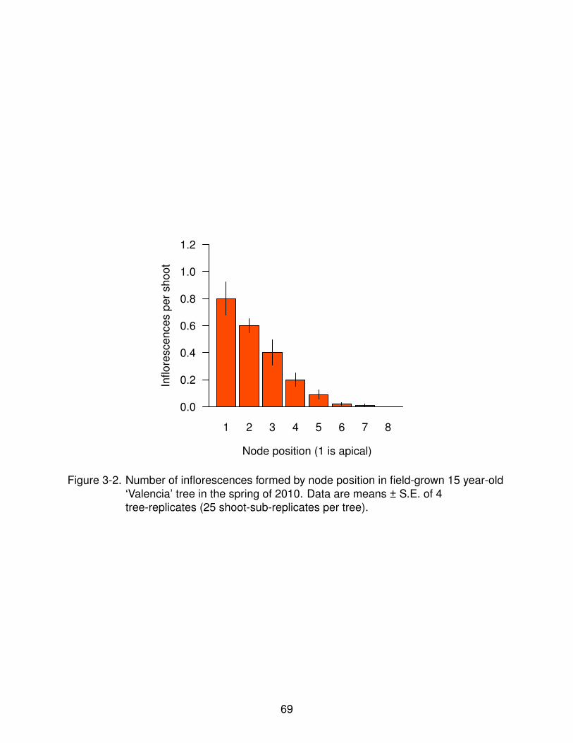
1 2 3 4 5 6 7 8
0.0
0.2
0.4
0.6
0.8
1.0
1.2
Node position (1 is apical)
Inflore
scen
ces p
er
sh
oot
Figure 3-2. Number of inflorescences formed by node position in field-grown 15 year-old‘Valencia’ tree in the spring of 2010. Data are means ± S.E. of 4tree-replicates (25 shoot-sub-replicates per tree).
69

1 2 3 4 5 7
0.0
0.5
1.0
1.5
2.0
Leaf position (1 is apical)
Rel
ativ
e tr
ansc
ript a
ccum
ulat
ion
OctoberDecember
A
1 3 5 7
0
1
2
3
4
Leaf position (1 is apical)
Rel
ativ
e tr
ansc
ript a
ccum
ulat
ion
23°C12°C
B
Figure 3-3. Expression of CsFT in leaves at different positions in shoots of field-grown‘Valencia’ trees in October and December of 2010 (A) and potted trees at23°C or 12°C (B). The level of expression of CsFT in leaves in October andat 23°C is representative of the levels of CsFT in leaves not exposed tofloral-induction whereas the level of expression of CsFT in December and at12°C is representative of levels of CsFT of leaves under floral-induction.Data are means ± S.E. of 4 tree-replicates (6 shoot-sub-replicates per tree).
70

0 30
23°C 12°CTemp.
Days
01234
CsAP1
0 29 33
Rel
ativ
e tr
ansc
ript a
ccum
ulat
ion
01234
CsLFY
1 3 5 7 1 3 5 7 1 3 5 7
Day of the experiment
Bud position (1 is apical)
Figure 3-4. Expression of CsAP1 and CsLFY in buds at different position in shoots of3-year-old potted ‘Valencia’ trees. Trees were transferred from a room at 23°C to a room at 12°C(Day 1). After 29 days, the trees were transferred backto the room at 23°C to promote growth. Data are means ± S.E. of 4tree-replicates (3 shoot-sub-replicates per tree).
71

1 2 3 4 5 6 7
0
1
2
3
4
5
Node position (1 is apical)
Num
ber
of in
flore
scen
ces
(cou
nts
out o
f 3 n
odes
per
pos
ition
)
ControlTIBA
Figure 3-5. Number of inflorescences formed by position in shoots of potted 3 year-old‘Valencia’ trees treated with 2,3,5-triiodobenzoic acid (TIBA). TIBA-treatedshoots had a lanolin paste containing 0.5% (w/w) of TIBA applied as a ringaround each internode, halfway between nodes. Control trees had a lanolinpaste without TIBA applied as before. Both sets of trees were transferred toa room at 12°C for 30 days and then transferred to another room at 23°C topromote growth. Each group consisted of 3 trees. Data are sums of thenumber of inflorescences formed in each position. The distributions ofinflorescences formed by bud position between TIBA-treated and controlshoots were statistically different (chi-square test).
72
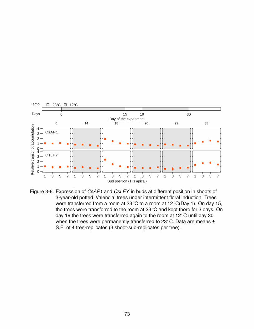
0 15 19 30
23°C 12°CTemp.
Days
01234
CsAP1
0 14 18 20 29 33
Rel
ativ
e tr
ansc
ript a
ccum
ulat
ion
01234
CsLFY
1 3 5 7 1 3 5 7 1 3 5 7 1 3 5 7 1 3 5 7 1 3 5 7
Day of the experiment
Bud position (1 is apical)
Figure 3-6. Expression of CsAP1 and CsLFY in buds at different position in shoots of3-year-old potted ‘Valencia’ trees under intermittent floral induction. Treeswere transferred from a room at 23°C to a room at 12°C(Day 1). On day 15,the trees were transferred to the room at 23°C and kept there for 3 days. Onday 19 the trees were transferred again to the room at 12°C until day 30when the trees were permanently transferred to 23°C. Data are means ±S.E. of 4 tree-replicates (3 shoot-sub-replicates per tree).
73

1 2 3 4 5 6 7 8
0.0
0.2
0.4
0.6
0.8
1.0
1.2
Node position (1 is apical)
Inflorescences per shoot Continuous induction
Intermittent induction
Figure 3-7. Number of inflorescences formed by position in 3 year-old ‘Valencia’ treesexposed to intermittent induction. Trees were transferred from a room at 23°C to a room at 12°C(Day 1). On day 15, the trees were transferred to theroom at 23°C and kept there for 3 days. On day 19 the trees weretransferred again to the room at 12°C until day 30 when the trees werepermanently transferred to 23°C. Data are means ± S.E. of 4 tree-replicates(3 shoot-sub-replicates per tree).
74

CHAPTER 4OTHER FACTORS ALTERING THE EXPRESSION OF SWEET ORANGE FLOWERING
GENES DURING FLORAL INDUCTION
Although low temperatures and water deficit are the only factors known to induce
flowering in C. sinensis, other factors like crop load, carbohydrate levels, gibberellins
and products of nitrogen metabolism can also alter the level of floral induction (Albrigo
and Galen-Sauco, 2004; Krajewsky and Rabe, 1995). Since these other factors (crop
load, etc.) modify the level of floral induction in C. sinensis trees, it was hypothesized
that signals initiated by these factors integrate with the regulatory pathway that controls
the expression of citrus flowering genes. Even though C. sinensis flowering genes are
probably orthologous to Arabidopsis flowering genes (Endo et al., 2005; Nishikawa
et al., 2010, 2009, 2007; Pillitteri et al., 2004a,b; Tan and Swain, 2007), the mechanisms
controlling floral induction are apparently different in both species. For instance,
although flowering in Arabidopsis is induced primarily by changes in photoperiod
(Valverde et al., 2004), changes in photoperiod do not seem to have any effect on floral
induction in citrus (Moss, 1969). Furthermore, gibberellins promote flowering in Ara-
bidopsis (Blazquez et al., 1998) but they produce the opposite effect in citrus (Monselise
et al., 1964). The effects of low temperatures and water deficit on the expression of C.
sinensis flowering genes have been reviewed and reported earlier in Chapter 2 of this
dissertation and in the published literature (Nishikawa et al., 2007; Pillitteri et al., 2004a).
In this chapter I present a collection of experiments designed to determine the
effects of crop load, gibberellins and light on the expression of flowering genes in C.
sinensis. Also, I present experiments that describe the changes in the level of transcript
accumulation of flowering genes early in the floral induction process. Results from
these experiments provide information about the molecular mechanism underlying
the effects of crop load and gibberellins on flowering in C. sinensis. In addition, these
results show that flowering genes respond rapidly and are highly sensitive to changes
75

in environmental conditions. Together, these results add more details to the proposed
hypothetical mechanism by which flowering is induced in C. sinensis.
4.1 Materials and Methods
4.1.1 Plant Material
Field experiments were conducted using mature ‘Valencia’ sweet orange trees
grafted on ‘Carrizo’ citrange in an orchard at the University of Florida’s Citrus Research
and Education Center in Lake Alfred, Florida (28°5’N, 81°43’W) during 2009 and
2010. The orchard received similar horticultural care as in neighboring commercial
groves throughout the experiments. Experiments under controlled environments were
conducted using either 2-3 year old potted ‘Valencia’ trees grafted on ‘Swingle’ citrumelo
or 2-3 year old potted ‘Washington Navel’ cuttings. All the trees used for experiments
were tested for floral competence and were maintained in a shaded greenhouse with
natural photoperiods, non-limiting irrigation and standard fertilization when not in use
for experiments. The growth rooms in which the controlled conditions experiments were
conducted were illuminated with with fluorescent lights (800µmoles·m-2·s-1 at canopy
level) with a 11/13h (day/night) photoperiod.
4.1.2 Experimental Conditions
To determine the effect of gibberellins on the accumulation of CsAP1, CsLFY,
CsSL1 and CsWUS transcripts in buds previously exposed to water deficit, transcripts of
these genes were quantified in buds of potted trees following a floral-inductive treatment
by low temperatures. Trees that had been kept at 23°C for at least one month were
transferred to a growth room at 12°C and kept at this temperatures for 30 days to induce
flowering. On the last day of the floral-inductive treatment, a 2.5µl drop of a 90ppm
aqueous solution of gibberellic acid (GA, Acros organics) and 0.5% Tween 80 (Fisher)
was applied to buds of previously selected shoots. Then trees were transferred to a
room at 23°C to promote bud growth. One day after the transfer to the room at 23°C, a
drop of the same gibberellic acid solution described before was applied to the buds of
76

another set of previously selected shoots. Another set of shoots was treated with a 0.5%
Tween 80 (Fisher) aqueous solution as a control. Three days after the final transfer to
23°C, the treated buds were sampled for transcript quantification. This experiment was
conducted using a completely randomized design with 4 tree replicates. Differences in
levels of transcript accumulation of CsAP1, CsLFY, CsSL1 and CsWUS in buds treated
with gibberellin and control buds were analyzed using analysis of variance.
To test determine the effect of gibberellins on the accumulation of CsFT transcripts,
leaves from the same trees of the experiment described in the previous paragraph were
treated with the same gibberellic acid solution described before after 25 days at 12°C.
The gibberellic acid solution was applied using cotton swabs and spreading it on both
the adaxial and abaxial surface of the leaves. Another group of leaves were treated
with the same solution without gibberellic acid as control. One day after the treatments
were applied the leaves were sampled for transcript quantification. This experiment was
conducted using a completely randomized design with 4 tree replicates. Differences
in levels of transcript accumulation of CsFT in leaves of gibberellin-treated and control
leaves were analyzed using a t-test.
To determine the effect of factors associated with fruit on the accumulation of CsFT
transcripts, leaves located at different distances from fruit on the same limb unit were
sampled in field-grown trees during the winter of 2009. This experiment was conducted
using a completely randomized design with 4 tree replicates. Differences in levels of
transcript accumulation of CsFT in leaves at different position relative to the nearest
fruit were analyzed using a general linear model. In the following spring, the number of
inflorescences formed in the shoots from which the leaves were sampled were recorded
and analyzed also using a general linear model.
To test whether cycles of light and dark alter the accumulation of CsFT transcripts,
CsFT transcripts were quantified in trees exposed to normal day/night conditions
(11/13h day/night), constant darkness or constant light in growth rooms at 12°C. Trees
77
that had been kept at 23°C for at least one month with a constant photoperiod (11/13h
day/night) were transferred to another room at 12°Cunder the 3 light regimes previously
indicated. Before the transfer, leaves were sampled to determine reference initial levels
of CsFT transcripts. Three days after the beginning of the treatments, leaf samples
were collected and CsFT transcripts quantified. This experiment was conducted using
a completely randomized design with 4 tree replicates. For each treatment, differences
in transcript levels between samples collected before the transfer and 3 days after
the transfer were analyzed using t-tests. Differences between the 3 light regimes on
samples collected 3 days after the transfer were analyzed using analysis of variance.
To determine whether the accumulation of CsFT transcripts changes throughout
the day, leaves samples were collected at different times under different conditions
known to modify floral induction in C. sinensis. To determine levels of CsFT transcripts
at non floral-inductive 23°C and floral-inductive 12°C, potted trees exposed to these
temperatures for 15 days were sampled every 2 hours from 06H00 until 18H00 for 3
consecutive days. To determine levels of CsFT in trees exposed to floral-inductive
water deficits and naturally occurring floral inductive temperatures, field-grown trees
were sampled during the summer of 2010 and the winter of 2010 at 08H00, 15H00,
18H00 and 21H00 for 3 consecutive days. This experiment was conducted using a
completely randomized design with 4 tree replicates. Differences in accumulation of
CsFT transcripts were analyzed using analysis of variance. To investigate whether the
time at which floral inductive temperatures occur has an effect on the accumulation of
CsFT transcripts, transcripts of CsFT of trees transferred from 23°C to 12°C at either
07H00 or 15H00 were quantified before the transfer, 2 hours after the transfer, and
at 07H00 and 15H00 on the next day. Differences in CsFT transcript accumulation
between trees transferred in the morning or in the afternoon were analyzed using
analysis of variance. To investigate the short-term response of CsFT transcript
accumulation to naturally occurring reductions in temperature under field conditions,
78

trees were sampled at 08H00 and 15H00 during the fall of 2010 2 days before and after
the passing of a cold front that reduced night temperatures below 20°C. Differences in
CsFT transcript accumulation in leaves before and after the passing of the cold front
were analyzed by t-test.
To determine the effect of levels of floral-inductive temperature on the accumulation
of CsFT transcripts, CsFT transcripts were quantified on leaves of trees after 3 days
of exposure to either 5, 10, 15, 20 or 23°C. Trees that had been kept at 23°C for at
least one month before the transfer to rooms at each temperature of the indicated
temperatures. Leaves were sampled before transfer and on day 3 after the transfer.
This experiment was conducted using a completely randomized design with 4 tree
replicates. Differences in CsFT transcript accumulation in leaves at the 5 temperatures
were analyzed by analysis of variance.
4.1.3 qRT-PCR
Total RNA was extracted using a phenol-chloroform precipitation method and
purified using silica membranes with on-column DNase digestion (Qiagen). Leaf
samples were used for analysis of CsFT expression, whereas bud samples where
used for analysis of CsSL1, CsAP1 and CsLFY expression. Five hundred nanograms
of total RNA were used for cDNA synthesis in a 20µl reaction with oligo dT primers
(SuperScriptIII®, Invitrogen). One microliter of the synthesized cDNA was used for
two-step (95°C denaturation and 60°C for 1 minute annealing and extension) qPCR
in a 20µl reaction (SYBR® Premix ExTaq™II, Takara) on a Applied Biosystems
7500 FAST real-time PCR system (Life Technologies) using optimized qPCR assays
(see Appendix). Primers for qPCR were: 5’-CGGCGGAAGGACTATGAC-3’ and
5’-TGTGAGAAAGCCAGAGAGGAA-3’ (CsFT ), 5’-CAGCCAGAGAATCTAACAAACG-3’
and 5’-TCAGTTTTGTGGTGGTATTGCC-3’ (CsSL1), 5’-CCCTGGAGTGCAACAACCT-3’
and 5’-CTGATGTGTTTGAGAGCGGT-3’ (CsAP1), 5’-TCTTGATCCAGGTCCAGAACATC-3’
and 5’-TAGTCACCTTGGTTGGGCATT-3’ (CsLFY ), and 5’-CCATGCACCAGAGACCAG-3’
79

and 5’-GTCTCCCATTTGACCACCA-3’ (CsWUS). CsGAPDH was used as reference
gene (5’-GGAAGGTCAAGATCGCAATCAA-3’ and 5’-CGTCCCTCTGCAAGATGACTCT-3’).
All qPCR assays were validated for specific amplification and optimized for amplification
efficiencies between 1.88 and 2.05 with a linear dynamic range of 6 log10 cycles.
The sequence of the primers to amplify CsLFY was obtained from Nishikawa et al.
(2009) whereas all other primer sequences were designed in-house. Relative gene
expression was calculated as a fold change ratio using Pfaffl’s method (Pfaffl, 2001) with
sliding-window efficiencies calculated for each reaction using the sliwin function in the
qpcR R package (Ritz and Spiess, 2008).
4.1.4 Data Analysis
Mean fold change of transcript levels were transformed to a logarithmic scale
(log2) for statistical analysis but data in the graphs represents the untransformed data.
Unless noted otherwise, all differences reported are statistically significant (p<0.05). All
statistical analyses were executed in R (R Development Core Team, 2011).
4.2 Results and Discussion
4.2.1 Gibberellins Down-regulate the Accumulation of Putative Flowering Signalsand Floral Identity Genes Transcripts
In Arabidopsis, gibberellins (GA) promote flowering by directly up-regulating the
floral identity gene LFY under short day conditions (Blazquez et al., 1998). In citrus
however, exogenous GA inhibit flowering when applied during floral induction (Cooper
and Peynado, 1958; Garcıa-Luis et al., 1986; Monselise et al., 1964). To test the
hypothesis that GA regulates the expression of floral identity genes (CsAP1 and CsLFY )
in C. sinensis and Arabidopsis although with opposite effects, 2.5µl of 90ppm solution
of GA were applied directly to buds of shoots 6-7 nodes long of potted C. sinensis
trees. Pillitteri et al. (2004a) reported that the expression of floral identity genes is kept
at basal levels during floral induction and their expression is only up-regulated after
transfer to growth promoting conditions. Similar results are also reported in Chapter
80
2 of this dissertation. Thus, it was assumed that the hypothetical effect of GA on the
expression would be easier to determine during the transition from floral-inductive to
growth-promoting conditions.
Figure 4-1 shows that application of GA consistently reduced the expression of
CsSL1, CsAP1 and CsLFY. These results support the hypothesis that GA signals
regulate the expression of the same genes in C. sinensis and Arabidopsis but in
opposite ways. Decreased expression of CsAP1 and CsLFY occurred both when GA
was applied before and after the transfer to growth-promoting conditions, however,
the reduction was greatest when GA was applied before the transfer. Besides CsSL1,
CsAP1 and CsLFY, the expression of the meristem identity gene CsWUS was also
measured and similarly reduced by GA application.
Guardiola et al. (1982) suggested that the application of exogenous GA during floral
induction could induce reversion of floral meristems to vegetative meristems. Lord and
Eckard (1987) reported that hypothetical inflorescence reversion did not occur if GA
was applied when sepals had already differentiated. Whereas there is no conclusive
evidence on whether actual inflorescence reversion occurs in C. sinensis, it is possible
that rather than inducing inflorescence reversion, GA disrupt floral identity determination
by down-regulating CsAP1 and CsLFY (reversion would not occur since no floral identity
has not been determined). Assuming that floral-bud differentiation occurs at the onset of
growth-promoting conditions, greater reduction of CsAP1 and CsLFY expression in buds
when GA is applied before the transfer to growth-promoting conditions than when it is
applied one day after the transfer supports the hypothesis that of GA disrupts acquisition
of floral identity. Since the samples used in the experiment reported here consisted of a
pool of bud tissue, higher expression of CsAP1 and CsLFY when GA was applied after
transfer than before transfer could indicate that some of the buds in the pool had already
acquired floral identity and thus were less affected by GA signals.
81

Application of GA also reduced the expression of CsWUS. In Arabidopsis, the
product of WUS, along with the products of STM and the CLAVATA clade, are key
components of the mechanism that maintains meristems undifferentiated and organized
(Gallois et al., 2002; Lenhard et al., 2002; Long et al., 1996; Mayer et al., 1998).
Furthermore, WUS and LFY activate the expression of AG (Lenhard et al., 2001;
Lohmann et al., 2001) which is required for floral organ identity in whorls 3 and 4 (Weigel
and Meyerowitz, 1994). Thus, reduced expression of CsWUS provides further support
to the hypothesis of GA disrupting the acquisition of floral meristem identity.
Besides reduced expression of floral and meristem identity genes, GA application to
leaves during induction also reduced the expression of floral signal integrator CsFT
(Figure 4-2). In Arabidopsis, application of GA is key to induce flowering under
non-floral-inductive short days (Blazquez et al., 1998). The role of GA on Arabidop-
sis floral induction under long days is less clear (Mutasa-Gottgens and Hedden, 2009),
but apparently GA signals are also required for up-regulating CsFT (Hisamatsu and
King, 2008). Thus, the down-regulation of CsFT in response to GA also supports the
hypothesis of GA inducing opposite effects on the expression of flowering genes in C.
sinensis and Arabidopsis.
4.2.2 Fruit Proximity
In C. sinensis, fruit inhibits floral induction in nearby shoots (Moss, 1971) and
trees carrying heavier crops usually flower less profusely than trees with lighter crops
(Valiente and Albrigo, 2004). The exact mechanism by which fruit inhibits flowering in
C. sinensis has not been elucidated. Fruit-induced reduction in carbohydrate levels
Goldschmidt et al. (1985), higher concentration of gibberellins in fruit-bearing branches
(Koshita et al., 1999) and changes in nitrogen metabolism in fruit-bearing branches
(Martınez-Fuentes et al., 2010) have been related to the inhibition of flowering. In this
experiment, the hypothesis that fruit inhibits the expression of the flowering signal
integrator gene CsFT was tested. Based on genetic transformation and gene expression
82

reports (Endo et al., 2005; Kobayashi et al., 1999; Nishikawa et al., 2007), it was
assumed that CsFT is in fact a flowering signal, similar to the role of its ortholog in
Arabidopsis (Corbesier et al., 2007), and thus reductions in the level of expression of
this gene in leaves nearby fruit would indicate disruption in the mechanism producing
flowering signals.
The expression of CsFT was proportional to the distance between fruit and leaves,
indicating that the presence of fruit probably reduces the production of flowering signals
(assuming that protein expression is proportional to transcript accumulation for this
gene, Figure 4-3. The inhibitory effect of fruit on CsFT occurs in a gradient and was
present at least as far as 30cm away from the fruit. It is possible that expression of
floral identity genes were also affected by nearby fruit, however this possibility was not
tested. Nonetheless, these results indicated that the flower-inhibiting factor associated
with fruit at least disrupts the production of flowering signals in nearby leaves and thus,
could partially explain the negative effect of fruit on flowering. At the whole tree level,
reduced flowering in trees carrying a heavy crop could be explained by overall reduced
production of flowering signals since heavier crops imply shorter average distances
between fruit and nearby leaves than lighter crops.
4.2.3 Effect of Light/Dark Cycles on CsFT Transcript Accumulation
Photoperiod is a key regulator of the transcription of FT in Arabidopsis (Turck
et al., 2008). In Arabidopsis, expression of FT is up-regulated by CONSTANS (CO)
in the phloem of leaves (An et al., 2004) and the FT protein is transported to the
shoot apical meristem (Corbesier et al., 2007) where, along with the product of FD
(Abe et al., 2005), activates the expression of floral identity genes LFY and AP1
(Samach et al., 2000). Expression of CO is controlled by the plant’s circadian clock
and peaks late in the day (Suarez-Lopez et al., 2001). CO transcription is followed
by translation of the CO protein, however, in the absence of light, the CO protein
is targeted for degradation in the proteosome (Valverde et al., 2004) and does not
83

up-regulate FT. Conversely, under floral-inductive long days, CO is stabilized by light and
effectively up-regulates FT and promote flowering (Valverde et al., 2004). Since CsFT
is apparently the citrus ortholog of Arabidopsis FT, it was hypothesized that light/dark
cycles could also regulate expression of CsFT even though floral induction in C. sinensis
is considered to be photoperiod-insensitive (Moss, 1969). In this experiment trees were
exposed to 2 extreme photoperiods for 3 days at floral-inductive temperatures to test
whether disrupting light/dark cycles altered the expression of CsFT compared to an
intermediate photoperiod.
Figure 4-4 shows that either constant light or constant darkness inhibited the
expression of CsFT compared to the intermediate photoperiod. This indicated that the
expression of CsFT requires alternation of periods of light and darkness. Moss (1969)
reported that flowering was induced in C. sinensis trees exposed to floral-inductive
temperatures at daylengths ranging from 8 to 16 hours, proposing that floral induction
in C. sinensis is probably insensitive to photoperiod. Furthermore, Cassin et al. (1969)
reported that C. sinensis trees are capable of flowering and produce commercial
crops even at tropical locations where seasonal changes in daylength are negligible.
The results in Figure 4-4 support a role for photoperiod regulating the expression
of CsFT and floral induction (assuming that CsFT is a floral-promoting signal as
in Arabidopsis). Since flowering in C. sinensis can be induced in a wide range of
photoperiods, floral-induction in C. sinensis may not be as sensitive as floral-induction in
Arabidopsis but still requires alternation of periods of light and darkness.
4.2.4 Accumulation of CsFT Transcripts Changes Throughout the Day
In Arabidopsis, the photoperiodic induction of flowering relies on the translation of
CO protein during periods of light (Turck et al., 2008). The expression of FT usually
follows the expression of CO under floral-inductive long days (Suarez-Lopez et al.,
2001). The transcription of CO is regulated by components of the circadian clock
(Suarez-Lopez et al., 2001) but the activity of CO is regulated after translation (Valverde
84

et al., 2004). Whereas no C. sinensis ortholog of CO has been characterized, the
genome of C. sinensis contains several sequences with high degree of sequence
similarity to CO and CO-LIKE genes in other species (data not shown). Furthermore,
as reported in the previous section (4.2.3), the up-regulation of CsFT by floral-inductive
temperatures requires the alternation of periods of light and darkness, a response that
could be mediated by a putative CsCO ortholog. In this experiment the hypothesis
was tested that the expression of CsFT changes throughout the day, similar to the
expression of FT in Arabidopsis (Suarez-Lopez et al., 2001).
Figure 4-5 shows that expression of CsFT changes throughout the day in C.
sinensis under the conditions evaluated. Expression of CsFT was highest between
13H00 and 15H00 local standard time (LST, UTC-5) and then decreased sharply.
The expression of CsFT oscillated also under non-floral-inductive conditions (23°C in
growth rooms and irrigated trees in Summer 2010) but the amplitude of the peak was
significantly smaller than at floral-inductive temperatures. Expression of CsFT started
to decrease before the onset of periods of dark suggesting that diurnal changes in
expression might be regulated by some other factor besides light. The up-regulation
of CsFT did not occur under either constant light or darkness (Subsection 4.2.3) so
light/dark cycles could still be involved in the regulation of CsFT, though less directly.
Complete inhibition of CsFT up-regulation under constant light or darkness conditions
did not support the hypothesis of direct regulation of CsFT expression by the tree’s
internal clock. Since diurnal changes in the expression of CsFT could be induced
by various factors, the diurnal cycling supports the hypothesis that CsFT could be
regulated by a putative CO ortholog.
4.2.5 Early Changes in Transcript Levels of CsFT in Response to Floral InductiveTemperatures
The expression of CsFT has been shown to change seasonally in trees growing
in the field and in response to artificial floral inductive treatments (Nishikawa et al.
85

2007, 2009 and Chapter 2 of this dissertation). However, details about the earliest
changes in transcription of CsFT after initial exposure to floral inductive stimuli are
lacking. Assuming that CsFT is a flowering signal as in Arabidopsis, describing the
earliest responses of CsFT could help elucidate characteristics of the mechanism
regulating CsFT expression and floral induction. In this set of experiments changes in
the expression of CsFT taking place within the first 3 days of exposure to floral-inductive
conditions are described.
Figure 4-6 shows the expression of CsFT within 24 hours of transfer to floral-inductive
temperatures. In this experiment trees were transferred either in the morning or in
the afternoon to test whether processes occurring early in the day (or during the
evening/night) could be related to immediate up-regulation of CsFT. Expression of CsFT
was significantly up-regulated regardless of the time at which trees were transferred,
indicating that the temperature-sensing mechanism regulating CsFT expression is
active throughout the day. However, the magnitude of the up-regulation of CsFT 2 hours
after the transfer was higher in trees transferred at 15H00 LST, indicating that CsFT
sensitivity to changes in temperature is higher at this time than earlier in the morning.
Furthermore, at 17H00 LST the level of expression CsFT in trees transferred at 07H00
was equivalent to the level of expression of CsFT in trees transferred at 15H00 LST,
indicating that the effect of the exposure to floral-inductive temperatures for 10h through
most of the day on CsFT was equivalent to the effect of exposing the trees for 2h to
floral-inductive temperatures in the afternoon.
On the next day, the level of expression of CsFT in trees transferred at 08H00 LST
the day before was no different than the level of CsFT expression in trees transferred
at 15H00 LST the day before at any of the times sampled. Diurnal changes in CsFT
expression also occurred in this experiment as reported before (Figure 4-5). The
level of expression of CsFT in morning samples on the day after the transfer were
equivalent to the level of expression of CsFT at 17H00 on the day of the transfer. This
86

may indicate that floral induction is intensified with time by maintaining the levels of
expression of CsFT reached by the end of the day overnight and re-setting the diurnal
cycle at continuously increasing levels. This response was also observed in field-grown
trees during the winter of 2010 before and after the passing of cold fronts that reduced
ambient temperatures below the hypothetical floral-inductive threshold of 20°C (Figure
4-7). In the latter case, cooling also occurred by the end of the day and the lowest level
of the diurnal oscillation in CsFT expression was continuously shifted in the two days
following the drop in ambient temperatures.
In C. sinensis, temperatures ranging from 5°C to about 20°C are considered to be
floral-inductive (Garcıa-Luis et al., 1992; Moss, 1969; Valiente and Albrigo, 2004). Within
this range, temperatures between 10°C and 15°C induce flowering most intensely.
To test whether transcript accumulation of CsFT is sensitive to levels of temperature,
trees were kept at growth-promoting temperatures and then transferred to rooms at
different floral-inductive temperatures and expression of CsFT was quantified three
days later. Figure 4-8 shows that the expression of CsFT was sensitive to levels
of floral-inductive temperatures. Consistent with earlier reports of floral induction
(Moss, 1969), transcript accumulation of CsFT was highest after transfer to 15°C. At
temperatures lower than 15°C transcript accumulation of CsFT was also increased
whereas transcript accumulation at 20°C was higher but not statistically different than
transcript accumulation at 23°C (growth-promoting non-floral-inductive). These results
support the hypothesis of CsFT acting as a quantitative and qualitative integrator of
temperature-regulated flowering signals.
Results of the experiments reported in this chapter show that the negative
regulation of flowering by gibberellins (Monselise et al., 1964) could be a consequence
of reduced levels of expression of CsFT (assumed to be a floral-promoting signal) and
reduced levels of expression of floral identity genes in buds. Also, these results show
that, different from Arabidopsis (Blazquez et al., 1998), gibberellins negatively regulate
87

the expression of CsLFY and CsAP1 in C. sinensis which supports a hypothesis for
diverging evolution of mechanisms regulating the response of flowering genes to
gibberellin signals. It was also shown that reduced levels of floral induction associated
with the presence of fruit and crop load (Moss, 1971; Valiente and Albrigo, 2004),
could be a consequence of reduced expression of CsFT in the proximity of fruit. Even
though gibberellin levels were not quantified in this study, higher concentration of
endogenous gibberellins near fruit (Koshita et al., 1999) could be related to the reduced
level of expression of CsFT since application of exogenous gibberellin reduce the
expression of CsFT. Other factors that have been proposed as possible factors that
reduce the level of floral induction in fruit-bearing branches such as carbohydrates
(Garcıa-Luis et al., 1988) and nitrogen metabolism (Martınez-Fuentes et al., 2010)
were not tested in this study. The results presented in this chapter also show that
C. sinensis requires alternation of cycles of light and darkness for floral induction,
indicating that floral induction in C. sinensis is not insensitive to photoperiod signals,
but instead sensitivity to photoperiod is much reduced compared to other species.
Furthermore, the mechanism regulating floral induction in C. sinensis seems also to
be regulated by diurnal cycles with sensitivity to floral inductive signals being highest
in the afternoon-evening. Continuous increase in the level of floral induction could
be explained by continuously shifting the lowest level of the oscillation overnight.
Together, the results presented in this Chapter offer details about the regulation of
flowering-related genes in C. sinensis by factors known to modify the level of floral
induction. Some of these factors (i.e. gibberellins and light) induce critical diverging
responses in C. sinensis and the model Arabidopsis. Thus, characterizing the response
of flowering-related genes to these factors could help elucidate where the differences
in the mechanisms regulating flowering in C. sinensis and Arabidopsis could be. In
addition, the earliest changes in transcript accumulation of CsFT (assumed to be the
floral-promoting signal in C. sinensis) in response to floral-inductive temperature, were
88

also described and provide insights about the mechanism regulating floral induction in C.
sinensis.
89

CsAP10.0
0.2
0.4
0.6
0.8
1.0
1.2
1.4
CsLFY CsSL1 CsWUS
Control +GA Pre−transfer +GA Post−transferR
elat
ive
tran
scrip
t acc
umul
atio
n
Figure 4-1. Expression of CsAP1, CsLFY, CsSL1 and CsWUS in buds of 2 year old‘Valencia’ trees treated with gibberellic acid one day before or one day aftertransfer from floral-inductive to growth-promoting conditions. Three sets of 3trees each were exposed to floral-inductive temperatures (12°C) for 30 days.On the 30th day of the experiment (“pre-transfer”) the buds of 4 shoots in oneset of trees were treated with 2.5µl of a 90ppm gibberellic acid 0.5% Tween80 solution using a micropipette. The 3 sets of trees were then transferred toa chamber at growth-promoting temperatures (23°C). On the 31st day of theexperiment (“post-transfer”), the buds of 4 shoots in another set of treeswere treated with gibberellic acid as before. The buds on the shoots of thetrees in the last set were treated with the same solution used before exceptthat the solution did not contain gibberellic acid (“Untreated”). Bud sampleswere collected on the 33rd day of the experiment (3 days after transfer).Figures are means of 3 tree-replicates ± S.E. Gene expression is relative tothe levels of each gene in the “Untreated” buds.
90

Untreated +GA
0.0
0.5
1.0
1.5
2.0
Rel
ativ
e tr
ansc
ript a
ccum
ulat
ion Before treatment
1 day after treatment
Figure 4-2. Expression of CsFT in leaves of 2 year old ‘Valencia’ trees treated withgibberellic acid during floral induction. Two sets of 4 trees were exposed tofloral-inductive temperatures (12°C) for 25 days. On day 25, the leaves of 4shoots in one set of trees were treated with a 90ppm gibberellic acid 0.5%Tween 80 solution using a cotton swab (“+GA”). The leaves of 4 shoots inthe other set of trees were treated using a similar solution without gibberellicacid (“Untreated”). Leaf discs from shoots in both set of trees were collectedbefore application of the treatments and on the next day. Figures are meansof 4 tree-replicates ± S.E. Gene expression is relative to the levels of CsFTin “Untreated” leaves before treatments were applied.
91

0
1
2
3
4
5
6
7
<10 10−20 20−30 >60
Distance to nearest fruit (cm)
Rel
ativ
e tr
ansc
ript a
ccum
ulat
ion
0 ± 0 0.58 ± 0.08 0.92 ± 0.08 4.25 ± 0.14
Inflorescences
Figure 4-3. Expression of CsFT in leaves of 15 year old ‘Valencia’ in the field located atdifferent distances from single fruit. Leaves at located at different distancesfrom single fruit in the same branch were sampled in December of 2009 on 4branches of three different trees from the same grove. The leaves sampledwere located in shoots formed during the previous year. The distance to thenearest fruit was measured adding up the length of individual shootsegments (formed in preceding growth cycles) separating the fruit from thesampled leaf. Table on top of the figure indicates the number ofinflorescences that formed in the sampled shoots in the spring. Figures aremeans of 3 tree-replicates ± S.E. (4 shoot-subreplicates). Gene expressionis relative to the levels of CsFT in leaves located in shoots at <10cm from afruit.
92

Constant dark Constant light Normal day
0
1
2
3
4
5
Rel
ativ
e tr
ansc
ript a
ccum
ulat
ion
Pre−transfer3 days after transfer
Figure 4-4. Expression of CsFT in leaves of 2 year old ‘Valencia’ trees under constantlight, constant darkness and an intermediate photoperiod (“Normal day”, 11hlight / 13h darkness) at floral-inductive conditions (12°C). Three sets of 3trees were exposed for 3 days to each of the light regimes. Leaves weresampled before transfer to the growth rooms and 3 days after. Figures aremeans of 3 tree-replicates ± S.E. Gene expression is relative to the levels ofCsFT before the transfer.
93
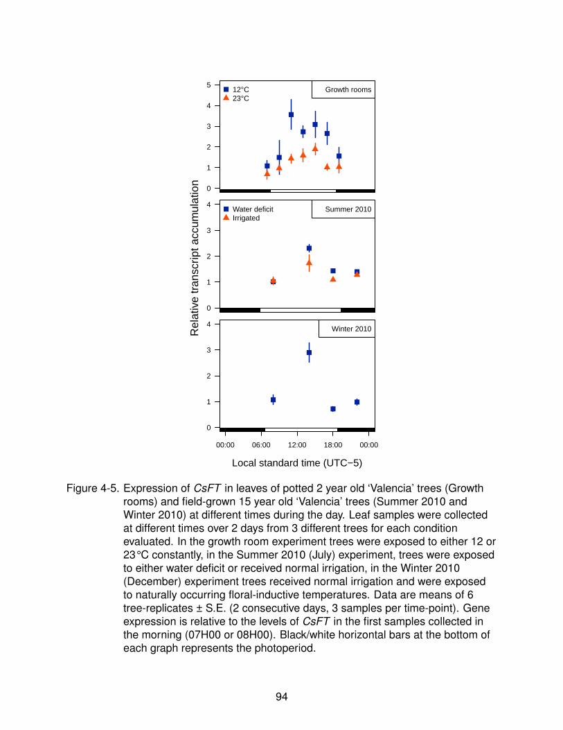
0
1
2
3
4
5Growth rooms12°C
23°C
0
1
2
3
4Summer 2010Water deficit
Irrigated
0
1
2
3
4
00:00 06:00 12:00 18:00 00:00
Winter 2010Rel
ativ
e tr
ansc
ript a
ccum
ulat
ion
Local standard time (UTC−5)
Figure 4-5. Expression of CsFT in leaves of potted 2 year old ‘Valencia’ trees (Growthrooms) and field-grown 15 year old ‘Valencia’ trees (Summer 2010 andWinter 2010) at different times during the day. Leaf samples were collectedat different times over 2 days from 3 different trees for each conditionevaluated. In the growth room experiment trees were exposed to either 12 or23°C constantly, in the Summer 2010 (July) experiment, trees were exposedto either water deficit or received normal irrigation, in the Winter 2010(December) experiment trees received normal irrigation and were exposedto naturally occurring floral-inductive temperatures. Data are means of 6tree-replicates ± S.E. (2 consecutive days, 3 samples per time-point). Geneexpression is relative to the levels of CsFT in the first samples collected inthe morning (07H00 or 08H00). Black/white horizontal bars at the bottom ofeach graph represents the photoperiod.
94

0
1
2
3
4
5
6
09:00 17:00
07:00 15:00 07:00 15:00
Local standard time (UTC−5)
Rel
ativ
e tr
ansc
ript a
ccum
ulat
ion
Morning transferAfternoon transfer
Figure 4-6. Expression of CsFT in leaves of 3 year old ‘Valencia’ trees after transfer tofloral-inductive temperatures (12°C). Two sets of 3 trees maintained at 23°Cwere transferred either at 07H00 or 15H00 to a growth room at 12°C(floral-inductive). Leaves were sampled before transfer, two hours after thetransfer and on the next day. Figures are means of 3 tree-replicates ± S.E.Gene expression is relative to the levels of CsFT before the 08H00 transfer.
95
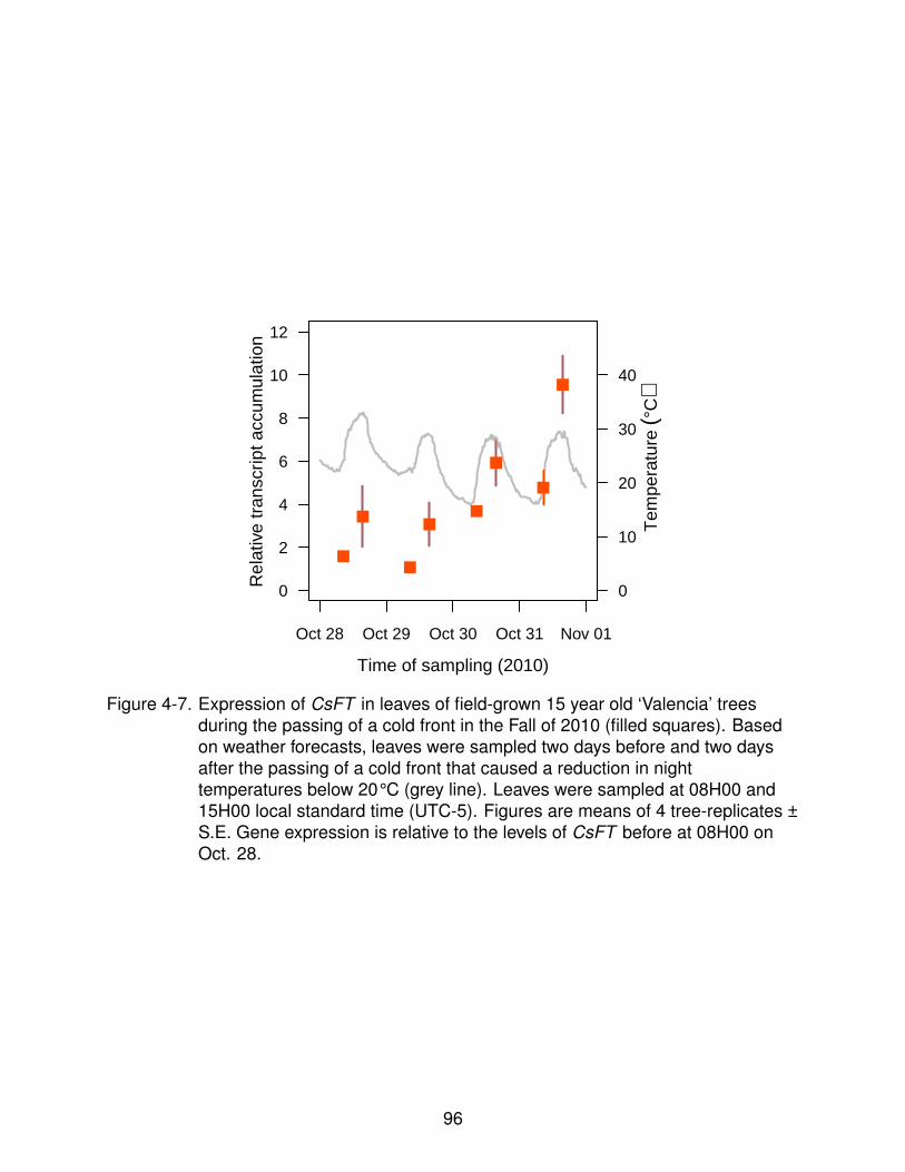
0
2
4
6
8
10
12
Oct 28 Oct 29 Oct 30 Oct 31 Nov 01
0
10
20
30
40
Time of sampling (2010)
Rel
ativ
e tr
ansc
ript a
ccum
ulat
ion
Tem
pera
ture
(°C
)
Figure 4-7. Expression of CsFT in leaves of field-grown 15 year old ‘Valencia’ treesduring the passing of a cold front in the Fall of 2010 (filled squares). Basedon weather forecasts, leaves were sampled two days before and two daysafter the passing of a cold front that caused a reduction in nighttemperatures below 20°C (grey line). Leaves were sampled at 08H00 and15H00 local standard time (UTC-5). Figures are means of 4 tree-replicates ±S.E. Gene expression is relative to the levels of CsFT before at 08H00 onOct. 28.
96
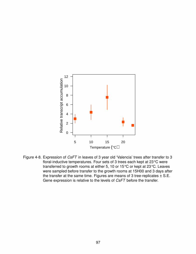
5 10 15 20
0
2
4
6
8
10
12
Temperature (°C)
Rel
ativ
e tr
ansc
ript a
ccum
ulat
ion
Figure 4-8. Expression of CsFT in leaves of 3 year old ‘Valencia’ trees after transfer to 3floral-inductive temperatures. Four sets of 3 trees each kept at 23°C weretransferred to growth rooms at either 5, 10 or 15°C or kept at 23°C. Leaveswere sampled before transfer to the growth rooms at 15H00 and 3 days afterthe transfer at the same time. Figures are means of 3 tree-replicates ± S.E.Gene expression is relative to the levels of CsFT before the transfer.
97

CHAPTER 5CONCLUSIONS
The objective of my study was to characterize changes in transcript accumulation
of C. sinensis flowering-related genes in response to treatments known to alter floral
induction and determination. Figure 5-1 shows a graphic summary of the conclusions
reached from the results of the experiments reported in previous chapters.
I found that the expression of CsFT is up-regulated by both known floral-inductive
stimuli of C. sinensis, water deficit and low temperatures. Up-regulation of CsFT after
exposure to low-temperature had been reported earlier (Nishikawa et al., 2007) but
there had been no reports about the response of CsFT (or its putative orthologs in other
species) in response to floral-inductive water deficit. Furthermore, when low temperature
and water deficit were present at the same time, accumulation of CsFT transcripts
was induced above the levels observed when each stimulus was present separately.
This, suggests that CsFT could be an integrator of flowering signals initiated by both
flowering stimuli in C. sinensis. Furthermore, I also reported that expression of CsFT
was increased as the time under floral-inductive conditions increased and was sensitive
to levels of temperature. Another effect of water deficit was to repress up-regulation of
floral identity genes CsAP1 and CsLFY during periods of warmer weather during the
winter. This repression of CsAP1 and CsLFY could keep buds undifferentiated and
responsive to additional floral-inductive signals when temperatures decrease again later
in the winter. Flowering in C. sinensis could be considered as a quantitative response
to levels of floral induction intensity. My results support a role for CsFT as a quantitative
marker of floral induction intensity.
In the model species Arabidopsis, CsFT ’s ortholog, FT, is a key component in the
mechanism that controls flowering (Turck et al., 2008). However, flowering responses
of each Arabidopsis and C. sinensis to floral-inductive stimuli in the other species do
not support the existence of an exact common mechanism regulating flowering in both
98
species (Amasino, 2010; Krajewsky and Rabe, 1995). Consequently, it is proposed that
regulation of expression of CsFT in C. sinensis and FT in Arabidopsis have evolved
to respond to different environmental stimuli. Evolution of flowering control mechanism
involving FT orthologs has been reported in short-day rice (Hayama and Coupland,
2004). Even if FT -mediated control of flowering in C. sinensis and Arabidopsis evolved
to respond to different environmental stimuli, some characteristics of the mechanism
related with FT are conserved such as diurnal changes in the level of expression of
CsFT and the dependence on light to enable up-regulation of CsFT. CsFT expression
dependence of light/dark cycles indicated that although not as sensitive as Arabidopsis
and other species induced to flower by specific photoperiods, floral induction in C.
sinensis is not totally insensitive to photoperiod.
In addition to CsFT, I also found that the response of CsAP1 and CsLFY to
exogenous gibberellins was opposite to the response of Arabidopsis’ AP1 and LFY
to similar treatments. Consistent with reduced formation of inflorescences after
the application of gibberellins (Monselise et al., 1964), expression of CsAP1 and
CsLFY (whose expression is assumed to determine floral identity and initiate floral
differentiation) was significantly reduced by the application of gibberellins to buds.
However, gibberellin application in Arabidopsis up-regulates the expression of these
genes and promote flowering under non-floral-inductive short days (Blazquez et al.,
1998). My results also show that the inhibitory effect of gibberellins on the transcript
accumulation of CsAP1 and CsLFY is greater before growth has been stimulated by
warmer temperatures, suggesting that floral determination and initiation might not
occur while under floral-inductive conditions and require the re-initiation of growth. The
pattern of expression of CsAP1 and CsLFY was more related to the establishment of
a flowering gradient and the initiation of multiple flowering cohorts than the pattern of
expression of CsFT. This suggested that flowering gradients are not a consequence
of biased production of flowering signals near the apex but maybe a consequence of
99

biased distribution of flowering signals and/or a gradient in the sensitivity of buds at
different position within the shoot to flowering signals.
A major limitation of this study was that it was restricted to characterize the
expression of flowering-related genes at the transcript level and did not explore
other levels of regulation of the expression of these genes. Regulation of the activity
of flowering genes before or after transcription is a key component of the flowering
regulatory mechanisms in Arabidopsis and other species in which these mechanisms
have been more thoroughly investigated (Adrian et al., 2010; Dennis and Peacock,
2007; Valverde et al., 2004). However, regulation of transcription is still a major factor in
the mechanisms controlling flowering in model species, and comparing those patterns of
transcript accumulation of flowering-related genes with those in C. sinensis and model
species can help identify where mechanisms regulating flowering in these species differ.
Another potential limitation of this study is that it relies on the assumption of orthology
between genes in C. sinensis and model species. Although reports of transgenic
over-expression, Arabidopsis mutant complementation, and expression patterns data
support this assumption (Endo et al., 2005; Kobayashi et al., 1999; Nishikawa et al.,
2007; Pena et al., 2001; Pillitteri et al., 2004a; Tan and Swain, 2007), the activity and
function of the genes used in this study has not been confirmed in citrus. Nonetheless,
this study provides useful information for developing models of how floral induction and
initiation is regulated at the genetic level in C. sinensis. Some of the findings of this
study could also be relevant to elucidating the flowering regulatory mechanisms in other
citrus cultivars and species originated in subtropical climates.
100

���
����
�������
� ������
�����������
����
���
�����������
����
�����
��
��������
�����
Figure 5-1. Graphical summary of the conclusions of this dissertation.
101

APPENDIXDESIGN, VALIDATION AND OPTIMIZATION OF qPCR ASSAYS
qPCR assays to quantify CsFT, CsSL1, CsAP1, CsLFY and CsWUS transcripts in
C. sinensis were designed from C. sinensis and C. unshiu sequences of these genes
available in the NCBI’s Nucleotide database and optimized using the algorithm in Figure
A-2.
For each assay, primer pairs that amplified products between 60 and 200 base pairs
(bp) were selected (Table A-1). Amplification specificity of these primers was checked
by agarose gel electrophoresis (Figure A-3A) and by generating dissociation curves of
the amplified products on a Applied Biosystems 7500 FAST real-time PCR system (Life
Technologies) (Figure A-3B). The amplified products were then cloned into a vector and
sequenced to confirm each amplicon’s identity. Amplification efficiencies for each assay
were calculated for each reaction using the sli.win function (Ritz and Spiess, 2008)
and by the dilution curve method (Figure A-4). The linear dynamic range of the qPCR
assays were determined by the dilution curve method (Figure A-4).
RNA was extracted as indicated in the Materials and Methods section of each
chapter in batches of 30 to 50 samples. From each batch, 6 samples were randomly
selected to check RNA integrity by electrophoresis of glyoxylated RNA. When degradation
of RNA was suspected, the rest of the samples of the batch were checked as already
indicated and the suspected degraded RNA samples discarded. Reverse transcription
reactions were run as indicated in the Materials and Methods section of each chapter
in batches of 30 samples. In each batch, control reactions that did not contain reverse
transcriptase in the reaction mix were run using 5 randomly selected samples to
check for potential DNA contamination after subsequent amplification by qPCR. No
amplification products were detected in any of these control reactions.
102

��������������
�������������������������
�����
�����
� !
����"�
#$��
����"!
���$"�#�$���������%&�����'
� !
����� ���(������
��������)������������
� $���������
��������$����
%*�
+��
*�
+��
*�
+��
*�
�"���$����������
$��"�,�$������������
����������
��� �!
+��
*�
Figure A-2. Algorithm for the design, validation and optimization of qPCR assays.
103
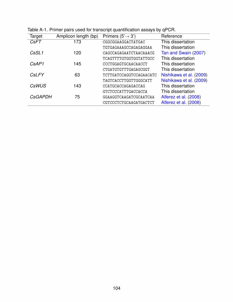
Table A-1. Primer pairs used for transcript quantification assays by qPCR.Target Amplicon length (bp) Primers (5’→ 3’) ReferenceCsFT 173 CGGCGGAAGGACTATGAC This dissertation
TGTGAGAAAGCCAGAGAGGAA This dissertationCsSL1 120 CAGCCAGAGAATCTAACAAACG Tan and Swain (2007)
TCAGTTTTGTGGTGGTATTGCC This dissertationCsAP1 145 CCCTGGAGTGCAACAACCT This dissertation
CTGATGTGTTTGAGAGCGGT This dissertationCsLFY 63 TCTTGATCCAGGTCCAGAACATC Nishikawa et al. (2009)
TAGTCACCTTGGTTGGGCATT Nishikawa et al. (2009)CsWUS 143 CCATGCACCAGAGACCAG This dissertation
GTCTCCCATTTGACCACCA This dissertationCsGAPDH 75 GGAAGGTCAAGATCGCAATCAA Alferez et al. (2008)
CGTCCCTCTGCAAGATGACTCT Alferez et al. (2008)
104
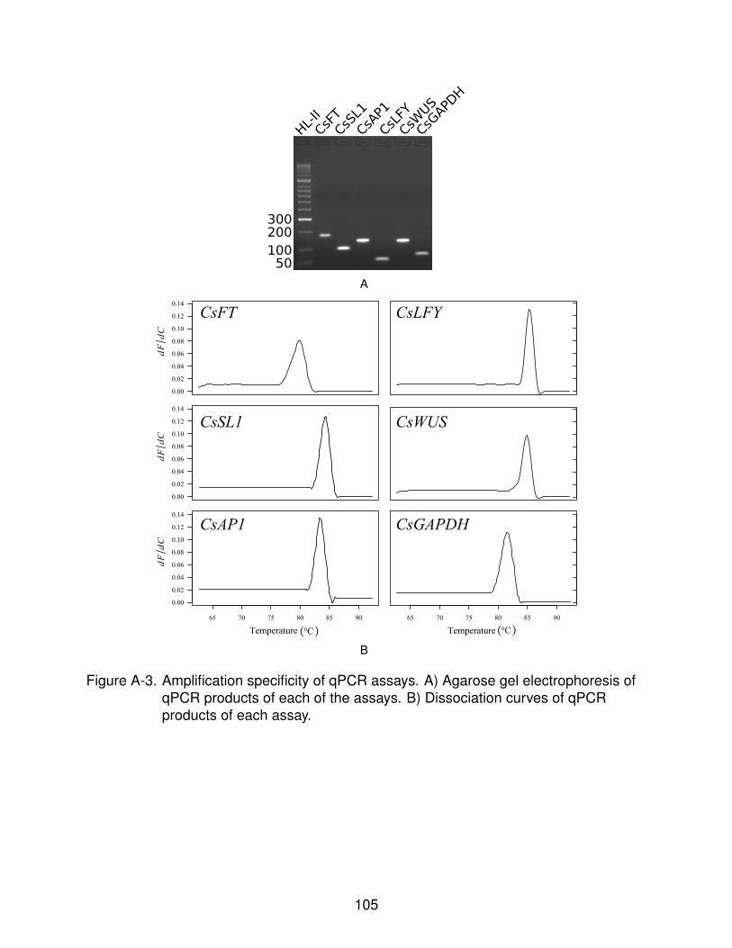
CsFT
CsSL1
CsAP1
CsLFY
CsWUS
CsGAPDH
HL-II
50100
200300
A
0.00
0.02
0.04
0.06
0.08
0.10
0.12
0.14
65 70 75 80 85 90
Temperature
dFdC
0.00
0.02
0.04
0.06
0.08
0.10
0.12
0.14
dFdC
dFdC
0.00
0.02
0.04
0.06
0.08
0.10
0.12
0.14
65 70 75 80 85 90
Temperature � C�O� C�O
CsFT CsLFY
CsSL1 CsWUS
CsAP1 CsGAPDH
B
Figure A-3. Amplification specificity of qPCR assays. A) Agarose gel electrophoresis ofqPCR products of each of the assays. B) Dissociation curves of qPCRproducts of each assay.
105

0
10
20
30
40
Cq
q
1.8
2
1.9
5
1.9
7
1.7
9
1.9
9
1.8
3
1.9
8
CsFT
y = 25.06−3.61x
R2� 0.99
0
10
20
30
40
Cq
q
1.8
6
1.9
2
2
1.9
1.9
1.9
1
2.0
5
CsSL1
y = 20.08−3.71x
R2� 1
−4 −2 0 2 4
0
10
20
30
40
log10 Relative template concentration
Cq
q
1.9
1
1.9
7
1.8
8
1.7
6
1.8
1
1.9
1
1.8
7
1.9
9
CsAP1
y = 25.97−3.58x
R2� 1
q
1.7
7
1.8
8
1.8
8
1.9
6
1.8
1.8
9
1.7
5
1.8
8
CsLFY
y = 26.45−3.49x
R2� 1
q
1.9
4
1.9
3
1.9
6
2.0
7
1.9
4
1.9
5
1.9
4
CsWUSy = 29.49−3.91x
R2� 0.99
−4 −2 0 2 4
q
2
2.0
7
1.9
1.9
3
1.9
5
1.3
8
1.9
9
CsGAPDH
y = 29.85−3.65x
R2� 0.99
log10 Relative template concentration
Figure A-4. Linear dynamic range and amplification efficiency of qPCR assays.Numbers above each symbol represent individual reaction efficiencycalculated using the sli.win function in the qpcR R package (Ritz andSpiess, 2008)
106

REFERENCES
Abe, M., Y. Kobayashi, S. Yamamoto, Y. Daimon, A. Yamaguchi, Y. Ikeda, H. Ichinoki,M. Notaguchi, K. Goto, and T. Araki. 2005. FD, a bZIP protein mediating signals fromthe floral pathway integrator FT at the shoot apex. Science 309:1052–1056.
Adrian, J., S. Farrona, J. J. Reimer, M. C. Albani, G. Coupland, and F. Turck. 2010.cis-Regulatory elements and chromatin state coordinately control temporal and spatialexpression of FLOWERING LOCUS T in Arabidopsis. Plant Cell 22:1425–1440.
Aida, M., T. Ishida, H. Fukaki, H. Fujisawa, and M. Tasaka. 1997. Genes involved inorgan separation in arabidopsis: An analysis of the cup-shaped cotyledon mutant.Plant Cell 9:841–857.
Albani, M. C. and G. Coupland. 2010. Comparative analysis of flowering in annual andperennial plants. In: M. C. Timmermans (ed.) Plant Development, volume Volume 91,pp. 323–348. Academic Press.
Albrigo, L.. 1999. Effects of foliar applications of urea or nutriphite on flowering andyields of Valencia orange trees. Proc. Fla. State Hort. Soc. 112:1–4.
Albrigo, L. G., H. W. Beck, and J. I. Valiente. 2006a. Testing a flowering expert systemfor the ’Decision Information System for Citrus’. Acta Hort. 707:17–24.
Albrigo, L. G. and V. Galen-Sauco. 2004. Flower bud induction, flowering and fruit-setof some tropical and subtropical fruit tree crops with special reference to citrus. ActaHorticulturae 632:81–91.
Albrigo, L. G., J. I. Valiente, and H. W. Beck. 2002. Flowering expert systemdevelopment for a phenology based citrus decision support system. Acta Horticulturae584:247–254.
Albrigo, L. G., J. I. Valiente, and C. Van Parys de Wit. 2006b. Influence of winter andspring weather on year-to-year citrus fruit set and yield variation in sao paulo, brazil.In: A. El-Otmani, M; Ait-Oubahou (ed.) Proceedings of the International Society ofCitriculture, volume 1, pp. 263–267. Agadir, Morocco.
Alferez, F., Y. Lluch, and J. K. Burns. 2008. Phospholipase A2 and postharvest peelpitting in citrus fruit. Postharvest Biology and Technology 49:69–76.
Ali, A. and C. Lovatt. 1995. Relationship of polyamines to low-temperaturestress-induced flowering of the Washington navel orange (Citrus Sinensis L. Osbeck).Journal of Horticultural Science & Biotechnology 70:491–498.
Ali, A. G. and C. J. Lovatt. 1994. Winter application of low-biuret urea to the foliage of‘Washington’ navel orange increased yield. J. Amer. Soc. Hort. Sci. 119:1144–1150.
Alvarez, J., C. L. Guli, X.-H. Yu, and D. R. Smyth. 1992. terminal flower : a gene affectinginflorescence development in Arabidopsis thaliana. The Plant Journal 2:103–116.
107
Amasino, R.. 2010. Seasonal and developmental timing of flowering. The Plant Journal61:1001–1013.
An, H., C. Roussot, P. Suarez-Lopez, L. Corbesier, C. Vincent, M. Pineiro, S. Hepworth,A. Mouradov, S. Justin, C. Turnbull, and G. Coupland. 2004. CONSTANS acts inthe phloem to regulate a systemic signal that induces photoperiodic flowering ofArabidopsis. Development 131:3615–3626.
Araki, T.. 2001. Transition from vegetative to reproductive phase. Current Opinion inPlant Biology 4:63–68.
Balasubramanian, S., S. Sureshkumar, J. Lempe, and D. Weigel. 2006. Potent inductionof Arabidopsis thaliana flowering by elevated growth temperature. PLoS Genetics2:e106.
Barton, M. K. and R. S. Poethig. 1993. Formation of the shoot apical meristem inArabidopsis thaliana: an analysis of development in the wild type and in the shootmeristemless mutant. Development 119:823–831.
Baurle, I. and C. Dean. 2008. Differential interactions of the autonomous pathway RRMproteins and chromatin regulators in the silencing of arabidopsis targets. PLoS ONE3:e2733.
Benlloch, R., A. Berbel, A. Serrano-Mislata, and F. Madueo. 2007. Floral initiation andinflorescence architecture: A comparative view. Annals of Botany 100:659–676.
Berardini, T. Z., K. Bollman, H. Sun, and R. Scott Poethig. 2001. Regulation ofvegetative phase change in Arabidopsis thaliana by cyclophilin 40. Science291:2405–2407.
Blazquez, M., C. Ferrandiz, F. Madueno, and F. Parcy. 2006. How floral meristems arebuilt. Plant Molecular Biology 60:855–870.
Blazquez, M. A., J. H. Ahn, and D. Weigel. 2003. A thermosensory pathway controllingflowering time in Arabidopsis thaliana. Nature Genetics 33:168–171.
Blazquez, M. A., R. Green, O. Nilsson, M. R. Sussman, and D. Weigel. 1998.Gibberellins promote flowering of arabidopsis by activating the LEAFY promoter.Plant Cell 10:791–800.
Blazquez, M. A. and D. Weigel. 1999. Independent regulation of flowering byphytochrome b and gibberellins in arabidopsis. Plant Physiol. 120:1025–1032.
Boss, P. K., R. M. Bastow, J. S. Mylne, and C. Dean. 2004. Multiple pathways in thedecision to flower: Enabling, promoting, and resetting. Plant Cell 16:S18–31.
Bowman, J., H. Sakai, T. Jack, D. Weigel, U. Mayer, and E. Meyerowitz. 1992. SUPER-MAN, a regulator of floral homeotic genes in Arabidopsis. Development 114:599–615.
108

Bowman, J. and D. Smyth. 1999. CRABS CLAW, a gene that regulates carpel andnectary development in Arabidopsis, encodes a novel protein with zinc finger andhelix-loop-helix domains. Development 126:2387–2396.
Bowman, J., D. Smyth, and E. Meyerowitz. 1991. Genetic interactions among floralhomeotic genes of arabidopsis. Development 112:1–20.
Bowman, J. L. and Y. Eshed. 2000. Formation and maintenance of the shoot apicalmeristem. Trends Plant Sci 5:110–115.
Bradley, D., O. Ratcliffe, C. Vincent, R. Carpenter, and E. Coen. 1997. Inflorescencecommitment and architecture in arabidopsis. Science 275:80–83.
Brand, U., J. C. Fletcher, M. Hobe, E. M. Meyerowitz, and R. Simon. 2000. Dependenceof stem cell fate in Arabidopsis on a feedback loop regulated by CLV3 activity. Science289:617–619.
Brunner, A. M. and O. Nilsson. 2004. Revisiting tree maturation and floral initiation in thepoplar functional genomics era. New Phytologist 164:43–51.
Bull-Herenu, K. and R. Claßen-Bockhoff. 2011. Open and closed inflorescences: morethan simple opposites. Journal of Experimental Botany 62:79–88.
Carles, C. C., K. Lertpiriyapong, K. Reville, and J. C. Fletcher. 2004. The ULTRA-PETALA1 gene functions early in Arabidopsis development to restrict shootapical meristem activity and acts through WUSCHEL to regulate floral meristemdeterminacy. Genetics 167:1893–1903.
Carraro, N., A. Peaucelle, P. Laufs, and J. Traas. 2006. Cell differentiation and organinitiation at the shoot apical meristem. Plant Molecular Biology 60:811–826.
Cassin, J., J. Bourdeuet, A. Fougue, V. Furon, J. P. Gailland, J. Bourdelles,G. Montaguad, and C. Moreuil. 1969. The influence of climate upon the blooming ofcitrus in tropical areas. In: H. D. Chapman (ed.) Proceedings of the First InternationalCitrus Symposium, volume 1, pp. 315–323. Riverside, California (USA).
Chatfield, S. P., P. Stirnberg, B. G. Forde, and O. Leyser. 2000. The hormonal regulationof axillary bud growth in Arabidopsis. The Plant Journal 24:159–169.
Chica, E.. 2007. Studies on Some Factors Affecting Flower Bud Induction in SweetOrange (Citrus sinensis Osbeck): Cold, Drought and Removal of Terminal Buds.University of Florida, Gainesville. Master’s thesis.
Chica, E. J. and L. G. Albrigo. 2011. Stimulating flowering in basal buds of sweet orangesummer shoots by removal of terminal buds early in the flower bud induction period.Proc. Fla. State Hort. Soc. (Accepted).
109

Chuck, G., A. M. Cigan, K. Saeteurn, and S. Hake. 2007. The heterochronic maizemutant Corngrass1 results from overexpression of a tandem microRNA. Nat Genet39:544–549.
Clark, S., S. Jacobsen, J. Levin, and E. Meyerowitz. 1996. The CLAVATA and SHOOTMERISTEMLESS loci competitively regulate meristem activity in Arabidopsis.Development 122:1567–1575.
Clark, S., M. Running, and E. Meyerowitz. 1993. CLAVATA1, a regulator of meristemand flower development in Arabidopsis. Development 119:397–418.
Clark, S. E., M. P. Running, and E. M. Meyerowitz. 1995. CLAVATA3 is a specificregulator of shoot and floral meristem development affecting the same processes asCLAVATA1. Development 121:2057–2067.
Clark, S. E., R. W. Williams, and E. M. Meyerowitz. 1997. The CLAVATA1 gene encodesa putative receptor kinase that controls shoot and floral meristem size in Arabidopsis.Cell 89:575–585.
Coen, E. S. and R. Carpenter. 1993. The metamorphosis of flowers. Plant Cell5:1175–1181.
Coen, E. S. and E. M. Meyerowitz. 1991. The war of the whorls: genetic interactionscontrolling flower development. Nature 353:31–37.
Conti, L. and D. Bradley. 2007. Terminal flower1 is a mobile signal controllingarabidopsis architecture. Plant Cell 19:767–778. 10.1105/tpc.106.049767.
Cooper, W. C. and A. Peynado. 1958. Effect of gibberellic acid on growth and dormancyin citrus. Proceedings of the American Society for Horticultural Science 72:284–289.
Corbesier, L., C. Vincent, S. Jang, F. Fornara, Q. Fan, I. Searle, A. Giakountis,S. Farrona, L. Gissot, C. Turnbull, and G. Coupland. 2007. FT protein movementcontributes to long-distance signaling in floral induction of Arabidopsis. Science316:1030–1033.
Davies, C. R. and P. F. Wareing. 1965-06-01. Auxin-directed transport ofradiophosphorus in stems. Planta 65:139–156–156.
Davies, F. S. and L. G. Albrigo. 1994. Citrus. CAB International, Wallingford, U.K.
Day, M. E., M. S. Greenwood, and C. Diaz-Sala. 2002. Age- and size-related trendsin woody plant shoot development: regulatory pathways and evidence for geneticcontrol. Tree Physiology 22:507–513.
Dennis, E. and W. Peacock. 2007. Epigenetic regulation of flowering. Current Opinion inPlant Biology 10:520–527.
110

Elliott, R. C., A. S. Betzner, E. Huttner, M. P. Oakes, W. Tucker, D. Gerentes, P. Perez,and D. R. Smyth. 1996. AINTEGUMENTA, an APETALA2-like gene of Arabidopsiswith pleiotropic roles in ovule development and floral organ growth. Plant Cell8:155–168.
Endo, T., T. Shimada, H. Fujii, Y. Kobayashi, T. Araki, and M. Omura. 2005. Ectopicexpression of an FT homolog from citrus confers an early flowering phenotype ontrifoliate orange (Poncirus trifoliata L. Raf.). Transgenic Research 14:703–712.
Esau, K.. 1977. Anatomy of Seed Plants. John Wiley and Sons, 2nd edition.
Everat-Bourbouloux, A. and J.-L. Bonnemain. 1980. Distribution of labelled auxin andderivatives in stem tissues of intact and decapitated broad-bean plants in relation toapical dominance. Physiologia Plantarum 50:145–152.
Flanagan, C. and H. Ma. 1994. Spatially and temporally regulated expression of theMADS-box gene AGL2 in wild-type and mutant arabidopsis flowers. Plant MolecularBiology 26:581–595.
Fletcher, J. C., U. Brand, M. P. Running, R. Simon, and E. M. Meyerowitz. 1999.Signaling of cell fate decisions by CLAVATA3 in Arabidopsis shoot meristems. Science283:1911–1914.
Gallois, J.-L., C. Woodward, G. V. Reddy, and R. Sablowski. 2002. Combined SHOOTMERISTEMLESS and WUSCHEL trigger ectopic organogenesis in Arabidopsis.Development 129:3207–3217.
Gandikota, M., R. P. Birkenbihl, S. Hhmann, G. H. Cardon, H. Saedler, and P. Huijser.2007. The miRNA156/157 recognition element in the 3’ UTR of the arabidopsis SBPbox gene SPL3 prevents early flowering by translational inhibition in seedlings. ThePlant Journal 49:683–693.
Garcıa-Luis, A., V. Almela, C. Monerri, M. Agusti, and J. L. Guardiola. 1986. Inhibitionof flowering in vivo by existing fruits and applied growth regulators in Citrus unshiu.Physiologia Plantarum 66:515–520.
Garcıa-Luis, A., F. Fornes, A. Sanz, and J. Guardiola. 1988. The regulation of floweringand fruit set in citrus: relationship with carbohydrate levels. Israel Journal of Botany37:189–201.
Garcıa-Luis, A., M. Kanduser, P. Santamarina, and J. L. Guardiola. 1992. Lowtemperature influence on flowering in citrus. The separation of inductive and buddormancy releasing effects. Physiologia Plantarum 86:648–652.
Geldner, N., J. Friml, Y.-D. Stierhof, G. Jurgens, and K. Palme. 2001. Auxin transportinhibitors block PIN1 cycling and vesicle trafficking. Nature 413:425–428.
Gendall, A. R., Y. Y. Levy, A. Wilson, and C. Dean. 2001. The VERNALIZATION 2 genemediates the epigenetic regulation of vernalization in Arabidopsis. Cell 107:525–535.
111

Gifford, J., Ernest M.. and J. Corson, George E.. 1971. The shoot apex in seed plants.Botanical Review 37:143–229.
Gmitter, F. and X. Hu. 1990. The possible role of Yunnan, China, in the origin ofcontemporary citrus species (Rutaceae). Economic Botany 44:267–277.
Goldschmidt, E., N. Aschkenazi, Y. Herzano, A. Schaffer, and S. Monselise. 1985. Arole for carbohydrate levels in the control of flowering in citrus. Scientia Horticulturae26:159 – 166.
Goldschmidt, E. E. and A. Golomb. 1982. The carbohydrate balance of alternate-bearingcitrus trees and the significance of reserves for flowering and fruiting. J. Amer. Soc.Hort. Sci. 107:206–208.
Greenwood, M. S., M. E. Day, and J. Schatz. 2010. Separating the effects of tree sizeand meristem maturation on shoot development of grafted scions of red spruce (Picearubens Sarg.). Tree Physiology 30:459–468.
Guardiola, J. L., C. Monerri, and M. Agusti. 1982. The inhibitory effect of gibberellic acidon flowering in citrus. Physiologia Plantarum 55:136–142.
Halliday, K. J., M. G. Salter, E. Thingnaes, and G. C. Whitelam. 2003. Phytochromecontrol of flowering is temperature sensitive and correlates with expression of thefloral integrator FT. The Plant Journal 33:875–885.
Havelange, A., P. Lejeune, G. Bernier, R. Kaur-Sawhney, and A. W. Galston. 1996.Putrescine export from leaves in relation to floral transition in Sinapis alba. PhysiologiaPlantarum 96:59–65.
Hayama, R. and G. Coupland. 2004. The molecular basis of diversity in thephotoperiodic flowering responses of arabidopsis and rice. Plant Physiol.135:677–684.
He, Y., S. D. Michaels, and R. M. Amasino. 2003. Regulation of flowering time by histoneacetylation in arabidopsis. Science 302:1751–1754.
Helliwell, C. A., C. C. Wood, M. Robertson, W. James Peacock, and E. S. Dennis.2006. The arabidopsis FLC protein interacts directly in vivo with SOC1 and FTchromatin and is part of a high-molecular-weight protein complex. The Plant Journal46:183–192.
Hempel, F., D. Weigel, M. Mandel, G. Ditta, P. Zambryski, L. Feldman, and M. Yanofsky.1997. Floral determination and expression of floral regulatory genes in Arabidopsis.Development 124:3845–3853.
Hempel, F. D., D. R. Welch, and L. J. Feldman. 2000. Floral induction and determination:where is flowering controlled? Trends in Plant Science 5:17–21.
112

Hepworth, S. R., F. Valverde, D. Ravenscroft, A. Mouradov, and G. Coupland. 2002.Antagonistic regulation of flowering-time gene SOC1 by CONSTANS and FLC viaseparate promoter motifs. EMBO J 21:4327–4337.
Hisamatsu, T. and R. W. King. 2008. The nature of floral signals in arabidopsis.ii. roles for flowering locus t (ft) and gibberellin. J. Exp. Bot 59:3821–3829.10.1093/jxb/ern232.
Hsu, C.-Y., Y. Liu, D. S. Luthe, and C. Yuceer. 2006. Poplar FT2 shortens the juvenilephase and promotes seasonal flowering. Plant Cell 18:1846–1861.
Huang, C.-K., B.-S. Chang, K.-C. Wang, S.-J. Her, T.-W. Chen, Y.-A. Chen, C.-L. Cho,L.-J. Liao, K.-L. Huang, W.-S. Chen, and Z.-H. Liu. 2004. Changes in polyaminepattern are involved in floral initiation and development in Polianthes tuberosa. Journalof Plant Physiology 161:709–713.
Hunter, C., H. Sun, and R. Poethig. 2003. The arabidopsis heterochronic gene ZIPPY isan ARGONAUTE family member. Current Biology 13:1734–1739.
Imaizumi, T. and S. A. Kay. 2006. Photoperiodic control of flowering: not only bycoincidence. Trends in Plant Science 11:550–558.
Jack, T.. 2001. Relearning our ABCs: new twists on an old model. Trends in PlantScience 6:310–316.
Jones, C. S.. 1999. An essay on juvenility, phase change, and heteroblasty in seedplants. International Journal of Plant Sciences 160:S105–S111.
Kim, D.-H., M. R. Doyle, S. Sung, and R. M. Amasino. 2009. Vernalization: Winter andthe timing of flowering in plants. Annu. Rev. Cell Dev. Biol. 25:277–299.
Kim, H.-J., Y. Hyun, J.-Y. Park, M.-J. Park, M.-K. Park, M. D. Kim, H.-J. Kim, M. H.Lee, J. Moon, I. Lee, and J. Kim. 2004. A genetic link between cold responses andflowering time through FVE in Arabidopsis thaliana. Nature Genetics 36:167–171.
Kim, S.-G., S.-Y. Kim, and C.-M. Park. 2007. A membrane-associated NAC transcriptionfactor regulates salt-responsive flowering via FLOWERING LOCUS T in Arabidopsis.Planta 226:647–654.
Kobayashi, Y., H. Kaya, K. Goto, M. Iwabuchi, and T. Araki. 1999. A pair of related geneswith antagonistic roles in mediating flowering signals. Science 286:1960–1962.
Kojima, S., Y. Takahashi, Y. Kobayashi, L. Monna, T. Sasaki, T. Araki, and M. Yano.2002. Hd3a, a rice ortholog of the Arabidopsis FT gene, promotes transitionto flowering downstream of Hd1 under short-day conditions. Plant Cell Physiol.43:1096–1105.
Komeda, Y.. 2004. Genetic regulation of time to flower in Arabidopsis thaliana. AnnualReview of Plant Biology 55:521–535.
113

Koshita, Y., T. Takahara, T. Ogata, and A. Goto. 1999. Involvement of endogenous planthormones (IAA, ABA, GAs) in leaves and flower bud induction of Satsuma mandarin(Citrus unshiu Marc.). Scientia Horticulturae 26:185–194.
Krajewsky, A. and E. Rabe. 1995. Citrus flowering: a critical evaluation. J. Hort. Sci.70:357–374.
Krizek, B. A., V. Prost, and A. Macias. 2000. AINTEGUMENTA promotes petal identityand acts as a negative regulator of AGAMOUS. Plant Cell 12:1357–1366.
Laufs, P., O. Grandjean, C. Jonak, K. Kieu, and J. Traas. 1998. Cellular parameters ofthe shoot apical meristem in Arabidopsis. Plant Cell 10:1375–1390.
Laux, T., K. Mayer, J. Berger, and G. Jurgens. 1996. The WUSCHEL gene is required forshoot and floral meristem integrity in Arabidopsis. Development 122:87–96.
Laux, T. and K. F. X. Mayer. 1998. Cell fate regulation in the shoot meristem. Seminarsin Cell & Developmental Biology 9:195–200.
Lawson, E. J. and R. S. Poethig. 1995. Shoot development in plants: time for a change.Trends in Genetics 11:263–268.
Lee, H., S.-S. Suh, E. Park, E. Cho, J. H. Ahn, S.-G. Kim, J. S. Lee, Y. M. Kwon, andI. Lee. 2000. The AGAMOUS-LIKE 20 MADS domain protein integrates floralinductive pathways in Arabidopsis. Genes and Development 14:2366–2376.
Lee, J. and I. Lee. 2010. Regulation and function of SOC1, a flowering pathwayintegrator. Journal of Experimental Botany 61:2247–2254.
Lee, J.-Y., S. F. Baum, J. Alvarez, A. Patel, D. H. Chitwood, and J. L. Bowman. 2005.Activation of CRABS CLAW in the nectaries and carpels of Arabidopsis. Plant Cell17:25–36.
Lenhard, M., A. Bohnert, G. Jurgens, and T. Laux. 2001. Termination of stem cellmaintenance in arabidopsis floral meristems by interactions between WUSCHEL andAGAMOUS. Cell 105:805–814.
Lenhard, M., G. Jrgens, and T. Laux. 2002. The WUSCHEL and SHOOTMERISTEM-LESS genes fulfil complementary roles in Arabidopsis shoot meristem regulation.Development 129:3195–3206.
Lenhard, M. and T. Laux. 1999. Shoot meristem formation and maintenance. CurrentOpinion in Plant Biology 2:44–50.
Levy, Y. Y., S. Mesnage, J. S. Mylne, A. R. Gendall, and C. Dean. 2002. Multiple roles ofArabidopsis VRN1 in vernalization and flowering time control. Science 297:243–246.
Leyser, O. H. M. and I. J. Furner. 1992. Characterisation of three shoot apical meristemmutants of Arabidopsis thaliana. Development 116:397–403.
114
Li, Z.-M., J.-Z. Zhang, L. Mei, X.-X. Deng, C.-G. Hu, and J.-L. Yao. 2010. PtSVP, an SVPhomolog from trifoliate orange (Poncirus trifoliata l. raf.), shows seasonal periodicity ofmeristem determination and affects flower development in transgenic Arabidopsis andtobacco plants. Plant Molecular Biology pp. 1–14.
Liljegren, S. J., C. Gustafson-Brown, A. Pinyopich, G. S. Ditta, and M. F. Yanofsky. 1999.Interactions among APETALA1, LEAFY, and TERMINAL FLOWER1 specify meristemfate. Plant Cell 11:1007–1018.
Lohmann, J. U., R. L. Hong, M. Hobe, M. A. Busch, F. Parcy, R. Simon, and D. Weigel.2001. A molecular link between stem cell regulation and floral patterning inarabidopsis. Cell 105:793–803.
Long, J. A., E. I. Moan, J. I. Medford, and M. K. Barton. 1996. A member of theKNOTTED class of homeodomain proteins encoded by the STM gene of Arabidopsis.Nature 379:66–69.
Longman, K. A. and P. F. Wareing. 1959. Early induction of flowering in birch seedlings.Nature 184:2037–2038.
Lord, E. M. and K. J. Eckard. 1987. Shoot development in Citrus sinensis l. (washingtonnavel orange). ii. alteration of developmental fate of flowering shoots after ga3treatment. Botanical Gazette 148:17–22.
Lovatt, C., O. Sagee, and A. Ali. 1992. Ammonia and/or its metabolites influenceflowering, fruit set, and yield of the Washington navel orange. In: Proc. Intl. Soc.Citricult, volume 1, pp. 412–416.
Lovatt, C., Y. Zheng, and K. Hake. 1988. Demonstration of a change in nitrogenmetabolism influencing flower initiation in citrus. Israel Journal of Botany 37:181–188.
Macknight, R., I. Bancroft, T. Page, C. Lister, R. Schmidt, K. Love, L. Westphal,G. Murphy, S. Sherson, C. Cobbett, and C. Dean. 1997. FCA, a gene controllingflowering time in arabidopsis, encodes a protein containing RNA-binding domains.Cell 89:737–745.
Mandel, M. A. and M. Yanofsky. 1995. A gene triggering flower formation in arabidopsis.Nature 377:522–524.
Mandel, M. A. and M. F. Yanofsky. 1998. The Arabidopsis AGL 9 MADS box gene isexpressed in young flower primordia. Sexual Plant Reproduction 11:22–28.
Martınez-Fuentes, A., C. Mesejo, C. Reig, and M. Agustı. 2010. Timing of the inhibitoryeffect of fruit on return bloom of Valencia sweet orange (Citrus sinensis (L.) Osbeck).J. Sci. Food Agric. 90:1936–1943.
Martınez-Zapater, J. M., J. A. Jarillo, M. Cruz-Alvarez, M. Roldan, and J. Salinas.1995. Arabidopsis late-flowering fve mutants are affected in both vegetative andreproductive development. The Plant Journal 7:543–551.
115

Mathieu, J., N. Warthmann, F. Kuttner, and M. Schmid. 2007. Export of ft protein fromphloem companion cells is sufficient for floral induction in Arabidopsis. Current Biology17:1055–1060.
Matsuda, N., K. Ikeda, M. Kurosaka, T. Takashina, K. Isuzugawa, T. Endo, andM. Omura. 2009. Early flowering phenotype in transgenic pears (Pyrus commu-nis l.) expressing the CiFT gene. Journal of the Japanese Society for HorticulturalScience 78:410–416.
Mayer, K. F., H. Schoof, A. Haecker, M. Lenhard, G. Jurgens, and T. Laux. 1998. Roleof WUSCHEL in regulating stem cell fate in the Arabidopsis shoot meristem. Cell95:805–815.
McCutchan, H. and K. Shackel. 1992. Stem water potential as a sensitive indicator ofwater stress in prune trees (Prunus domestica L. cv. French). Journal of the AmericanSociety for Horticultural Science 117:607–611.
McDaniel, C.. 1980. Influence of leaves and roots on meristem development in Nicotianatabacum L. cv. Wisconsin 38. Planta 148:462–467.
McDaniel, C. N., S. R. Singer, and S. M. E. Smith. 1992. Developmental statesassociated with the floral transition. Developmental Biology 153:59–69.
Meinke, D. W., J. M. Cherry, and M. Koornneef. 1998. Arabidopsis thaliana. Trends inGenetics 14:S.24 – S.25.
Michaels, S. D. and R. M. Amasino. 1999. FLOWERING LOCUS C encodes a novelMADS domain protein that acts as a repressor of flowering. Plant Cell 11:949–956.
Michaels, S. D. and R. M. Amasino. 2001. Loss of FLOWERING LOCUS C activityeliminates the late-flowering phenotype of FRIGIDA and autonomous pathwaymutations but not responsiveness to vernalization. The Plant Cell 13:935–941.
Michaels, S. D., E. Himelblau, S. Y. Kim, F. M. Schomburg, and R. M. Amasino.2005. Integration of flowering signals in winter-annual arabidopsis. Plant Physiol.137:149–156.
Ming, F. and H. Ma. 2009. A terminator of floral stem cells. Genes & Development23:1705–1708.
Mizukami, Y. and H. Ma. 1995. Separation of AG function in floral meristem determinacyfrom that in reproductive organ identity by expressing antisense AG rna. PlantMolecular Biology 28:767–784.
Monselise, S. P., R. Goren, and A. Halevy. 1964. Chemical inhibition and promotionof citrus flower bud induction. Proceedings of the American Society for HorticulturalScience 84:141–146.
116

Moon, J., H. Lee, M. Kim, and I. Lee. 2005. Analysis of flowering pathway integrators inarabidopsis. Plant and Cell Physiology 46:292–299. 10.1093/pcp/pci024.
Moss, G. I.. 1969. Influence of temperature and photoperiod on flower induction andinflorescence development in sweet orange (Citrus sinensis l. osbeck). Journal ofHorticultural Research 44:311–320.
Moss, G. I.. 1971. Effect of fruit on flowering in relation to biennial bearing in sweetorange (Citrus sinensis). Journal of Horticultural Science 46:177–184.
Moussian, B., H. Schoof, A. Haecker, G. Jurgens, and T. Laux. 1998. Role of theZWILLE gene in the regulation of central shoot meristem cell fate during arabidopsisembryogenesis. EMBO J 17:1799–1809.
Mutasa-Gottgens, E. and P. Hedden. 2009. Gibberellin as a factor in floral regulatorynetworks. Journal of Experimental Botany 60:1979–1989.
Nebauer, S. G., C. Avila, A. Garca-Luis, and J. L. Guardiola. 2006. Seasonal variationin the competence of the buds of three cultivars from different citrus species to flower.Trees 20:507–514.
Niedergang-Kamien, E. and F. Skoog. 1956. Studies on polarity and auxin transport inplants1,2. Physiologia Plantarum 9:60–73.
Nishikawa, F., T. Endo, T. Shimada, H. Fujii, T. Shimizu, Y. Kobayashi, T. Araki, andM. Omura. 2010. Transcriptional changes in CiFT -introduced transgenic trifoliateorange (Poncirus trifoliata L. Raf.). Tree Physiol. 30:431–439.
Nishikawa, F., T. Endo, T. Shimada, H. Fujii, T. Shimizu, and M. Omura. 2009.Differences in seasonal expression of flowering genes between deciduous trifoliateorange and evergreen Satsuma mandarin. Tree Physiol 29:921–926.
Nishikawa, F., T. Endo, T. Shimada, H. Fujii, T. Shimizu, M. Omura, and Y. Ikoma.2007. Increased CiFT abundance in the stem correlates with floral induction by lowtemperature in Satsuma mandarin (Citrus unshiu Marc.). Journal of ExperimentalBotany 58:3915–3927.
Noh, B., S.-H. Lee, H.-J. Kim, G. Yi, E.-A. Shin, M. Lee, K.-J. Jung, M. R. Doyle,R. M. Amasino, and Y.-S. Noh. 2004. Divergent roles of a pair of homologousJumonji/Zinc-Finger-Class transcription factor proteins in the regulation of arabidopsisflowering time. Plant Cell 16:2601–2613.
Olivera, C. and G. Browning. 1993. Studies on the induction of flowering in juvenilePrunus Avium L. . Journal of Horticultural Science & Biotechnology 68:731739.
Otsuga, D., B. DeGuzman, M. J. Prigge, G. N. Drews, and S. E. Clark. 2001. REVO-LUTA regulates meristem initiation at lateral positions. The Plant Journal 25:223–236.
117

Parcy, F., K. Bomblies, and D. Weigel. 2002. Interaction of LEAFY, AGAMOUS and TER-MINAL FLOWER1 in maintaining floral meristem identity in Arabidopsis. Development129:2519–2527.
Parcy, F., O. Nilsson, M. A. Busch, I. Lee, and D. Weigel. 1998. A genetic framework forfloral patterning. Nature 395:561–566.
Patrick, J. W. and K. H. Steains. 1987. Auxin-promoted transport of metabolites in stemsof Phaseolus vulgaris l.: Auxin dose-response curves and effects of inhibitors of polarauxin transport. Journal of Experimental Botany 38:203–210.
Pena, L., M. Martin-Trillo, J. Juarez, J. A. Pina, L. Navarro, and J. M. Martinez-Zapater.2001. Constitutive expression of arabidopsis LEAFY or APETALA1 genes in citrusreduces their generation time. Nat Biotech 19:263–267.
Pelaz, S., G. S. Ditta, E. Baumann, E. Wisman, and M. F. Yanofsky. 2000. B and C floralorgan identity functions require SEPALLATA MADS-box genes. Nature 405:200–203.
Peragine, A., M. Yoshikawa, G. Wu, H. L. Albrecht, and R. S. Poethig. 2004. SGS3and SGS2/SDE1/RDR6 are required for juvenile development and the production oftrans-acting siRNAs in Arabidopsis. Genes & Development 18:2368–2379.
Pfaffl, M. W.. 2001. A new mathematical model for relative quantification in real-timeRT-PCR. Nucl. Acids Res. 29:e45.
Pillitteri, L. J., C. J. Lovatt, and L. L. Walling. 2004a. Isolation and characterization ofa TERMINAL FLOWER homolog and its correlation with juvenility in citrus. PlantPhysiology 135:1540–1551.
Pillitteri, L. J., C. J. Lovatt, and L. L. Walling. 2004b. Isolation and characterization ofLEAFY and APETALA1 homologues from Citrus sinensis L. Osbeck ’Washington’.Journal of the American Society for Horticultural Science 129:846–856.
Poethig, R. S.. 1990. Phase change and the regulation of shoot morphogenesis inplants. Science 250:923–930.
Poethig, R. S.. 2003. Phase change and the regulation of developmental timing inplants. Science 301:334–336.
Prunet, N., P. Morel, A.-M. Thierry, Y. Eshed, J. L. Bowman, I. Negrutiu, and C. Trehin.2008. REBELOTE, SQUINT, and ULTRAPETALA1 function redundantly in thetemporal regulation of floral meristem termination in Arabidopsis thaliana. Plant Cell20:901–919.
Putterill, J., R. Laurie, and R. Macknight. 2004. It’s time to flower: the genetic control offlowering time. Bioessays 26:363–373.
118

R Development Core Team. 2011. R: A Language and Environment for StatisticalComputing. R Foundation for Statistical Computing, Vienna, Austria. ISBN3-900051-07-0.
Ratcliffe, O., I. Amaya, C. Vincent, S. Rothstein, R. Carpenter, E. Coen, and D. Bradley.1998. A common mechanism controls the life cycle and architecture of plants.Development 125:1609–1615.
Ratcliffe, O., D. Bradley, and E. Coen. 1999. Separation of shoot and floral identity inarabidopsis. Development 126:1109–1120.
Ratcliffe, O. J., R. W. Kumimoto, B. J. Wong, and J. L. Riechmann. 2003. Analysisof the arabidopsis MADS AFFECTING FLOWERING gene family: MAF2 preventsvernalization by short periods of cold. Plant Cell 15:1159–1169.
Reddy, G. V., M. G. Heisler, D. W. Ehrhardt, and E. M. Meyerowitz. 2004. Real-timelineage analysis reveals oriented cell divisions associated with morphogenesis at theshoot apex of Arabidopsis thaliana. Development 131:4225–4237.
Reddy, G. V. and E. M. Meyerowitz. 2005. Stem-cell homeostasis and growth dynamicscan be uncoupled in the Arabidopsis shoot apex. Science 310:663–667.
Ritz, C. and A. N. Spiess. 2008. qpcR: an R package for sigmoidal model selectionin quantitative real-time polymerase chain reaction analysis. Bioinformatics24:1549–1551.
Ruiz-Garcia, L., F. Madueno, M. Wilkinson, G. Haughn, J. Salinas, and J. M.Martinez-Zapater. 1997. Different roles of flowering-time genes in the activationof floral initiation genes in arabidopsis. Plant Cell 9:1921–1934.
Sablowski, R.. 2007. Flowering and determinacy in Arabidopsis. Journal of ExperimentalBotany 58:899–907.
Samach, A., H. Onouchi, S. E. Gold, G. S. Ditta, Z. Schwarz-Sommer, M. F. Yanofsky,and G. Coupland. 2000. Distinct roles of CONSTANS target genes in reproductivedevelopment of Arabidopsis. Science 288:1613–1616.
Sauer, M. R.. 1954. Flowering in the sweet orange. Australian Journal of AgriculturalResearch 5:649–657.
Savidge, B., S. D. Rounsley, and M. F. Yanofsky. 1995. Temporal relationship betweenthe transcription of two arabidopsis MADS box genes and the floral organ identitygenes. Plant Cell 7:721–733.
Scholander, P. F., A. D. Hammel, A. D. Bradstreet, and E. A. Hemmingsen. 1965. Sappressure in vascular plants. Science 148:339–346.
119

Schomburg, F. M., D. A. Patton, D. W. Meinke, and R. M. Amasino. 2001. FPA,a gene involved in floral induction in Arabidopsis, encodes a protein containingRNA-recognition motifs. Plant Cell 13:1427–1436.
Schultz, E. A. and G. W. Haughn. 1993. Genetic analysis of the floral initiation process(FLIP) in arabidopsis. Development 119:745–765.
Schultz, E. A., F. B. Pickett, and G. W. Haughn. 1991. The FLO10 gene productregulates the expression domain of homeotic genes AP3 and PI in Arabidopsisflowers. Plant Cell 3:1221–1237.
Schwab, R., J. F. Palatnik, M. Riester, C. Schommer, M. Schmid, and D. Weigel. 2005.Specific effects of microRNAs on the plant transcriptome. Developmental Cell8:517–527.
Schwabe, W. W. and A. H. Al-Doori. 1973. Analysis of a juvenile-like condition affectingflowering in the black currant (Ribes nigrum). Journal of Experimental Botany24:969–981.
Schwarz, S., A. Grande, N. Bujdoso, H. Saedler, and P. Huijser. 2008. The microRNAregulated SBP-box genes SPL9 and SPL15 control shoot maturation in arabidopsis.Plant Molecular Biology 67:183–195.
Scortecci, K. C., S. D. Michaels, and R. M. Amasino. 2001. Identification of a MADS-boxgene, FLOWERING LOCUS M, that represses flowering. The Plant Journal26:229–236.
Scott, D. B., W. Jin, H. K. Ledford, H.-S. Jung, and M. A. Honma. 1999. EAF1regulates vegetative-phase change and flowering time in arabidopsis. Plant Physiol.120:675–684.
Searle, I., Y. He, F. Turck, C. Vincent, F. Fornara, S. Krober, R. A. Amasino, andG. Coupland. 2006. The transcription factor FLC confers a flowering response tovernalization by repressing meristem competence and systemic signaling in Arabidop-sis. Genes and Development 20:898–912.
Shannon, S. and D. R. Meeks-Wagner. 1991. A mutation in the Arabidopsis TFL1 geneaffects inflorescence meristem development. Plant Cell 3:877–892.
Shannon, S. and D. R. Meeks-Wagner. 1993. Genetic interactions that regulateinflorescence development in arabidopsis. Plant Cell 5:639–655.
Simanton, W. A.. 1969. Seasonal pattern of citrus bloom. Proceedings of the FloridaState Horticultural Society 82:96–98.
Simon, R., M. I. Igeno, and G. Coupland. 1996. Activation of floral meristem identitygenes in Arabidopsis. Nature 384:59–62.
120

Smyth, D. R., J. L. Bowman, and E. M. Meyerowitz. 1990. Early flower development inArabidopsis. Plant Cell 2:755–767.
Snyder, W. E.. 1949. Some responses of plants to 2,3,5-triiodobenzoic acid. PlantPhysiology 24:195–206.
Southwick, S. M. and T. L. Davenport. 1986. Characterization of water stress and lowtemperature effects on flower induction in citrus. Plant Physiology 81:26–29.
Stewart, R. N. and H. Dermen. 1970. Determination of number and mitotic activityof shoot apical initial cells by analysis of mericlinal chimeras. American Journal ofBotany 57:816–826.
Steynen, Q. J., D. A. Bolokoski, and E. A. Schultz. 2001. Alteration in flowering timecauses accelerated or decelerated progression through Arabidopsis vegetativephases. Canadian Journal of Botany 79:657–665.
Stone, J. M., A. E. Trotochaud, J. C. Walker, and S. E. Clark. 1998. Control ofmeristem development by CLAVATA1 receptor kinase and kinase-associated proteinphosphatase interactions. Plant Physiol. 117:1217–1225.
Suarez-Lopez, P., K. Wheatley, F. Robson, H. Onouchi, F. Valverde, and G. Coupland.2001. CONSTANS mediates between the circadian clock and the control of floweringin Arabidopsis. Nature 410:1116–1120.
Sung, S. and R. M. Amasino. 2004. Vernalization in Arabidopsis thaliana is mediated bythe PHD finger protein VIN3. Nature 427:159–164.
Sussex, I. M.. 1989. Developmental programming of the shoot meristem. Cell56:225–229.
Takada, S. and K. Goto. 2003. TERMINAL FLOWER2, an arabidopsis homolog ofHETEROCHROMATIN PROTEIN1, counteracts the activation of FLOWERINGLOCUS T by constans in the vascular tissues of leaves to regulate flowering time.Plant Cell 15:2856–2865.
Tan, F.-C. and S. M. Swain. 2007. Functional characterization of AP3, SOC1 and WUShomologues from citrus (Citrus sinensis). Physiologia Plantarum 131:481–495.
Tanaka, M., K. Takei, M. Kojima, H. Sakakibara, and H. Mori. 2006. Auxin controlslocal cytokinin biosynthesis in the nodal stem in apical dominance. The Plant Journal45:1028–1036.
Telfer, A., K. Bollman, and R. Poethig. 1997. Phase change and the regulation oftrichome distribution in Arabidopsis thaliana. Development 124:645–654.
Telfer, A. and R. Poethig. 1998. HASTY : a gene that regulates the timing of shootmaturation in Arabidopsis thaliana. Development 125:1889–1898.
121
Thimann, K. V. and F. Skoog. 1933. Studies on the growth hormone of plants.Proceedings of the National Academy of Sciences 19:714–716.
Thimann, K. V. and F. Skoog. 1934. On the inhibition of bud development and otherfunctions of growth substance in vicia faba. Proceedings of the Royal Society ofLondon. Series B, Containing Papers of a Biological Character 114:317–339.
Turck, F., F. Fornara, and G. Coupland. 2008. Regulation and identity of florigen:Flowering locus t moves center stage. Annual Review of Plant Biology 59:573–594.
Valiente, J. I. and L. G. Albrigo. 2003. Modeling flowering date of sweet orange trees incentral florida based on historical weather. In: F. S. Davies, J. J. Ferguson, P. Timmer,E. Etxeberria, L. Duncan, C. McCoy, C. C. Childers, E. Baldwin, M. Ritenour, andF. Gmitter (eds.) Proceedings of the International Society of Citriculture, volume 1, pp.296–299. Orlando, Florida (USA).
Valiente, J. I. and L. G. Albrigo. 2004. Flower bud induction of sweet orange trees [Citrussinensis (L.) Osbeck]: effect of temperature, crop load and bud age. J. Amer. Soc.Hort. Sci. 129:158–164.
Valverde, F., A. Mouradov, W. Soppe, D. Ravenscroft, A. Samach, and G. Coupland.2004. Photoreceptor regulation of CONSTANS protein in photoperiodic flowering.Science 303:1003–1006.
Veley, K. M. and S. D. Michaels. 2008. Functional redundancy and new roles for genesof the autonomous floral-promotion pathway. Plant Physiol. 147:682–695.
Vernoux, T., J. Kronenberger, O. Grandjean, P. Laufs, and J. Traas. 2000. PIN-FORMED1 regulates cell fate at the periphery of the shoot apical meristem. Development127:5157–5165.
Vroemen, C. W., A. P. Mordhorst, C. Albrecht, M. A. C. J. Kwaaitaal, and S. C. de Vries.2003. The CUP-SHAPED COTYLEDON3 gene is required for boundary and shootmeristem formation in arabidopsis. Plant Cell 15:1563–1577.
Wada, N., M. Shinozaki, and H. Iwamura. 1994. Flower induction by polyamines andrelated compounds in seedlings of morning glory (Pharbitis nil cv. Kidachi). Plant andCell Physiology 35:469–472.
Wagner, D., R. W. M. Sablowski, and E. M. Meyerowitz. 1999. Transcriptional activationof APETALA1 by LEAFY. Science 285:582–584.
Wang, J.-W., B. Czech, and D. Weigel. 2009. miR156-regulated SPL transcription factorsdefine an endogenous flowering pathway in Arabidopsis thaliana. Cell 138:738–749.
Wang, X., Y. Zhang, Q. Ma, Z. Zhang, Y. Xue, S. Bao, and K. Chong. 2007.SKB1-mediated symmetric dimethylation of histone H4R3 controls flowering timein Arabidopsis. EMBO J 26:1934–1941.
122

Weigel, D., J. Alvarez, D. R. Smyth, M. F. Yanofsky, and E. M. Meyerowitz. 1992. LEAFYcontrols floral meristem identity in Arabidopsis. Cell 69:843–859.
Weigel, D. and E. M. Meyerowitz. 1994. The ABCs of floral homeotic genes. Cell78:203–209.
Weigel, D. and O. Nilsson. 1995. A developmental switch sufficient for flower initiation indiverse. Nature 377:495–500.
Wigge, P. A., M. C. Kim, K. E. Jaeger, W. Busch, M. Schmid, J. U. Lohmann, andD. Weigel. 2005. Integration of spatial and temporal information during floral inductionin arabidopsis. Science 309:1056–1059.
Willmann, M. R. and R. S. Poethig. 2011. The effect of the floral repressor FLC on thetiming and progression of vegetative phase change in Arabidopsis. Development138:677–685.
Wilson, R. N., J. W. Heckman, and C. R. Somerville. 1992. Gibberellin is required forflowering in Arabidopsis thaliana under short days. Plant Physiology 100:403–408.
Wu, G., M. Y. Park, S. R. Conway, J.-W. Wang, D. Weigel, and R. S. Poethig. 2009.The sequential action of miR156 and miR172 regulates developmental timing inarabidopsis. Cell 138:750–759.
Wu, G. and R. S. Poethig. 2006. Temporal regulation of shoot development in Arabidop-sis thaliana by miR156 and its target SPL3. Development 133:3539–3547.
Yamaguchi, A., M.-F. Wu, L. Yang, G. Wu, R. S. Poethig, and D. Wagner. 2009. ThemicroRNA-regulated SBP-Box transcription factor SPL3 is a direct upstream activatorof LEAFY, FRUITFULL, and APETALA1. Developmental Cell 17:268–278.
Yang, L., S. R. Conway, and R. S. Poethig. 2011. Vegetative phase change is mediatedby a leaf-derived signal that represses the transcription of miR156. Development138:245–249.
Yano, M., Y. Katayose, M. Ashikari, U. Yamanouchi, L. Monna, T. Fuse, T. Baba,K. Yamamoto, Y. Umehara, Y. Nagamura, and T. Sasaki. 2000. Hd1, a majorphotoperiod sensitivity quantitative trait locus in rice, is closely related to thearabidopsis flowering time gene CONSTANS. Plant Cell 12:2473–2484.
Yanovsky, M. J. and S. A. Kay. 2002. Molecular basis of seasonal time measurement inarabidopsis. Nature 419:308–312.
Yoo, S. K., K. S. Chung, J. Kim, J. H. Lee, S. M. Hong, S. J. Yoo, S. Y. Yoo, J. S. Lee, andJ. H. Ahn. 2005. CONSTANS activates SUPPRESSOR OF OVEREXPRESSION OFCONSTANS 1 through FLOWERING LOCUS T to promote flowering in arabidopsis.Plant Physiology 139:770–778.
123
Zhang, J., X. Ai, L. Sun, D. Zhang, W.-W. Guo, X. Deng, and C. Hu. 2011. Transcriptomeprofile analysis of flowering molecular processes of early flowering trifoliate orangemutant and the wild-type [Poncirus trifoliata (L.) Raf.] by massively parallel signaturesequencing. BMC Genomics 12:63.
Zhang, J.-Z., Z.-M. Li, L. Liu, L. Mei, J.-L. Yao, and C.-G. Hu. 2008. Identification ofearly-flower-related ests in an early-flowering mutant of trifoliate orange (Poncirustrifoliata) by suppression subtractive hybridization and macroarray analysis. TreePhysiol 28:1449–1457.
Zhang, J.-Z., Z.-M. Li, L. Mei, J.-L. Yao, and C.-G. Hu. 2009a. PtFLC homologfrom trifoliate orange (Poncirus trifoliata) is regulated by alternative splicing andexperiences seasonal fluctuation in expression level. Planta 229:847–859.
Zhang, J.-Z., Z.-M. Li, J.-L. Yao, and C.-G. Hu. 2009b. Identification of flowering-relatedgenes between early flowering trifoliate orange mutant and wild-type trifoliateorange (Poncirus trifoliata l. raf.) by suppression subtraction hybridization (ssh)and macroarray. Gene 430:95 – 104.
124

BIOGRAPHICAL SKETCH
Eduardo Jose Chica Martınez was born in 1982 in Guayaquil, Ecuador. In 2005, he
obtained the degree of Ingeniero Agropecuario from the Escuela Superior Politecnica
del Litoral in Guayaquil. In 2007, he obtained his M.Sc. degree from the Horticultural
Science Department at the University of Florida. In 2011, he obtained his Ph.D. degree
from the Horticultural Science Department at the University of Florida.
125


![Predistorter for Power Amplifier using Flower Pollination ... · flower pollination algorithm ([3] and [6]) which is developed based on the flower pollination process of flowering](https://static.fdocuments.net/doc/165x107/6036a860c9608f042126548f/predistorter-for-power-amplifier-using-flower-pollination-flower-pollination.jpg)