Expression of TIGIT in splenic and circulatory T cells ...
9
Expression of TIGIT in splenic and circulatory T cells from mice acutely infected with Toxoplasma gondii Shuai Wang 1,a,* , Haoran Li 1,a , Fuqiang Zhang 1 , Yuankai Jiao 2 , Qing Xie 1 , Zhenchao Zhang 1 , and Xiangrui Li 1,3,* 1 Xinxiang Key Laboratory of Pathogenic Biology, School of Basic Medical Sciences, Xinxiang Medical University, Xinxiang, 453003 Henan, PR China 2 Second Clinical Medical College, Xinxiang Medical University, Xinxiang, 453003 Henan, PR China 3 MOE Joint International Research Laboratory of Animal Health and Food Safety, College of Veterinary Medicine, Nanjing Agricultural University, Nanjing, 210095 Jiangsu, PR China Received 29 August 2020, Accepted 4 February 2021, Published online 25 February 2021 Abstract – The surface protein TIGIT (T cell immunoglobulin and immunoreceptor tyrosine-based inhibitory motif (ITIM) domain) has been characterized as an important regulator of cell-mediated immune responses in various infections. However, TIGIT expression in immune cells of mice infected with Toxoplasma gondii has not been investigated. Here, we detected TIGIT expression and related phenotypes by flow cytometry and real-time PCR in splenic and circulatory T cells of mice infected with the T. gondii RH strain. We found that the expression of TIGIT on the surface of CD4 + T cells and CD8 + T cells from the spleen and peripheral blood mononuclear cells decreased in the early stage, but increased significantly in the late stage of acute T. gondii infection in mice. Importantly, TIGIT expression was positively correlated with lesions in the murine spleen. In addition, T. gondii-specific TIGIT + T CM cells in the spleen were activated and transformed into TIGIT + T EM cells. Hematoxylin and eosin staining of spleen sections and real-time PCR showed that the severity of splenic lesions was positively correlated with the T. gondii load. This study demonstrates that acute T. gondii infection can regulate the expression of TIGIT in T cells and affect immune cell function. Key words: Toxoplasma gondii, T cells, TIGIT, CD226. Re ´ sume ´– Expression de TIGIT dans les cellules T spléniques et circulatoires de souris lourdement infectées par Toxoplasma gondii. La protéine de surface TIGIT a été caractérisée comme un régulateur important des réponses immunitaires à médiation cellulaire dans diverses infections. Cependant, l’expression de TIGIT dans les cellules immunitaires de souris infectées par Toxoplasma gondii n’a pas été étudiée. Ici, nous avons détecté l’expression de TIGIT et les phénotypes associés par cytométrie en flux et PCR en temps réel dans les cellules T spléniques et circulatoires de souris infectées par la souche RH de T. gondii. Nous avons constaté que l’expression de TIGIT à la surface des cellules T CD4 + et des cellules T CD8 + de la rate et des cellules mononucléées du sang périphérique diminuait au stade précoce, mais augmentait de manière significative au stade avancé de l’infection aiguë à T. gondii chez la souris. Surtout, l’expression de TIGIT était positivement corrélée avec les lésions de la rate de la souris. De plus, des cellules TIGIT + T CM spécifiques de T. gondii dans la rate ont été activées et transformées en cellules T EM . La coloration à l’hématoxyline et à l’éosine (H&E) des coupes de rate et la PCR en temps réel ont montré que la gravité des lésions spléniques était positivement corrélée à la charge en T. gondii. Cette étude démontre qu’une infection aiguë par T. gondii peut réguler à la hausse l’expression de TIGIT dans les cellules T et affecter la fonction des cellules immunitaires. Introduction The obligate intracellular parasitic protozoan Toxoplasma gondii (T. gondii) infects most warm-blooded animals and seriously threatens the health of human beings and animals [7, 20]. It has been reported that more than one-third of people around the world are infected with T. gondii, and its incidence is increasing each year [9, 22]. The transmission of T. gondii in humans may result from ingestion of food or water contami- nated with oocysts excreted by infected cats or ingestion of raw or undercooked meat containing tissue cysts. Transplacen- tal or vertical transmission from the mother to the fetus occurs when tachyzoites pass through the placenta during preg- nancy, or medical intervention (e.g., blood transfusion or organ *Corresponding authors: [email protected], [email protected] a These authors contributed equally to this paper. Parasite 28, 13 (2021) Ó S. Wang et al., published by EDP Sciences, 2021 https://doi.org/10.1051/parasite/2021010 Available online at: www.parasite-journal.org This is an Open Access article distributed under the terms of the Creative Commons Attribution License (https://creativecommons.org/licenses/by/4.0), which permits unrestricted use, distribution, and reproduction in any medium, provided the original work is properly cited. OPEN ACCESS RESEARCH ARTICLE
Transcript of Expression of TIGIT in splenic and circulatory T cells ...

Expression of TIGIT in splenic and circulatory T cells frommice acutely infected with Toxoplasma gondii
Shuai Wang1,a,*, Haoran Li1,a, Fuqiang Zhang1, Yuankai Jiao2, Qing Xie1, Zhenchao Zhang1, and Xiangrui Li1,3,*
1 Xinxiang Key Laboratory of Pathogenic Biology, School of Basic Medical Sciences, Xinxiang Medical University,Xinxiang, 453003 Henan, PR China
2 Second Clinical Medical College, Xinxiang Medical University, Xinxiang, 453003 Henan, PR China3 MOE Joint International Research Laboratory of Animal Health and Food Safety, College of Veterinary Medicine, Nanjing AgriculturalUniversity, Nanjing, 210095 Jiangsu, PR China
Received 29 August 2020, Accepted 4 February 2021, Published online 25 February 2021
Abstract – The surface protein TIGIT (T cell immunoglobulin and immunoreceptor tyrosine-based inhibitory motif(ITIM) domain) has been characterized as an important regulator of cell-mediated immune responses in variousinfections. However, TIGIT expression in immune cells of mice infected with Toxoplasma gondii has not beeninvestigated. Here, we detected TIGIT expression and related phenotypes by flow cytometry and real-time PCR insplenic and circulatory T cells of mice infected with the T. gondii RH strain. We found that the expression of TIGITon the surface of CD4+ T cells and CD8+ T cells from the spleen and peripheral blood mononuclear cells decreased inthe early stage, but increased significantly in the late stage of acute T. gondii infection in mice. Importantly, TIGITexpression was positively correlated with lesions in the murine spleen. In addition, T. gondii-specific TIGIT+TCM cellsin the spleen were activated and transformed into TIGIT+ TEM cells. Hematoxylin and eosin staining of spleen sectionsand real-time PCR showed that the severity of splenic lesions was positively correlated with the T. gondii load. Thisstudy demonstrates that acute T. gondii infection can regulate the expression of TIGIT in T cells and affect immune cellfunction.
Key words: Toxoplasma gondii, T cells, TIGIT, CD226.
Resume – Expression de TIGIT dans les cellules T spléniques et circulatoires de souris lourdement infectéespar Toxoplasma gondii. La protéine de surface TIGIT a été caractérisée comme un régulateur important des réponsesimmunitaires à médiation cellulaire dans diverses infections. Cependant, l’expression de TIGIT dans les cellulesimmunitaires de souris infectées par Toxoplasma gondii n’a pas été étudiée. Ici, nous avons détecté l’expression deTIGIT et les phénotypes associés par cytométrie en flux et PCR en temps réel dans les cellules T spléniques etcirculatoires de souris infectées par la souche RH de T. gondii. Nous avons constaté que l’expression de TIGIT à lasurface des cellules T CD4 + et des cellules T CD8 + de la rate et des cellules mononucléées du sang périphériquediminuait au stade précoce, mais augmentait de manière significative au stade avancé de l’infection aiguë à T. gondiichez la souris. Surtout, l’expression de TIGIT était positivement corrélée avec les lésions de la rate de la souris.De plus, des cellules TIGIT+TCM spécifiques de T. gondii dans la rate ont été activées et transformées en cellules TEM.La coloration à l’hématoxyline et à l’éosine (H&E) des coupes de rate et la PCR en temps réel ont montré que la gravitédes lésions spléniques était positivement corrélée à la charge en T. gondii. Cette étude démontre qu’une infection aiguëpar T. gondii peut réguler à la hausse l’expression de TIGIT dans les cellules T et affecter la fonction des cellulesimmunitaires.
Introduction
The obligate intracellular parasitic protozoan Toxoplasmagondii (T. gondii) infects most warm-blooded animals andseriously threatens the health of human beings and animals
[7, 20]. It has been reported that more than one-third of peoplearound the world are infected with T. gondii, and its incidenceis increasing each year [9, 22]. The transmission of T. gondii inhumans may result from ingestion of food or water contami-nated with oocysts excreted by infected cats or ingestion ofraw or undercooked meat containing tissue cysts. Transplacen-tal or vertical transmission from the mother to the fetusoccurs when tachyzoites pass through the placenta during preg-nancy, or medical intervention (e.g., blood transfusion or organ
*Corresponding authors: [email protected],[email protected] authors contributed equally to this paper.
Parasite 28, 13 (2021)� S. Wang et al., published by EDP Sciences, 2021https://doi.org/10.1051/parasite/2021010
Available online at:www.parasite-journal.org
This is an Open Access article distributed under the terms of the Creative Commons Attribution License (https://creativecommons.org/licenses/by/4.0),which permits unrestricted use, distribution, and reproduction in any medium, provided the original work is properly cited.
OPEN ACCESSRESEARCH ARTICLE

transplantation) [8]. Toxoplasmosis is often asymptomaticwhen host immune function is normal. When the immune func-tion of the host is impaired, T. gondii will spread widely withinthe host via blood circulation and repeatedly invades and prolif-erates within host cells, resulting in damage to multiple organsand even death of the host [16].
Cell-mediated immune responses play important roles inT. gondii infection [10, 12]. T cells, NK cells, macrophages,INF-c, TNF-a and NO are all key components of this protectiveimmune response, among which T cells are particularly criticalduring T. gondii infection [10, 17]. A significant feature of thestrong immune response of the host to intracellular pathogens isthe rapid proliferation of specific T cells and the secretion ofmultiple functional cytokines. After T. gondii infection, specificT cells and NK cells proliferate, mediate cytotoxicity andproduce many cytokines, such as TNF-a and IFN-c, that playcritical roles in anti-T. gondii infection [19, 23].
Studies have shown that chronic T. gondii infection cancause host T cell exhaustion. High expression of inhibitoryreceptors such as PD-1, TIM-3 and TIGIT [T cell immunoglob-ulin and immunoreceptor tyrosine-based inhibitory motif(ITIM) domain] on the surface of exhausted T cells significantlyinhibited the effector function of T cells [2, 3]. A study foundthat the proliferative activity of T cells and their ability tosecrete factors were restored in a mouse model of T. gondiiinfection after blocking the PD-1-PDL-1 pathway, and thesurvival rate of the mice increased to 90%. Therefore, it isnecessary to identify other checkpoint receptors that mediateT cell exhaustion caused by T. gondii infection [3].
TIGIT is encoded on human chromosome 16 and is mainlyexpressed on NK cells, Treg cells and helper T cells [4, 13].TIGIT binds to the ligands CD155 (PVR) and CD112 (PVRL2,nectin-2). Both ligands are expressed on antigen presenting cells(APCs) andmany non-hematopoietic cell types, including tumorcells, and share a signaling network with the costimulatoryreceptor CD226 (DNAM-1) [5, 6]. The binding affinity ofCD226 for these ligands is approximately 10 times weaker thanthat of TIGIT. TIGIT can inhibit the interaction between CD226and CD155 in a dose-dependent manner. In addition to ligandcompetition, TIGIT can also directly bind to cis isomers ofCD226 and disrupt its costimulatory function [11, 15]. Studieshave shown that the expression of TIGIT on T cells increasedafter infection with Echinococcus multilocularis, Plasmodiumberghei or Schistosoma japonicum, and was negativelycorrelated with the immune function of T cells [14, 24, 25].Nevertheless, it has not been reported whether TIGIT affectsthe T cell immune response during acute T. gondii infection.
In the present study, we aimed to investigate the expressionof TIGIT on splenic and circulatory lymphocyte populationsand related phenotypes of these cells during acute T. gondiiinfection.
Materials and methods
Mice and parasites
TheRH strain (type I, virulent strain) of T. gondii used in thisexperiment was preserved by the Department of Human Para-sitology, Xinxiang Medical University. Male C57BL/6 mice
(7–8 weeks old) were purchased from Beijing Vital RiverExperimental Animal Technology Co., Ltd., and maintained inanimal facility under specific pathogen-free conditions.All animal experiments were reviewed and approved by theEthics Committee of Xinxiang Medical University (ref.No. 20170305).
Infection experiments
C57BL/6 mice were divided into an infection group (n = 75)and a control group (n = 75). The tachyzoites of the RH strainwere obtained from the peritoneal fluid of C57BL/6 mice thathad previously been inoculated with the RH strain. Mice inthe infection group were injected intraperitoneally with200 tachyzoites of the RH strain, and the same volume ofphosphate buffered saline (PBS) solution was injected into thecontrol group.
Separation and preservation of tissue
Tenmice in each group were randomly sacrificed at 0, 1, 3, 5and 7 days after infection. Anticoagulated blood was collectedaseptically from the mouse orbit, and the red blood cells wereremoved with erythrocyte lytic solution and then frozen at�80 �C for future use. The spleens of five mice were removedaseptically, groundwith liquid nitrogen and frozen at�80 �C forfuture use. For the other five mice, mouse spleens were removedand immediately fixed in 10% formalin, and 5-lm-thick spleensections from each mouse were stained with hematoxylin andeosin (H&E) and evaluated for histopathological changes.
Preparation of peripheral blood mononuclearcells (PBMCs) and splenic mononuclear cells(SMCs)
Five mice in each group were randomly sacrificed at 0, 1, 3,5 and 7 days after infection. Peripheral blood mononuclear cell(PBMC) isolation was performed by aseptic orbital acquisitionof fresh ethylenediaminetetraacetic acid (EDTA) anticoagulantperipheral blood, which was then diluted with PBS preheatedto 37 �C. Then, the anticoagulant was slowly spread on theupper layer of lymphocyte separation solution that waspreheated to 37 �C and centrifuged at 500� g for 20 min; sub-sequently, different layers were observed. The middle layercontaining lymphocytes was adsorbed and transferred to a15 mL centrifuge tube preloaded with 10 mL of PBS. Thesample was centrifuged for 10 min at 4 �C and 400� g, andthe supernatant was removed. The cell precipitate was resus-pended in flow cytometry staining (FACS) buffer (PBS with2% FCS), and the cells were counted.
Splenic mononuclear cell (SMC) isolation included asepticisolation of the mouse spleen. Once removed, the spleen wasplaced in a Petri dish with a diameter of 6 cm (preloaded with200 mesh/25.4 mm2 nylon mesh), and 5 mL of FACS bufferwas added. Next, the spleen was cut, and the filter cloth wastightened (the filter cloth was half submerged in PBS). A20 mL syringe piston was used to grind the spleen until therewere no obvious masses, and the cell suspension was collected
2 S. Wang et al.: Parasite 2021, 28, 13
in a 15 mL centrifuge tube prefilled with 5 mL of 37 �Clymphocyte separation solution. Then, the samples werecentrifuged at 500� g and 25 �C for 20 min, followed by thesame steps described for peripheral blood.
Flow cytometry
The SMCs and PBMCs were incubated with FcR BlockingReagent (Miltenyi Biotec, 130-092-575) at 4 �C for 10 min toblock non-specific immunoglobulin binding to Fc receptors,stained with Viobility 405/520 Fixable Dye (Miltenyi Biotec,130-092-575) at 4 �C for 30 min, and washed once with FACSbuffer. The cells were incubated with specific antibodies orisotype controls, according to the manufacturer’s guidelines.The antibodies used were as follows: anti-mouse Abs againstCD3e-APC-Vio770, mouse (Miltenyi Biotec, 130-117-676),CD8a-PE-Vio770, mouse (Miltenyi Biotec, 130-102-358),PE/Dazzle™ 594 anti-mouse CD4 Antibody (BioLegend,100456), Brilliant Violet 421™ anti-mouse TIGIT (Vstm3)Antibody (BioLegend, 142111), Brilliant Violet 421™ MouseIgG1, j Isotype Ctrl Antibody (BioLegend, 400157), FITCanti-mouse CD226 (DNAM-1) (BioLegend, 128803), FITCRat IgG2b, j Isotype Ctrl Antibody (BioLegend, 400634),PE anti-mouse/human CD44 (BioLegend, 103007), and AlexaFluor� 488 anti-mouse CD62L Antibody (BioLegend,104420). All flow cytometry acquisitions were performed usingCytoFLEX (Beckman Coulter, Brea, CA, USA) under the sameapplication settings. Flow cytometry data analysis wasperformed using CytExpert 2.1 software.
Quantitative real-time PCR
Total RNA was extracted from PBMCs and spleen tissuesusing TRIzol reagent (Yi Fei Xue Biotechnology, Nanjing, PRChina) and then converted to first-strand cDNA using a Script1st Strand cDNA Synthesis Kit (Yi Fei Xue Biotechnology,Nanjing, PR China), according to the manufacturer’s protocol.cDNA was obtained by reverse transcription PCR on a thermalcycler (Eppendorf, Hamburg, Germany). The product wasdirectly used for quantitative real-time PCR (RT-PCR).
As a measure of parasite load, the T. gondii tachyzoite-specific gene SAG1 (TgSAG1) was amplified by RT-PCR
[3]. To determine mRNA expression levels of TIGIT andCD226 in the PBMCs and spleens from mice, RT-PCR wasperformed using ChamQ Universal SYBR qPCR Master Mix(Vazyme Biotech, Nanjing, PR China), and QuantStudioTM 5(Applied Biosystems, Foster City, CA, USA) was used toanalyze the amplification products. The PCR primers used here-in are listed in Table 1. The reaction conditions were as follows:initial denaturation at 95 �C for 30 s, followed by 40 amplifica-tion cycles (denaturation at 95 �C for 10 s, annealing at 60 �Cfor 30 s, and extension at 72 �C for 30 s). Values are meansfrom triplicate measurements; specific mRNA expression levelswere normalized to the housekeeping gene b-actin mRNA. Theresults were calculated using the 2�44Ct method.
Statistical analysis
Statistical analysis was performed using SPSS 20 softwarefor Windows (SPSS Inc., Chicago, IL, USA). Student’s t testwas used to compare the differences between the two groups,and one-way ANOVA was used to compare the differencesbetween multiple groups (one-way ANOVA). The results wereconsidered significantly different when p < 0.05.
Results
Acute T. gondii infection specifically regulatedthe expression of TIGIT on T cells
Compared with that of the control group, the TIGIT expres-sion of CD4+ and CD8+T cells among PBMCs of the infectiongroup was significantly upregulated on the 7th day post infec-tion (TIGIT+CD4+T cell: 5.58% ± 1.30% vs. 72.72% ± 0.64%,p < 0.001; TIGIT+CD8+T cells: 13.15% ± 2.32% vs.78.72% ± 2.06%, p < 0.001). Similarly, the proportion ofTIGIT+T cells in the spleen of infected mice was much higherthan that of the control mice (TIGIT+CD4+T cells:19.85% ± 0.41% vs. 59.50% ± 2.5%, p < 0.001; TIGIT+CD8+Tcells: 8.07% ± 1.19% vs. 79.48% ± 3.40%, p < 0.001). Duringthe infection process, TIGIT expression in T cell subsets withinPBMCs and spleens from the infection group decreased on thethird day post infection; this trend was more obvious in CD4+ Tcells, but it only lasted until the fifth day after infection inCD4+T cells within PBMCs (Fig. 1).
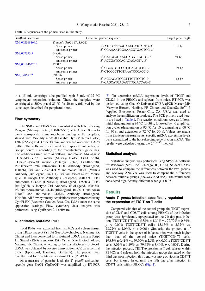
Table 1. Sequences of the primers used in this study.
GenBank accession Gene and primer sequence Target gene length
XM_002368164.2 T. gondii SAG1 (TgSAG1)Sense primer 50–ATCGCCTGAGAAGCATCACTG–30 101 bpAntisense primer 50–CGAAAATGGAAACGTGACTGG–30
NM_007393.5 b-actinSense primer 50–GATGCAGAAGGAGATTACTG–30 91 bpAntisense primer 50–ACCGATCCACACAGAGTA–30
NM_001146325.1 TIGITSense primer 50–GGCATGTCGCTTCAGTCTTC–30 139 bpAntisense primer 50–CTCCCCTTGTAAATCCCACC–30
NM_178687.2 CD226Sense primer 50–ACCACATGGCTTTCTTGCTC–30 112 bpAntisense primer 50–CAGCATGAGAGTTGGACCAG–30
S. Wang et al.: Parasite 2021, 28, 13 3

Acute T. gondii infection selectivelydownregulated the expression of CD226 on T cells
In contrast to that of TIGIT, even at 7 days after infection, theexpression of CD226 on T cells was hardly affected by T. gondiiinfection (CD226+CD4+ T cells among PBMCs: 27.31% ±5.671% vs. 31.74% ± 4.430%, p = 0.48; CD226+CD8+ T cellsamong PBMCs: 93.28% ± 3.949% vs. 86.42% ± 6.86%,p = 0.16; CD226+CD4+ T cells in the spleen: 32.53% ±3.249% vs. 28.80% ± 3.73%, p = 0.31; CD226+CD8+ T cellsin the spleen: 84.03% ± 0.725% vs. 83.31% ± 2.275%,
p = 0.77). CD226 appeared more frequently on the cell surfaceof CD8+ T cells than CD4+ T cells. However, on the third dayafter infection, the number of CD226+ T cells was lower in theinfection group than that in the Nc group (Fig. 2).
Acute T. gondii infection triggers an increasein TEM cells among TIGIT+T cells
Memory T cells play a critical role in providing long-termimmunity. Memory T cells are divided into four subsets based
Figure 1. Changes in TIGIT expression on T cells in different tissuesafter T. gondii infection. (A) Proportions of TIGIT+ cells amongCD4+ and CD8+ T cells in T. gondii-infected (RH) and normal mice(Nc) at 7 days after infection. (B) Results of statistical analysis of thepercentage of TIGIT+ cells among CD4+T and CD8+T cells in RHand Nc mice at 7 days after infection. (C) Dynamic changes in thepercentages of TIGIT+ in T cells at different time points. The resultsare representative of three independent experiments with five mice ineach group per experiment, with data denoting means ± SDs.
Figure 2. Changes in CD226 expression on T cells in different tissuesafter T. gondii infection. (A) Proportions of CD226+ cells amongCD4+T and CD8+T cells in T. gondii-infected (RH) and normal mice(Nc) at 7 days after infection. (B) Results of statistical analysis of thepercentage of CD226+ cells among CD4+T and CD8+T in RH andNc mice at 7 days after infection. (C) Dynamic changes in thepercentages of CD226+ T cells at different time points. The resultsare representative of three independent experiments with five mice ineach group per experiment, with data denoting means ± SDs.
4 S. Wang et al.: Parasite 2021, 28, 13

on the expression of the cell surface markers CD44 andCD62L. We classified cells based on the expectation thatcentral memory T cells are CD44hiCD62Lhi, effector memorycells are CD44hi CD62Llo, effector memory T cells are
CD44loCD62Llo, and naive memory cells are CD44loCD62Lhi.Memory T cells subset analysis was performed on bothTIGIT+CD4+ and TIGIT+CD8+T cells. As shown in Figure 3,we found that TCM and TEM cells were the predominant subsets
Figure 3. Relative contributions of memory T cell subsets of TIGIT+ T cells after T. gondii infection. (A) Dynamic changes in memory T cellsubsets of TIGIT+ T cell in PBMCs at different time points following RH infection. (B) Dynamic changes in memory T cell subsets ofTIGIT+T cells in the spleen at different time points following RH infection. The results are representative of three independent experimentswith five mice in each group per experiment, with data denoting means ± SDs.
S. Wang et al.: Parasite 2021, 28, 13 5

among T. gondii-specific TIGIT+T cells. The phenotypicchanges of TIGIT+T cells in PBMCs were specific. The TCMsubset was activated and transformed into TEM cells on the3rd day after infection in TIGIT+CD8+ T cells and on the 5thday after infection in TIGIT+CD4+ T cells. On the 3rd and7th days post infection, the TCM subset of T. gondii-specificTIGIT+CD4+ T cells in the spleen was activated and trans-formed into TEM cells. For the TIGIT+CD8+ T cells in thespleen, the TCM subset was activated and transformed intoTEM cells on the 3rd day post infection, but the oppositehappened on the 7th day post infection (Fig. 3).
Histopathological changes in the spleen wereaggravated by an increasing T. gondii parasiteload
The relative expression of the T. gondii tachyzoite-specificgene SAG1 in PBMCs and spleen increased significantly fromthe first day post infection (Fig. 4A). Meanwhile, from the 3rdday after infection, the spleens of mice in the infected grouprapidly enlarged, and the spleen index was significantly higherthan in the control group (Fig. 4B). Through H&E staining of
spleen sections, we observed little change in the spleen onthe first day post infection; the splenic sinus was intact, andthe course of the splenic cord was normal. On the 3rd day postinfection, infiltration of inflammatory cells, congestion in themiddle of the splenic cord, and a compensatory increase inlymphocytes were observed. On the 7th day after infection,there was necrosis of spleen cells, complete destruction of thespleen structure, a decrease in lymphocytes, and a large numberof tachyzoites and pseudocysts (Fig. 4C). The dynamic changesin TgSAG1 gene expression appeared consistent with thespleen pathology from days 3 to 7 post infection. These resultsshowed that T. gondii tachyzoites quickly invaded the circula-tory system and were then transported to various organs. Thentachyzoites further proliferated in the spleen by means of animmune escape mechanism and gradually destroyed spleenfunction.
TIGIT and CD226 mRNA expression in PBMCsand spleen
As shown in Figure 5, the relative expression level ofTIGIT downregulated in the PBMCs and spleens of infected
Figure 4. Dynamic pathological changes in the spleen during acute T. gondii infection. (A) Relative expression of TgSAG1 at 0, 1, 3, 5, and7 days post infection in the PBMCs and spleen. (B) Spleen index values of the RH and Nc groups. The results are representative of threeindependent experiments with five mice per group per experiment; with data denoting means ± SDs; (C) H&E staining of spleen sections atdifferent time points in the RH infection group. The original magnification was 100�, and the corresponding images on the right weremagnified at 400�.
6 S. Wang et al.: Parasite 2021, 28, 13

mice on the first day after infection, while the expression of cos-timulatory receptor CD226 in the spleen of infected miceincreased significantly. From the 3rd day after infection, theexpression of TIGIT in PBMCs and spleens of the infectedgroup was significantly higher than in the control group, andthe down-regulation of CD226 was also observed at thesame time point. These results showed that with an increasingT. gondii parasite load, the increased expression of TIGIT com-petitively suppressed the expression of CD226, resulting in asignificant inhibition of cell-mediated immunity in miceinfected with T. gondii.
Discussion
Previous studies have shown that TIGIT plays a pivotalinhibitory role in the immune system. However, research onTIGIT in the context of parasite infection is relatively scarce.Zhang et al. [24] found that the expression of TIGIT, CD3e,CD4 and CD8B was upregulated in the central liver tissue ofpatients with alveolar echinococcosis, and the expression ofTIGIT was positively correlated with the expression of thesefactors. The same results were also observed in the mouseinfection model. Further studies showed that blocking treatmentwith a TIGIT monoclonal antibody could significantly increasethe content of CD4-Teffs (CD4+Foxp3� T cells) and the ability
of liver infiltrating T cells to secrete cytokines such as IL-2,IFN-c and TNF-a. Infection with Plasmodium yoelii couldinduce high expression of TIGIT on splenic CD4+T cells ininfected mice [21]. Zhang et al. [25] further demonstrated thatthe compensatory increase in TIGIT in P. berghei ANKA-infected mice was caused by blockade of the TIM-3-Gal-9 path-way with the TIM-3 ligand Galectin (Gal)-9 blocker a-lactose,which may be one of the causes of death in mice. Zhang et al.[14] found that TIGIT can enhance the proliferation of CD4+ Tcells in the spleen of mice infected with S. japonicum and theninduce higher expression of IL-4 and lower expression of IFN-cto promote activation of the Th2 immune response. However,the expression pattern and function of TIGIT in the immunecells of mice infected with T. gondii were unknown.
In this study, we found for the first time that the expressionof TIGIT on the surface of CD4+ and CD8+T cells in the spleenand PBMCs decreased in the early stage but increased signifi-cantly in the late stage of acute T. gondii infection in a mousemodel. On the 3rd day after acute infection, the expression ofTIGIT and CD226 decreased in the PBMCs and spleens ofthe mice, suggesting that the immune system was activatedand functional in the early stage of T. gondii infection, produc-ing a large number of immune cells, enhancing immuneactivity, and eliminating free tachyzoites; however, at the sametime, T. gondii formed pseudocysts to achieve immune escape.In the later stage of infection, T. gondii continuously disrupted
Figure 5. mRNA expression of TIGIT and CD226 in the PBMCs and spleens from mice infected with the T. gondii RH strain, as assessed byqRT-PCR. A: Data obtained from PBMCs. B: Data obtained from the spleen. Values are the means from triplicate measurements, with datadenoting means ± SDs; three independent experiments were performed with five mice per group. *p < 0.05, **p < 0.01 and ***p < 0.001(compared to the control).
S. Wang et al.: Parasite 2021, 28, 13 7

the immune system, resulting in impaired immune activity. Onthe 7th day after infection, the expression of TIGIT in PBMCsand the spleen was significantly upregulated, eventually leadingto immune failure and death. Studies have shown that the hostT cell-mediated immune response plays an important role in theresponse to pathogen infection. Ackermann et al. [1] found thatthe number of TIGIT+CD4+T cells in patients with acutehepatitis C virus (HCV) infection was significantly higher thanin healthy controls, and TIGIT was highly expressed in allCD4+Tm subsets (memory T cells). However, the mechanismof immune cell failure caused by pathogen infection, as wellas whether this process is related to TIGIT, has not been wellstudied until now.
To further explore the effect of T. gondii infection onthe phenotypic changes of TIGIT+T cells, we analyzed theexpression of CD44 and CD62L on TIGIT+ T cells by flowcytometry. The results showed that the TIGIT+ T cells of boththe Nc and RH groups were mainly TCM cells, and the rest wereTEM cells. TCM cells usually reside in T cell areas of secondarylymphoid organs and readily proliferate and differentiate intoTEM cells, which are the predominant population elicited inresponse to antigenic stimulation during parasitic infection.These cells have little or no effector function, but persistentantigen stimulation maintains the effector function of memoryT cells at a high level, which eventually leads to T cell exhaus-tion [18]. In this study, T. gondii-specific TIGIT+ TEM cells inthe spleen increased on the 3rd and 7th days post infection,indicating that TCM cells were triggered to differentiate intoTEM cells due to the increase in parasite load and antigenstimulation.
In addition, specific TgSAG1 expression in PBMCs andspleen increased significantly after T. gondii infection, andthe expression of TIGIT was also considerably up-regulated,indicating that T. gondii infection can induce a large amountof expression of immunosuppressive receptors in the host, thusactivating an immune escape mechanism and massive prolifer-ation in the host, which eventually leads to destruction of theimmune system and damage to immune function. Histopatho-logical changes in the spleen were aggravated by an increasingT. gondii parasite load. By the 7th day after infection, thesplenic structure of infected mice was completely destroyed,and the relative expression of TIGIT increased significantly,while T. gondii was still proliferating. These results are consis-tent with reports that T. gondii can cause host T cell exhaustion.However, the effect of TIGIT on the proliferation and cytokinesecretion of T. gondii-specific T cells is not clear, and whetherT cell function can be restored after blocking the TIGITpathway remains to be studied further.
Conclusion
Conclusively, our results indicated that acute T. gondiiinfection can increase the expression of TIGIT in host T cellsand stimulate the transformation of TIGIT+ TCM cells intoTIGIT+ TEM cells. Whether the increase in TIGIT expressioninduces exhaustion of host T cells during acute T. gondiiinfection, and whether TIGIT signaling blockade reverses thefunctional impairment of T cells needs to be studied further.
Conflict of interest
The authors declare that they have no conflict of interest.
Acknowledgements. The current work received support from theNational Natural Science Foundation of China (No. 81702025),and the Science and Technology Planning Project of Henan Province(Nos. 182102310220 and 182102310431).
References
1. Ackermann C, Smits M, Woost R, Eberhard JM, Peine S,Kummer S, Marget M, Kuntzen T, Kwok WW, Lohse AW,Jacobs T, Boettler T, Schulze Zur Wiesch J. 2019. HCV-specificCD4+ T cells of patients with acute and chronic HCV infectiondisplay high expression of TIGIT and other co-inhibitorymolecules. Scientific Reports, 9(1), 10624.
2. Berrocal Almanza LC, Muñoz M, Kühl AA, Kamradt T,Heimesaat MM, Liesenfeld O. 2013. Tim-3 is differentlyexpressed in genetically susceptible C57BL/6 and resistantBALB/c mice during oral infection with Toxoplasma gondii.European Journal of Microbiology & Immunology, 3(3), 211–221.
3. Bhadra R, Gigley JP, Weiss LM, Khan IA. 2011. Control ofToxoplasma reactivation by rescue of dysfunctional CD8+ T-cell response via PD-1-PDL-1 blockade. Proceedings of theNational Academy of Sciences of the United States of America,108(22), 9196–9201.
4. Boles KS, Vermi W, Facchetti F, Fuchs A, Wilson TJ, DiacovoTG, Cella M, Colonna M. 2009. A novel molecular interactionfor the adhesion of follicular CD4 T cells to follicular DC.European Journal of Immunology, 39(3), 695–703.
5. Bottino C, Castriconi R, Pende D, Rivera P, Nanni M,Carnemolla B, Cantoni C, Grassi J, Marcenaro S, Reymond N,Vitale M, Moretta L, Lopez M, Moretta A. 2003. Identification ofPVR (CD155) and Nectin-2 (CD112) as cell surface ligands forthe human DNAM-1 (CD226) activating molecule. Journal ofExperimental Medicine, 198(4), 557–567.
6. Casado JG, Pawelec G, Morgado S, Sanchez-Correa B, DelgadoE, Gayoso I, Duran E, Solana R, Tarazona R. 2009. Expressionof adhesion molecules and ligands for activating and costim-ulatory receptors involved in cell-mediated cytotoxicity in alarge panel of human melanoma cell lines. Cancer Immunology,Immunotherapy, 58(9), 1517–1526.
7. Edwards JF, Dubey JP. 2013. Toxoplasma gondii abortion stormin sheep on a Texas farm and isolation of mouse virulent atypicalgenotype T. gondii from an aborted lamb from a chronicallyinfected ewe. Veterinary Parasitology, 192(1–3), 129–136.
8. Flegr J, Prandota J, Sovickova M, Israili ZH. 2014. Toxoplas-mosis–a global threat. Correlation of latent toxoplasmosis withspecific disease burden in a set of 88 countries. PLoS One, 9(3),e90203.
9. Guirelli PM, Angeloni MB, Barbosa BF, Gomes AO, CastroAS, Franco PS, Silva RJ, Oliveira JG, Martins-Filho OA, MineoJR, Ietta F, Ferro EA. 2015. Trophoblast-macrophage crosstalkon human extravillous under Toxoplasma gondii infection.Placenta, 36(10), 1106–1114.
10. Hwang S, Khan IA. 2015. CD8+ T cell immunity in anencephalitis model of Toxoplasma gondii infection. Seminars inImmunopathology, 37(3), 271–279.
11. Johnston RJ, Comps-Agrar L, Hackney J, Yu X, Huseni M,Yang Y, Park S, Javinal V, Chiu H, Irving B, Eaton DL, GroganJL. 2014. The immunoreceptor TIGIT regulates antitumor andantiviral CD8(+) T cell effector function. Cancer Cell, 26(6),923–937.
8 S. Wang et al.: Parasite 2021, 28, 13
12. Landrith TA, Harris TH, Wilson EH. 2015. Characteristics andcritical function of CD8+ T cells in the Toxoplasma-infectedbrain. Seminars in Immunopathology, 37(3), 261–270.
13. Levin SD, Taft DW, Brandt CS, Bucher C, Howard ED,Chadwick EM, Johnston J, Hammond A, Bontadelli K,Ardourel D, Hebb L, Wolf A, Bukowski TR, Rixon MW,Kuijper JL, Ostrander CD, West JW, Bilsborough J, Fox B, GaoZ, Xu W, Ramsdell F, Blazar BR, Lewis KE. 2011. Vstm3 is amember of the CD28 family and an important modulator ofT-cell function. European Journal of Immunology, 41(4),902–915.
14. Zhang L, Wang X, Qi Q, Dong L, Xu L, Pu Y, Wei C, Zhu J,Zhou S, Li Y, Liu F, Chen X, Su C. 2018. Study on role ofTIGIT signal in Th1/Th2 balance in Schistosoma japonicum-infected mice. Chinese Journal of Schistosomiasis Control,30(2), 136–139.
15. Lozano E, Dominguez-Villar M, Kuchroo V, Hafler DA. 2012.The TIGIT/CD226 axis regulates human T cell function. Journalof Immunology, 188(8), 3869–3875.
16. Montoya JG, Liesenfeld O. 2004. Toxoplasmosis. Lancet, 363(9425), 1965–1976.
17. Ochiai E, Sa Q, Perkins S, Grigg ME, Suzuki Y. 2016. CD8(+)T cells remove cysts of Toxoplasma gondii from the brainmostly by recognizing epitopes commonly expressed by orcross-reactive between type II and type III strains of the parasite.Microbes and Infection, 18(7–8), 517–522.
18. Opata MM, Stephens R. 2013. Early decision: Effector andeffector memory T cell differentiation in chronic infection.Current Immunology Reviews, 9(3), 190–206.
19. Schietinger A, Greenberg PD. 2014. Tolerance and exhaustion:defining mechanisms of T cell dysfunction. Trends in Immunol-ogy, 35(2), 51–60.
20. Vado-Solis IA, Suarez-Solis V, Jimenez-Delgadillo B, Zavala-Velazquez JE, Segura-Correa JC. 2013. Toxoplasma gondiipresence in women with spontaneous abortion in Yucatan,Mexico. Journal for Parasitology, 99(2), 383–385.
21. Villegas-Mendez A, Inkson CA, Shaw TN, Strangward P, CouperKN. 2016. Long-Lived CD4+IFN-c+ T Cells rather than short-lived CD4+IFN-c+IL-10+ T cells initiate rapid IL-10 productionto suppress anamnestic T cell responses during secondary malariainfection. Journal of Immunology, 197(8), 3152–3164.
22. Xiao Y, Yin J, Jiang N, Xiang M, Hao L, Lu H, Sang H, Liu X,Xu H, Ankarklev J, Lindh J, Chen Q. 2010. Seroepidemiologyof human Toxoplasma gondii infection in China. BMCInfectious Diseases, 10, 4.
23. Ye B, Liu X, Li X, Kong H, Tian L, Chen Y. 2015. T-cellexhaustion in chronic hepatitis B infection: current knowledgeand clinical significance. Cell Death & Disease, 6, e1694.
24. Zhang C, Lin R, Li Z, Yang S, Bi X, Wang H, Aini A,Zhang N, Abulizi A, Sun C, Li L, Zhao Z, Qin R, Li X, Li L,Aji T, Shao Y, Vuitton DA, Tian Z, Wen H. 2020. Immuneexhaustion of T Cells in alveolar echinococcosis patients and itsreversal by blocking checkpoint receptor TIGIT in a murinemodel. Hepatology, 71(4), 1297–1315.
25. Zhang Y, Jiang N, Zhang T, Chen R, Feng Y, Sang X, Yang N,Chen Q. 2019. Tim-3 signaling blockade with alpha-lactoseinduces compensatory TIGIT expression in Plasmodium bergheiANKA-infected mice. Parasites & Vectors, 12(1), 534.
Cite this article as: Wang S, Li H, Zhang F, Jiao Y, Xie Q, Zhang Z & Li X. 2021. Expression of TIGIT in splenic and circulatory T cellsfrom mice acutely infected with Toxoplasma gondii. Parasite 28, 13.
An international open-access, peer-reviewed, online journal publishing high quality paperson all aspects of human and animal parasitology
Reviews, articles and short notes may be submitted. Fields include, but are not limited to: general, medical and veterinary parasitology;morphology, including ultrastructure; parasite systematics, including entomology, acarology, helminthology and protistology, andmolecularanalyses; molecular biology and biochemistry; immunology of parasitic diseases; host-parasite relationships; ecology and life history ofparasites; epidemiology; therapeutics; new diagnostic tools.All papers in Parasite are published in English. Manuscripts should have a broad interest and must not have been published or submittedelsewhere. No limit is imposed on the length of manuscripts.
Parasite (open-access) continues Parasite (print and online editions, 1994-2012) and Annales de Parasitologie Humaine et Comparée(1923-1993) and is the official journal of the Société Française de Parasitologie.
Editor-in-Chief: Submit your manuscript atJean-Lou Justine, Paris http://parasite.edmgr.com/
S. Wang et al.: Parasite 2021, 28, 13 9