
Exposure Factors or Prime Factors 1. PRIME FACTORS What is “technique” ? How does it affect the...
49
Exposure Factors or Prime Factors 1
-
Upload
horatio-allison -
Category
Documents
-
view
220 -
download
3
Transcript of Exposure Factors or Prime Factors 1. PRIME FACTORS What is “technique” ? How does it affect the...
Exposure Factors or Prime Factors
1
PRIME FACTORS
What is “technique” ?
How does it affect the “image”
2
Exposure Factors – 3 or 4
The four prime exposure factors are: Voltage = kVp* Current = mA* Exposure time = seconds or fractions of a sec* Source-to-image distance = SID
3
PRIME FACTORS
• KVP
• MAS
• DISTANCE
4
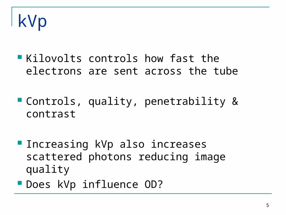
kVp
Kilovolts controls how fast the electrons are sent across the tube
Controls, quality, penetrability & contrast
Increasing kVp also increases scattered photons reducing image quality
Does kVp influence OD?
5
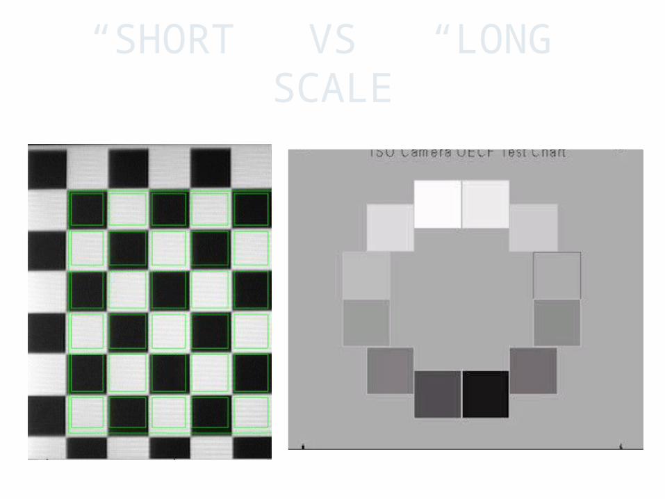
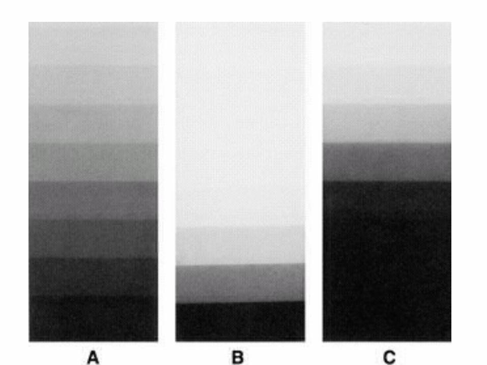
“SHORT” VS “LONG” SCALE
6
kVp
• Low kVp (50 – 60)
• Short scale
• High contrast
• “Bone work”
7
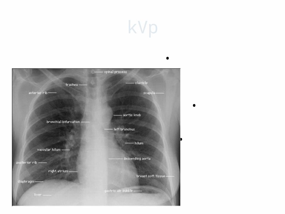
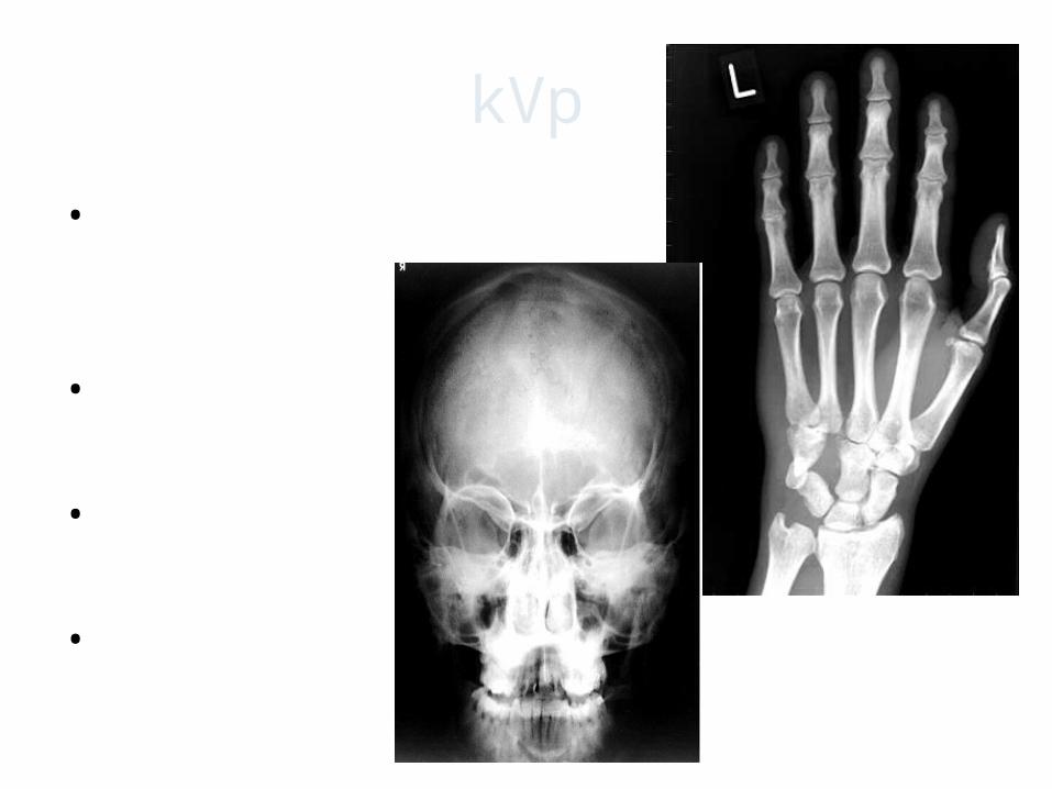
kVp
• High kVp (90 – 120)
• Long scale
• Low contrast
• “Chest images”
8
9
10
11
12
mA
Determines the number of photons, radiation quantity, OD & patient dose
Changing mA does not change the kinetic energy of e-
Available mA stations are usually 50, 100, 200, 300, 400 & 600
13
14
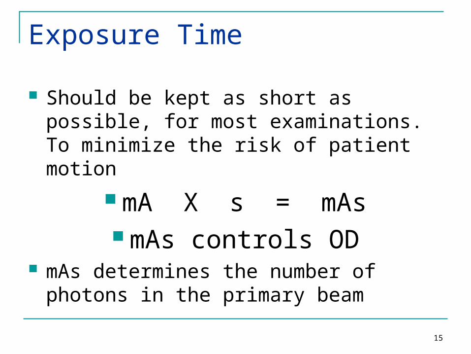
Exposure Time
Should be kept as short as possible, for most examinations. To minimize the risk of patient motion
mA X s = mAs mAs controls OD
mAs determines the number of photons in the primary beam
15
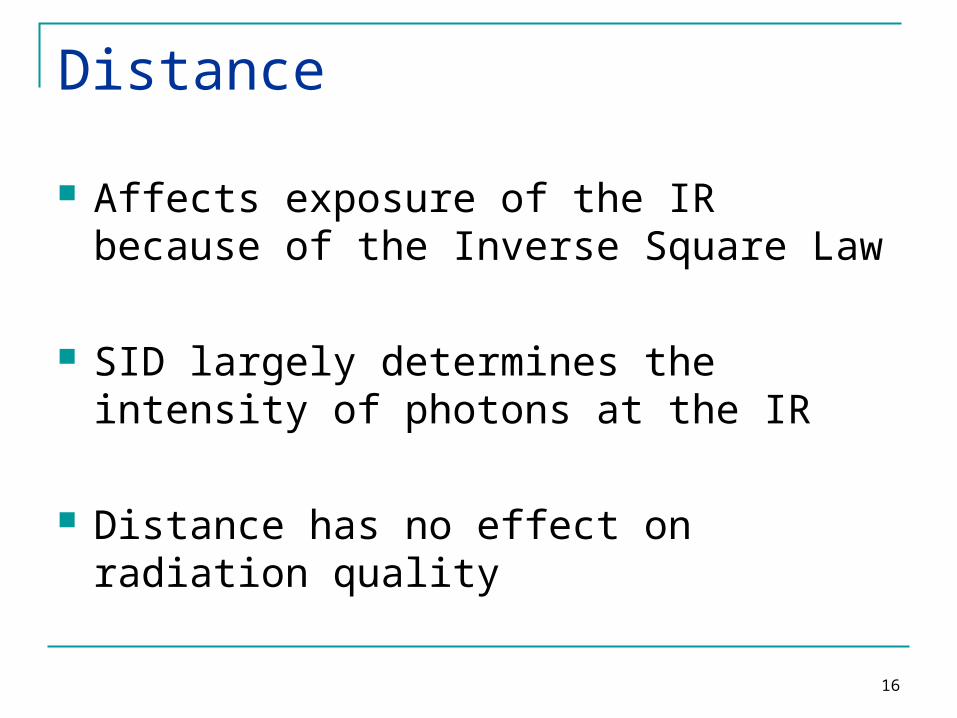
Distance
Affects exposure of the IR because of the Inverse Square Law
SID largely determines the intensity of photons at the IR
Distance has no effect on radiation quality
16
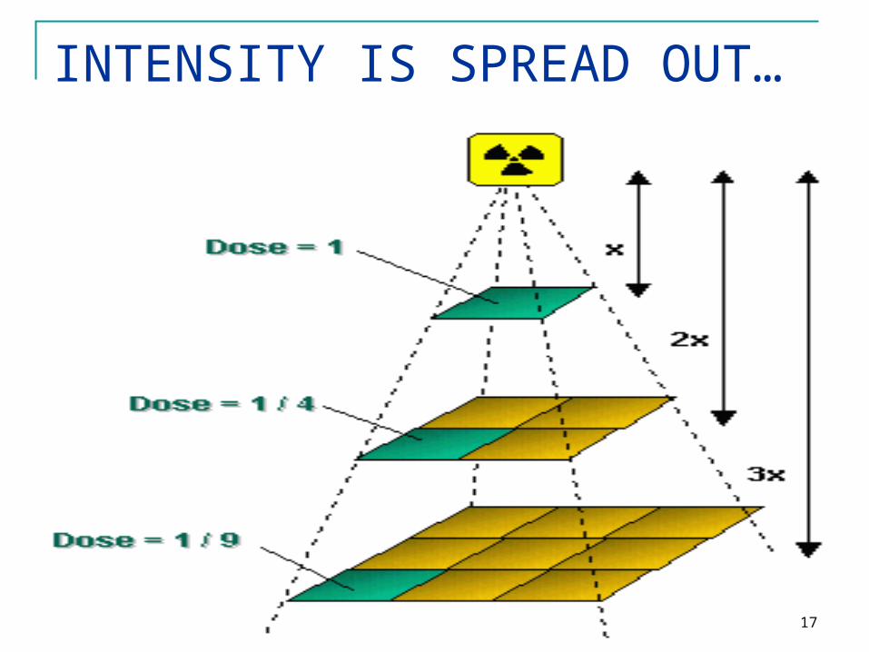
INTENSITY IS SPREAD OUT…
17
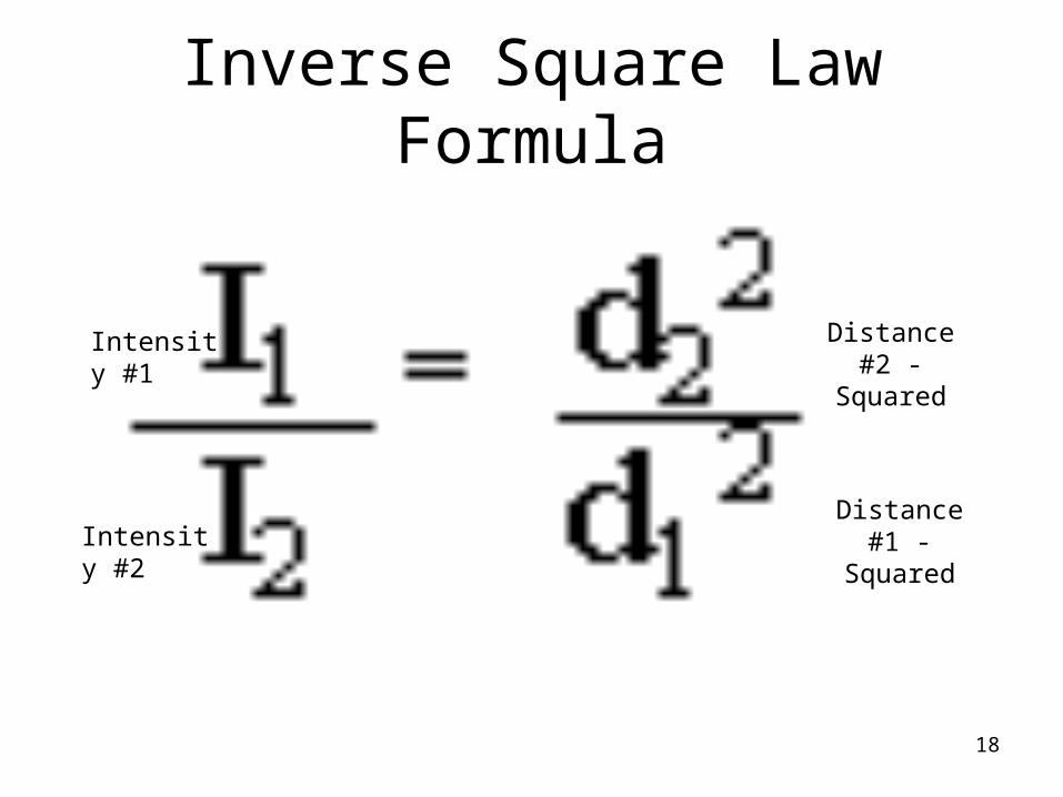
Inverse Square Law Formula
Intensity #1
Intensity #2
Distance #2 - Squared
Distance #1 - Squared
18
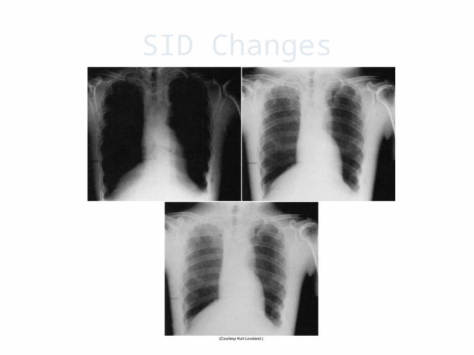
SID Changes
19

Direct Square Law
• New mAs = New distance 2
Old mAs Old distance 2
20
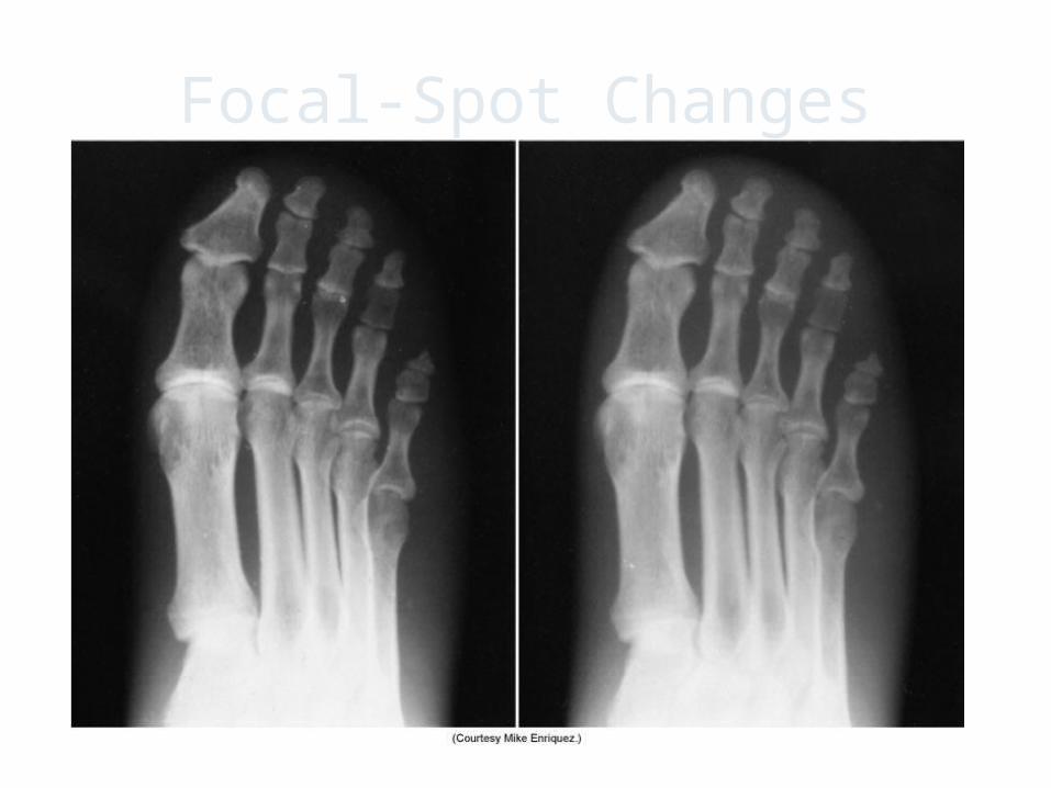
Focal-Spot Changes
21
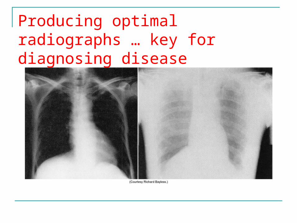
Producing optimal radiographs … key for diagnosing disease
22
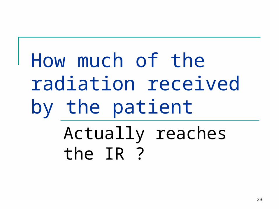
How much of the radiation received by the patient
Actually reaches the IR ?
23
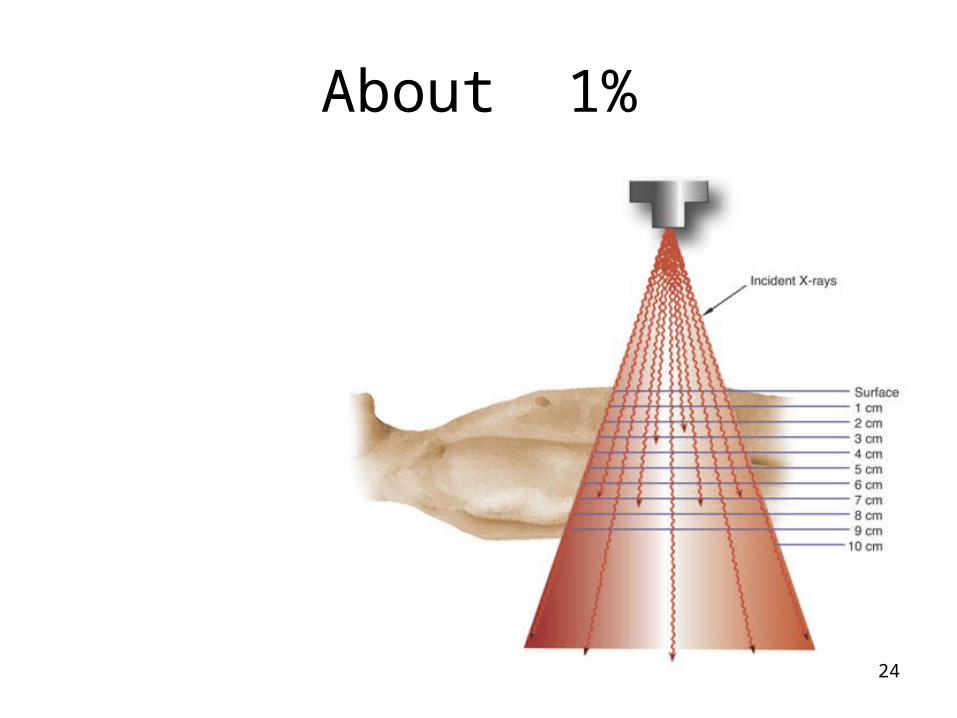
About 1%
24
25

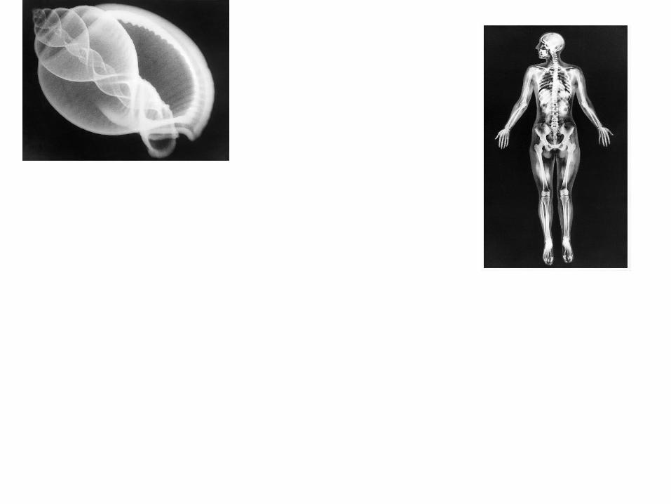
Creating the IMAGE
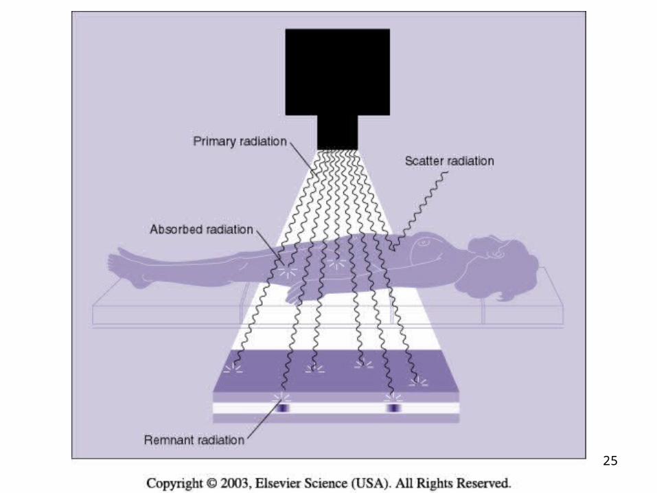
• When x-rays pass through a patient's body, three things can happen:
• (1) the x-ray photon is transmitted, passing through the body, interacting with the film, and producing a dark area on the film;
• (2) the x-ray photon is absorbed in an area of greater tissue density, producing lighter areas on the film; and
• (3) the x-ray photon is scattered and reaches the film causing an overall gray fog.
26

IMAGES
• DENSITY = THE AMOUNT OF BLACKENING “DARKNESS” ON THE RADIOGRAPH
• CONTRAST – THE DIFFERENCES BETWEEN THE BLACKS TO THE WHITES
27
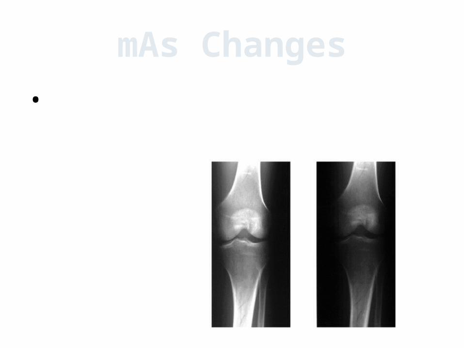
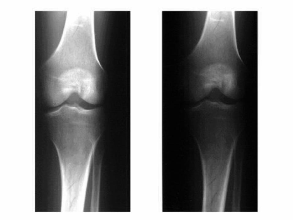
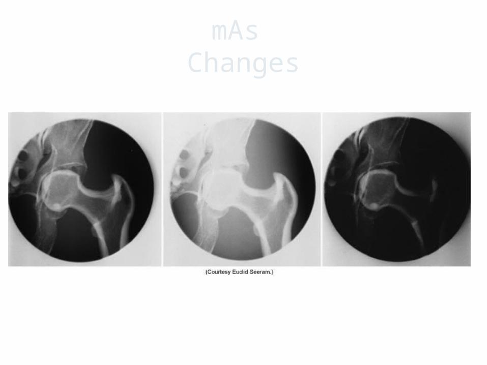
mAs Changes
• at least 20 - 30 % mas change needed to see a visible change in density
28
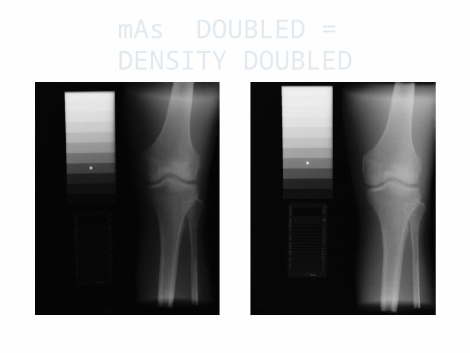
mAs DOUBLED = DENSITY DOUBLED
29
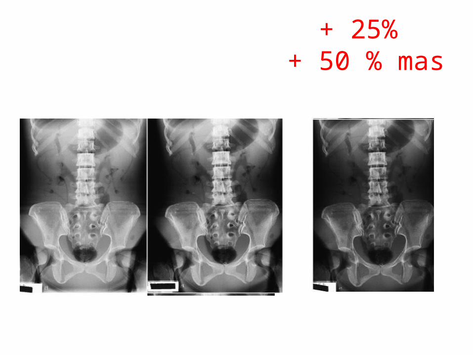
+ 25% + 50 % mas
30
mAs Changes
31
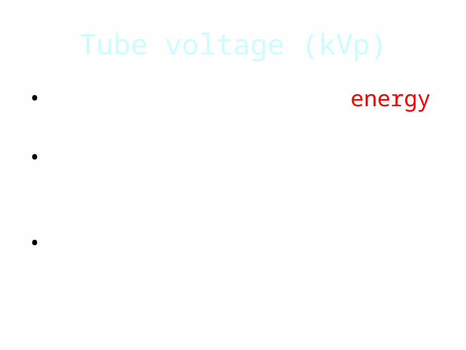
Tube voltage (kVp)
• Determines the maximum energy in the beam
• spectrum and affects the quality of the output spectrum
• Efficiency of x-ray production is directly related to tube voltage
32
Influencing factors: kVp
15% rule: 15% kVp = doubling of exposure to the film
15% kVp = halving of exposure to the film
15% rule will always change the contrast of the image because kV is the primary method of changing image contrast.
Remember : 15% change ( ) KVP has the same effect as
doubling or ½ the MAS on density
33
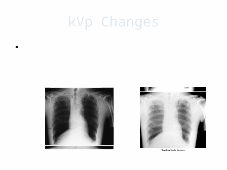
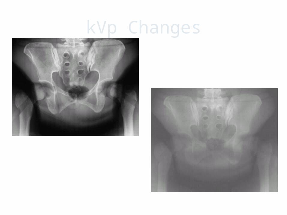
kVp Changes
• The kVp setting must be changed by at least 4% to produce visual changes an image
34
kVp Changes
35
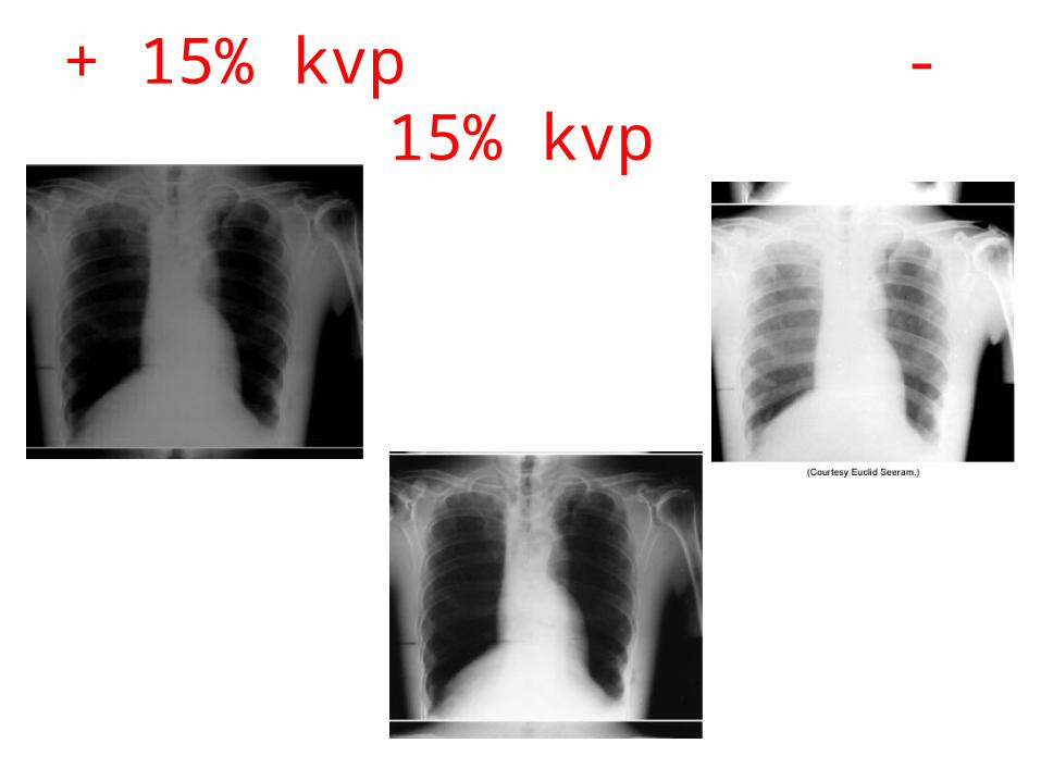
+ 15% kvp - 15% kvp
36
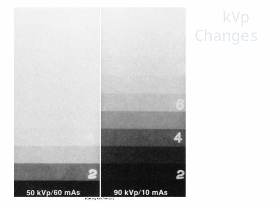
kVp Changes
37
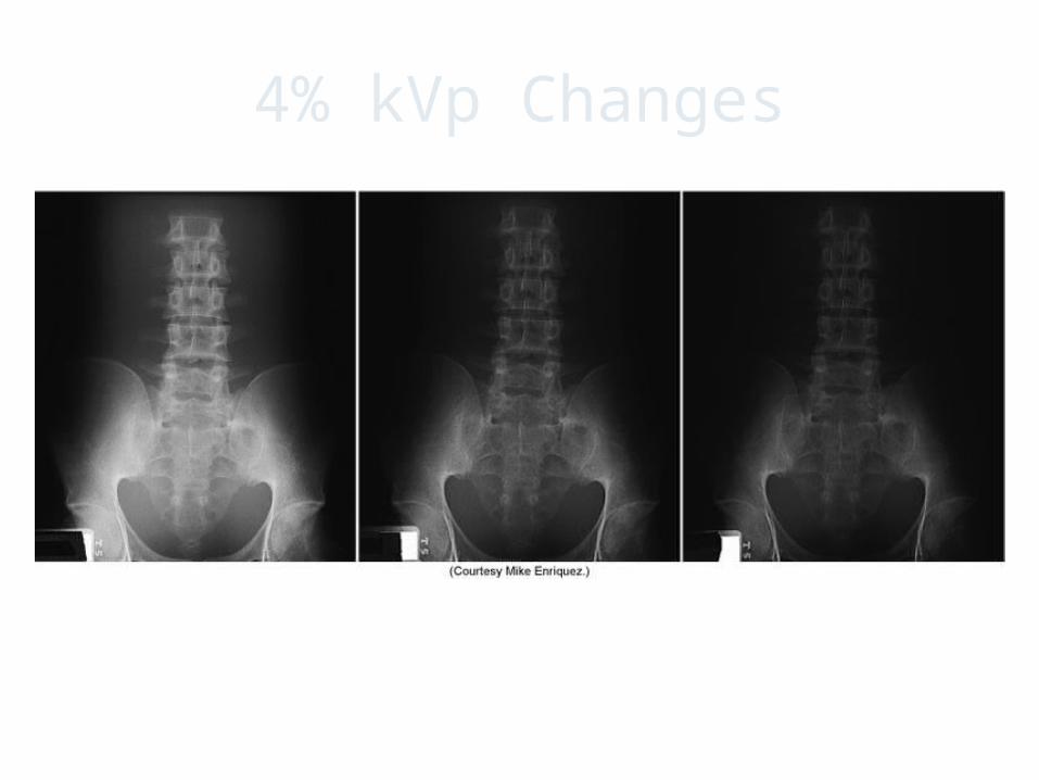
4% kVp Changes
38
Determining Radiographic TechniqueThe Patient Factor The most difficult task for technologists…
evaluating your patient
The patient size, shape, and physical condition greatly influences the technique selection
39
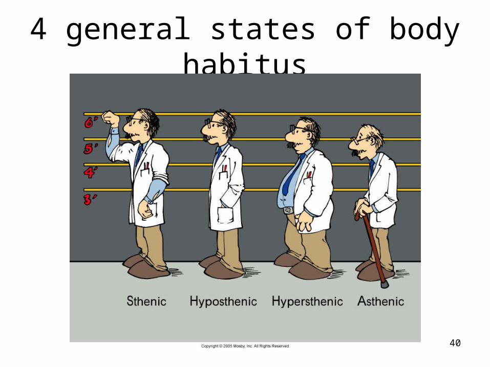
4 general states of body habitus
40
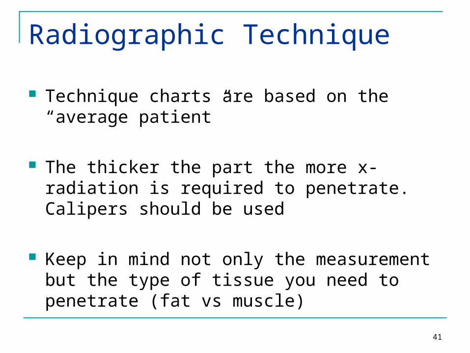
Radiographic Technique
Technique charts are based on the “average patient”
The thicker the part the more x-radiation is required to penetrate. Calipers should be used
Keep in mind not only the measurement but the type of tissue you need to penetrate (fat vs muscle)
41
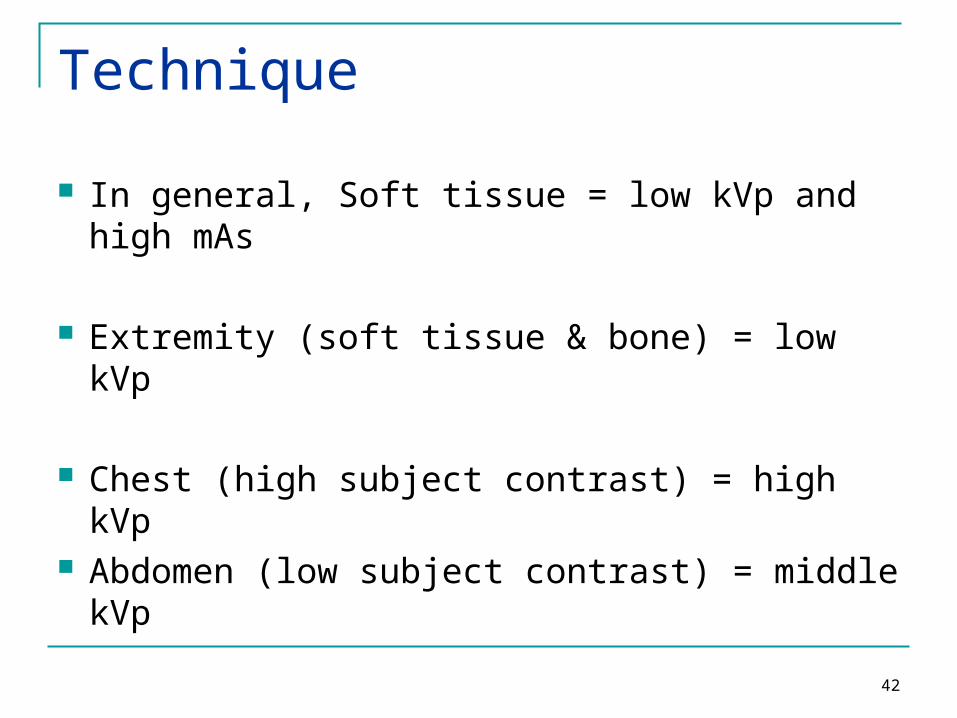
Technique
In general, Soft tissue = low kVp and high mAs
Extremity (soft tissue & bone) = low kVp
Chest (high subject contrast) = high kVp Abdomen (low subject contrast) = middle kVp
42
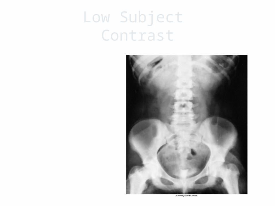
Low Subject Contrast
43
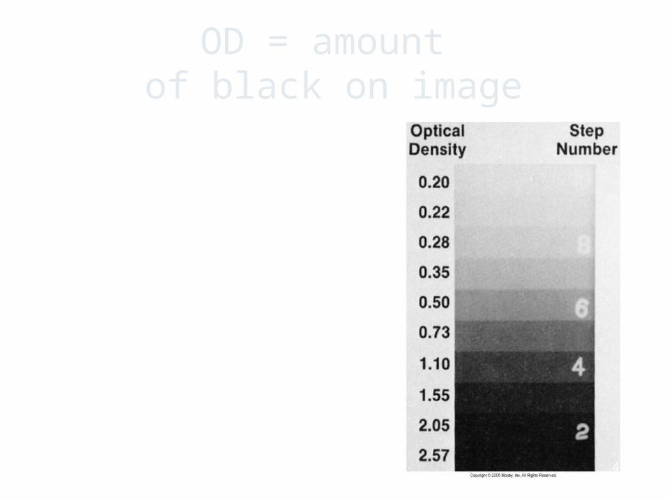
OD = amount of black on image
44
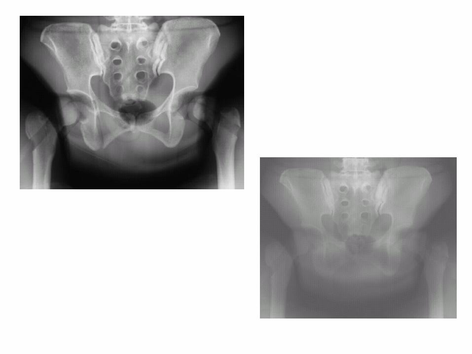
Film Screen• Overexposed
• Referring to a radiograph that is too dark because too much x-radiation reached the image receptor
• Underexposed
• Referring to a radiograph that is too light because too little x-radiation reached the image receptor
45
Technique - Pathology
Pathology can severely affect the technologist technique selection
Always question your patients about health status
If prior images are available…check them!
46
Pathology
Can appear with increased radiolucency or radiopacity
Some pathology is destructive causing tissue to be radiolucent
Others can be additive causing tissue to be radiopaque
47
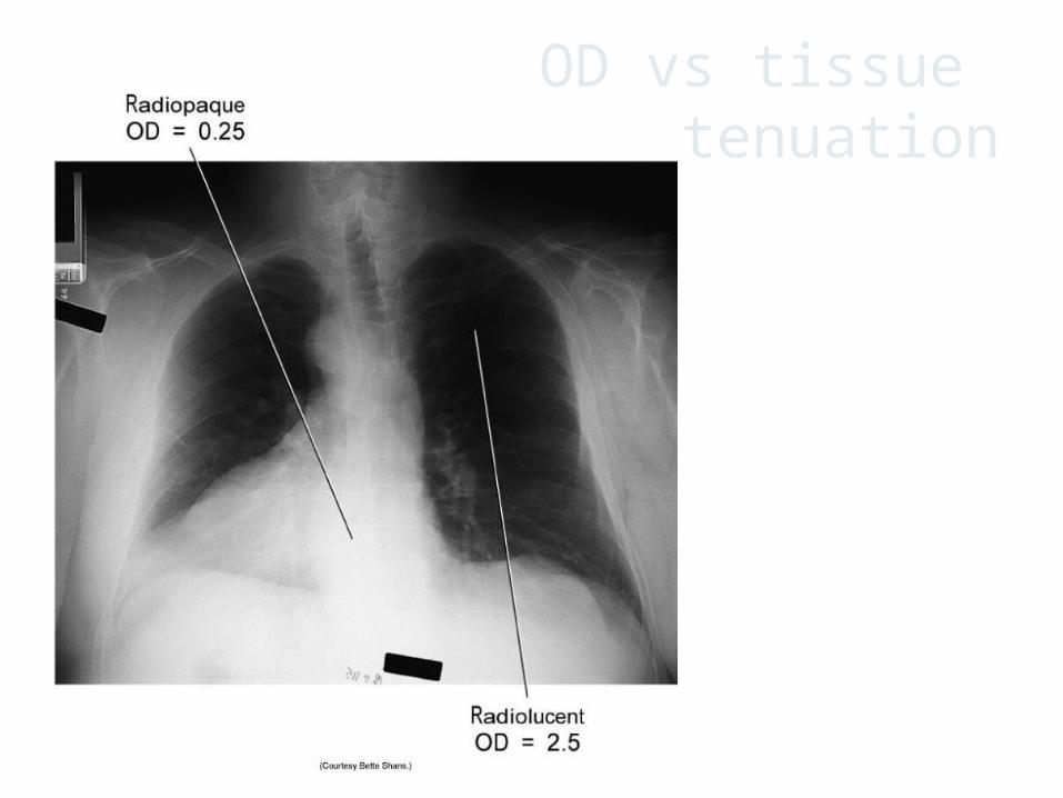
OD vs tissue attenuation
48
Technique selection – Fixed kVp For each anatomic part there is an optimum
kVp
mAs is varied based on part thickness or pathological condition
49


![Factors tht affect recruitment[ppt]](https://static.fdocuments.net/doc/165x107/54541a88b1af9f84228b493f/factors-tht-affect-recruitmentppt.jpg)


