Exploratory proteomic analysis implicates the...
13
ARTICLE Exploratory proteomic analysis implicates the alternative complement cascade in primary CNS vasculitis Caleigh Mandel-Brehm, PhD,* Hanna Retallack, AB,* Giselle M. Knudsen, PhD, Alex Yamana, BS, Rula A. Hajj-Ali, MD, Leonard H. Calabrese, DO, Tarik Tihan, MD, PhD, Hannah A. Sample, BS, Kelsey C. Zorn, MHS, Mark P. Gorman, MD, Jennifer Madan Cohen, MD, Antoine G. Sreih, MD, Jacqueline F. Marcus, MD, S. Andrew Josephson, MD, Vanja C. Douglas, MD, Jeffrey M. Gelfand, MD, Michael R. Wilson, MD, and Joseph L. DeRisi, PhD Neurology ® 2019;93:e1-e12. doi:10.1212/WNL.0000000000007850 Correspondence Dr. DeRisi [email protected] Abstract Objective To identify molecular correlates of primary angiitis of the CNS (PACNS) through proteomic analysis of CSF from a biopsy-proven patient cohort. Methods Using mass spectrometry, we quantitatively compared the CSF proteome of patients with biopsy-proven PACNS (n = 8) to CSF from individuals with noninflammatory conditions (n = 11). Significantly enriched molecular pathways were identified with a gene ontology workflow, and high confidence hits within enriched pathways (fold change >1.5 and concordant Benja- mini-Hochberg–adjusted p < 0.05 on DeSeq and t test) were identified as differentially regu- lated proteins. Results Compared to noninflammatory controls, 283 proteins were differentially expressed in the CSF of patients with PACNS, with significant enrichment of the complement cascade pathway (C4- binding protein, CD55, CD59, properdin, complement C5, complement C8, and complement C9) and neural cell adhesion molecules. A subset of clinically relevant findings were validated by Western blot and commercial ELISA. Conclusions In this exploratory study, we found evidence of deregulation of the alternative complement cascade in CSF from biopsy-proven PACNS compared to noninflammatory controls. More specifically, several regulators of the C3 and C5 convertases and components of the terminal cascade were significantly altered. These preliminary findings shed light on a previously un- appreciated similarity between PACNS and systemic vasculitides, especially anti-neutrophil cytoplasmic antibody–associated vasculitis. The therapeutic implications of this common bi- ology and the diagnostic and therapeutic utility of individual proteomic findings warrant validation in larger cohorts. *These authors contributed equally to this work. From the Departments of Biochemistry and Biophysics (C.M.-B., H.R., H.A.S., K.C.Z., J.L.D.), Pharmaceutical Chemistry (G.M.K., A.Y.), Pathology and Laboratory Medicine (T.T.), and Neurology (S.A.J., V.C.D., J.M.G., M.R.W.), University of California, San Francisco; Department of Rheumatology/Immunology (R.A.H.-A., L.H.C.), Cleveland Clinic, OH; Department of Neurology (M.P.G.), Boston Children’s Hospital, MA; Division of Neurology (J.M.C.), Connecticut Children’s Medical Center, Hartford; Division of Rheumatology (A.G.S.), University of Pennsylvania, Philadelphia; Kaiser Permanente (J.F.M.), San Francisco Medical Center; UCSF Weill Institute for Neurosciences (S.A.J., V.C.D., J.M.G., M.R.W.); and Chan Zuckerberg Biohub (J.L.D.), San Francisco, CA. Go to Neurology.org/N for full disclosures. Funding information and disclosures deemed relevant by the authors, if any, are provided at the end of the article. Copyright © 2019 American Academy of Neurology e1 Copyright © 2019 American Academy of Neurology. Unauthorized reproduction of this article is prohibited. Published Ahead of Print on July 3, 2019 as 10.1212/WNL.0000000000007850
Transcript of Exploratory proteomic analysis implicates the...
ARTICLE
Exploratory proteomic analysis implicates thealternative complement cascade in primary CNSvasculitisCaleigh Mandel-Brehm, PhD,* Hanna Retallack, AB,* Giselle M. Knudsen, PhD, Alex Yamana, BS,
Rula A. Hajj-Ali, MD, Leonard H. Calabrese, DO, Tarik Tihan, MD, PhD, Hannah A. Sample, BS,
Kelsey C. Zorn, MHS, Mark P. Gorman, MD, Jennifer Madan Cohen, MD, Antoine G. Sreih, MD,
Jacqueline F. Marcus, MD, S. Andrew Josephson, MD, Vanja C. Douglas, MD, Jeffrey M. Gelfand, MD,
Michael R. Wilson, MD, and Joseph L. DeRisi, PhD
Neurology® 2019;93:e1-e12. doi:10.1212/WNL.0000000000007850
Correspondence
Dr. DeRisi
AbstractObjectiveTo identify molecular correlates of primary angiitis of the CNS (PACNS) through proteomicanalysis of CSF from a biopsy-proven patient cohort.
MethodsUsing mass spectrometry, we quantitatively compared the CSF proteome of patients withbiopsy-proven PACNS (n = 8) to CSF from individuals with noninflammatory conditions (n =11). Significantly enriched molecular pathways were identified with a gene ontology workflow,and high confidence hits within enriched pathways (fold change >1.5 and concordant Benja-mini-Hochberg–adjusted p < 0.05 on DeSeq and t test) were identified as differentially regu-lated proteins.
ResultsCompared to noninflammatory controls, 283 proteins were differentially expressed in the CSFof patients with PACNS, with significant enrichment of the complement cascade pathway (C4-binding protein, CD55, CD59, properdin, complement C5, complement C8, and complementC9) and neural cell adhesion molecules. A subset of clinically relevant findings were validatedby Western blot and commercial ELISA.
ConclusionsIn this exploratory study, we found evidence of deregulation of the alternative complementcascade in CSF from biopsy-proven PACNS compared to noninflammatory controls. Morespecifically, several regulators of the C3 and C5 convertases and components of the terminalcascade were significantly altered. These preliminary findings shed light on a previously un-appreciated similarity between PACNS and systemic vasculitides, especially anti-neutrophilcytoplasmic antibody–associated vasculitis. The therapeutic implications of this common bi-ology and the diagnostic and therapeutic utility of individual proteomic findings warrantvalidation in larger cohorts.
*These authors contributed equally to this work.
From the Departments of Biochemistry and Biophysics (C.M.-B., H.R., H.A.S., K.C.Z., J.L.D.), Pharmaceutical Chemistry (G.M.K., A.Y.), Pathology and Laboratory Medicine (T.T.), andNeurology (S.A.J., V.C.D., J.M.G., M.R.W.), University of California, San Francisco; Department of Rheumatology/Immunology (R.A.H.-A., L.H.C.), Cleveland Clinic, OH; Department ofNeurology (M.P.G.), Boston Children’s Hospital, MA; Division of Neurology (J.M.C.), Connecticut Children’s Medical Center, Hartford; Division of Rheumatology (A.G.S.), University ofPennsylvania, Philadelphia; Kaiser Permanente (J.F.M.), San Francisco Medical Center; UCSF Weill Institute for Neurosciences (S.A.J., V.C.D., J.M.G., M.R.W.); and Chan ZuckerbergBiohub (J.L.D.), San Francisco, CA.
Go to Neurology.org/N for full disclosures. Funding information and disclosures deemed relevant by the authors, if any, are provided at the end of the article.
Copyright © 2019 American Academy of Neurology e1
Copyright © 2019 American Academy of Neurology. Unauthorized reproduction of this article is prohibited.
Published Ahead of Print on July 3, 2019 as 10.1212/WNL.0000000000007850
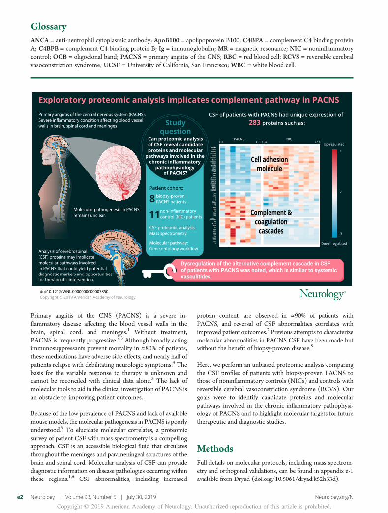
Primary angiitis of the CNS (PACNS) is a severe in-flammatory disease affecting the blood vessel walls in thebrain, spinal cord, and meninges.1 Without treatment,PACNS is frequently progressive.2,3 Although broadly actingimmunosuppressants prevent mortality in ≈80% of patients,these medications have adverse side effects, and nearly half ofpatients relapse with debilitating neurologic symptoms.4 Thebasis for the variable response to therapy is unknown andcannot be reconciled with clinical data alone.3 The lack ofmolecular tools to aid in the clinical investigation of PACNS isan obstacle to improving patient outcomes.
Because of the low prevalence of PACNS and lack of availablemousemodels, themolecular pathogenesis in PACNS is poorlyunderstood.5 To elucidate molecular correlates, a proteomicsurvey of patient CSF with mass spectrometry is a compellingapproach. CSF is an accessible biological fluid that circulatesthroughout the meninges and parameningeal structures of thebrain and spinal cord. Molecular analysis of CSF can providediagnostic information on disease pathologies occurring withinthese regions.1,6 CSF abnormalities, including increased
protein content, are observed in ≈90% of patients withPACNS, and reversal of CSF abnormalities correlates withimproved patient outcomes.7 Previous attempts to characterizemolecular abnormalities in PACNS CSF have been made butwithout the benefit of biopsy-proven disease.8
Here, we perform an unbiased proteomic analysis comparingthe CSF profiles of patients with biopsy-proven PACNS tothose of noninflammatory controls (NICs) and controls withreversible cerebral vasoconstriction syndrome (RCVS). Ourgoals were to identify candidate proteins and molecularpathways involved in the chronic inflammatory pathophysi-ology of PACNS and to highlight molecular targets for futuretherapeutic and diagnostic studies.
MethodsFull details on molecular protocols, including mass spectrom-etry and orthogonal validations, can be found in appendix e-1available from Dryad (doi.org/10.5061/dryad.k52h33d).
GlossaryANCA = anti-neutrophil cytoplasmic antibody; ApoB100 = apolipoprotein B100; C4BPA = complement C4 binding proteinA; C4BPB = complement C4 binding protein B; Ig = immunoglobulin; MR = magnetic resonance; NIC = noninflammatorycontrol; OCB = oligoclonal band; PACNS = primary angiitis of the CNS; RBC = red blood cell; RCVS = reversible cerebralvasoconstriction syndrome; UCSF = University of California, San Francisco; WBC = white blood cell.
e2 Neurology | Volume 93, Number 5 | July 30, 2019 Neurology.org/N
Copyright © 2019 American Academy of Neurology. Unauthorized reproduction of this article is prohibited.
Patient recruitment and study protocolPatients with PACNS and NICs were recruited as part ofa larger study analyzing biological samples from patientswith suspected neuroinflammatory disease at the Universityof California, San Francisco (UCSF). The UCSF In-stitutional Review Board approved the study protocol, andparticipants or their surrogates provided written informedconsent. RCVS controls were recruited as part of a largerstudy analyzing biological samples from patients with CNSvascular disorders at Cleveland Clinic. The Cleveland ClinicInstitutional Review Board approved the study protocol, andparticipants or their surrogates provided written informedconsent. Patients with PACNS and RCVS were diagnosedaccording to standard clinical diagnostic criteria, includingneuropathology evaluation in all of the patients diagnosedwith PACNS.
PACNS clinical vignettes
Patient 1A previously healthy 50-year-old woman developed mildheadaches with episodic, migrainous features, including visualauras together with worsening mental fogging independent ofthe headaches, all of which worsened over 4 years. At thattime, she was discovered to have a thoracic myelopathy onexamination and inflammatory CSF of unclear etiology. OnMRI, she was found to have a thoracic myelitis with nodularleptomeningeal enhancement throughout the spine and thebrain. A magnetic resonance (MR) angiogram of the head andneck was unremarkable. A brain biopsy revealed evidence fora small vessel vasculitis and chronic meningitis. An extensiveworkup for neoplastic, infectious, and other autoimmuneetiologies was unrevealing. The CSF profile from a samplefrom later in the patient’s clinical course was used for thisstudy and showed a white blood cell (WBC) count of 2 cells/μL (66% lymphocytes, 34% monocytes) (0–5 cells/μL), redblood cell (RBC) count of 0 cells/μL (0–5 cells/μL), glucoseof 45 mg/dL (45–80 mg/dL), total protein of 192 mg/dL(15–45 mg/dL), immunoglobulin (Ig) G index of 1.8 (<0.6),and <5 unique oligoclonal bands (OCBs) (≤1 band).
Patient 2A 39-year-old woman with a history of ulcerative colitis wellcontrolled on mesalamine and oral budesonide developedincreasing fatigue and increasingly painful, new left-sidedheadaches and left facial paresthesias over 6 weeks to the pointthat they prompted hospitalization. A brain MRI revealed T2hyperintensities in a gyriform pattern over the left parietal andtemporal lobes with associated leptomeningeal enhancement.A CSF examination revealed a WBC count of 15 cells/μL(54% lymphocytes, 39% granulocytes, 7% other), an RBCcount of 0 cells/μL, glucose of 65 mg/dL, total protein of 50mg/dL, and 0 OCBs. A CT angiogram of the head and neckwas unremarkable except for the suggestion of mild smoothnarrowing of the left carotid artery terminus and left M1segment of the middle cerebral artery. Over the next few days,the patient developed aphasia and apraxia, prompting a brain
biopsy that revealed a small vessel vasculitis. Extensiveworkup for neoplastic, infectious, and other autoimmuneetiologies was unrevealing.
Patient 3A 56-year-old man was hospitalized for rapid cognitive declineand was found to have bilateral papilledema and a left abdu-cens nerve palsy on examination. He had extensive confluentwhite matter T2 hyperintensities and multiple small areas ofrestricted diffusion consistent with acute infarcts on brainMRI. An MR angiogram of the head and neck was un-remarkable. A CSF examination showed a WBC count of 26cells/μL (53% neutrophils, 39% lymphocytes, 8% mono-cytes), RBC count of 3,275 cells/μL, glucose 60 mg/dL, totalprotein 141 mg/dL and an IgG index of 0.9. An extra-ventricular drain was placed for elevated intracranial pressure,and a brain biopsy revealed a small vessel vasculitis. Extensiveworkup for neoplastic, infectious, and other autoimmuneetiologies was unrevealing.
Patient 4A 55-year-old woman with a history of non–insulin-dependentdiabetes mellitus had progressive difficulty walking over 6months before she acutely lost sensation in her right leg anddeveloped severe urinary retention. She was found to havea longitudinally extensive transverse myelitis on MRI, anda CSF examination revealed a WBC count of 8 cells/μL, anRBC count of 1 cell/μL, glucose of 92 mg/dL, total proteinof 99 mg/dL, an IgG index of 0.57, and 1 unique OCB.Despite initial attempts at immunosuppression with glu-cocorticoids, the patient developed new weakness in her leftleg and urinary and fecal incontinence. Serial imagingrevealed new inflammatory lesions in the cerebellum andoverlying leptomeninges. All vascular imaging, includinga cerebral and spinal angiogram, was unremarkable. A brainbiopsy revealed a small vessel vasculitis. Extensive workupfor neoplastic, infectious, and other autoimmune etiologieswas unrevealing.
Patient 5A 36-year-old man with a history of possible relapsing poly-chondritis, with 1 episode of ear chondritis, presented witha fewweeks of new-onset daily headaches and fatigue followedby visual hallucinations that prompted a neurologic evalua-tion. A CSF examination revealed a WBC count of 6 cells/μL(60% lymphocytes, 22% monocytes, 18% neutrophils), anRBC count of 3 cells/μL, glucose of 51 mg/dL, total proteinof 27 mg/dL, and an IgG index of 1.62. A brain MRI revealedpatchy leptomeningeal enhancement, multiple areas of T2hyperintensity with patchy gadolinium enhancement, andmultifocal areas of restricted diffusion consistent with acuteinfarcts. An MR angiogram of the head was normal, buta cerebral angiogram showed diffuse vasculopathy bilaterallyin the distal vasculature (i.e., M3, M4, ophthalmic arteries, P3and P4 vessels). A brain biopsy showed small vessel vasculitis.Extensive workup for neoplastic, infectious, and other auto-immune etiologies was unrevealing.
Neurology.org/N Neurology | Volume 93, Number 5 | July 30, 2019 e3
Copyright © 2019 American Academy of Neurology. Unauthorized reproduction of this article is prohibited.
Patient 6A previously healthy 57-year-old woman presented with 2months of new-onset headaches with visual aura, 10 days ofdizziness and vertigo, and an isolated episode of hemi-bodysensory symptoms and was found to have a subarachnoid T2hyperintensity on brain MRI and faint leptomeningeal en-hancement. A CT angiogram of the head and neck was nor-mal. CSF examination revealed a WBC count of 31 cells/μL(90% lymphocytes, 6% monocytes, 2% neutrophils, 1%eosinophils, and 1% unidentified), an RBC count of 102 cells/μL, glucose of 54 mg/dL, total protein of 75 mg/dL, an IgGindex of 2.02, and >2 unique OCBs. A brain biopsy revealeda small vessel vasculitis. Extensive workup for neoplastic, in-fectious, and other autoimmune etiologies was unrevealing.
Patient 7A previously healthy 13-year-old girl presented with fever,headache, altered mental status, and seizure and was found tohave unihemispheric, subcortical T2 hyperintense lesions onbrain MRI, many of which enhanced with gadolinium. AnMRangiogram of the head and neck was unremarkable. A CSFexamination revealed a WBC count of 11 cells/μL (84% lym-phocytes, 16% monocytes), an RBC count of 6 cells/μL, glu-cose of 49 mg/dL, total protein of 37 mg/dL, and >5 uniqueOCBs. Brain biopsy revealed a small vessel vasculitis. Extensiveworkup for neoplastic, infectious, and other autoimmune eti-ologies was unrevealing.
Patient 8A 51-year-old man with a history of atrial fibrillation, hyper-tension, and seizure presented with bony aches and migratoryjoint pains that went away and were followed months later bybilateral episcleritis, fever, numbness in his feet, and confusion.A brain MRI showed leptomeningeal enhancement and over-lying multifocal areas of swollen and T2-hyperintense cortexand possible subcortical U-fiber enhancement. An MR angio-gram of the head and neck was unremarkable. CSF examinationrevealed a WBC count of 3 cells/μL (82% lymphocytes, 12%monocytes, 6% neutrophils), an RBC count of 1 cell/μL,glucose of 59 mg/dL, total protein of 42 mg/dL, an IgGindex 0.7, and 5 unique OCBs. The clinical impression by thetreating neurologist was that the systemic symptoms wereunrelated to the patient’s neuroinflammatory disease. A brainbiopsy revealed a small vessel vasculitis. Extensive workup forneoplastic, infectious, and other autoimmune etiologies wasunrevealing.
RCVS clinical vignettes
Patient 9A 54-year-old woman with a history of hepatitis C virus in-fection and epilepsy presented with altered mental status,dysarthria, expressive aphasia, and left- greater than right-sidedweakness. AnMR angiogram of the head revealed irregularitiesof the right anterior cerebral artery and left internal carotidartery. A cerebral angiogram showed narrowing and beading inmultiple vessels, including the basilar artery, bilateral posterior
cerebral arteries, and left M1 and A1 segments. There was alsofocal beading in the distal left anterior and middle cerebralarteries and an aneurysm at the origin of the right temporalartery. Her CSF profile revealed a WBC count of 11 cells/μL(93% lymphocytes, 3% monocytes, 4% other), an RBCcount of 340 cells/μL, glucose of 57 mg/dL, total protein of22 mg/dL, and no OCBs. She started on calcium channelblockers, and repeated MR angiogram of intracranial vessel10 days later showed marked improvement in the intracranialvessel abnormalities.
Patient 10A 33-year-old woman presented with a sudden-onset, thun-derclap headache that was clearly different from her typicalmigraine headaches. A CT angiogram of the head and a non-contrast head CT revealed diffuse beading of the vasculaturethroughout the anterior and posterior circulation, and a parietalsubarachnoid hemorrhage, respectively. A cerebral angiogramsimilarly found segmental irregularities of the intracranial ves-sels of the distal left anterior circulation. Her CSF profilerevealed a WBC count of 1 cell/μL (82% lymphocytes, 8%monocytes, 10% other), an RBC count of 1,150 cells/μL,glucose of 65 mg/dL, total protein of 115 mg/dL, and an IgGindex of 1.0. The patient was started on calcium channelblockers and had rapid resolution of her symptoms and nodisease recurrence.
Patient 11A 57-year-old woman with a history of depression treated withcitalopram and bupropion hydrochloride presented with 4 daysof dizziness, lightheadedness, left greater than right leg weak-ness, and falls followed by a rapid decline in mental status. Shewas found to have large areas of restricted diffusion in thebilateral parietal lobes on brain MRI. She became unresponsiveand was intubated and transferred to the intensive care unit. ACT angiogram of the head revealed irregularities in the distalportions of the anterior cerebral arteries, and a cerebral an-giogram was similarly consistent with vasospasm. A CSFexamination revealed a WBC count of 1 cell/μL, an RBCcount of 29 cells/μL, glucose of 95 mg/dL, and total proteinof 22 mg/dL. The patient improved clinically and radio-logically after administration of intra-arterial nicardipine andverapamil. She was started on a calcium channel blocker, andcitalopram and bupropion hydrochloride were discontinued.
Patient 12A previously healthy 30-year-old woman was admitted withnew left-sided weakness and severe hypothermia after expe-riencing new-onset, recurrent thunderclap headaches for 2weeks. A CT angiogram of the head and neck showed mul-tiple foci of intracranial vascular narrowing in the bilateralanterior cerebral arteries, middle cerebral arteries, and pos-terior cerebral arteries, which was corroborated by a cerebralangiogram. A brain MRI showed acute bilateral subcorticalinfarcts. A high-resolution brain MRI found no evidence ofabnormal vessel wall enhancement. Her CSF examinationrevealed a WBC count of 0 cells/μL, an RBC count of 133
e4 Neurology | Volume 93, Number 5 | July 30, 2019 Neurology.org/N
Copyright © 2019 American Academy of Neurology. Unauthorized reproduction of this article is prohibited.
cells/μL, glucose of 78 mg/dL, and total protein of 33 mg/dL.She was started on calcium channel blockers and improvedclinically and radiographically.
Mass spectrometryTotal protein concentration in patient CSF was determinedto be 0.1 to 0.6 mg/mL by Bradford assay (Sigma, B6916). Atotal of 5 μg protein was used from each patient’s CSFsample for liquid chromatography–dual mass spectrometryanalysis. Appendix e-1 available from Dryad (doi.org/10.5061/dryad.k52h33d) provides full details on sample pro-cessing and data acquisition.
Statistical analysesThe following statistical analyses were performed in R ver-sion 3.4.1. For comparative analyses of the individual CSFproteomes, spectral counts were aggregated by protein(i.e., protein abundance) for each sample. The proteinabundances for the unique 1,043 proteins identified in CSFwere compared between the PACNS (n = 8) and NIC (n =11) cohorts with 2 statistical approaches commonly usedfor mass spectrometry datasets, DESeq2 version 3.7 and thet test.9,10 DESeq2 uses a method based on the negativebinomial distribution to assess differential expression incount data. Spectral counts for all 1,043 unique proteinswere used as the input for this package, which was then runwith default settings. For the t test, spectral count values ofzero were first replaced with counts of 0.16, a value empir-ically determined to best approximate normal distributionsfor each protein within the NIC samples. Spectral countswere then divided by the sum of spectral counts for eachsample and multiplied by 10,000, generating normalizedspectral counts. Only proteins at sufficient abundance wereconsidered in the t test, defined as having a sum across allNIC and PACNS samples of ≥10 normalized spectral countsand being observed in at least 5 of the combined group ofNIC and PACNS samples. Normalized spectral counts forthese 713 abundant proteins were then transformed bynatural logarithm to avoid large differences in variances fordifferent proteins and then analyzed with 2-sided t tests as-suming unequal variance to compare abundances betweenthe PACNS and NIC cohorts for each protein. The resultingp values were then adjusted for multiple comparisons withthe Benjamini-Hochberg method. Fold changes were cal-culated as the ratio between the mean of PACNS and NICsamples for each protein. In our conservative approach, onlyproteins that were significantly different (fold change >1.5and Benjamini-Hochberg–adjusted p < 0.05) betweenPACNS and NIC in both tests were considered differentiallyregulated proteins in PACNS. Because of the low samplenumber in the RCVS cohort (n = 4), this comparative sta-tistical analysis was restricted to the PACNS and NICcohorts only.
Unbiased hierarchical clustering was performed with thenumpy (version 1.15.0) and seaborn (version 0.9.0) packagesin python (version 3.7.0). For this analysis, abundant proteins
across PACNS, NIC, and RCVS samples were grouped tocombine counts for isoforms of the same protein with sharedpeptides (peptides mapping to multiple isoforms). Proteinsand samples were then clustered by applying an unweightedpair group method with arithmetic mean to the correlationdistance matrix.
Molecular pathway enrichment analysisPathway enrichment analysis was performed with the Data-base for Annotation, Visualization and Integrated DiscoveryBioinformatics resource (david.ncifcrf.gov/). For enrichmentanalysis, the 222 downregulated proteins and 61 upregulatedproteins in PACNS were analyzed against the CSF back-ground of all 1,043 observed proteins. For 50 of the proteinsmost highly downregulated in PACNS, annotations fortransmembrane and topologic domains were retrieved fromUniProt. The locations of these domains and the locations ofpeptides observed in the NIC samples by mass spectrometrywere then mapped onto the protein chains.
Data availabilityRawmass spectrometry data files and peak list files have beendeposited at ProteoSAFE (massive.ucsd.edu) with accessionnumber MSV000082129.
ResultsSummary of clinical characteristicsIn addition to the individual patient vignettes above, an ag-gregated summary of patient demographics, clinical features,imaging abnormalities, and clinical CSF parameters for thePACNS, NIC, and RCVS cohorts is provided for comparisonin the table. The majority of our patients with biopsy-provenPACNS were refractory to initial treatment strategies or re-quired continual immunosuppression to achieve remission.Notably, all patients with PACNS had a chronic diseasecourse, compared to the monophasic nature of RCVS.
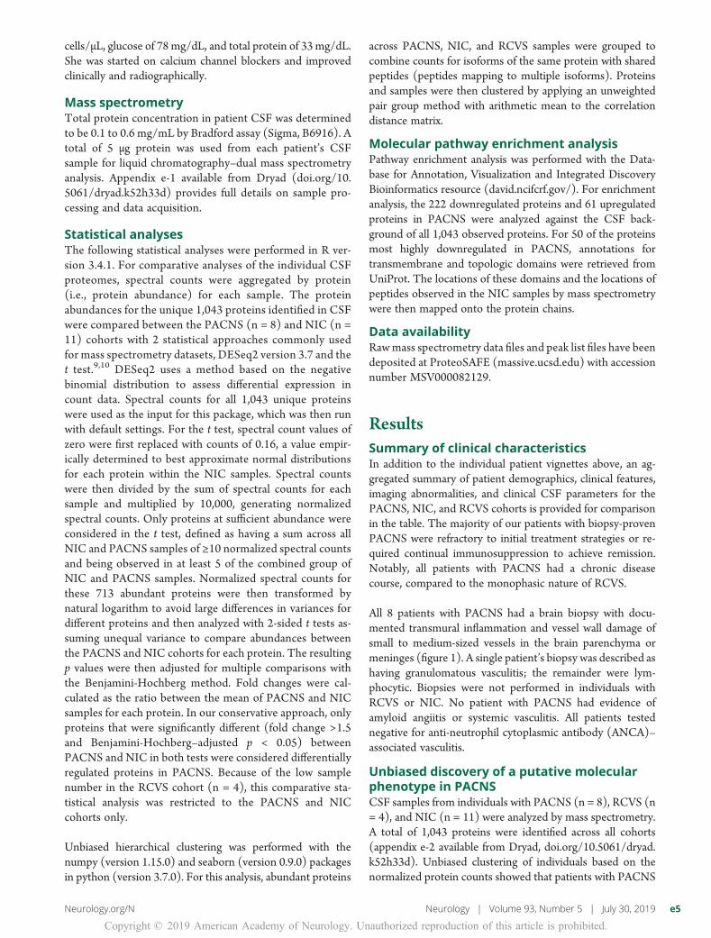
All 8 patients with PACNS had a brain biopsy with docu-mented transmural inflammation and vessel wall damage ofsmall to medium-sized vessels in the brain parenchyma ormeninges (figure 1). A single patient’s biopsy was described ashaving granulomatous vasculitis; the remainder were lym-phocytic. Biopsies were not performed in individuals withRCVS or NIC. No patient with PACNS had evidence ofamyloid angiitis or systemic vasculitis. All patients testednegative for anti-neutrophil cytoplasmic antibody (ANCA)–associated vasculitis.
Unbiased discovery of a putative molecularphenotype in PACNSCSF samples from individuals with PACNS (n = 8), RCVS (n= 4), and NIC (n = 11) were analyzed by mass spectrometry.A total of 1,043 proteins were identified across all cohorts(appendix e-2 available from Dryad, doi.org/10.5061/dryad.k52h33d). Unbiased clustering of individuals based on thenormalized protein counts showed that patients with PACNS
Neurology.org/N Neurology | Volume 93, Number 5 | July 30, 2019 e5
Copyright © 2019 American Academy of Neurology. Unauthorized reproduction of this article is prohibited.
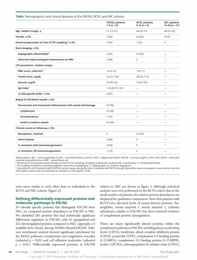
were more similar to each other than to individuals in theRCVS and NIC cohorts (figure 2).
Defining differentially expressed proteins andmolecular pathways in PACNSTo identify specific proteins that distinguish PACNS fromNIC, we compared protein abundances in PACNS vs NIC.We identified 283 proteins that had statistically significantdifferential regulation in PACNS, with 61 upregulated and222 downregulated proteins compared to NIC (appendix e-3available from Dryad, doi.org/10.5061/dryad.k52h33d). Path-way enrichment analysis showed significant enrichment forthe KEGG pathways complement and coagulation cascades(adjusted p < 0.05) and cell adhesion molecules (adjustedp < 0.01). Differentially expressed proteins in PACNS
relative to NIC are shown in figure 3. Although statisticalanalyses were not performed on the RCVS cohort due to thesmall number of patients, the relative protein abundances aredisplayed for qualitative comparison. Note that patients withRCVS have elevated levels of serum-derived proteins (he-moglobin, serum amyloid 1, serum amyloid 2, carbonicanhydrase), similar to PACNS, but show minimal evidenceof complement protein dysregulation.
There are many significantly altered proteins within thecomplement pathway in PACNS, including decay acceleratingfactor (CD55), membrane attack complex inhibitory protein(CD59), properdin (CFP), complement C4 binding proteinA (C4BPA), complement C4 binding protein B (C4BPB),ficolin-3 (FCN3), carboxypeptidase N catalytic chain (CPN1),
Table Demographics and clinical features of the PACNS, RCVS, and NIC cohorts
PACNS, patients1–8 (n = 8)
RCVS, patients9–12 (n = 4)
NIC, patients13–23 (n = 11)
Age, median (range), y 51 (13–57) 44 (30–57) 48 (31–62)
Female, n (%) 5 (63) 4 (100) 8 (73)
Immunosuppression at time of CSF sampling,a n (%) 5 (63) 1 (25) 0
Brain imaging, n (%)
Angiographic abnormalityb 2 (25) 4 (100) —
Abnormal leptomeningeal enhancement on MRI 7 (88) 0 —
CSF parameters, median (range)
WBC count, cells/mm3 10 (2–31) 1 (0–11) —
Protein level, mg/dL 63 (27–192) 28 (22–115) —
Glucose, mg/dL 57 (45–92) 72 (57–95) —
IgG indexc 1.26 (0.57–2.02) — —
≥2 CNS-specific OCBs,c n (%) 4 (67) — —
Biopsy of CNS blood vessels, n (%)
Perivascular and intramural inflammation with vessel wall damage 8 (100) — —
Lymphocytic 8 (100) — —
Granulomatous 1 (13) — —
Small to medium vessels 8 (100) — —
Clinical course on follow-up, n (%)
Monophasic, resolved 0 4 (100) —
Active disease 3 (38) 0 —
In remission with immunosuppression 4 (50) 0 —
In remission, off immunosuppression 1 (13) 0 —
Abbreviations: IgG = immunoglobulin G; NIC = noninflammatory control; OCB = oligoclonal bands; PACNS = primary angiitis of the CNS; RCVS = reversiblecerebral vasoconstriction; WBC = white blood cell.a Receiving immunosuppressive therapy at time of CSF sampling, including prednisone, budesonide, mesalamine, or cyclophosphamide.b On imaging modalities, including magnetic resonance angiography, CT angiography, or cerebral angiogram.c Calculated for 6 of 8 patients with PACNS in whom assay was performed. In patients with RCVS, the IgG index either was not assayed or was normal, and theOCB pattern either was not assessed or showed no CNS-specific OCBs.
e6 Neurology | Volume 93, Number 5 | July 30, 2019 Neurology.org/N
Copyright © 2019 American Academy of Neurology. Unauthorized reproduction of this article is prohibited.
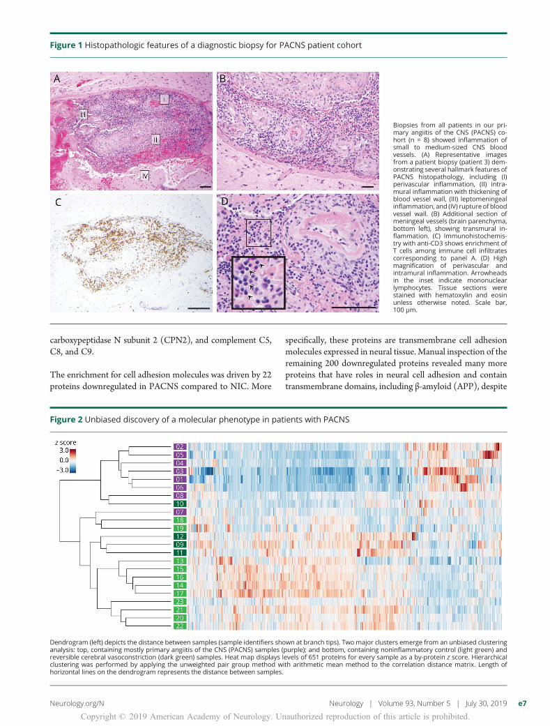
carboxypeptidase N subunit 2 (CPN2), and complement C5,C8, and C9.
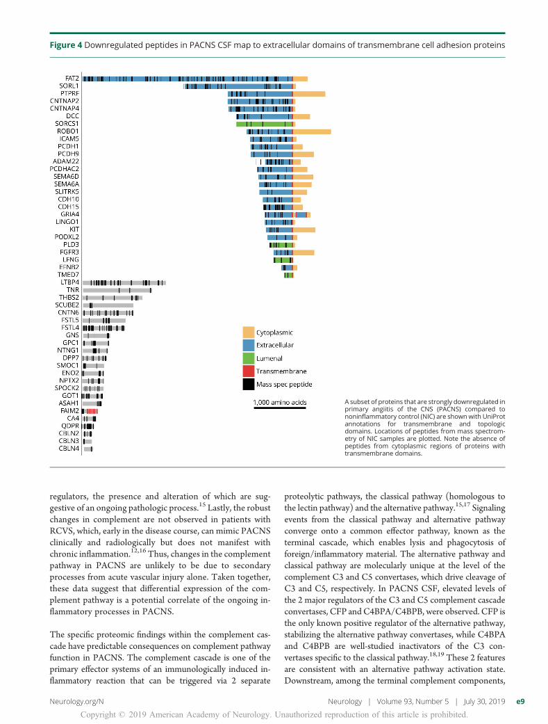
The enrichment for cell adhesion molecules was driven by 22proteins downregulated in PACNS compared to NIC. More
specifically, these proteins are transmembrane cell adhesionmolecules expressed in neural tissue. Manual inspection of theremaining 200 downregulated proteins revealed many moreproteins that have roles in neural cell adhesion and containtransmembrane domains, including β-amyloid (APP), despite
Figure 1 Histopathologic features of a diagnostic biopsy for PACNS patient cohort
Biopsies from all patients in our pri-mary angiitis of the CNS (PACNS) co-hort (n = 8) showed inflammation ofsmall to medium-sized CNS bloodvessels. (A) Representative imagesfrom a patient biopsy (patient 3) dem-onstrating several hallmark features ofPACNS histopathology, including (I)perivascular inflammation, (II) intra-mural inflammation with thickening ofblood vessel wall, (III) leptomeningealinflammation, and (IV) rupture of bloodvessel wall. (B) Additional section ofmeningeal vessels (brain parenchyma,bottom left), showing transmural in-flammation. (C) Immunohistochemis-try with anti-CD3 shows enrichment ofT cells among immune cell infiltratescorresponding to panel A. (D) Highmagnification of perivascular andintramural inflammation. Arrowheadsin the inset indicate mononuclearlymphocytes. Tissue sections werestained with hematoxylin and eosinunless otherwise noted. Scale bar,100 μm.
Figure 2 Unbiased discovery of a molecular phenotype in patients with PACNS
Dendrogram (left) depicts the distance between samples (sample identifiers shown at branch tips). Two major clusters emerge from an unbiased clusteringanalysis: top, containing mostly primary angiitis of the CNS (PACNS) samples (purple); and bottom, containing noninflammatory control (light green) andreversible cerebral vasoconstriction (dark green) samples. Heat map displays levels of 651 proteins for every sample as a by-protein z score. Hierarchicalclustering was performed by applying the unweighted pair group method with arithmetic mean method to the correlation distance matrix. Length ofhorizontal lines on the dendrogram represents the distance between samples.
Neurology.org/N Neurology | Volume 93, Number 5 | July 30, 2019 e7
Copyright © 2019 American Academy of Neurology. Unauthorized reproduction of this article is prohibited.
the absence of these proteins in the core set of the KEGG celladhesion molecules pathway. We localized the recoveredpeptides from mass spectrometry according to protein
domain annotations assigned by UniProt and found that NICCSF contained peptides specifically from the extracellulardomains of transmembrane proteins that are downregulatedin PACNS CSF (figures 4 and 5).
In addition, we noted several significant findings that do notcorrespond to a specific pathway but pique clinical interest.These findings include elevated levels of immunoglobulins(IgM and IgA) and apolipoproteins [including apolipoproteinB100 (ApoB100) and lipoprotein(a)] in PACNS.
Orthogonal confirmationTo validate our technical approach, we reproduced a subset offindings by Western blot and commercial ELISA. Because ofthe potential clinical and therapeutic implications of identi-fying a role for the alternative complement cascade inPACNS, specifically complement C5, we validated elevatedC5 levels in PACNS CSF through Western blotting withcommercial antibody and commercial ELISA (figures e-1 ande-2 available from Dryad, doi.org/10.5061/dryad.k52h33d).Notably, the relative C5 levels by mass spectrometry analysisand by ELISA are correlated, suggesting that the variation inthese data across patients is reproducible across technicalapproaches. The substantial changes in IgM and IgA levelsidentified by mass spectrometry were also reproduced or-thogonally through Western blotting.
DiscussionIn this exploratory study, a mass spectrometry–based ap-proach was used to characterize the CSF proteome associatedwith ongoing PACNS pathology relative to noninflammatorydisease, with the intention to discover new diagnostic ortherapeutic candidates. Currently, the diagnosis of PACNSremains challenging.3 The diagnostic criteria for PACNS,including CSF cytology, imaging abnormalities, and neuro-logic manifestations, are largely nonspecific, and long-termoutcomes are variable.1,11 Several of these features, includingelevated protein and imaging abnormalities, are also observedin individuals diagnosed with an early mimic, RCVS, as de-tailed in the clinical summary of our patient cohorts (table)and individualized clinical vignettes (Methods) for patientswith PACNS and RCVS.12,13 We restricted our analyses topatients with biopsy-proven PACNS to enhance the rigor ofthis exploratory study and to ensure that our molecular datawere most closely correlated to the key features that are as-sociated with PACNS pathology.14
Our findings highlight the complement cascade as a signifi-cant feature of PACNS CSF. We found up to 12 significantlydysregulated proteins in PACNS CSF that function withinthe complement cascade pathway, and these changes arehighly reproducible across the PACNS patient cohort. Inaddition, the proteomic changes within the complementpathway are specific, affecting the alternative and terminalcascade only, and include changes in transient, fluid-phase
Figure 3 Differentially expressed proteins and molecularpathways in PACNS
Relative protein abundances for a subset of differentially regulated proteins inprimary angiitis of the CNS (PACNS) vs noninflammatory control (NIC) arereported. Proteins representing the statistically significantly enrichedpathwayscell adhesion molecules and complement and coagulation cascades are in-cluded, as well as a subset of proteins manually curated to reflect findings ofsignificant clinical interest, with functional classifiers informed by Database forAnnotation, Visualization and Integrated Discovery and KEGG annotations.Heat map displays relative protein abundance across individual samples(PACNSn = 8,NIC n= 11, and reversible cerebral vasoconstriction [RCVS], n = 4),plottedas the z score of the normalized spectral counts. Differential expressionwas evaluated with DESeq2 and the t test (see Methods), with significancedefined as having fold change >1.5 and Benjamini-Hochberg–adjusted p < 0.05for both tests (*p < 0.05, **p ≤ 0.01, ***p ≤ 0.001). n.s. = not significant.
e8 Neurology | Volume 93, Number 5 | July 30, 2019 Neurology.org/N
Copyright © 2019 American Academy of Neurology. Unauthorized reproduction of this article is prohibited.
regulators, the presence and alteration of which are sug-gestive of an ongoing pathologic process.15 Lastly, the robustchanges in complement are not observed in patients withRCVS, which, early in the disease course, can mimic PACNSclinically and radiologically but does not manifest withchronic inflammation.12,16 Thus, changes in the complementpathway in PACNS are unlikely to be due to secondaryprocesses from acute vascular injury alone. Taken together,these data suggest that differential expression of the com-plement pathway is a potential correlate of the ongoing in-flammatory processes in PACNS.
The specific proteomic findings within the complement cas-cade have predictable consequences on complement pathwayfunction in PACNS. The complement cascade is one of theprimary effector systems of an immunologically induced in-flammatory reaction that can be triggered via 2 separate
proteolytic pathways, the classical pathway (homologous tothe lectin pathway) and the alternative pathway.15,17 Signalingevents from the classical pathway and alternative pathwayconverge onto a common effector pathway, known as theterminal cascade, which enables lysis and phagocytosis offoreign/inflammatory material. The alternative pathway andclassical pathway are molecularly unique at the level of thecomplement C3 and C5 convertases, which drive cleavage ofC3 and C5, respectively. In PACNS CSF, elevated levels ofthe 2 major regulators of the C3 and C5 complement cascadeconvertases, CFP and C4BPA/C4BPB, were observed. CFP isthe only known positive regulator of the alternative pathway,stabilizing the alternative pathway convertases, while C4BPAand C4BPB are well-studied inactivators of the C3 con-vertases specific to the classical pathway.18,19 These 2 featuresare consistent with an alternative pathway activation state.Downstream, among the terminal complement components,
Figure 4 Downregulated peptides in PACNS CSF map to extracellular domains of transmembrane cell adhesion proteins
A subset of proteins that are strongly downregulated inprimary angiitis of the CNS (PACNS) compared tononinflammatory control (NIC) are shown with UniProtannotations for transmembrane and topologicdomains. Locations of peptides from mass spectrom-etry of NIC samples are plotted. Note the absence ofpeptides from cytoplasmic regions of proteins withtransmembrane domains.
Neurology.org/N Neurology | Volume 93, Number 5 | July 30, 2019 e9
Copyright © 2019 American Academy of Neurology. Unauthorized reproduction of this article is prohibited.
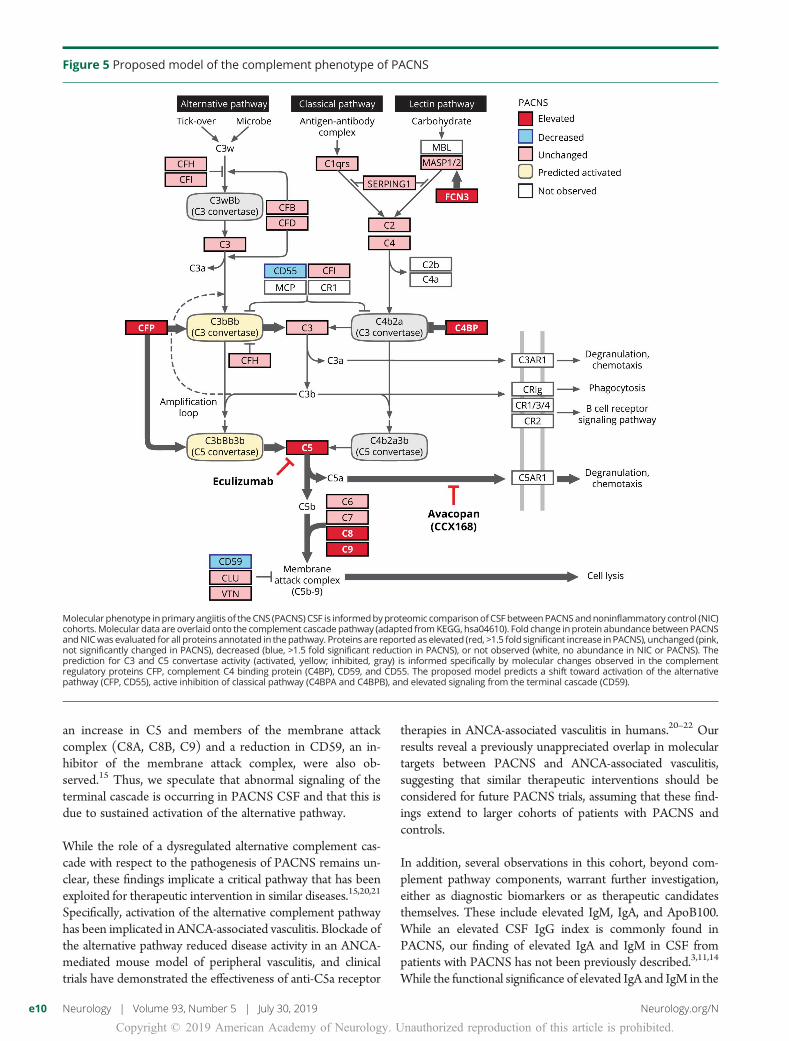
an increase in C5 and members of the membrane attackcomplex (C8A, C8B, C9) and a reduction in CD59, an in-hibitor of the membrane attack complex, were also ob-served.15 Thus, we speculate that abnormal signaling of theterminal cascade is occurring in PACNS CSF and that this isdue to sustained activation of the alternative pathway.
While the role of a dysregulated alternative complement cas-cade with respect to the pathogenesis of PACNS remains un-clear, these findings implicate a critical pathway that has beenexploited for therapeutic intervention in similar diseases.15,20,21
Specifically, activation of the alternative complement pathwayhas been implicated in ANCA-associated vasculitis. Blockade ofthe alternative pathway reduced disease activity in an ANCA-mediated mouse model of peripheral vasculitis, and clinicaltrials have demonstrated the effectiveness of anti-C5a receptor
therapies in ANCA-associated vasculitis in humans.20–22 Ourresults reveal a previously unappreciated overlap in moleculartargets between PACNS and ANCA-associated vasculitis,suggesting that similar therapeutic interventions should beconsidered for future PACNS trials, assuming that these find-ings extend to larger cohorts of patients with PACNS andcontrols.
In addition, several observations in this cohort, beyond com-plement pathway components, warrant further investigation,either as diagnostic biomarkers or as therapeutic candidatesthemselves. These include elevated IgM, IgA, and ApoB100.While an elevated CSF IgG index is commonly found inPACNS, our finding of elevated IgA and IgM in CSF frompatients with PACNS has not been previously described.3,11,14
While the functional significance of elevated IgA and IgM in the
Figure 5 Proposed model of the complement phenotype of PACNS
Molecular phenotype inprimary angiitis of the CNS (PACNS) CSF is informedbyproteomic comparison of CSF betweenPACNSandnoninflammatory control (NIC)cohorts.Molecular data are overlaid onto the complement cascade pathway (adapted fromKEGG, hsa04610). Fold change inprotein abundance betweenPACNSandNICwas evaluated for all proteins annotated in thepathway. Proteins are reported as elevated (red, >1.5 fold significant increase inPACNS), unchanged (pink,not significantly changed in PACNS), decreased (blue, >1.5 fold significant reduction in PACNS), or not observed (white, no abundance in NIC or PACNS). Theprediction for C3 and C5 convertase activity (activated, yellow; inhibited, gray) is informed specifically by molecular changes observed in the complementregulatory proteins CFP, complement C4 binding protein (C4BP), CD59, and CD55. The proposed model predicts a shift toward activation of the alternativepathway (CFP, CD55), active inhibition of classical pathway (C4BPA and C4BPB), and elevated signaling from the terminal cascade (CD59).
e10 Neurology | Volume 93, Number 5 | July 30, 2019 Neurology.org/N
Copyright © 2019 American Academy of Neurology. Unauthorized reproduction of this article is prohibited.
context of PACNS is unclear, elevated IgA and IgM levels maybe explored as a separate diagnostic differentiator.
In contrast, several anecdotal pieces of evidence implicateApoB100 with a subset of PACNS pathological features. Forone, an immune response to ApoB100, mediated by T cells andIgM antibodies, is commonly observed in the progressive de-velopment of atherosclerotic lesions.23,24 Furthermore, ele-vated low-density lipoprotein and antibodies to ApoB100 havebeen identified in alternative non-PACNS vasculitides, in-cluding ANCA-associated vasculitis. In the case of both MPO-ANCA and PR3-ANCA, elevated levels of anti-apoB100 anti-bodies are thought to be an indirect result of the chronic in-flammatory disease.25 Given the robust elevation of ApoB100in this PACNS cohort, further investigation into the direct orindirect role of ApoB100 in PACNS pathology is warranted.
Finally, an unexpected finding of this study was the loss ofneural cell adhesion molecules in PACNS CSF. For non-inflammatory CSF, we observe peptides almost exclusivelyfrom the extracellular domains (ectodomains) of trans-membrane proteins. Changes in these proteins in PACNSmay be the result of transcriptional, translational, or post-translational regulatory differences.26–28 The last may includeabnormal regulation of the normal process of ectodomainshedding.26,29,30 The clear absence of ectodomain peptides inPACNS CSF suggests a loss of these proteins or a loss ofproteolytic homeostasis associated with shedding. While theroles of ectodomain shedding are diverse, the mechanism andimpact of dysregulated shedding in CSF are unknown.
Overall, these exploratory findings suggest potential newbiomarkers of PACNS, subject to validation in larger cohorts.These results also underline the importance of future mech-anistic studies around the role of complement pathways inPACNS disease pathobiology, with the ultimate goal of cre-ating targeted therapeutic interventions for this devastatingand poorly understood disease.
Study fundingFunded by the NIH National Institutes for NeurologicalDisorders and Stroke (award K08NS096117); the UCSFCenter for Next-Gen Precision Diagnostics supported by theSandler Foundation and William K. Bowes, Jr. Foundation;UCSF Medical Scientist Training Program; the RachleffFoundation; and Chan Zuckerberg Biohub.
DisclosureC. Mandel-Brehm, H. Retallack, G. Knudsen, A. Yamana, R.Hajj-Ali, L. Calabrese, T. Tihan, H. Sample, K. Zorn, M.Gorman, J. Madan Cohen, A. Sreih, J. Marcus, S. Josephson,and V. Douglas report no disclosures relevant to the man-uscript. J. Gelfand reports personal compensation for con-sulting for Biogen and Alexion, research support to UCSFfrom Genentech, service contract support from MedDay,and personal compensation for medical legal consulting/expert witness. M. Wilson reports no disclosures relevant to
the manuscript. J. DeRisi is a scientific consultant for Allen &Company. Go to Neurology.org/N for full disclosures.
Publication historyReceived by Neurology July 29, 2018. Accepted in final formMarch 12, 2019.
Appendix Authors
Name Location Role Contribution
CaleighMandel-Brehm,PhD
University ofCalifornia SanFrancisco
Author Study design,experiments, datainterpretation,manuscript drafting
HannaRetallack,AB
University ofCalifornia SanFrancisco
Author Study design, experiments,statistical analyses, datainterpretation, figures andtables
Giselle M.Knudsen,PhD
University ofCalifornia SanFrancisco
Author Mass spectrometry dataacquisition and analysis
AlexYamana,BS
University ofCalifornia SanFrancisco
Author Mass spectrometry dataacquisition and analysis
Rula A.Hajj-Ali,MD
Cleveland Clinic,OH
Author Study design, manuscriptrevisions for intellectualcontent
Leonard H.Calabrese,DO
Cleveland Clinic,OH
Author Study design, manuscriptrevisions for intellectualcontent
TarikTihan, MD,PhD
University ofCalifornia SanFrancisco
Author Manuscript revisions forintellectual content
Hannah A.Sample, BS
University ofCalifornia SanFrancisco
Author Clinical data acquisition
Kelsey C.Zorn, MHS
University ofCalifornia SanFrancisco
Author Clinical data acquisition
Mark P.Gorman,MD
Boston Children’sHospital, MA
Author Manuscript revisions forintellectual content
JenniferMadanCohen, MD
ConnecticutChildren’s MedicalCenter, Hartford
Author Manuscript revisions forintellectual content
Antoine G.Sreih, MD
University ofPennsylvania,Philadelphia
Author Manuscript revisions forintellectual content
JacquelineF. Marcus,MD
KaiserPermanente SanFrancisco MedicalCenter, CA
Author Manuscript revisions forintellectual content
S. AndrewJosephson,MD
University ofCalifornia SanFrancisco
Author Manuscript revisions forintellectual content
Vanja C.Douglas,MD
University ofCalifornia SanFrancisco
Author Manuscript revisions forintellectual content
Continued
Neurology.org/N Neurology | Volume 93, Number 5 | July 30, 2019 e11
Copyright © 2019 American Academy of Neurology. Unauthorized reproduction of this article is prohibited.
References1. Hajj-Ali RA, Calabrese LH. Primary angiitis of the central nervous system. Auto-
immun Rev 2013;12:463–466.2. Salvarani C, Brown RD, Christianson TJH, et al. Adult primary central nervous system
vasculitis treatment and course: analysis of one hundred sixty-three patients. ArthritisRheumatol 2015;67:1637–1645.
3. Byram K, Hajj-Ali RA, Calabrese L. CNS vasculitis: an approach to differential di-agnosis and management. Curr Rheumatol Rep 2018;20:37.
4. Hutchinson C, Elbers J, Halliday W, et al. Treatment of small vessel primary CNSvasculitis in children: an open-label cohort study. Lancet Neurol 2010;9:1078–1084.
5. Alba MA, Espigol-Frigole G, Prieto-Gonzalez S, et al. Central nervous system vas-culitis: still more questions than answers. Curr Neuropharmacol 2011;9:437–448.
6. Bastos P, Ferreira R, Manadas B, Moreira PI, Vitorino R. Insights into the humanbrain proteome: disclosing the biological meaning of protein networks in cerebro-spinal fluid. Crit Rev Clin Lab Sci 2017;54:185–204.
7. Oliveira V, Povoa P, Costa A, Ducla-Soares J. Cerebrospinal fluid and therapy ofisolated angiitis of the central nervous system. Stroke 1994;25:1693–1695.
8. Ruland T, Wolbert J, Gottschalk MG, et al. Cerebrospinal fluid concentrations ofneuronal proteins are reduced in primary angiitis of the central nervous system. FrontNeurol 2018;9:1–9.
9. Langley SR, Mayr M. Comparative analysis of statistical methods used for detectingdifferential expression in label-free mass spectrometry proteomics. J Proteomics 2015;129:83–92.
10. Zybailov B, Mosley AL, Sardiu ME, Coleman MK, Florens L, Washburn MP. Sta-tistical analysis of membrane proteome expression changes in Saccharomyces cer-evisiae. J Proteome Res 2006;5:2339–2347.
11. Salvarani C, Brown RD, Calamia KT, et al. Primary central nervous system vasculitis:analysis of 101 patients. Ann Neurol 2007;62:442–451.
12. Ducros A, Boukobza M, Porcher R, Sarov M, Valade D, Bousser MG. The clinical andradiological spectrum of reversible cerebral vasoconstriction syndrome: a prospectiveseries of 67 patients. Brain 2007;130:3091–3101.
13. Rocha EA, Topcuoglu MA, Silva GS, Singhal AB. RCVS2 score and diagnosticapproach for reversible cerebral vasoconstriction syndrome. Neurology 2019;92:e639–e647.
14. Miller DV, Salvarani C, Hunder GG, Brown JE, Christianson TJ, Giannini C.Biopsy findings in primary angiitis of the central nervous system. Am J SurgPathol 2009;33:35–43.
15. Thurman JM, Holers VM. The central role of the alternative complement pathway inhuman disease. J Immunol 2006;176:1305–1310.
16. de Boysson H, Parienti J-J, Mawet J, et al. Primary angiitis of the CNS and reversiblecerebral vasoconstriction syndrome: a comparative study. Neurology 2018;91:e1468–e1478.
17. Gigli I. Immunochemistry and immunobiology of the complement system. J InvestDermatol 1976;67:346–353.
18. Blatt AZ, Pathan S, Ferreira VP. Properdin: a tightly regulated critical inflammatorymodulator. Immunol Rev 2016;274:172–190.
19. Gigli I, Fujita T, Nussenzweig V. Modulation of the classical pathway C3 convertaseby plasma proteins C4 binding protein and C3b inactivator. Am J Physiol 1979;273:C883–C892.
20. Xiao H, Schreiber A, Heeringa P, Falk RJ, Jennette JC. Alternative complementpathway in the pathogenesis of disease mediated by anti-neutrophil cytoplasmicautoantibodies. Am J Pathol 2007;170:52–64.
21. Jayne DRW, Bruchfeld AN, Harper L, et al. Randomized trial of C5a receptorinhibitor avacopan in ANCA-associated vasculitis. J Am Soc Nephrol 2017;28:2756–2767.
22. Bekker P, Dairaghi D, Seitz L, et al. Characterization of pharmacologic and phar-macokinetic properties of CCX168, a potent and selective orally administered com-plement 5a receptor inhibitor, based on preclinical evaluation and randomized phase 1clinical study. PLoS One 2016;11:1–19.
23. Nilsson J, Bjorkbacka H, Fredrikson GN. Apolipoprotein B100 autoimmunity andatherosclerosis-disease mechanisms and therapeutic potential. Curr Opin Lipidol2012;23:422–428.
24. Nilsson J, Hansson GK. Autoimmunity in atherosclerosis: a protective response losingcontrol? J Intern Med 2008;263:464–478.
25. Slot MC, Theunissen R, van Paassen P, Damoiseaux JG, Tervaert JW. Anti-oxidizedlow-density lipoprotein antibodies in myeloperoxidase-positive vasculitis patientspreferentially recognize hypochlorite-modified low density lipoproteins. Clin ExpImmunol 2007;149:257–264.
26. Tsumagari K, Shirakabe K, Ogura M, Sato F, Ishihama Y, Sehara-Fujisawa A. Secre-tome analysis to elucidate metalloprotease-dependent ectodomain shedding of gly-coproteins during neuronal differentiation. Genes Cells 2017;22:237–244.
27. Shirakabe K, Omura T, Shibagaki Y, et al. Mechanistic insights into ectodomainshedding: susceptibility of CADM1 adhesion molecule is determined by alternativesplicing and O-glycosylation. Sci Rep 2017;7:46174.
28. Lichtenthaler SF, Lemberg MK, Fluhrer R. Proteolytic ectodomain shedding ofmembrane proteins in mammals: hardware, concepts, and recent developments.EMBO J 2018;37:e99456.
29. Waldera-Lupa DM, Etemad-Parishanzadeh O, Brocksieper M, et al. Proteomicchanges in cerebrospinal fluid from primary central nervous system lymphomapatients are associated with protein ectodomain shedding. Oncotarget 2017;8:110118–110132.
30. Miller MA, Sullivan RJ, Lauffenburger DA. Molecular pathways: receptor ectodomainshedding in treatment, resistance, and monitoring of cancer. Clin Cancer Res 2017;23:623–629.
Appendix (continued)
Name Location Role Contribution
Jeffrey M.Gelfand,MD
University ofCalifornia SanFrancisco
Author Study design, manuscriptrevisions for intellectualcontent
Michael R.Wilson, MD
University ofCalifornia SanFrancisco
Author Study design, figures andtables, manuscriptrevisions for intellectualcontent
Joseph L.DeRisi, PhD
University ofCalifornia SanFrancisco
Author Study design, statisticalanalyses, manuscriptrevisions for intellectualcontent
e12 Neurology | Volume 93, Number 5 | July 30, 2019 Neurology.org/N
Copyright © 2019 American Academy of Neurology. Unauthorized reproduction of this article is prohibited.
DOI 10.1212/WNL.0000000000007850 published online July 3, 2019Neurology
Caleigh Mandel-Brehm, Hanna Retallack, Giselle M. Knudsen, et al. primary CNS vasculitis
Exploratory proteomic analysis implicates the alternative complement cascade in
This information is current as of July 3, 2019
ServicesUpdated Information &
850.fullhttp://n.neurology.org/content/early/2019/07/03/WNL.0000000000007including high resolution figures, can be found at:
Subspecialty Collections
http://n.neurology.org/cgi/collection/vasculitisVasculitis
http://n.neurology.org/cgi/collection/all_clinical_neurologyAll Clinical Neurologyfollowing collection(s): This article, along with others on similar topics, appears in the
Permissions & Licensing
http://www.neurology.org/about/about_the_journal#permissionsits entirety can be found online at:Information about reproducing this article in parts (figures,tables) or in
Reprints
http://n.neurology.org/subscribers/advertiseInformation about ordering reprints can be found online:
rights reserved. Print ISSN: 0028-3878. Online ISSN: 1526-632X.1951, it is now a weekly with 48 issues per year. Copyright © 2019 American Academy of Neurology. All
® is the official journal of the American Academy of Neurology. Published continuously sinceNeurology