EXPLORATION OF NEURAL CORRELATES OF...
1
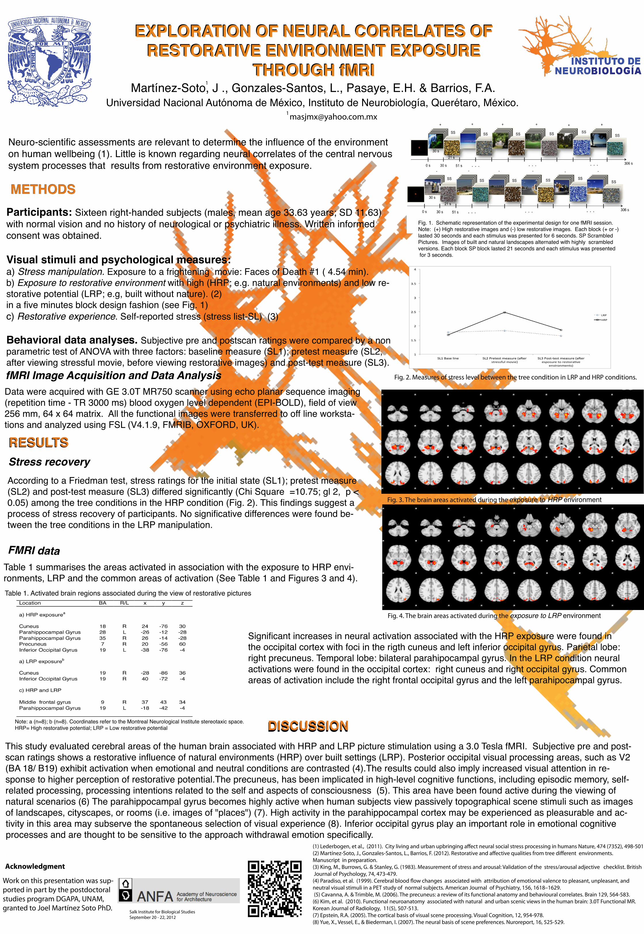
DISCUSSION RESULTS METHODS EXPLORATION OF NEURAL CORRELATES OF RESTORATIVE ENVIRONMENT EXPOSURE THROUGH fMRI EXPLORATION OF NEURAL CORRELATES OF RESTORATIVE ENVIRONMENT EXPOSURE THROUGH fMRI Martínez-Soto, J ., Gonzales-Santos, L., Pasaye, E.H. & Barrios, F.A. Universidad Nacional Autónoma de México, Instituto de Neurobiología, Querétaro, México. Neuro-scientific assessments are relevant to determine the influence of the environment on human wellbeing (1). Little is known regarding neural correlates of the central nervous system processes that results from restorative environment exposure. METHODS Participants: Sixteen right-handed subjects (males, mean age 33.63 years; SD 11.63) with normal vision and no history of neurological or psychiatric illness. Written informed consent was obtained. Visual stimuli and psychological measures: a) Stress manipulation. Exposure to a frightening movie: Faces of Death #1 ( 4.54 min). b) Exposure to restorative environment with high (HRP; e.g. natural environments) and low re- storative potential (LRP; e.g, built without nature). (2) in a five minutes block design fashion (see Fig. 1) c) Restorative experience. Self-reported stress (stress list-SL) (3) Behavioral data analyses. Subjective pre and postscan ratings were compared by a non parametric test of ANOVA with three factors: baseline measure (SL1); pretest measure (SL2, after viewing stressful movie, before viewing restorative images) and post-test measure (SL3). fMRI Image Acquisition and Data Analysis Data were acquired with GE 3.0T MR750 scanner using echo planar sequence imaging (repetition time - TR 3000 ms) blood oxygen level dependent (EPI-BOLD), field of view 256 mm, 64 x 64 matrix. All the functional images were transferred to off line worksta- tions and analyzed using FSL (V4.1.9, FMRIB, OXFORD, UK). RESULTS Stress recovery According to a Friedman test, stress ratings for the initial state (SL1); pretest measure (SL2) and post-test measure (SL3) differed significantly (Chi Square =10.75; gl 2, p < 0.05) among the tree conditions in the HRP condition (Fig. 2). This findings suggest a process of stress recovery of participants. No significative differences were found be- tween the tree conditions in the LRP manipulation. FMRI data Table 1 summarises the areas activated in association with the exposure to HRP envi- ronments, LRP and the common areas of activation (See Table 1 and Figures 3 and 4). DISCUSSION Work on this presentation was sup- ported in part by the postdoctoral studies program DGAPA, UNAM, granted to Joel Martínez Soto PhD. Acknowledgment (1) Lederbogen, et al., (2011). City living and urban upbringing affect neural social stress processing in humans Nature, 474 (7352), 498-501. (2) Martínez-Soto, J., Gonzales-Santos, L., Barrios, F. (2012). Restorative and affective qualities from tree different environments. Manuscript in preparation. (3) King, M., Burrows, G. & Stanley, G. (1983). Measurement of stress and arousal: Validation of the stress/arousal adjective checklist. British Journal of Psychology, 74, 473-479. (4) Paradiso, et al. (1999). Cerebral blood flow changes associated with attribution of emotional valence to pleasant, unpleasant, and neutral visual stimuli in a PET study of normal subjects. American Journal of Psychiatry, 156, 1618–1629. (5) Cavanna, A. & Trimble, M. (2006). The precuneus: a review of its functional anatomy and behavioural correlates. Brain 129, 564-583. (6) Kim, et al. (2010). Functional neuroanatomy associated with natural and urban scenic views in the human brain: 3.0T Functional MR. Korean Journal of Radiology, 11(5), 507-513. (7) Epstein, R.A. (2005). The cortical basis of visual scene processing. Visual Cognition, 12, 954-978. (8) Yue, X., Vessel, E., & Biederman, I. (2007). The neural basis of scene preferences. Nuroreport, 16, 525-529. Fig. 2. Measures of stress level between the tree condition in LRP and HRP conditions. Fig. 3. The brain areas activated during the exposure to HRP environment 1 1 Salk Institute for Biological Studies September 20 - 22, 2012 Fig. 1. Schematic representation of the experimental design for one fMRI session. Note: (+) High restorative images and (-) low restorative images. Each block (+ or -) lasted 30 seconds and each stimulus was presented for 6 seconds. SP Scrambled Pictures. Images of built and natural landscapes alternated with highly scrambled versions. Each block SP block lasted 21 seconds and each stimulus was presented for 3 seconds. * n.s Fig. 4. The brain areas activated during the exposure to LRP environment This study evaluated cerebral areas of the human brain associated with HRP and LRP picture stimulation using a 3.0 Tesla fMRI. Subjective pre and post- scan ratings shows a restorative influence of natural environments (HRP) over built settings (LRP). Posterior occipital visual processing areas, such as V2 (BA 18/ B19) exhibit activation when emotional and neutral conditions are contrasted (4).The results could also imply increased visual attention in re- sponse to higher perception of restorative potential.The precuneus, has been implicated in high-level cognitive functions, including episodic memory, self- related processing, processing intentions related to the self and aspects of consciousness (5). This area have been found active during the viewing of natural scenarios (6) The parahippocampal gyrus becomes highly active when human subjects view passively topographical scene stimuli such as images of landscapes, cityscapes, or rooms (i.e. images of "places") (7). High activity in the parahippocampal cortex may be experienced as pleasurable and ac- tivity in this area may subserve the spontaneous selection of visual experience (8). Inferior occipital gyrus play an important role in emotional cognitive processes and are thought to be sensitive to the approach withdrawal emotion specifically. Table 1. Activated brain regions associated during the view of restorative pictures Note: a (n=8); b (n=8). Coordinates refer to the Montreal Neurological Institute stereotaxic space. HRP= High restorative potential; LRP = Low restorative potential Significant increases in neural activation associated with the HRP exposure were found in the occipital cortex with foci in the rigth cuneus and left inferior occipital gyrus. Parietal lobe: right precuneus. Temporal lobe: bilateral parahipocampal gyrus. In the LRP condition neural activations were found in the occipital cortex: right cuneus and right occipital gyrus. Common areas of activation include the right frontal occipital gyrus and the left parahipocampal gyrus.
-
Upload
phungkhanh -
Category
Documents
-
view
220 -
download
2
Transcript of EXPLORATION OF NEURAL CORRELATES OF...
DISCUSSION
RESULTS
METHODS
EXPLORATION OF NEURAL CORRELATES OF RESTORATIVE ENVIRONMENT EXPOSURE
THROUGH fMRI
EXPLORATION OF NEURAL CORRELATES OF RESTORATIVE ENVIRONMENT EXPOSURE
THROUGH fMRIMartínez-Soto, J ., Gonzales-Santos, L., Pasaye, E.H. & Barrios, F.A.
Universidad Nacional Autónoma de México, Instituto de Neurobiología, Querétaro, México.
Neuro-scientific assessments are relevant to determine the influence of the environment on human wellbeing (1). Little is known regarding neural correlates of the central nervous system processes that results from restorative environment exposure.
METHODSParticipants: Sixteen right-handed subjects (males, mean age 33.63 years; SD 11.63) with normal vision and no history of neurological or psychiatric illness. Written informed consent was obtained.
Visual stimuli and psychological measures: a) Stress manipulation. Exposure to a frightening movie: Faces of Death #1 ( 4.54 min). b) Exposure to restorative environment with high (HRP; e.g. natural environments) and low re-storative potential (LRP; e.g, built without nature). (2)in a five minutes block design fashion (see Fig. 1)c) Restorative experience. Self-reported stress (stress list-SL) (3)
Behavioral data analyses. Subjective pre and postscan ratings were compared by a non parametric test of ANOVA with three factors: baseline measure (SL1); pretest measure (SL2, after viewing stressful movie, before viewing restorative images) and post-test measure (SL3). fMRI Image Acquisition and Data AnalysisData were acquired with GE 3.0T MR750 scanner using echo planar sequence imaging (repetition time - TR 3000 ms) blood oxygen level dependent (EPI-BOLD), field of view 256 mm, 64 x 64 matrix. All the functional images were transferred to off line worksta-tions and analyzed using FSL (V4.1.9, FMRIB, OXFORD, UK).
RESULTSStress recoveryAccording to a Friedman test, stress ratings for the initial state (SL1); pretest measure (SL2) and post-test measure (SL3) differed significantly (Chi Square =10.75; gl 2, p < 0.05) among the tree conditions in the HRP condition (Fig. 2). This findings suggest a process of stress recovery of participants. No significative differences were found be-tween the tree conditions in the LRP manipulation.
FMRI dataTable 1 summarises the areas activated in association with the exposure to HRP envi-ronments, LRP and the common areas of activation (See Table 1 and Figures 3 and 4).
DISCUSSION
Work on this presentation was sup-ported in part by the postdoctoral studies program DGAPA, UNAM, granted to Joel Martínez Soto PhD.
Acknowledgment
(1) Lederbogen, et al., (2011). City living and urban upbringing a�ect neural social stress processing in humans Nature, 474 (7352), 498-501.(2) Martínez-Soto, J., Gonzales-Santos, L., Barrios, F. (2012). Restorative and a�ective qualities from tree di�erent environments. Manuscript in preparation. (3) King, M., Burrows, G. & Stanley, G. (1983). Measurement of stress and arousal: Validation of the stress/arousal adjective checklist. British Journal of Psychology, 74, 473-479.(4) Paradiso, et al. (1999). Cerebral blood �ow changes associated with attribution of emotional valence to pleasant, unpleasant, and neutral visual stimuli in a PET study of normal subjects. American Journal of Psychiatry, 156, 1618–1629. (5) Cavanna, A. & Trimble, M. (2006). The precuneus: a review of its functional anatomy and behavioural correlates. Brain 129, 564-583.(6) Kim, et al. (2010). Functional neuroanatomy associated with natural and urban scenic views in the human brain: 3.0T Functional MR. Korean Journal of Radiology, 11(5), 507-513.(7) Epstein, R.A. (2005). The cortical basis of visual scene processing. Visual Cognition, 12, 954-978. (8) Yue, X., Vessel, E., & Biederman, I. (2007). The neural basis of scene preferences. Nuroreport, 16, 525-529.
Fig. 2. Measures of stress level between the tree condition in LRP and HRP conditions.
Fig. 3. The brain areas activated during the exposure to HRP environment
1
1
Salk Institute for Biological StudiesSeptember 20 - 22, 2012
Fig. 1. Schematic representation of the experimental design for one fMRI session. Note: (+) High restorative images and (-) low restorative images. Each block (+ or -) lasted 30 seconds and each stimulus was presented for 6 seconds. SP Scrambled Pictures. Images of built and natural landscapes alternated with highly scrambled versions. Each block SP block lasted 21 seconds and each stimulus was presented for 3 seconds.
*n.s
Fig. 4. The brain areas activated during the exposure to LRP environment
This study evaluated cerebral areas of the human brain associated with HRP and LRP picture stimulation using a 3.0 Tesla fMRI. Subjective pre and post-scan ratings shows a restorative influence of natural environments (HRP) over built settings (LRP). Posterior occipital visual processing areas, such as V2 (BA 18/ B19) exhibit activation when emotional and neutral conditions are contrasted (4).The results could also imply increased visual attention in re-sponse to higher perception of restorative potential.The precuneus, has been implicated in high-level cognitive functions, including episodic memory, self-related processing, processing intentions related to the self and aspects of consciousness (5). This area have been found active during the viewing of natural scenarios (6) The parahippocampal gyrus becomes highly active when human subjects view passively topographical scene stimuli such as images of landscapes, cityscapes, or rooms (i.e. images of "places") (7). High activity in the parahippocampal cortex may be experienced as pleasurable and ac-tivity in this area may subserve the spontaneous selection of visual experience (8). Inferior occipital gyrus play an important role in emotional cognitiveprocesses and are thought to be sensitive to the approach withdrawal emotion specifically.
Table 1. Activated brain regions associated during the view of restorative pictures
Note: a (n=8); b (n=8). Coordinates refer to the Montreal Neurological Institute stereotaxic space. HRP= High restorative potential; LRP = Low restorative potential
Significant increases in neural activation associated with the HRP exposure were found in the occipital cortex with foci in the rigth cuneus and left inferior occipital gyrus. Parietal lobe: right precuneus. Temporal lobe: bilateral parahipocampal gyrus. In the LRP condition neural activations were found in the occipital cortex: right cuneus and right occipital gyrus. Common areas of activation include the right frontal occipital gyrus and the left parahipocampal gyrus.