Experimental study of the pharmacological modulation...
26
Marcelo Duarte Dias Mendonça de Sousa Experimental study of the pharmacological modulation of nociception in an animal model of Osteoarthritis 2009/2010 Abril, 2010
Transcript of Experimental study of the pharmacological modulation...

Marcelo Duarte Dias Mendonça de Sousa
Experimental study of the pharmacological modulation of nociception in
an animal model of Osteoarthritis
2009/2010
Abril, 2010

Marcelo Duarte Dias Mendonça de Sousa
Experimental study of the pharmacological modulation of nociception in
an animal model of Osteoarthritis
Mestrado Integrado em Medicina
Área: Neurobiologia da Dor
Trabalho efectuado sobre a Orientação de:
Prof. Dr. José Manuel Pereira Dias Castro Lopes
De acordo com os critérios da revista Pain
Abril, 2010

Projecto de Opção do 6º ano - DECLARAÇÃO DE REPRODUÇÃO
Nome: Marcelo Duarte Dias Mendonça de Sousa
Endereço electrónico: [email protected]
Título da Dissertação: Experimental study of the pharmacological modulation of nociception
in an animal model of Osteoarthritis
Nome completo do Orientador: José Manuel Pereira Dias Castro Lopes
Ano de conclusão: 2010
Designação da área do projecto de opção: Neurobiologia da Dor
É autorizada a reprodução integral desta Dissertação apenas para efeitos de investigação,
mediante declaração escrita do interessado, que a tal se compromete.
Faculdade de Medicina da Universidade do Porto, 19/04/2010
Assinatura:

Projecto de Opção do 6º ano - DECLARAÇÃO DE INTEGRIDADE
Eu, Marcelo Duarte Dias Mendonça de Sousa, abaixo assinado, nº mecanográfico 040801095,
aluno do 6º ano do Mestrado Integrado em Medicina, na Faculdade de Medicina da
Universidade do Porto, declaro ter actuado com absoluta integridade na elaboração deste
projecto de opção.
Neste sentido, confirmo que NÃO incorri em plágio (acto pelo qual um indivíduo, mesmo por
omissão, assume a autoria de um determinado trabalho intelectual, ou partes dele). Mais
declaro que todas as frases que retirei de trabalhos anteriores pertencentes a outros autores,
foram referenciadas, ou redigidas com novas palavras, tendo colocado, neste caso, a citação da
fonte bibliográfica.
Faculdade de Medicina da Universidade do Porto, 19/04/2010
Assinatura:

1
Experimental study of the pharmacological modulation of
nociception in an animal model of Osteoarthritis
Marcelo Duarte Dias Mendonça de Sousa
Institute of Histology and Embryology, Faculty of Medicine of Porto and Institute of
Molecular and Cellular Biology (IBMC), University of Porto, Portugal
22 Pages
6 Figures
Corresponding author
Adress: Institute of Histology and Embryology, Faculty of Medicine, Alameda Prof.
Hernani Monteiro, 4200-319 Porto, Portugal
Tel: +351 22 551 36 54 Fax: +351 22 551 35 55
E-mail adress: [email protected]

2
Abstract
Pain in osteoarthritis (OA) is characterized by being present at rest but typically worsening
with weight bearing and movement of the affected joint. Despite the high prevalence of pain
associated with OA, it remains the least studied feature of this pathology, with the
pharmacological control of OA-associated chronic pain being far from optimal. Previous studies
have shown that the Knee-Bend and CatWalk tests are clinically relevant and useful to assess
movement-induced nociception in the mono-iodoacetate (MIA) model of OA. The aim of this
study was to investigate the effect of the administration of the opioid morphine (6 mg/Kg,
subcutaneous), the non-steroidal anti-inflammatory drug (NSAID) diclofenac (30 mg/Kg per os)
and the local anaesthetic lidocaine (5 mg, 10% solution, intra-articular) in the nociceptive
behavior assessed by those two tests in control and OA animals.
The effect of drug administration on nociceptive behavior was evaluated at days 3, 20 and
31 post MIA administration. The three drugs significantly reduced the Knee Bend score at all
time points, although the diclofenac effect at day 20 was much less pronounced. On the
CatWalk test, all drugs showed an increase on the ipsilateral paw intensity at day 3, but at day
20 only morphine showed to be significantly effective.
These results further validate the use of the Knee-Bend and CatWalk tests in the evaluation
of movement-induced nociception, showing their potential usefulness in the study of the
analgesic efficacy of drugs in OA treatment.
Keywords: Osteoarthritis; Nociception; Pain; CatWalk; Knee-Bend; Pharmacology
3
Introduction
Osteoarthritis (OA) is the most common type of articular disorders [1,14]. It involves the
whole joint and is characterized by focal areas of damage in the articular cartilage, sclerosis of
the subchondral bone, outgrowth of osteophytes and variable synovitis and capsular
thickening [9,14]. Pain worsened by weight bearing and movement of the affected joint is one
of the cardinal symptoms of OA. It is responsible for the high rate of disability and quality of
life impairment, especially in the elderly, due to functional limitations [5,16].
Articular cartilage has a striking role in the pathogenesis of OA but, since it is not
innervated, it cannot be the tissue that directly generates pain. By contrast, there are
suggestions that the afferent innervation of subchondral bone, periosteum, synovium,
ligaments and the joint capsule could be the source of nociception [7,9,11]. In the lack of
disease-modifying therapeutic actions, pain management becomes a critical cornerstone of
symptomatic OA treatment, contributing to the improvement of joint mobility and reduction
of functional impairment. Unfortunately, even the pharmacological control of OA-associated
chronic pain is far from optimal with the current pharmacological approaches (including non-
steroidal anti-inflammatory drugs (NSAIDs) and opioid formulations) not providing the
adequate relief for this condition [5,6,14].
Several experimental animal models have been developed to mimic human OA. The intra-
articular injection of monosodium iodoacetate (MIA) in the rat knee has been described as the
best model for studying OA pain [10,11,17,25]. MIA is a glycolysis inhibitor that disturbs the
condrocyte metabolism, inducing progressive cartilage degeneration and histopathological and
behavioral changes similar to the human disease [13].
Ferreira-Gomes et al [11] described the CatWalk and Knee-Bend assays as feasible methods
to measure joint-related pain in the MIA model. These tests allow a clinically relevant
evaluation of weight-bearing and movement-related pain. To better validate the usefulness of
these tests, we performed a study on the pharmacological modulation of nociception in this
model, using three analgesic drugs from different classes: a non-selective NSAID, diclofenac,
the opioid morphine, and lidocaine, a blocker of sodium channels commonly used as a local
anesthetic. Doses were chosen based on data retrieved from other animal models of OA [10].
As previously shown in this model [2,6], at day 3 there is an inflammatory state that seems
to be resolved by day 7. After day 14 a sustained state of pain is established [6], therefore, we
evaluate the drug effect on the behavioral tests at day 3, an early and inflammatory stage of

4
the disease, and at day 20, when the disease is fully established, presenting the typical
histological changes.
In this study, by addressing the question of how the analgesic drugs administration affect
noxious-stimuli evoked behavioral response we try to better evaluate the usefulness of the
CatWalk and Knee-Bend test as clinically relevant tools for pain evaluation in an animal model
of OA
Materials and Methods
Experimental animals
Adult male Wistar rats (Charles River, Lyon, France) weighing 250 ± 50 g at the time of the
knee injection (day 0) were used in these experiments. Animals were housed at a maximum of
three per solid-bottom cage, with water and food ad libitum, and the animal room
temperature was kept at constant temperature of 22º C on a 12-hour light/12-hour dark cycle.
Adequate measures were taken to minimize pain or discomfort of the animals, and all
experimental procedures were performed in accordance with the ethical guidelines for the
study of pain in conscious animals, [28] as well as the European Communities Council Directive
86/609/EEC.
Induction of Osteoarthritis
Under brief isoflurane anaesthesia, animals were injected with the use of a Hamilton
syringe (Hamilton, Reno, NV) inserted through the patellar ligament into the joint space of the
left knee, with 25 μL of either saline or 2 mg of MIA in saline (Sigma-Aldrich, St. Louis, MO).
Contralateral knees did not receive any treatment.
Behavioral Testing
Animals were habituated/adapted to the experimenter and to the testing situation for at
least 1 week before the start of the experiment and 5 to 10 minutes before each testing, until
exploration activities ceased. For each rat, Knee-Bend and CatWalk testing was applied before
the knee injection (day 0) to assess the baseline response of each animal and 3, 20 and 31 days
after the injection. All tests were done bilaterally.
Knee-Bend Test
To evaluate knee movement nociception, a variation of the ankle-bend test of nociception
for monoarthritic rats was performed [18]. For this purpose, animal movements were

5
restricted while preserving access to both hind limbs. The test consists of 5 flexions and
extensions of the knee joint, performed by the experimenter, with the recording of the
number of vocalizations and/or struggle reactions in response to movement. The test score is
determined according to the type of the reaction and manipulation that instigated that
reaction following this evaluation scale: Score 0 is given to no response to any kind of
flexion/extension of the joint; score 0.5 when struggle occurs to maximal manipulation; score 1
when struggle occurs to moderate and also when squeak reaction occurs to maximal
manipulation; and score 2 is given to vocalization in response to moderate flexion/extension of
the knee joint. The sum of the reactions, giving the maximal value of 20, represents the knee-
bend score, an indication of animal’s nociception.
CatWalk
The CatWalk test measures the total intensity of the contact area of each paw, becoming a
tool to evaluate animal disability and nociception. Animals were allowed to walk freely, over a
glass platform in a dark compartment, while a light beam illuminated the platform in a way
that light was reflected downward only at the points where the paw touched the glass surface,
providing us an image of the paw print. The animal behavior was monitored by a video camera
placed under the glass platform and connected to a computer equipped with video acquisition
software (Ulead Video Studio, Freemont, CA). The intensity is dependent on the area of the
paw in contact with the platform and the pressure applied by the paw. The higher the pressure
applied and the paw contact area, the higher the intensity of the signal. Six random frames of
the videos recorded during rat evaluation, three with the animal walking and three with the
animal standing still, were analyzed using ImageJ 1.37 (available at
www.tucows.com/preview/510562). The number and intensity of pixels above a defined
threshold were quantified. Results are expressed as the percentage of the ipsilateral hind paw
print over the total intensity of both hind paws.
Pharmacological Evaluation
Sodium diclofenac (Sigma-Aldrich, St. Louis, MO) and morphine (Labesfal, Portugal) were
dissolved in double-distilled water and saline respectively. Lidocaine (Sigma-Aldrich, St. Louis,
MO) was administered as a 10% saline solution. After baseline testing, drugs were
administered to OA rats either orally (p.o; diclofenac, 30 mg/Kg, n=5), subcutaneously (s.c;
morphine, 6 mg/Kg, n=5) or intra-articularly (i.a; Lidocaine, 5mg, n=5 or saline, n=5), 3, 20 and
31 days after OA induction. Lidocaine effect was also tested in control animals at the same
dose (n=5). At days 3 and 20 of disease progression, behavioral responses were assessed 10,

6
20 and 30 minutes after lidocaine’s intra-articular injection. Morphine’s effect was evaluated
30, 60, 90, 120 and 180 minutes after the drug administration and diclofenac’s at 30, 60, 90
and 120 minutes after the treatment. At 31 days, behavioral response was only evaluated at
drug’s maximum effect time – 10 minutes for lidocaine, 60 minutes for morphine and 30
minutes for diclofenac as shown by days 3 and 20 results.
Tissue Processing
At 31 days, 2 hours after Knee-Bend testing, the rats were deeply anesthetized with Chloral
Hydrate (35%, i.p.) and sacrificed by intracardiac perfusion with 250 mL of Tyrode’s solution
and 1 L of a fixative solution containing 4% paraformaldehyde in 0.1 M phosphate buffer (PB).
After perfusion the knee joints were dissected, postfixed for 72 hours in the same fixative
solution and then decalcified for 8 hours with a decalcification solution containing 7% AlCl3; 5%
formic acid and 8.5% HCl. After this, the joint was washed in 0.1 M PB and cryoprotected in a
30% sucrose solution in 0.1 M phosphate buffer saline (PBS) with 0.01% sodium azide until
they were cut into 20-µm sections using a cryostat. Knee sections were stained by Fast Green
and Safranin-O method. They were mounted with Eukitt (Kindler, Freiburg, Germany) and
photographed with the use of a Nikon Eclipse E200 microscope with a DFK 41F02 color digital
camera (Imaging Source, Bremen, Germany) attached.
Data analysis
Data is expressed as mean ± SEM. The comparison between ipsi and contralateral knees
was analyzed using a paired t-test. Disease progression (time-course) and drug response was
compared using repeated-measures ANOVA. Statistical significance was considered at the p
value inferior to 0.05.
Results
Histological analysis of the knee joint
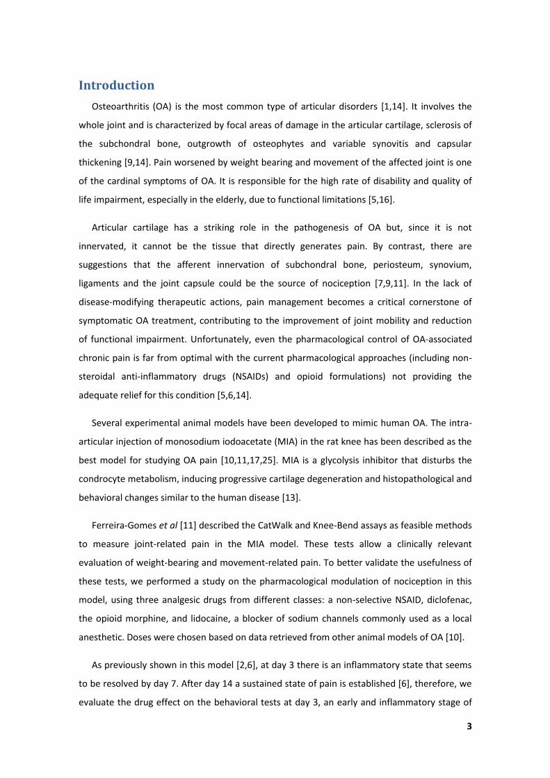
Joint histology was evaluated in all animals at day 31 through Fast-Green and Safranin-O
stained sections (Fig. 1). The observed changes were similar to those previously described [24].
In MIA injected animals, it was observed a decrease in the articular cartilage thickness due
to chondrocytes and matrix proteoglycan’s loss, as indicated by the marked reduction of
Safranin-O stain. There was also exposure of subchondral bone and subchondral bone
thickening. These alterations were similar and confirmed in all OA cases. Control animals
showed no alterations in cartilage or subchondral bone structure.

7
Behavioral Testing
Control animals show no nociceptive response when evaluated by Knee-Bend and CatWalk
tests.
The injection of MIA into the left knee induced a significant increase in the Knee-Bend score
of the ipsilateral knee verified at day 3, which was maintained throughout the study (Fig. 2). In
OA animals, the ipsilateral paw-print intensity, as assessed by the CatWalk test, was
significantly decreased from day 3 until day 31 (Fig. 3).
Pharmacological evaluation
Lidocaine
The effect of intra-articular administration of lidocaine on OA-induced nociception was
evaluated at 10, 20 and 30 minutes after its injection (Fig. 4). At day 3, 10 minutes after
lidocaine injection there was a significant reduction of the knee-bend score of the affected
knee from 11,2 ± 1,5 to 1,2 ± 0,7 (p<0.01). Afterwards, the effect starts to diminish and at 30
minutes after the administration it is completely reverted to baseline levels (Fig 4A). This was
also verified at day 20 (p<0.01) and at day 31 (p<0.05).
Lidocaine effect on CatWalk at day 3 was also observed 10 minutes after the drug injection
with an increase from 25,2 ± 4,8 to 38,2 ± 4,4 (p<0.05) immediately reversing at 20 minutes
(24,9 ± 2,9). At days 20 and 31, although there was a similar trend, no significance was
observed (Fig. 4B).
Lidocaine did not produce any effect on the control animals (data not shown).
Morphine
Morphine’s Knee-Bend ipsilateral response was observed 30 minutes after drug
administration at both days 3 and 20. There was a reduction from 11,4 ± 0,7 to 0,0 ± 0,0 at day
3 (p<0.001) and from 11,0 ± 1,1 to 0,6 ± 0,4 at day 20 (p<0.001). This effect is still observed at
90 minutes, and starts to revert at 120 minutes after morphine administration (Fig. 5A). A
score reduction is observed 60 minutes after drug administration at 31 days (p<0.01) to near-
zero levels (0,6 ± 0,4).
Morphine effect on CatWalk score at days 3 and 20 starts at 30 minutes after drug
administration but only reaches significant values at 60 minutes (p<0.05 at day 3 and p<0.001

8
at day 20). Drug effect starts to revert at 120 minutes (Fig. 5B). At 31 days, there was a
significant increase on left-hindpaw total intensity 60 minutes after drug administration
(p<0.05).
Diclofenac
Diclofenac maximum effect is observed at 30 minutes post drug administration, with
ipsilateral Knee-Bend scores at day 3 significantly reducing from 14,6 ± 0,9 to 2,0 ± 0,4
(p<0.001; Fig. 6A). Complete drug effect reversion is noted at 120 minutes. At 20 days there
was a significant decrease 30 minutes after drug administration (p<0.05) reversing at 120
minutes (10,6 ± 1,8 before drug injection to 6,2 ± 1,1, 30 minutes later). Nevertheless the
effect was much less pronounced than at day 3 (Fig. 6A). No effects of diclofenac on Knee-
Bend score were noted at day 31.
At day 3, 30 minutes after diclofenac administration there is an increase in CatWalk score
from 22,6 ± 4,0 to 37,8 ± 6,2 (p<0.01). At 20 or 31 days, diclofenac administration didn’t show
any effect on the CatWalk test (Fig. 6B)
Discussion
Chronic pain is usually accompanied by an increased response to painful stimuli,
hyperalgesia, and innocuous stimuli, allodynia. Such hypersensitivity could be related to
peripheral pain sensitization, mediated by cytokines, and/or central pain sensitization at
subcortical or cortical level, already described in the pathogenesis of OA [7]. Various
behavioral tools have been used to evaluate OA-related pain in the MIA model [2,5,17],
however none of them has been validated as a significant mean to study damaged tissues-
related (primary) hyperalgesia, as most of them focus on secondary hyperalgesia and
secondary allodynia. With the pharmacological study here described in this model during 31
days, we try to validate and better understand the usefulness of Knee-Bend and Catwalk to
evaluate knee-joint movement-elicited nociception, a common trait of OA [12].
Morphine is an opioid with strong analgesic properties that exerts its effects mostly at CNS
[26]. Its analgesic effect is mainly due to its activation of the inhibitory mu-opioid receptor
located both at spinal cord and supraspinal sites. This activation leads to reduced transmitter
release from nociceptive afferent fibers as well as an attenuation of neuronal processing of
pain messages [26]. In our study, morphine provided a remission of Knee-Bend score to nearly
zero-values in ipsilateral knee, due to its efficacy as an analgesic drug (Fig. 5A). The increase in

9
ipsilateral paw contribution to total weight bearing could easily be compared to that of non-
OA animals, supporting the nociceptive pathways block promoted by this drug (Fig. 5A). This
morphine nociception reversing effect, provide a support to the Knee-Bend testing and to the
CatWalk approach as valid methods to measure MIA-induced OA-related pain. Morphine-
related ipsilateral knee reduction on nociception can contribute to the reduction of
contralateral nociceptive response. In humans, the only published study on morphine efficacy
revealed improvement in the overall quality of sleep and reduced pain in patients with
moderate-to-severe OA pain [4]. This, as other preclinical studies, favor clinical research on
opioids role in OA pain.
Pain presents direct afferent, intrinsic regulatory and higher symbolism, associating cortical
components with many biological and psychological regulating elements. Morphine efficacy in
this model may result, not only, from reduced sensitized peripheral neurons activation of
central neurons but also from its interaction with descending control pathways. Morphine
psychoactive effects (in prefrontal opioid pathways) could add an increase to the ipsilateral
paw contribution to the weight-bearing activity. Detailed comprehension of morphine-acting
pathways would be very useful in the OA-related pain preclinical study as for the management
of OA patients.
Lidocaine exerts its effects by blocking the fast voltage-gated sodium (Na+) channels in the
neuronal membrane [3]. The excitable membrane of nerve axons maintains a resting
transmembrane potential of -90 to -60 mV. During excitation, the sodium channels open and a
fast inward sodium current quickly depolarizes the membrane. As a result of depolarization,
the sodium channels close and the potassium channels open [21]. The outward potassium
current repolarizes the membrane. With sufficient blockade, the membrane of the
postsynaptic neuron will not depolarize and so it fails to transmit the action potential leading
to lidocaine anesthetic effects. Lidocaine effectively reverted the nociception-related behavior
as assessed by the significant reduction in the Knee-Bend score (Fig. 4A). This is consistent with
the blockage of signal transmission to central structures, reflecting this test significance in the
evaluation of primary hyperalgesia.
CatWalk provides an indirect weight-load analysis in the context of movement related-
nociception study. In human unilateral knee OA it was observed an increase in contralateral leg
load [7], so, weight-load analysis can reflect the significance of OA pain in human daily life
activities. Facing that, OA-patients gait depends not only on the primary joint hyperalgesia but,
also, on the combination of that movement restricting factor with complex neural interacting

10
pathways, some of them with supraspinal origin. A change in one of the locomotion modulator
stimuli (as the nociceptive stimuli removal) implies the reorganization of the march related
pathways and the “learning” of a new way of walk. This learning process is certainly time-
dependent and is necessary a significant time period to allow the habituation to the new non-
painful condition and, with that, the development of a new “walking way” with a statistically
significant difference. The quick acting lidocaine, with its effect peaking at 10 minutes and
reverting 20 to 30 minutes after administration, does not provide the necessary time period to
the occurrence of this adaptation process, which may explain the lack of contribution of
ipsilateral hindpaw weightbearing at days 20 and 31. In contrast, morphine effect is way more
sustained (felt from 30 to 120 minutes) and this longer time period allows the rat to adapt to
its new “non-nociceptive” situation, resulting in the significant detection of new weight-load
pattern 60 minutes after drug administration.
Diclofenac, as other non-steroidal anti-inflammatory drugs (NSAIDs), is a common
therapeutic option in the management of middle to moderate OA pain. During the progression
of the inflammatory process, many prostanoid products (Prostaglandin E2, D2, F2, I2 and
thromboxane) are synthesized by the action of cyclo-oxygenase (COX) enzymes, thereby, the
blockage of the enzymes COX-1 and COX-2 is helpful in the management of an inflammatory
pain situation [8,15,27]. Pulichino and colleagues [20] documented, following MIA injection, a
rise in prostaglandins levels that return to baseline by day 7. Histological analysis also
demonstrates the presence of early inflammation in the MIA model and the subsequent
resolution of the inflammation by day 7 [2,6,10]. In the present study, diclofenac was highly
effective at day 3, in a period of disease progression characterized by the inflammatory state.
The absence of inflammation at days 20 or 31 may explain the decrease of efficacy of
diclofenac in this model at these time points. A slight significant decrease in Knee-Bend score
at day 20 thirty-minutes testing, was observed but was not accompanied by any CatWalk
increase in ipsilateral hindpaw contribution increase. This helps to illustrate the difference
between these tests properties, the Knee-Bend has most likely a higher sensitivity perceiving
primary hyperalgesia comparing to CatWalk (Fig. 6). The lack of relevant diclofenac’s effect at
days 20 or 31 argues in favor of the lack of inflammatory mechanisms in OA pain at later time
points.
Previous studies *5,10+ tried to assess acute diclofenac’s antinociceptive effect on various
behavioral endpoint and we can find some discordant results. Fernihough et al. [10] have
noticed a significant secondary mechanical hyperalgesia reduction with diclofenac at day 3 but
none at days 14 or 28; this is similar to our primary hyperalgesia reduction pattern. Twenty-

11
one days Pomonis et al. [17] results showed no significant change in weight bearing with the
administration of celecoxib or indomethacin. These results contrast with Chandran and
collegues [5], that, using the grip-force test, observed, 20 days after 3 mg MIA injection, an
antinociceptive effect attributable to 30 mg/Kg (100µmol/Kg) doses of diclofenac. Ivanavicius
et al. [12], aware of these works used weight-bearing methods and proved celecoxib
inefficiency after day 14; remarkably he found significant naproxene antinociceptive effects at
day 14. These authors used different MIA doses ranging from 1 to 3 mg and there seems to be
no trendline relating doses, disease progression and its dependent response to NSAIDs.
Further studies are necessary to understand the role of different clinical relevant NSAIDs in the
early inflammatory pain state observed in the MIA-induced model and a standardized
approach with the Knee-Bend and CatWalk tests would provide a deeper insight in this subject.
The study of the pharmacological modulation of a known pain-related molecular marker
would provide a powerful support to the validation of any pain-related behavioral study.
Noxious stimuli have been shown to induce Fos expression in sub-populations of spinal cord
neurons located predominatly in laminae I, IIo and V [20]. Despite low Fos specificity for these
stimuli, previous works [19,23] showed that pre-treatment with morphine prevent spinal Fos
activation. Studies to evaluate different drug-induced Fos pattern changes in this model are
already in progress.
The pain behavior associated with intra-articular MIA injection is biphasic in nature with an
early inflammatory phase that lasts until day 7 [6] and a second, chronic phase beginning at
day 14. Bove et al. [2] showed that bone damage only appears after day 7. The delayed onset
in human OA pain suggest that, even fronting the scarcity of knowledge regarding OA-
nociceptive mechanisms, a large part of pain afferents could come from subchondral bone. In
this model, the study of the efficacy of therapeutically relevant agents becomes clinically
significant when done at later time points such as 20 days after MIA injection. In this study we
provide compelling evidence of the high sensitivity and strength of the CatWalk and the Knee-
Bend tests to evaluate movement-induced nociception. The different antinociceptive profiles
of morphine, lidocaine and diclofenac, deeply linked and in agreement to their different OA-
associated pain reversal mechanisms, provides useful information about these tests’ value in
the study of the analgesic efficacy of drugs in OA treatment.

12
Acknowledgments
The author thanks Joana Ferreira-Gomes and Sara Adães for their valuable guidance in the
laboratorial work and precious contribution in the manuscript preparation.

13
Reference List
[1] American College of Rheumatology. Subcommittee on Osteoarthitis Guidelines.
Recommendations for the medical management of osteoarthritis of the hip and knee: 2000
update. Arthritis Rheum, 2000. 43(9):1905-15
[2] Bove SE, Calcaterra SL, Brooker RM, Huber CM, Guzman RE, Juneau PL, Schrier DJ,
Kilgore KS. Weight bearing as a measure of disease progression and efficacy of anti-
inflammatory compounds in a model of monosodium iodoacetate-induced osteoarthritis.
Osteoarthritis Cartilage, 2003. 11(11):821-30.
[3] Butterworth JF, Strichartz GR. Molecular Mechanisms of Local Anesthesia: A review.
Anesthesiology, 1990. 72(4): 711-34
[4] Caldwell JR, Rapoport RJ, Davis JC, Offenberg HL, Marker HW, Roth SH, Yuan W, Eliot L,
Babul N, Lynch PM. Efficacy and Safety of a Once-Daily Morphine Formulation in Chronic,
Moderate-to-Severe Osteoarthritis Pain: Results from a Randomized, Placebo-Controlled,
Double-Blind Trial and an Open-Label Extension Trial. J Pain Symptom Manage, 2002.
23(4):278-91
[5] Chandran P, Madhavi P, Blomme EA, Hsieh GC, Michael WS, Honore P. Pharmacological
modulation of movement-evoked pain in a rat model of osteoarthritis. Eur J Pharmacol, 2009.
613(1-3):39-45.
[6] Combe R, Bramwell S, Field MJ. The monosodium iodoacetate model of osteoarthritis: a
model of chronic nociceptive pain in rats? Neurosci Lett, 2004. 370(2-3):236-40.
[7] Dieppe PA, Lohmander LS. Pathogenesis and management of pain in osteoarthritis.
Lancet, 2005. 365(9463):965-73.

14
[8] Dray A, Read SJ. Arthritis and Pain: Future targets to control osteoarthritis pain. Arthritis
Res Ther, 2007. 9(3):212-25
[9] Felson DT. Clinical Practice. Osteoarthritis of the Knee. N Engl J Med, 2006. 354(8):841-8.
[10] Fernihough J, Gentry C, Malcangio M, Fox A, Rediske J, Pellas T, Kidd B, Bevan S, Winter
J. Pain related behavior in two models of osteoarthritis in the rat knee. Pain, 2004. 112(1-
2):83-93.
[11] Ferreira-Gomes J, Adães S, Castro-Lopes JM. Assessment of Movement-Evoked Pain in
Osteoarthritis by the Knee-Bend and CatWalk Tests: A Clinically Relevant Study. J Pain, 2008.
9(10):945-54
[12] Ivanavicius SP, Ball AD, Heapy CG, Westwood FR, Murray F, Read SJ. Structural
pathology in a rodent model of osteoarthritis is associated with neuropathic pain: Increased
expression of ATF-3 and pharmacological characterization. Pain 2007; 128(3): 272-82
[13] Janusz MJ, Hoofkin EB, Heitmeyer SA, Woessner JF, Freemont AJ, Hoyland JA, Brown
KK, Hsieh LC, Almstead NG, De B, Natchus MG, Pikul S, Taiwo YO. Moderation of iodoacetate-
induced experimental osteoarthritis in rats by matrix metalloproteinase inhibitors.
Osteoarthritis Cartilage, 2001. 9(8):751-60.
[14] Kidd BL. Osteoarthritis and joint pain. Pain 2006; 123(1-2):6-9
[15] Kokki H, Kumpulainen E, Laisalmi M, Savolainen J, Rautio J, Lehtonen M. Diclofenac
readily penetrates the cerebrospinal fluid in children. Br J Clin Pharmacol 2008. 65(6), 879-84.
*16+ O’Reilly SC, Muir KR, Doherty M. Knee pain and disability in the Nottingham
community: associated with poor health status and psychological distress. Br J Rheumatol,
1998. 37(8):870-3.

15
[17] Pomonis JD, Boulet JM, Gottshall SL, Phillips S, Sellers R, Bunton T, Walker K.
Development and pharmacological characterization of a rat model of osteoarthritis pain. Pain,
2005. 114(3):339-46.
[18] Potes CS, Neto FL, Castro-Lopes JM. Administration of baclofen, a gamma-aminobutyric
acid type B agonist in the thalamic ventrolbasal complex, attenuates allodynia in monoarthritic
rats subjected to the ankle-bend test. J Neurosci Res 2006. 83: 515-23.
[19] Presley RW, Menétrey D, Levine JD, Basbaum AI. Systemic morphine suppresses
noxious stimulus-evoked fos protein-like immunoreactivity in the rat spinal cord. J Neurosci,
1990. 10(1):323-35
[20] Pulichino AM, Rowland S, Wu T, Clark P, Xu D, Mathieu MC, Riendeau D, Audoly LP.
Prostacyclin antagonism reduces pain and inflammation in rodent models of hyperalgesia and
chronic arthritis. J Pharmacol Exp Ther, 2006. 319, 1043–1050.
[21] Scholtz A. Mechanisms of (local) anaesthetics on voltage-gated sodium and other ion
channels. Br J Anaesth 2002. 89: 52-61.
[22] Sun X, Yokoyama M, Mizobuchi S, Kaku R, Nakatsuka H, Takahashi T, Morita K. The
effects of pretreatment with lidocaine or bupivaxaine on the spatial and temporal expression
of c-Fos protein in the spinal cord caused by plantar incision in the rat. Anesth Analg, 2004.
98(4):1093-8.
[23] Tölle T.R., Castro-Lopes J.M., Coimbra A. and Zieglgänsberger W. Opiates modify
induction of c-fos proto-oncogene in the spinal cord following noxious stimulation. Neurosci
Lett, 1990. 111(1-2):46-51.
[24] van der Kraan PM, Vitters EL, van Beuningen HM, van den Berg WB. Proteoglycan
synthesis and osteophyte formation in ‘metabolically’ and ‘mechanically’ induced murine

16
degenerative joint disease: an in-vivo autoradiographic study. Int J Exp Pathol, 1992. 73, 335–
50.
[25] Vonsy JL, Ghandehari J, Dickenson AH. Differential analgesic effects of morphine and
gabapentin on behavioural measures of pain and disability in a model of osteoarthritis pain in
rats. Eur J Pain, 2009. 13(8):786-93.
[26] Yaksh TL. Pharmacology and mechanisms of opioid analgesic activity. Acta Anaesth
Scand, 1997. 41:94–111.
[27] Yaksh TL, Dirig DM, Conway CM, Svensson C, Luo ZD, Isakson PC. The acute
hyperalgesic action of non-steroidal, anti-inflammatory drugs and release of spinal
prostaglandin E2 is mediated by the inhibition of constitutive spinal cyclooxygenase-2 (COX-2)
but not COX-1. J Neurosci, 2001. 21:5847-53.
[28] Zimmermann M, Ethical guidelines for investigations of experimental pain in conscious
Animals. Pain, 1983. 16:109–10
17
Fig. 1 – Fast-Green and Safranin-O stainig of knee joint 31 days after saline (A) or 2 mg MIA (B)
injection. MIA injected animals show destruction of articular cartilage (C) with subchondral
bone (SB) changes (arrows). M: Menisci.

18
Fig. 2 – Time course changes in Knee-Bend score in MIA (A, n = 20) and saline (B, n = 5) injected
rats (mean ± S.E.M.). Baseline scores were determined in both knees for all animals before
injection (day 0). *** P < .001, significantly different from day 0 levels (repeated measures
ANOVA). †††P < .001, significantly different from contralateral knee (paired t-test). No
difference is observed, during disease progression time, between ipsi and contralateral paw in
saline injected animals (B).
A
†††
††† †††
Ipsilateral
Contralateral
B

19
Fig. 3 – Time course changes in the percentage of the ipsilateral paw print over total intensity
assessed by the CatWalk test. Paw print intensity baseline scores were determined for all
animals before injection (day 0). *** P < .001, significantly different from baseline levels
(repeated measures ANOVA). †P < .05, significantly different from control animals (1-way
ANOVA). No difference is observed, during disease progression time, between ipsi and
contralateral paw in saline injected animals (B).
***
OA animals
Control animals
*** *** † ***

20
Fig. 4 – Effect of lidocaine on MIA-injected rats assessed by Knee-Bend (A) and CatWalk (B)
tests on days 3, 20 and 31. Day 31 results were plotted as a bar graph. * and ** represent
significance levels of p < .05, and p < .01 at day 3. †† represent significance levels of p < .01 at
day 20. x represents significance levels of p < .05 at day 31. Effect on Knee-Bend test is verified
both at days 3, 20 and 31. CatWalk-assessed reduction on nociception is only verified at day 3.
A
B
A

21
Fig. 5 – Effect of morphine on MIA-injected rats assessed by Knee-Bend (A) and CatWalk (B)
tests on days 3, 20 and 31. Day 31 results were plotted as a bar graph. *, ** and *** represent
significance levels of p < .05, p < .01 and p < .001 at day 3. † and ††† represent significance
levels of p < .05 and p < .001 at day 20. x and xx represents significance levels of p < .05 and p
< .01 at day 31. Morphine nociception reduction, as evaluated by Knee-Bend test, is observed
at 3, 20 and 31 days and already detected 30 minutes after administration. CatWalk test detect
significant nociception reduction at days 3, 20 and 31.
B
A
x

22
Fig. 6 – Effect of diclofenac on MIA-injected rats assessed by Knee-Bend (A) and CatWalk (B)
tests on days 3, 20 and 31. Day 31 results were plotted as a bar graph. ** and *** represent
significance levels of p < .01 and p < .001 at day 3. † and †† represent significance levels of p <
.05 and p < .01 at day 20. At day 3, significant changes are noted on CatWalk and Knee-Bend
tests. At day 20 changes are only observed on Knee-Bend tests. No significant changes were
found at day 31.
A
B