
12 Supervision of Patients With Acute Renal Failure, Renal Colic.
Upload
vuongquynhCategory
view
222download
1
136
Lab Anim Res 2014: 30(3), 136-141
http://dx.doi.org/10.5625/lar.2014.30.3.136
Experimental model of tympanic colic (acute abdomen) in chinchillas (Chinchilla lanigera)
Malcon Andrei Martinez-Pereira1,2, Raphaela da Cunha Franceschi2,3, Bárbara Paranhos Coelho2,Gustavo da Rosa Fünkler2, Denise Maria Zancan2,3*
1Centro Ciências da Saúde, Universidade de Cruz Alta, Cruz Alta, Brazil2Laboratório de Neurobiologia Comparada, Departamento de Fisiologia, Instituto de Ciências Básicas da Saúde,
Universidade Federal do Rio Grande do Sul, Porto Alegre, Brazil3Programa de Pós-Graduação em Neurociências, Instituto de Ciências Básicas da Saúde (ICBS), Universidade Federal do Rio
Grande do Sul, Porto Alegre, Brazil
Digestive disorders caused by sudden changes in diet or inappropriate diet are among the most commondisorders of the digestive system. Cecal or intestinal tympany, one consequence of inappropriate diet, ischaracterized by the accumulation of gases, marked distension of the cecum and colon and the inductionof inflammatory processes. To know the effects of intestinal tympany on the enteric plexuses, wedeveloped a method of experimental tympanic colic (TC) in the Chinchilla lanigera. This species was usedin view of its susceptibility to TC. TC was induced with a diet rich in alfalfa associated with grain overloadfor two weeks. Physical and clinical examination including the von Frey test confirmed the diagnosis. Thechinchillas with acute abdomen were treated with 1% ketoprofen and resumption of a balanced diet.Necropsy and histopathological analysis showed tympany-induced alterations mainly in the cecum andcolon. After treatment, the control conditions were restored. The TC protocol is proposed as anexperimental approach designed to aid the study of the effects of acute intestinal inflammation andobstruction caused by an inappropriate diet.
Keywords: Intestinal tympany, acute abdomen, bowel inflammation, Chinchilla lanigera
Received 26 May 2014; Revised version received 4 September 2014; Accepted 11 September 2014
Tympanic colic (TC) is characterized by the inability
to expel gases produced during digestion by microbial
fermentation, resulting in marked distension of the walls
of cecum and colon. Colic is a major cause of illness and
death in the horse [1]. Gastric or intestinal tympany can
be associated with inappropriate diet, overeating hay rich
in clover or alfalfa, sudden dietary changes and gastro-
intestinal inflammation [2,3]. A change in the quality or
the quantity of food, as well as a change in the schedule
of feeding also results in an increased risk of colic [4].
In some cases, such as grain overload, the proximate
cause may be evident, but the underlying mechanism or
physiologic problems often remain unknown [5]. Acute
distention of the gut wall and the consequent intestinal
inflammation can lead to pronounced changes to the
structure and physiology of the enteric nervous system
(ENS). Considering the importance of the ENS in
controlling secretion and motility in the gastrointestinal
tract [6-8], knowledge of the etiology of TC can contribute
towards our understanding of the effects of similar
digestive disorders in the ENS of other mammals, such
as chronic constipation and bloat in ruminants or equine
colic.
Various aspects of the anatomy, physiology and
reproduction [9-13] of Chinchilla lanigera have been
studied. Chinchillas are monogastric hindgut fermenting
Letter
*Corresponding author: Denise Maria Zancan, Laboratório de Neurobiologia Comparada, Departamento de Fisiologia, ICBS, UFRGS,Rua Sarmento Leite, 500. Porto Alegre, Rio Grande do Sul, Brazil, 90050-170.Tel: +55-51-33083305; Fax: +55-51-33083155; E-mail: [email protected]
This is an Open Access article distributed under the terms of the Creative Commons Attribution Non-Commercial License (http://creativecommons.org/licenses/by-nc/3.0) which permits unrestricted non-commercial use, distribution, and reproduction in any medium, provided the original work is properly cited.
Experimental acute abdomen in chinchilla 137
Lab Anim Res | September, 2014 | Vol. 30, No. 3
herbivores. Some authors compare them to horses
(“miniature horses”) in terms of gastrointestinal disease
management [14,15]. The gastrointestinal anatomy of
the chinchilla, such as a highly sacculated cecum and a
large colon, predisposes it to TC [9]. As chinchillas are
more accessible to experimental approaches than equines
or ruminants, we are proposing chinchillas as an
experimental model for investigating the etiopathogeny
of abdominal tympany.
Fifteen Chinchilla lanigera from the Chillacenter
Farm (Viamão, RS, Brazil) were used, (13 females and
2 males), aged 18 to 32 months and weighing between
400 to 700 g. The animals were kept individually in
cages with free access to water and commercial food
(Supra Chinchila, Alisul Alimentos SA, Brazil), a
photoperiod regimen (12 h light/12 h dark) and controlled
temperature (16-24oC). All animals were acclimated for
three days before any experimental procedure (D0 at
D3). They were then divided into control (5 animals),
tympanic (5 animals) and recovery (5 animals) groups.
All animals were euthanatized by an overdose of
ketamine and xylazine (Pfizer, Brazil). The experimental
approach was approved by the local Committee for
Ethics in Research (no 2008148) and all animal procedures
were in accordance with the Brazilian law (Federal Law
no 11.794/2008) on procedures for the scientific use of
animals. The experimental design is depicted in Figure 1.
The TC inducing protocol for chinchillas was adapted
from the protocols of colic in equines [16] and by
analyzing the feeding practices that increase the risk of
colic [4]. Weiss et al. [16] induces a laminitis secondary
to intestinal impaction by grain overload. Among all the
feeding patterns studied, changing the type of hay
remains one of the most significant factors for colic
development [4]. As such, the TC protocol consisted of
a colic-inducing diet (15 % commercial food, 45% corn
grains, 20% carrots, 5% sunflower seeds and 15% alfalfa
daily). The chinchillas were fed at a fixed level (5% of
body weight, BW; as-fed basis) that averaged 23.5 g/day,
while water was freely accessible. The tympanic and
recovery groups were fed with the colic-inducing diet
daily for fifteen days (D3 to D18), while the control
group was fed commercial food daily (5% of BW; as-fed
basis) and 1.5% of BW of alfalfa (as-fed) once a week.
This mixed diet was based on Wolf et al. [17]. Alfalfa
was cited as one of the most common cause of excessive
gas production in equine and bovine. Many plant
compounds, such as the triterpene saponins, have been
suggested as contributing factors to the occurrence of
equine colic or bovine bloat foam [5,18]. Vieira et al.
[19] analyzed 28 different Brazilian cultivars of alfalfa
(Medicago sativa L.) and found a maximum 1.78% of
saponin in alfalfa. Regardless of the low or moderate
level of saponins in alfalfa, the consumption of only
small amounts of this leguminous is recommended for
chinchillas. Once TC had been diagnosed (D18), the
recovery group returned to the control feeding and were
medicated with 1% ketoprofen (2 mg/kg Ketofen®, IM,
Merial, Brazil) once a day, for five days (D18 to D23),
when the clinical examinations were completed.
The clinical diagnosis was based on physical examination,
using palpation, auscultation and abdominal percussion.
The animals were examined on the first day (D0), after
the acclimation period (D4), during the colic-inducing
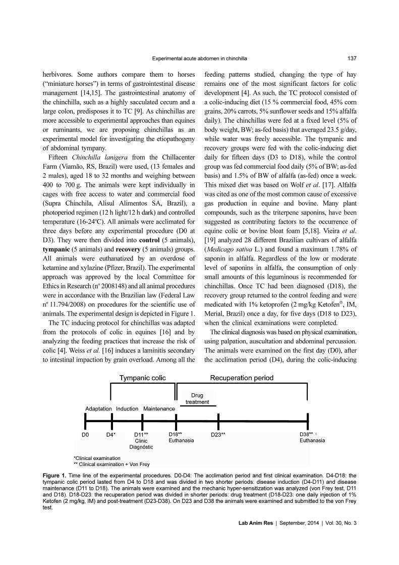
Figure 1. Time line of the experimental procedures. D0-D4: The acclimation period and first clinical examination. D4-D18: thetympanic colic period lasted from D4 to D18 and was divided in two shorter periods: disease induction (D4-D11) and diseasemaintenance (D11 to D18). The animals were examined and the mechanic hyper-sensitization was analyzed (von Frey test, D11and D18). D18-D23: the recuperation period was divided in shorter periods: drug treatment (D18-D23: one daily injection of 1%Ketofen (2 mg/kg, IM) and post-treatment (D23-D38). On D23 and D38 the animals were examined and submitted to the von Freytest.
138 Malcon Andrei Martinez-Pereira et al.
Lab Anim Res | September, 2014 | Vol. 30, No. 3
feed period for confirmation of disease onset (D11 and
D18), and during the recuperation period of the recovery
group (D23 and D38). The analysis of abdominal
sensitization was tested with a mechanical force transducer
(digital von Frey, Insight, Brazil) on the D11, D18, D23
and D38. Foot withdrawal frequencies in response to
von Frey stimuli were measured and used to indicate
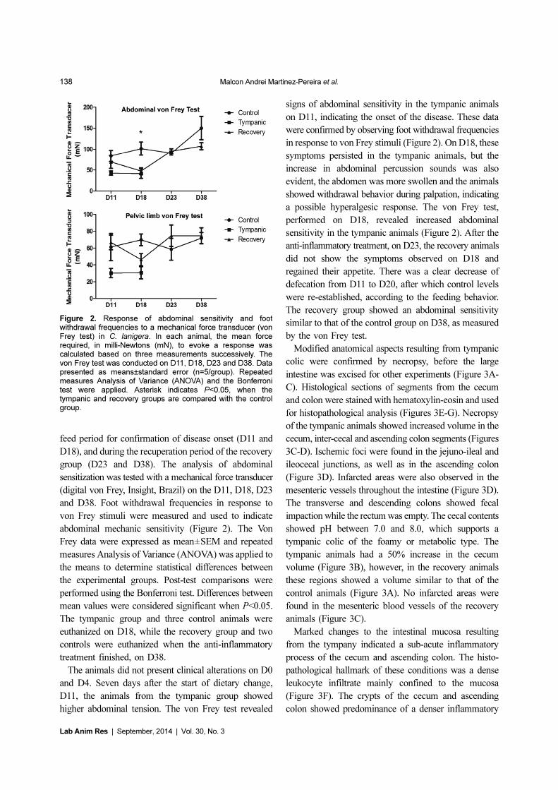
abdominal mechanic sensitivity (Figure 2). The Von
Frey data were expressed as mean±SEM and repeated
measures Analysis of Variance (ANOVA) was applied to
the means to determine statistical differences between
the experimental groups. Post-test comparisons were
performed using the Bonferroni test. Differences between
mean values were considered significant when P<0.05.
The tympanic group and three control animals were
euthanized on D18, while the recovery group and two
controls were euthanized when the anti-inflammatory
treatment finished, on D38.
The animals did not present clinical alterations on D0
and D4. Seven days after the start of dietary change,
D11, the animals from the tympanic group showed
higher abdominal tension. The von Frey test revealed
signs of abdominal sensitivity in the tympanic animals
on D11, indicating the onset of the disease. These data
were confirmed by observing foot withdrawal frequencies
in response to von Frey stimuli (Figure 2). On D18, these
symptoms persisted in the tympanic animals, but the
increase in abdominal percussion sounds was also
evident, the abdomen was more swollen and the animals
showed withdrawal behavior during palpation, indicating
a possible hyperalgesic response. The von Frey test,
performed on D18, revealed increased abdominal
sensitivity in the tympanic animals (Figure 2). After the
anti-inflammatory treatment, on D23, the recovery animals
did not show the symptoms observed on D18 and
regained their appetite. There was a clear decrease of
defecation from D11 to D20, after which control levels
were re-established, according to the feeding behavior.
The recovery group showed an abdominal sensitivity
similar to that of the control group on D38, as measured
by the von Frey test.
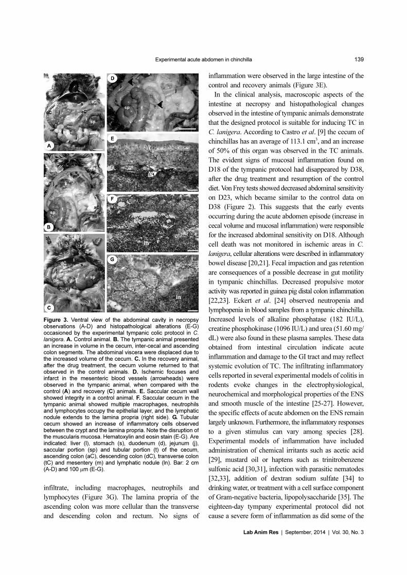
Modified anatomical aspects resulting from tympanic
colic were confirmed by necropsy, before the large
intestine was excised for other experiments (Figure 3A-
C). Histological sections of segments from the cecum
and colon were stained with hematoxylin-eosin and used
for histopathological analysis (Figures 3E-G). Necropsy
of the tympanic animals showed increased volume in the
cecum, inter-cecal and ascending colon segments (Figures
3C-D). Ischemic foci were found in the jejuno-ileal and
ileocecal junctions, as well as in the ascending colon
(Figure 3D). Infarcted areas were also observed in the
mesenteric vessels throughout the intestine (Figure 3D).
The transverse and descending colons showed fecal
impaction while the rectum was empty. The cecal contents
showed pH between 7.0 and 8.0, which supports a
tympanic colic of the foamy or metabolic type. The
tympanic animals had a 50% increase in the cecum
volume (Figure 3B), however, in the recovery animals
these regions showed a volume similar to that of the
control animals (Figure 3A). No infarcted areas were
found in the mesenteric blood vessels of the recovery
animals (Figure 3C).
Marked changes to the intestinal mucosa resulting
from the tympany indicated a sub-acute inflammatory
process of the cecum and ascending colon. The histo-
pathological hallmark of these conditions was a dense
leukocyte infiltrate mainly confined to the mucosa
(Figure 3F). The crypts of the cecum and ascending
colon showed predominance of a denser inflammatory
Figure 2. Response of abdominal sensitivity and footwithdrawal frequencies to a mechanical force transducer (vonFrey test) in C. lanigera. In each animal, the mean forcerequired, in milli-Newtons (mN), to evoke a response wascalculated based on three measurements successively. Thevon Frey test was conducted on D11, D18, D23 and D38. Datapresented as means±standard error (n=5/group). Repeatedmeasures Analysis of Variance (ANOVA) and the Bonferronitest were applied. Asterisk indicates P<0.05, when thetympanic and recovery groups are compared with the controlgroup.
Experimental acute abdomen in chinchilla 139
Lab Anim Res | September, 2014 | Vol. 30, No. 3
infiltrate, including macrophages, neutrophils and
lymphocytes (Figure 3G). The lamina propria of the
ascending colon was more cellular than the transverse
and descending colon and rectum. No signs of
inflammation were observed in the large intestine of the
control and recovery animals (Figure 3E).
In the clinical analysis, macroscopic aspects of the
intestine at necropsy and histopathological changes
observed in the intestine of tympanic animals demonstrate
that the designed protocol is suitable for inducing TC in
C. lanigera. According to Castro et al. [9] the cecum of
chinchillas has an average of 113.1 cm3, and an increase
of 50% of this organ was observed in the TC animals.
The evident signs of mucosal inflammation found on
D18 of the tympanic protocol had disappeared by D38,
after the drug treatment and resumption of the control
diet. Von Frey tests showed decreased abdominal sensitivity
on D23, which became similar to the control data on
D38 (Figure 2). This suggests that the early events
occurring during the acute abdomen episode (increase in
cecal volume and mucosal inflammation) were responsible
for the increased abdominal sensitivity on D18. Although
cell death was not monitored in ischemic areas in C.
lanigera, cellular alterations were described in inflammatory
bowel disease [20,21]. Fecal impaction and gas retention
are consequences of a possible decrease in gut motility
in tympanic chinchillas. Decreased propulsive motor
activity was reported in guinea pig distal colon inflammation
[22,23]. Eckert et al. [24] observed neutropenia and
lymphopenia in blood samples from a tympanic chinchilla.
Increased levels of alkaline phosphatase (182 IU/L),
creatine phosphokinase (1096 IU/L) and urea (51.60 mg/
dL) were also found in these plasma samples. These data
obtained from intestinal circulation indicate acute
inflammation and damage to the GI tract and may reflect
systemic evolution of TC. The infiltrating inflammatory
cells reported in several experimental models of colitis in
rodents evoke changes in the electrophysiological,
neurochemical and morphological properties of the ENS
and smooth muscle of the intestine [25-27]. However,
the specific effects of acute abdomen on the ENS remain
largely unknown. Furthermore, the inflammatory responses
to a given stimulus can vary among species [28].
Experimental models of inflammation have included
administration of chemical irritants such as acetic acid
[29], mustard oil or haptens such as trinitrobenzene
sulfonic acid [30,31], infection with parasitic nematodes
[32,33], addition of dextran sodium sulfate [34] to
drinking water, or treatment with a cell surface component
of Gram-negative bacteria, lipopolysaccharide [35]. The
eighteen-day tympany experimental protocol did not
cause a severe form of inflammation as did some of the
Figure 3. Ventral view of the abdominal cavity in necropsyobservations (A-D) and histopathological alterations (E-G)occasioned by the experimental tympanic colic protocol in C.lanigera. A. Control animal. B. The tympanic animal presentedan increase in volume in the cecum, inter-cecal and ascendingcolon segments. The abdominal viscera were displaced due tothe increased volume of the cecum. C. In the recovery animal,after the drug treatment, the cecum volume returned to thatobserved in the control animals. D. Ischemic focuses andinfarct in the mesenteric blood vessels (arrowheads) wereobserved in the tympanic animal, when compared with thecontrol (A) and recovery (C) animals. E. Saccular cecum wallshowed integrity in a control animal. F. Saccular cecum in thetympanic animal showed multiple macrophages, neutrophilsand lymphocytes occupy the epithelial layer, and the lymphaticnodule extends to the lamina propria (right side). G. Tubularcecum showed an increase of inflammatory cells observedbetween the crypt and the lamina propria. Note the disruption ofthe muscularis mucosa. Hematoxylin and eosin stain (E-G). Areindicated: liver (l), stomach (s), duodenum (d), jejunum (j),saccular portion (sp) and tubular portion (t) of the cecum,ascending colon (aC), descending colon (dC), transverse colon(tC) and mesentery (m) and lymphatic nodule (ln). Bar: 2 cm(A-D) and 100 µm (E-G).
140 Malcon Andrei Martinez-Pereira et al.
Lab Anim Res | September, 2014 | Vol. 30, No. 3
above-mentioned substances, but instead represents an
alteration that is more likely to occur due to the feeding
mistakes occasionally observed in animal husbandry.
These mistakes can result in unbalanced diets and
digestive disorders in chinchillas or other small,
domesticated mammals. The extent of the damage to
intrinsic and extrinsic gut innervation evoked by
experimental tympany is the object of our further
studies.
Acknowledgments
This research was supported by grants from the
Brazilian Coordination of Improvement of Higher
Education (Capes) and Brazilian National Research
Council (CNPq). We are grateful to Cabanha Chillacenter
(Viamão, RS, Brazil) for kindly providing the animals
used in this study.
References
1. Tinker MK, White NA, Lessard P, Thatcher CD, Pelzer KD,Davis B, Carmel DK. Prospective study of equine colic incidenceand mortality. Equine Vet J 1997; 29(6): 448-453.
2. Hoefer HL. Chinchillas. Vet Clin North Am Small Anim Pract1994; 24(1): 103-111.
3. Donnelly TM, Schaeffer DO. Disease problems of guinea pigsand chinchillas. In: Ferrets, Rabbits, and Rodents, ClinicalMedicine and Surgery (Hillyer EV, Quesenberry KQ, Eds.), 1997;pp 270-281.
4. Gonçalves S, Julliand V, Leblond A. Risk factors associated withcolic in horses. Vet Res 2002; 33(6): 641-652.
5. White NA II: Causes and risks for colic. AAEP Proceedings 2006;52: 115-119.
6. Kunze WA, Furness JB, Bertrand PP, Bornstein JC. Intracellularrecording from myenteric neurons of the guinea-pig ileum thatrespond to stretch. J Physiol 1998; 506: 827-842.
7. Saper CB. The central autonomic nervous system: consciousvisceral perception and autonomic pattern generation. Annu RevNeurosci 2002; 25: 433-469.
8. Jänig W. The integrative action of the autonomic nervous system:Neurobiology of homeostasis. Cambridge University Press,Cambridge, 2006; pp 168-208.
9. Castro TF, Dummer RJ, Rickes EM, Pereira MAM.Morphological, morphometric and topographical description ofthe digestive tract in Chinchilla lanigera. Braz J Vet Res Anim Sci2010; 47: 86-94.
10. Martinez-Pereira MA, Rickes EM. The spinal nerves thatconstitute the lumbosacral plexus and their distribution in thechinchilla. J S Afr Vet Assoc 2011; 82(3): 150-154.
11. Brown TA, Harrison RV. Responses of neurons in chinchillaauditory cortex to frequency-modulated tones. J Neurophysiol2009; 101(4): 2017-2029.
12. Jakubów K, Gromadzka-Ostrowska J, Zalewska B. Seasonalchanges in the haematological indices in peripheral blood ofchinchilla (Chinchilla laniger L.). Comp Biochem Physiol AComp Physiol 1984; 78(4): 845-885.
13. Busso JM, Ponzio MF, Fiol de Cuneo M, Ruiz RD. Reproductionin chinchilla (Chinchilla lanigera): current status of environmentalcontrol of gonadal activity and advances in reproductive
techniques. Theriogenology 2012; 78(1): 1-11.14. O’Malley B. Introduction to small mammals. In: Clinical anatomy
and physiology of exotic species, Saunders, New York, 2005, pp165-171.
15. Klaphake E. Common rodent procedures. Vet Clin North AmExot Anim Pract 2006; 9(2): 389-413.
16. Weiss DJ, Evanson OA, McClenahan D, Fagliari JJ, DunnwiddieCT, Wells RE. Effect of a competitive inhibitor of plateletaggregation on experimentally induced laminitis in ponies. Am JVet Res 1998; 59(7): 814-817.
17. Wolf P, Schröder A, Wenger A, Kamphues J. The nutrition of thechinchilla as a companion animal--basic data, influences anddependences. J Anim Physiol Anim Nutr (Berl) 2003; 87(3-4):129-133.
18. Clarke RT, Reid CS. Foamy bloat of cattle. A review. J Dairy Sci1974; 57(7): 753-785.
19. Vieira MEQ, Costa C, Silveira AC, Arrigoni MB. Porcentagensde saponinas e taninos em vinte e oito cultivares de alfafa(Medicago sativa L.) em duas épocas de corte -Botucatu- SP. RevBras Zootec 2001; 30: 1432-1438.
20. Johnson-Delaney, CA. 2008. Exotic Companion MedicineHandbook for Veterinarians. Zoological Education Network.Florida. pp 98.
21. Sanovic S, Lamb DP, Blennerhassett MG. Damage to the entericnervous system in experimental colitis. Am J Pathol 1999; 155(4):1051-1057.
22. Sharkey KA, Kroese AB. Consequences of intestinal inflammationon the enteric nervous system: neuronal activation induced byinflammatory mediators. Anat Rec 2001; 262(1): 79-90.
23. Linden DR, Sharkey KA, Ho W, Mawe GM. Cyclooxygenase-2contributes to dysmotility and enhanced excitability of myentericAH neurones in the inflamed guinea pig distal colon. J Physiol2004; 557: 191-205.
24. Eckert BS, Funkler GR, Rodrigues SV. Case report: Acuteabdomen in chinchilla. UFRGS 2011.
25. Collins SM. The immunomodulation of enteric neuromuscularfunction: implications for motility and inflammatory disorders.Gastroenterology 1996; 111(6): 1683-1699.
26. Lomax AE, Fernández E, Sharkey KA. Plasticity of the entericnervous system during intestinal inflammation. NeurogastroenterolMotil 2005; 17(1): 4-15.
27. Vasina V, Barbara G, Talamonti L, Stanghellini V, Corinaldesi R,Tonini M, De Ponti F, De Giorgio R. Enteric neuroplasticityevoked by inflammation. Auton Neurosci 2006; 126-127: 264-272.
28. Mawe GM, Strong DS, Sharkey KA. Plasticity of enteric nervefunctions in the inflamed and postinflamed gut. NeurogastroenterolMotil 2009; 21(5): 481-491.
29. Valentine JF, Tannahill CL, Stevenot SA, Sallustio JE, Nick HS,Eaker EY. Colitis and interleukin 1beta up-regulate induciblenitric oxide synthase and superoxide dismutase in rat myentericneurons. Gastroenterology 1996; 111(1): 56-64.
30. Morris GP, Beck PL, Herridge MS, Depew WT, Szewczuk MR,Wallace JL. Hapten-induced model of chronic inflammation andulceration in the rat colon. Gastroenterology 1989; 96(3): 795-803.
31. Reinshagen M, Rohm H, Steinkamp M, Lieb K, Geerling I, VonHerbay A, Flämig G, Eysselein VE, Adler G. Protective role ofneurotrophins in experimental inflammation of the rat gut.Gastroenterology 2000; 119(2): 368-376.
32. Barbara G, Vallance BA, Collins SM. Persistent intestinalneuromuscular dysfunction after acute nematode infection inmice. Gastroenterology 1997; 113(4): 1224-1232.
33. Chen Z, Suntres Z, Palmer J, Guzman J, Javed A, Xue J, Yu JG,Cooke H, Awad H, Hassanain HH, Cardounel AJ, Christofi FL.Cyclic AMP signaling contributes to neural plasticity andhyperexcitability in AH sensory neurons following intestinalTrichinella spiralis-induced inflammation. Int J Parasitol 2007;37(7): 743-761.
Experimental acute abdomen in chinchilla 141
Lab Anim Res | September, 2014 | Vol. 30, No. 3
34. Kimball ES, Schneider CR, Wallace NH, Hornby PJ. Agonists ofcannabinoid receptor 1 and 2 inhibit experimental colitis inducedby oil of mustard and by dextran sulfate sodium. Am J PhysiolGastrointest Liver Physiol 2006; 291(2): G364-G371.
35. Yuan PQ, Wu SV, Wang L, Taché Y. Corticotropin releasing factorin the rat colon: expression, localization and upregulation byendotoxin. Peptides 2010; 31(2): 322-331.


![Nephrolithiasis K17 .ppt [Read-Only]ocw.usu.ac.id/course/download/1110000119-genitourinary-system/gus... · PRESENTATION Renal colic-- classically : flank pain, often acute inclassically](https://static.fdocuments.net/doc/165x107/5b7b6f017f8b9ae7368dbea5/nephrolithiasis-k17-ppt-read-onlyocwusuacidcoursedownload1110000119-genitourinary-systemgus.jpg)


![Acute Otitis Media and Acute Coalescent Mastoiditis 2the fluid persisted more than 3 months, it is considered as COME [1]. CSOM is defined as an ear discharge with tympanic membrane](https://static.fdocuments.net/doc/165x107/602d555392edf157af38a866/acute-otitis-media-and-acute-coalescent-mastoiditis-2-the-fluid-persisted-more-than.jpg)


