Exercise conditioning after 10 bed rest -...
7
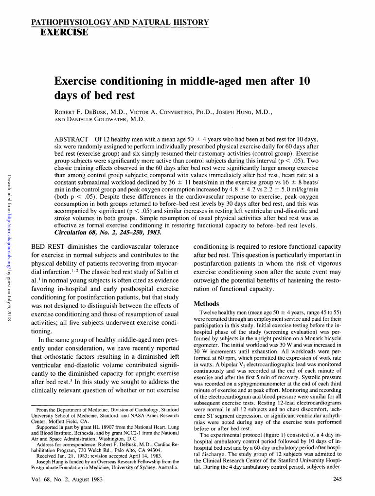
PATHOPHYSIOLOGY AND NATURAL HISTORY EXERCISE Exercise conditioning in middle-aged men after 10 days of bed rest ROBERT F. DEBUSK, M.D., VICTOR A. CONVERTINO, PH.D., JOSEPH HUNG, M.D., AND DANIELLE GOLDWATER, M.D. ABSTRACT Of 12 healthy men with a mean age 50 ± 4 years who had been at bed rest for 10 days, six were randomly assigned to perform individually prescribed physical exercise daily for 60 days after bed rest (exercise group) and six simply resumed their customary activities (control group). Exercise group subjects were significantly more active than control subjects during this interval (p < .05). Two classic training effects observed in the 60 days after bed rest were significantly larger among exercise than among control group subjects; compared with values immediately after bed rest, heart rate at a constant submaximal workload declined by 36 ± 11 beats/min in the exercise group vs 16 + 8 beats/ min in the control group and peak oxygen consumption increased by 4.8 ± 4.2 vs 2.2 ± 5.0 ml/kg/min (both p < .05). Despite these differences in the cardiovascular response to exercise, peak oxygen consumption in both groups returned to before-bed rest levels by 30 days after bed rest, and this was accompanied by significant (p < .05) and similar increases in resting left ventricular end-diastolic and stroke volumes in both groups. Simple resumption of usual physical activities after bed rest was as effective as formal exercise conditioning in restoring functional capacity to before-bed rest levels. Circulation 68, No. 2, 245-250, 1983. BED REST diminishes the cardiovascular tolerance for exercise in normal subjects and contributes to the physical debility of patients recovering from myocar- dial infarction. 1 2 The classic bed rest study of Saltin et al. I in normal young subjects is often cited as evidence favoring in-hospital and early posthospital exercise conditioning for postinfarction patients, but that study was not designed to distinguish between the effects of exercise conditioning and those of resumption of usual activities; all five subjects underwent exercise condi- tioning. In the same group of healthy middle-aged men pres- ently under consideration, we have recently reported that orthostatic factors resulting in a diminished left ventricular end-diastolic volume contributed signifi- cantly to the diminished capacity for upright exercise after bed rest.3 In this study we sought to address the clinically relevant question of whether or not exercise From the Department of Medicine, Division of Cardiology, Stanford University School of Medicine, Stanford, and NASA-Ames Research Center, Moffett Field, CA. Supported in part by grant HL 18907 from the National Heart, Lung and Blood Institute, Bethesda, and by grant NCC2- 1 from the National Air and Space Administration, Washington, D.C. Address for correspondence: Robert F. DeBusk, M.D., Cardiac Re- habilitation Program, 730 Welch Rd., Palo Alto, CA 94304. Received Jan. 21, 1983; revision accepted April 14, 1983. Joseph Hung is funded by an Overseas Research Fellowship from the Postgraduate Foundation in Medicine, University of Sydney, Australia. Vol. 68, No. 2, August 1983 conditioning is required to restore functional capacity after bed rest. This question is particularly important in postinfarction patients in whom the risk of vigorous exercise conditioning soon after the acute event may outweigh the potential benefits of hastening the resto- ration of functional capacity. Methods Twelve healthy men (mean age 50 -+- 4 years, range 45 to 55) were recruited through an employment service and paid for their participation in this study. Initial exercise testing before the in- hospital phase of the study (screening evaluation) was per- formed by subjects in the upright position on a Monark bicycle ergometer. The initial workload was 30 W and was increased in 30 W increments until exhaustion. All workloads were per- formed at 60 rpm, which permitted the expression of work rate in watts. A bipolar V5 electrocardiographic lead was monitored continuously and was recorded at the end of each minute of exercise and after the first 5 min of recovery. Systolic pressure was recorded on a sphygmomanometer at the end of each third minute of exercise and at peak effort. Monitoring and recording of the electrocardiogram and blood pressure were similar for all subsequent exercise tests. Resting 12-lead electrocardiograms were normal in all 12 subjects and no chest discomfort, isch- emic ST segment depression, or significant ventricular arrhyth- mias were noted during any of the exercise tests performed before or after bed rest. The experimental protocol (figure 1) consisted of a 4 day in- hospital ambulatory control period followed by 10 days of in- hospital bed rest and by a 60-day ambulatory period after hospi- tal discharge. The study group of 12 subjects was admitted to the Clinical Research Center of the Stanford University Hospi- tal. During the 4 day ambulatory control period, subjects under- 245 by guest on July 6, 2018 http://circ.ahajournals.org/ Downloaded from
Transcript of Exercise conditioning after 10 bed rest -...
PATHOPHYSIOLOGY AND NATURAL HISTORYEXERCISE
Exercise conditioning in middle-aged men after 10days of bed restROBERT F. DEBUSK, M.D., VICTOR A. CONVERTINO, PH.D., JOSEPH HUNG, M.D.,AND DANIELLE GOLDWATER, M.D.
ABSTRACT Of 12 healthy men with a mean age 50 ± 4 years who had been at bed rest for 10 days,six were randomly assigned to perform individually prescribed physical exercise daily for 60 days afterbed rest (exercise group) and six simply resumed their customary activities (control group). Exercisegroup subjects were significantly more active than control subjects during this interval (p < .05). Twoclassic training effects observed in the 60 days after bed rest were significantly larger among exercisethan among control group subjects; compared with values immediately after bed rest, heart rate at a
constant submaximal workload declined by 36 ± 11 beats/min in the exercise group vs 16 + 8 beats/min in the control group and peak oxygen consumption increased by 4.8 ± 4.2 vs 2.2 ± 5.0 ml/kg/min(both p < .05). Despite these differences in the cardiovascular response to exercise, peak oxygen
consumption in both groups returned to before-bed rest levels by 30 days after bed rest, and this wasaccompanied by significant (p < .05) and similar increases in resting left ventricular end-diastolic andstroke volumes in both groups. Simple resumption of usual physical activities after bed rest was as
effective as formal exercise conditioning in restoring functional capacity to before-bed rest levels.Circulation 68, No. 2, 245-250, 1983.
BED REST diminishes the cardiovascular tolerancefor exercise in normal subjects and contributes to thephysical debility of patients recovering from myocar-dial infarction. 1 2 The classic bed rest study of Saltin etal. I in normal young subjects is often cited as evidencefavoring in-hospital and early posthospital exerciseconditioning for postinfarction patients, but that studywas not designed to distinguish between the effects ofexercise conditioning and those of resumption of usualactivities; all five subjects underwent exercise condi-tioning.
In the same group of healthy middle-aged men pres-ently under consideration, we have recently reportedthat orthostatic factors resulting in a diminished leftventricular end-diastolic volume contributed signifi-cantly to the diminished capacity for upright exerciseafter bed rest.3 In this study we sought to address theclinically relevant question of whether or not exercise
From the Department of Medicine, Division of Cardiology, StanfordUniversity School of Medicine, Stanford, and NASA-Ames ResearchCenter, Moffett Field, CA.
Supported in part by grant HL 18907 from the National Heart, Lungand Blood Institute, Bethesda, and by grant NCC2- 1 from the NationalAir and Space Administration, Washington, D.C.
Address for correspondence: Robert F. DeBusk, M.D., Cardiac Re-habilitation Program, 730 Welch Rd., Palo Alto, CA 94304.
Received Jan. 21, 1983; revision accepted April 14, 1983.Joseph Hung is funded by an Overseas Research Fellowship from the
Postgraduate Foundation in Medicine, University of Sydney, Australia.
Vol. 68, No. 2, August 1983
conditioning is required to restore functional capacityafter bed rest. This question is particularly important inpostinfarction patients in whom the risk of vigorousexercise conditioning soon after the acute event mayoutweigh the potential benefits of hastening the resto-ration of functional capacity.
MethodsTwelve healthy men (mean age 50 -+- 4 years, range 45 to 55)
were recruited through an employment service and paid for theirparticipation in this study. Initial exercise testing before the in-hospital phase of the study (screening evaluation) was per-formed by subjects in the upright position on a Monark bicycleergometer. The initial workload was 30W and was increased in30 W increments until exhaustion. All workloads were per-formed at 60 rpm, which permitted the expression of work ratein watts. A bipolar V5 electrocardiographic lead was monitoredcontinuously and was recorded at the end of each minute ofexercise and after the first 5 min of recovery. Systolic pressurewas recorded on a sphygmomanometer at the end of each thirdminute of exercise and at peak effort. Monitoring and recordingof the electrocardiogram and blood pressure were similar for allsubsequent exercise tests. Resting 12-lead electrocardiogramswere normal in all 12 subjects and no chest discomfort, isch-emic ST segment depression, or significant ventricular arrhyth-mias were noted during any of the exercise tests performedbefore or after bed rest.The experimental protocol (figure 1) consisted of a 4 day in-
hospital ambulatory control period followed by 10 days of in-hospital bed rest and by a 60-day ambulatory period after hospi-tal discharge. The study group of 12 subjects was admitted tothe Clinical Research Center of the Stanford University Hospi-tal. During the 4 day ambulatory control period, subjects under-
245
by guest on July 6, 2018http://circ.ahajournals.org/
Dow
nloaded from

DEBUSK et al.
HAMBNLATORY BED REST ,| RECOVERY P.
9 1 30 60DAYS 0 1 2 3 4 1 2 3 4 5 6 7 8 9 10 1 30 60
V02 HOSPITAL
ECHO DISCHARGE
+V02ECHO
V02ECHO
FIGURE 1. Experimental protocol. Echo = resting supine echocardiogram.
went baseline studies for determination of endocrine levels andmetabolic function and M mode echographic imaging of theheart during supine rest. Standard procedures were used forechographic imaging and for calculation of cardiac volume.4On the morning of the second day before bed rest, subjects
underwent upright symptom-limited bicycle ergometric exer-
cise testing in conjunction with the recording of radionuclideventriculograms. A specially constructed Lucite brace was usedto minimize movement of the arms and torso during exercise. Inorder to produce physical exhaustion within the 12 to 15 minavailable for imaging of left ventricular function, workloadincrements during testing before bed rest were larger than thoseused on the screening evaluation (approximately 300 vs 180 kg-m/min). As a result of these differences in protocol, the valuesfor peak oxygen consumption (VO2) were 21 % lower duringtesting before bed rest than during the screening exercise test,despite similar peak heart rates during both tests (170 + 3 and173 + 4 beats/min). Actual value for peak V02 for the entiregroup of 12 subjects were 25.8 + 5.2 and 31.3 + 5.9 ml/kg/min on before-bed rest and screening evaluations, respectively(p < .05).
Exercise tests were conducted with the use of a Schwinnelectrically braked bicycle ergometer that provided standardizedworkload increments of 150 kg-m/min. Although the same se-
quence of workloads was used for exercise testing before andafter bed rest, the duration of testing and the number of stagesvaried, tending to be greater at 60 days after than immediatelyafter bed rest (table 1). For all subjects exercise testing consistedof three uninterrupted 3 min stages (I, II, and III of submaximaleffort) that approximated 20%, 45%, and 70% of the workloadsduring the prehospital screening evaluation. Immediately afterbed rest both groups completed 1.5 to 2 min of stage IV effort,which approximated 100% of the highest exercise workloadduring the prehospital screening evaluation. The absolute work-load for stage IV effort was 1156 + 194 kg/min. Sixty daysafter bed rest almost all of the trained subjects were capable ofstage IV effort and some completed as much as 2 min of stage V
TABLE 1
effort, representing an increase of approximately 1 min over
before-bed rest values (table 1). Whereas most control subjectsalso completed stage IV 60 days after bed rest, few reachedstage V, and values for exercise duration in this group did notexceed values before bed rest.Oxygen consumption was measured during the last 30 sec of
each ergometric workload by standard techniques.3 During ex-
ercise testing subjects underwent radionuclide ventriculographywith a gamma camera positioned to record left ventricular per-formance in the left anterior oblique position.
At the conclusion of the bed rest period all subjects were
advised to resume their customary living habits and routines. Onthe first day of ambulation after bed rest the 12 subjects were
sorted into six pairs matched on the basis of age, height, weight,body surface area, and peak V02. One member of each pair wasrandomly assigned to undergo exercise conditioning (exercisegroup) and one was randomly assigned to receive no instruc-tions for exercise conditioning (control group).
Exercise and control subjects were evenly matched with re-
spect to age (49 + 3 years, range 45 to 54 vs 51 + 4 years,range 45 to 55), height (177 + 8 cm, range 167 to 188 vs 178 +
7 cm, range 170 to 188), weight (82.1 + 13.3 kg, range 65.0 to99.3 vs 84.5 9.7 kg, range 72.4 to 96.1), and peak VO2 (31.6+ 7.1 ml/kg/min, 26.0 to 43.4 vs 31.0 + 5.1 ml/kg/min, range23.9 to 37.0).
Although it was not used for randomization, each subject'scustomary pattern of physical activity before entry into the studywas obtained by questionnaire on the day before randomizationinto exercise and control groups. We have used this question-naire, modified from that used by Shapiro et al.,5 to evaluatephysical activity patterns in patients with myocardial infarc-tion.6
In order to assess the relative levels of physical activity in thetwo groups, all 12 subjects completed a daily activity logthroughout the entire 60 day period after bed rest. Subjects were
asked to indicate their most strenuous physical activity for eachof three time periods during each day: wake up to noon. noon to
V02, test duration, heart rate (HR), systolic blood pressure (SBP), and rate-pressure product (RPP) during maximal exercise before bed restand 0, 30, and 60 days after bed rest (mean ± SD)
Before bed rest After bed rest R = 30 R = 60
Control Exercise Control Exercise Control Exercise Control Exercise
Peak V02 (l/min)A 2.15+0.52 2.13+0.36 1.81 +0.34 1.75+0.34 2.24+0.54 2.03+0.38 2.10+0.48 2.21 +0.32Peak V02 (ml/kg/min)A 25.4 +6.2 26.3 +4.7 21.9 +4.9 22.0 +4.2 26.0 6.5 24.5 +5.0 24.1 5.3 26.8 +4.6Test duration (sec)B 698 +109 685 +48 654 +78 632 49 722 113 741 +96 706 +56 762 91HR (bpm) 170+6 171 +16 182+ 15 178+ 12 173+ 12 164+ 15 169+ 12 164 17SBP (mm Hg)A 213+34 208+ 19 195+22 200+24 217+24 223+29 235+22 218+27RPP x 10 -2 361 +61 351 +30 357+48 355+56 375+48 366+64 397+47 358+59
AThe difference between value after bed rest and 60 day response significantly differentiated exercise and control groups, with p < .02 (interaction oftime and training).
BTest duration at both 30 and 60 days after bed rest was significantly longer for exercise than for control groups, p < .05 (main effect of training).R = recovery day 30 or 60 after bed rest.
246 CIRCULATION
tHOSPITAL VoADMISSION ECHO
by guest on July 6, 2018http://circ.ahajournals.org/
Dow
nloaded from

PATHOPHYSIOLOGY AND NATURAL HISTORY-EXERCISE
6 P.M., and 6 P.M. to bedtime. These peak activities were con-
sidered in four categories: cycle/walk/jog, lift/push/move, com-
petitive sports, and "other. " The intensity of these peak activi-ties was evaluated with a Borg scale that rates activity levelscontinuously from 7 (very very light) to 19 (very very hard).This scale has been widely used in the classification of the levelof exertion during physical activities in ambulatory subjects.7Subjects also reported the duration of their peak activities andthe highest heart rate attained during these activities. An overallrating of daily physical exertion was obtained by multiplying theduration of peak activity in minutes by the highest heart ratenoted during the peak activity during each of the three recordingperiods each day. All 12 subjects were instructed in recordingcarotid pulses at rest and during stationary cycling or immedi-ately after running or jogging. Subjects were given pread-dressed envelopes in which to return daily activity logs at theend of each week of participation in the study.The home exercise training program consisted of riding a
Monark bicycle ergometer in the upright position for 30 mindaily for 60 consecutive days. Each training session consisted ofa 5 min warm-up at a relatively low workload followed by 20min of continuous cycling at an intensity sufficient to elicit a
heart rate of 70% to 85% of the peak heart rate measured on thescreening exercise test. This corresponded to a training range ofapproximately 120 to 150 beats/min. Peak heart rates duringscreening exercise tests were used because these data were
available for all subjects at the time of randomization to exercisetraining. A 5 min cool-down period of low-resistance cyclingconcluded each exercise session.
In order to ensure compliance to the home training program,the same staff member who had instructed subjects in the use ofbicycle ergometers telephoned them twice a week during theirexercise sessions in order to record their peak heart rate duringtraining periods. Exercise sessions were held at a fixed prear-ranged time each day but subjects were not forewarned as to thedays on which they would be telephoned. Subjects responded to90 of the 96 investigator-initiated telephone calls during the 60days of training. In all but eight of these 90 calls, subjectsreported having initiated or completed their planned trainingsessions; intercurrent illness or injury accounted for the remain-der.
Thirty and 60 days after bed rest all subjects returned to thehospital for M mode echographic imaging at rest and for symp-tom-limited ergometric exercise testing. The methods usedwere identical to those employed during previous tests. Radio-nuclide ventriculographic measurements were recorded beforeand after bed rest and are reported separately.8 To maintaincomparability, at 30 and 60 days subjects were tested whilestrapped to the gamma camera in the usual manner.
Data were analyzed by analysis of variance for multiple com-parisons and by paired and unpaired t tests for two-group com-
parisons. Differences were considered significant at p < .05.
Results
Self-reported physical activity during the 3 monthsbefore bed rest was not significantly different in sub-jects in the exercise and in control groups, all of whichhad ratings categorized as "moderately active." Dur-ing the 60 days after bed rest, self-reported activitylevels were significantly higher in exercise than incontrol group subjects; the highest heart rates duringpeak activity were 141 ± 12 vs 106 + 22 beats/minand the product of highest heart rate and duration ofpeak activity was 4225 + 333 vs 3152 1370 beats
Vol. 68, No. 2, August 1983
(both p < .05). Mean values for peak rating of per-
ceived exertion corresponded to "hard" and tosomewhat hard" in exercise and in control subjects,
respectively.VO2s measured 30 days after bed rest were signifi-
cantly (p < .05) higher in both groups than thosemeasured immedately after bed rest (table 1, figure 2).The increase in peak V02 that occurred between days 0and 60 after bed rest was significantly greater in theexercise than in the control group (4.8 ± 4.2 vs 2.25.0 ml/kg/min and 0.46 0.32 vs 0.29 + 0.34 I/min;p < .05). The pattern of change in peak VO2 in the 60days after bed rest was significantly different in the twogroups; whereas exercise subjects demonstrated a con-
tinuing rise throughout this interval, control subjectsdemonstrated an increase in the first 30 days and a
decline in the second 30 days after bed rest (p < .05).Peak V02 at 60 days significantly exceeded valuesbefore bed rest in exercise subjects but not in controlsubjects (p < .05; table 1, figure 2).Compared with values immediately after bed rest,
test duration increased in both groups after bed rest.The increment in test duration was significantly greaterin the exercise group than in the control group, i.e.,130 vs 52 sec at 60 days after bed rest (p < .05; table1). Test duration at 60 days significantly (p < .05)exceeded that before bed rest in exercise-trained sub-jects but not in control subjects (77 vs 8 sec).
Peak heart rate in both groups was lower at 30 daysthan immediately after bed rest and showed little de-cline thereafter (table 1). Peak systolic blood pressure
during exercise was higher in both groups at 30 daysthan immediately after bed rest and rose further by 60days in the control group but not in the exercise group
(p < .05; table 1). The peak rate-pressure productshowed a similar pattern, but the differences betweengroups were not significant.Compared with pooled values immediately after bed
2.2
PEAK 2.0V02
(1/min) 1.8
1.6
0 10 30 60
DAYS
FBR - RECOVERY I
FIGURE 2. Peak VO2s. BR = bed rest.
247
I I ~ ~ ~~~II
I I
- I I- I '**IRIIN RU
I~ ~ ~I
by guest on July 6, 2018http://circ.ahajournals.org/
Dow
nloaded from

DEBUSK et al.
rest, values of heart rate during submaximal effort atthe same workload were significantly lower in exercisesubjects than in control subjects 30 and 60 days afterbed rest (p < .05; figure 3).
Echographically measured left ventricular end-dia-stolic volume at rest increased significantly (p < .05)after bed rest, mostly during the first 30 days, withoutsignificant differences between groups (table 2). Rest-ing stroke volume after bed rest increased significantlyin both groups (p < .05), without significant inter-group differences or further significant increases by 60days. Echographically measured resting left ventricu-lar ejection fraction did not change signficantly fromimmediately after to 60 days after bed rest in eithergroup.
Mean body weights measured immediately beforeand after bed rest were 85.2 + 9.5 and 83.5 ± 9.3 kgin the control group and 81.9 13.4 and 80.5 ± 12.7kg in the exercise group, corresponding to a percentagedecrease of approximately 2% in each group (p < .05).Thirty days after bed rest body weight had risen to 86.5
10.0 and 83.5 ± 13.1 kg in control and in exercisesubjects, corresponding to a percentage increase over
values immediately after bed rest of 3.5% and 3.7% (p< .05). No further significant increase in body weightwas noted 60 days after bed rest. For the entire group
of 12 subjects the increase in body weight and peakVO2 (expressed in ml/kg/min) correlated significantly(r = .74, p < .05).
Discussion
In the same group of 12 healthy middle-aged men
participating in this study we have previously reportedthat peak VO2 declined 15% during upright exercisebut only 6% during supine exercise after 10 days of bed
180
HR 140
(bpm)
100
60REST I II III IV
(max)- STAGE I
FIGURE 3. Heart rate (HR) after bed rest. Dotted lines indicate thatnot all subjects contributed to stage IV data.
248
rest. In the previous study echographically measuredleft ventricular end-diastolic volume during supine restdiminished 17% after bed rest and undoubtedly more
in the upright position as a result of orthostatic stress.Since orthostatic factors related to bed rest seemed toplay an important role in the decline of oxygen trans-port capacity during upright exercise we began to con-
sider the question of whether the reversal of thesefactors by simple exposure to the orthostatic stress ofusual ambulatory activities might be effective in restor-ing functional capacity after bed rest independent ofthe effects of vigorous physical exercise.The primary purpose of this study was to determine
the additive effects of vigorous exercise conditioningon the cardiovascular response to exercise in subjectswho had resumed their usual physical activities afterbed rest. It is therefore important to establish that therewas an actual difference in the "dose" of physicalactivity the two groups of subjects received. Subjectsin both groups were rated as moderately active beforethe study and several in both groups were involved inpersonal fitness activities such as jogging. It was con-
sidered important that usual activity habits be alteredas little as possible in order to enhance applicability ofstudy results; increased physical activity in the controlsubjects would bias the study against demonstratingsignificant differences between the two groups withrespect to their cardiovascular responses to exercisetesting. On the other hand, training was relatively in-tensive and heart rates during peak activity were sub-stantially higher in exercise subjects than in controlsubjects, i.e., 141 vs 106 during the first 60 days afterbed rest.
It is important to note that the experimental condi-tions under which exercise testing was performed im-mediately before and after bed rest exerted a constrainton values of peak VO,. Compared with the screeningexercise test, in which subjects were allowed to grasp
the handlebars and in which relatively small workloadincrements equivalent to 180 kg-m were used, testsperformed in conjunction with the recording of radio-nuclide ventriculograms required restraint of the arms
and torso and the use of large workload increments of300 kg-m. These factors resulted in peak VO2 valuesbefore bed rest that were 21% lower than on the screen-
ing evaluation. For these reasons the peak V02 in our
subjects is slightly lower than that reported in healthymiddle-aged men9' '° and it seems likely that the effectsof exercise training have been partially masked in our
subjects.By 60 days after bed rest peak V02 increased in both
groups, but to a greater extent in exercise than in con-
CIRCULATION
TI I I I
a POST-BR POOLED VALUESO CONTROL GP 30 DAYS* EXERCISE GP POST-BR
I I I . I- -
by guest on July 6, 2018http://circ.ahajournals.org/
Dow
nloaded from

PATHOPHYSIOLOGY AND NATURAL HISTORY-EXERCISE
TABLE 2Resting echocardiographic determinations of heart rate, end-diastolic volume, stroke volume, and ejection fraction(mean ± SD)
After bed rest R + 30 R + 60
Control Exercise Control Exercise Control Exercise
Heartrate (bpm) 73±11 73±11 69±15 71±11 68±6 70±13End-diastolic volume (ml) 103 ± 22B 98 ± 39B 141 ±49A 136 ±44A 148 ± 34A 132± 44AStroke volume (ml) 71 ± 27 72 ± 15B 88± 27A 95 37A 98 ± l5A 98 ± 37AEjection fraction (%) 69.8 ± 5.6 73.3 ± 5.1 63.8 ± 9.3 69.8 6.4 64.5 ± 12.5 73.3 ± 8.1
Ap < .05 vs after bed rest.Bp < .05 vs before bed rest.
trol subjects. Heart rate at fixed submaximal workloaddiminished significantly in both the exercise and con-trol groups 30 and 60 days after bed rest, with a signifi-cantly greater decrement in the exercise group at 30and at 60 days. Despite these differences in the cardio-vascular response to exercise, in both groups of sub-jects peak V02 returned to before-bed rest levels by 30days. Apart from its association with the upright pos-ture, formal exercise training appeared to contributerelatively little to the restoration of oxygen transportcapacity 30 days after bed rest.
These data should not be interpreted as disparagingthe value of exercise training in general, especially incardiac patients in whom declines in submaximal heartrate and rate-pressure product may significantly reducemyocardial oxygen demand and thus delay or preventthe onset of angina pectoris. Also, exercise training isassociated with an increase in peak aerobic capacity inpatients with coronary heart disease.'2 Finally, in pa-tients with chronic ischemic heart disease even verylow-level physical activity such as walking has beenshown to lower the submaximal heart rate-pressureresponse to exercise, even in the absence of an increasein peak VO2.'3The magnitude of the decrease in body weight that
occurred in our subjects after bed rest is similar to thatmeasured in other studies,116 in which the loss of bodyweight after bed rest was closely related to the loss ofplasma volume. Mean values of echographically mea-sured left ventricular end-diastolic and stroke volumeshowed a consistent increase of nearly 30% in bothgroups of subjects within 30 days after bed rest, withlittle further increase by 60 days. As in the study ofSaltin et al., ' the increase in peak V02 that occurred inour subjects 0 to 30 days after bed rest was associatedwith an increase in body weight and in cardiac volumethat occurred during this interval. However, we dem-onstrated that this can occur as a result of the assump-tion of the normal upright position and a return to
Vol. 68, No. 2, August 1983
customary activities without the necessity for formalexercise training. Although restoration of intravascularvolume is important in reversing the cardiovasculareffects of bed rest, volume repletion alone is not suffi-cient to accomplish this purpose. In normal subjectsHyatt et al.'7 found that oral saline did not restoreintravascular fluid volume or tolerance for the gravita-tional stress of lower body negative pressure after 7days of bed rest. In contrast, oral saline given in con-junction with exposure to lower body negative pres-sure, which simulates gravitational stress, helped tosustain fluid volume and gravitational tolerance. Thesedata underscore the importance of exposure to ortho-static stress in preventing and reversing the cardiovas-cular effects of bed rest.The design of this study did not permit the detection
of any differences in oxygen transport capacity thatmay have existed between the two groups in the inter-val 0 to 30 days after bed rest. In the three habituallysedentary subjects studied by Saltin et al. before-bedrest levels of peak VO2 were restored within 10 to 14days.' These subjects had lower peak VO2s that weresimilar to those noted in our older normal subjects. Incontrast, the two habitually active subjects of Saltin etal. required 30 to 40 days of physical activity to restorepeak V02 to before-bed rest levels. It is reasonable toconsider that the time course of recovery in our sub-jects may also have been dependent on their peak VO2sbefore bed rest.The major clinical implication of this study is that
resumption of usual physical activities is as effective asexercise conditioning in restoring the functional capac-ity of normal individuals by 30 days after bed rest.Although these data cannot be directly extrapolated topatients recovering from myocardial infarction, itseems likely that the mechanisms responsible for im-provement in their functional capacity are also moreclosely related to orthostatic factors such as a restora-tion of intravascular and left ventricular volume than to
249
by guest on July 6, 2018http://circ.ahajournals.org/
Dow
nloaded from

DEBUSK et al.
the intensity of physical activity to which they areexposed after bed rest.
Results of a recent randomized trial of in-hospitalexercise conditioning in postinfarction patients did notsupport the role of exercise conditioning in improvingtreadmill performance 10 days after the acute event. 18
These authors suggested two reasons for the failure todemonstrate a superior treadmill performance in pa-tients who had received in-hospital exercise condition-ing: (1) a duration of bed rest that was too short (2.7 to4. 1 days) to result in cardiovascular deconditioning ineither patient group and (2) an inadequate differentialin the intensity of physical activity in the two groups.To these reasons we would add a third, that restorationof functional capacity after bed rest is more closelyrelated to exposure to the orthostatic stress of usualphysical activities than to exposure to higher levels ofexercise. Our data provide physiologic support for apolicy of early ambulation in mitigating the deleteriouseffects of bed rest on cardiovascular performance aftermyocardial infarction but do not support the need forformal exercise training to accomplish this purpose.
We express our appreciation to Helena C. Kraemer for statis-tical consultation, to Dorothy Potter for typing the manuscript,and to Elizabeth Nash for processing the data.
References1. Saltin B, Blomqvist G, Mitchell JH, Johnson RL, Wildenthal K,
Capman CB: Response to exercise after bedrest and after training.Circulation 38 (suppl VII): VII-l, 1968
2. Levine SA, Lown B: Armchair treatment of acute coronary throm-bosis. JAMA 148: 1365, 1972
3. Convertino V, Hung J, Goldwater D, DeBusk RF: Cardiovascular
response to exercise in middle-aged men after 10 days of bedrest.Circulation 65: 134, 1982
4. Teicholz LE, Cohen MD, Sonnenblick EH, Gorlin R: Study of leftventricular geometry and function by B-scan untrasonography inpatients with and without asynergy. N Engi J Med 291: 1220, 1974
5. Shapiro S, Weinblatt E, Frank CW, Sager RV: The H.I.P. study ofincidence and prognosis of coronary heart disease. J Chron Dis 18:527, 1965
6. DeBusk RF, Houston N, Haskell W, Fry G, Parker M: Exercisetraining soon after myocardial infarction. Am J Cardiol 44: 1223,1979
7. Borg G: Physical performance and perceived exertion. Lund, Swe-den, 1962, Gleerup
8. Hung J, Goldwater D, Convertino V, McKillop J, Goris M, De-Busk R: Mechanisms for decreased exercise capacity after bed restin normal middle-aged men. Am J Cardiol 51: 344, 1983
9. Bruce RA, Kusumi F, Hosmer D: Maximal oxygen intake andnomographic assessment of functional aerobic impairment in car-diovascular disease. Am Heart J 83: 546, 1973
10. Wolthuis RA, Froelicher VF, Fischer J, Triebwasser JH: The re-sponse of healthy men to treadmill exercise. Circulation 55: 153,1977
11. Redwood DR, Rosing DR, Epstein SE: Circulatory and sympto-matic effects of physical training in patients with coronary arterydisease and angina pectoris. N Engl J Med 286: 959, 1972
12. Mitchell JH: Exercise training in the treatment of coronary heartdisease. Adv Intern Med 20: 249, 1975
13. Dressendorfer RH, Smith JL, Amsterdam EA, Mason DT: Reduc-tion of submaximal exercise myocardial oxygen demand post-walktraining program in coronary patients due to improved physicalwork efficiency. Am Heart J 103: 358, 1982
14. Deitrick JE, Whedon GD, Shorr E, Toscani V. Davis VB: Effectsof immobilization upon various metabolic and physiologic func-tions of normal men. Am J Med 4: 3, 1948
15. Chobanian AV, Lille RD, Tercyak A, Blevins P: The metabolicand hemodynamic effects of prolonged bed rest in normal subjects.Circulation 49: 551, 1974
16. Convertino VA, Stremel RW, Bernauer EM, Greenleaf JE: Cardio-respiratory responses to exercise after bed rest in men and women.Acta Astonautica 4: 895, 1977
17. Hyatt KH, West DA: Reversal of bedrest-induced orthostatic intol-erance by lower body negative pressure and saline. Aviation SpaceEnviron Med Feb: 120, 1977
18. Sivarajan ES, Bruce RA, Almes MJ, Green P, Belanger L, Linds-kog BD, Newton KM. Mansfield LW: In-hospital exercise aftermyocardial infarction does not improve treadmill performance. NEngl J Med 305: 357, 1981
CIRCULATION250
by guest on July 6, 2018http://circ.ahajournals.org/
Dow
nloaded from

R F DeBusk, V A Convertino, J Hung and D GoldwaterExercise conditioning in middle-aged men after 10 days of bed rest.
Print ISSN: 0009-7322. Online ISSN: 1524-4539 Copyright © 1983 American Heart Association, Inc. All rights reserved.
is published by the American Heart Association, 7272 Greenville Avenue, Dallas, TX 75231Circulation doi: 10.1161/01.CIR.68.2.245
1983;68:245-250Circulation.
http://circ.ahajournals.org/content/68/2/245the World Wide Web at:
The online version of this article, along with updated information and services, is located on
http://circ.ahajournals.org//subscriptions/
is online at: Circulation Information about subscribing to Subscriptions:
http://www.lww.com/reprints Information about reprints can be found online at: Reprints:
document. Permissions and Rights Question and Answer information about this process is available in the
located, click Request Permissions in the middle column of the Web page under Services. FurtherEditorial Office. Once the online version of the published article for which permission is being requested is
can be obtained via RightsLink, a service of the Copyright Clearance Center, not theCirculationpublished in Requests for permissions to reproduce figures, tables, or portions of articles originallyPermissions:
by guest on July 6, 2018http://circ.ahajournals.org/
Dow
nloaded from