
Exercise 35 The Lymphatic System & Immune Response.
31
Exercise 35 The Lymphatic System & Immune Response
-
Upload
denis-lang -
Category
Documents
-
view
369 -
download
8
Transcript of Exercise 35 The Lymphatic System & Immune Response.

Exercise 35
The Lymphatic System & Immune
Response
Objectives• Components & functions of lymphatic
system
• Roles of B-cells, T-cells
• Structure & function of lymph nodes
• Localization of T-cells, B-cells, macrophages in lymph nodes
• Antigen vs. antibody
• Immunological memory, specificity, differentiation of “self vs. nonself”

Lymphatic SystemLymphatic System
• Cells, tissues, & organs Cells, tissues, & organs responsible for defending the responsible for defending the body against body against –Environmental hazards Environmental hazards
(pathogens)(pathogens)
– Internal threats (cancers)Internal threats (cancers)

Lymphatic SystemLymphatic System
• LymphocytesLymphocytes are primary cells are primary cells responding to specific threatsresponding to specific threats “specific defense” is the immune “specific defense” is the immune responseresponse

Lymphatic SystemLymphatic System• Lymph (fluid c.t.)Lymph (fluid c.t.)
• Lymphatic vesselsLymphatic vessels
• Lymphoid tissuesLymphoid tissues
– TonsilsTonsils
• Lymphoid organsLymphoid organs
– Lymph nodes, Lymph nodes, thymus, spleenthymus, spleen
Fig. 22-1

Lymphatic System FunctionsLymphatic System Functions– Transports LYMPH to the bloodTransports LYMPH to the blood (lymphatic (lymphatic
capillaries pick up this “leaked” interstitial capillaries pick up this “leaked” interstitial fluid fluid from tissuesfrom tissues & takes it & takes it to the veinsto the veins))
Fig. 22-2

Lymphatic System FunctionsLymphatic System Functions– Protects the body:Protects the body:
• Removes foreign material (Removes foreign material (i.e.:i.e.: bacteria) from lymphatic systembacteria) from lymphatic system
• ““Police” of the body fluidsPolice” of the body fluids
• Replication/cell division of Replication/cell division of lymphocytes (T-cells, B-cells, etc.)lymphocytes (T-cells, B-cells, etc.)
SEE MODELS!!!Lymphatic VesselsLymphatic Vessels
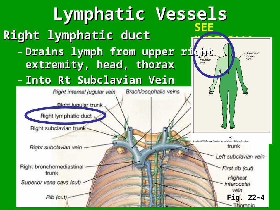
Fig. 22-4
Right lymphatic ductRight lymphatic duct– Drains lymph from upper right Drains lymph from upper right
extremity, head, thoraxextremity, head, thorax– Into Rt Subclavian VeinInto Rt Subclavian Vein
Lymphatic VesselsLymphatic Vessels
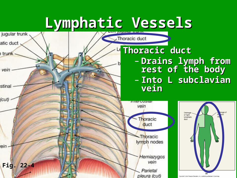
Fig. 22-4
Thoracic ductThoracic duct– Drains lymph from Drains lymph from
rest of the bodyrest of the body– Into L subclavian Into L subclavian
veinvein

Lymphatic VesselsLymphatic VesselsRt lymphatic & thoracic ductsRt lymphatic & thoracic ducts
– Both empty lymph into Both empty lymph into venousvenous circulationcirculation of blood of blood
– One-way system—only One-way system—only TOWARDTOWARD the heartthe heart
Fig. 22-4
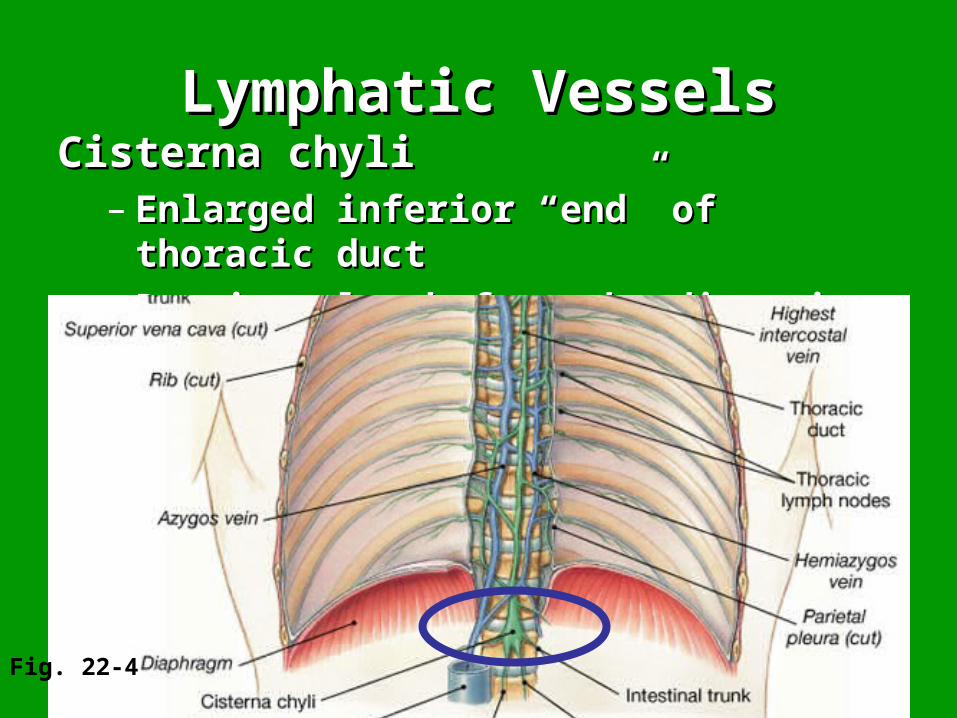
Lymphatic VesselsLymphatic VesselsCisterna chyliCisterna chyli
– Enlarged inferior “end” of thoracic duct Enlarged inferior “end” of thoracic duct – Receives lymph from the digestive visceraReceives lymph from the digestive viscera
Fig. 22-4

Lymph NodesLymph Nodes• Filter lymph as it’s transported through Filter lymph as it’s transported through
the lymphatic vesselsthe lymphatic vessels
• 1000s throughout the body1000s throughout the body
• Macrophages inside—phagocytize Macrophages inside—phagocytize bacteria, cancer cells, etc. in lymph bacteria, cancer cells, etc. in lymph before entering bloodstreambefore entering bloodstream
• Cervical, axillary, inguinal regions; Cervical, axillary, inguinal regions; Peyer’s patches in intestinePeyer’s patches in intestine

Fig. 22-1
SEE MODELS!!!

uhaweb.hartford.edu/ BUGL/immune.htm
Peyer’s patchesPeyer’s patches clusters of lymph clusters of lymph
nodules in small nodules in small intestine intestine
SEE MODELS!!!

Structure of Lymph nodeStructure of Lymph node
Fig. 22-7

Peyer’s patch histologyPeyer’s patch histology
http://www.uky.edu/LCC/BSN/BIO/BiologyLabs/BSL111/111Lab6/Lab6DigestiveSlides.html
Collection of lymphoid nodules in Collection of lymphoid nodules in mucosa of small intestinemucosa of small intestine
Peyer's Patches are largish, rounded areas Peyer's Patches are largish, rounded areas located in the submucosa. They can be a bit located in the submucosa. They can be a bit difficult to spot: look for the slightly darker, difficult to spot: look for the slightly darker, purpler color and more speckled or grainy purpler color and more speckled or grainy
texture.texture.
Lumen of small intestine

Lymph node histologyLymph node histology
http://science.nhmccd.edu/biol/lymphatic/lymph.htm

Lymphoid TissueLymphoid Tissue
Fig. 22-6
• TonsilsTonsils– LingualLingual
• (2) Base of tongue(2) Base of tongue
– PalatinePalatine• L, R: posterior L, R: posterior
inferior palateinferior palate
– Pharyngeal Pharyngeal (adenoids)(adenoids)• (1) posterior superior (1) posterior superior
wall of nasopharynxwall of nasopharynx
SEE MODELS!!!

• Thymus: primary lymphoid organThymus: primary lymphoid organ
Lymph OrgansLymph Organs
Fig. 22-8
SEE MODELS!!!

• Spleen: secondary lymphoid organSpleen: secondary lymphoid organ
Lymph OrgansLymph Organs
Fig. 22-9
SEE MODELS!!!

Lymphocytes: 2Lymphocytes: 2ndnd-most abundant -most abundant WBCsWBCs
Fig. 22-15

B cellsB cells
– Differentiate in Differentiate in
bonebone marrow marrow
T cellsT cells
– Differentiate in Differentiate in thymusthymus
Stem Cells originate in Stem Cells originate in bone marrowbone marrow
Then enter blood stream with Then enter blood stream with specific things on cell’s surface, specific things on cell’s surface,
and are cloned in lymph organsand are cloned in lymph organs

Fig. 22-15

Lymphocytes, continued: Lymphocytes, continued:
After cloned and made many more After cloned and made many more replicates, each can form replicates, each can form memory cellsmemory cells or effector/regulatory cellsor effector/regulatory cells

Lymphocytes, continued: Lymphocytes, continued:
B cell clones:B cell clones:– Memory B cellsMemory B cells– Antibody-producing plasma cellsAntibody-producing plasma cells– ““humoral immunity”humoral immunity” (work indirectly (work indirectly
through antibodies released into through antibodies released into blood/lymphstreamsblood/lymphstreams

Lymphocytes: Lymphocytes: • T cell clones:T cell clones:
– Memory T cellsMemory T cells– Cytotoxic T cellsCytotoxic T cells (directly attack virus- (directly attack virus-
infected tissue cells)infected tissue cells)– Helper T cellsHelper T cells (activate B cells & cytotoxic (activate B cells & cytotoxic
T cells)T cells)– Suppressor cellsSuppressor cells (can inhibit immune (can inhibit immune
response)response)– ““cellular immunity”cellular immunity” (act directly, attack (act directly, attack
bacteria/viruses/parasites/cancer cells)bacteria/viruses/parasites/cancer cells)

• Regions of T and B cells in lymph nodes
Fig. 22-7
Macrophages in
“FILTER”: ~99% of antigens are removed before enters bloodstream

Antigen (Ag) vs. Antibody (Ab)Antigen (Ag) vs. Antibody (Ab)
AntigensAntigens– Provoke an immune responseProvoke an immune response– ANTIANTI body body GENGEN eratorserators
AntibodiesAntibodies (immunoglobulins—Igs) (immunoglobulins—Igs)– Produced by sensitized B cells or plasma Produced by sensitized B cells or plasma
cells in response to antigen presencecells in response to antigen presence– (proteins; IgM, IgG, IgD, IgA, IgE)(proteins; IgM, IgG, IgD, IgA, IgE)– Antigen-binding site is specificAntigen-binding site is specific

Fig. 22-21

Important Immune Response Important Immune Response CharacteristicsCharacteristics
• Immunological MemoryImmunological Memory– Remembers foreign antigens it’s previously Remembers foreign antigens it’s previously
encountered, can react faster 2encountered, can react faster 2ndnd time ( time (i.e.:i.e.: chicken pox)chicken pox)
• SpecificitySpecificity– Memory and antibodies very specificMemory and antibodies very specific
• Differentiation of “self” and “non-self”Differentiation of “self” and “non-self”– Self = our own tissues, don’t alert immune Self = our own tissues, don’t alert immune
responseresponse– non-self = foreign antigens, triggers immune non-self = foreign antigens, triggers immune
responseresponse

Important Immune Response Important Immune Response CharacteristicsCharacteristics
Just FYI---Just FYI---
Differentiation of “self” and “non-self”Differentiation of “self” and “non-self”– CanCan malfunction malfunction– Autoimmune diseases (MS, Lupus, Autoimmune diseases (MS, Lupus,
rheumatoid arthritis, juvenile diabetes, rheumatoid arthritis, juvenile diabetes, etc.)etc.)


