EXCISION OF SACCULAR ANEURYSMS OF THE ...thorax.bmj.com/content/thoraxjnl/15/4/309.full.pdfEXCISION...
8
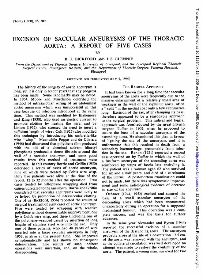
Thorax (1960), 15, 309. EXCISION OF SACCULAR ANEURYSMS OF THE THORACIC AORTA: A REPORT OF FIVE CASES BY B. J. BICKFORD AND J. S. GLENNIE From the Department of Thoracic Surgery, University of Liverpool, and the Liverpool Regional Thoracic Surgical Centre, Broadgreen Hospital, and the Department of Thoracic Surgery, Victoria Hospital, Blackpool (RECEIVED FOR PUBLICATION JULY 5, 1960) The history of the surgery of aortic aneurysm is long, yet it is only in recent years that any progress has been made. Some landmarks may be noted. In 1864, Moore and Murchison described the method of intrasaccular wiring of an abdominal aortic aneurysm which was unsuccessful in this case because of infection introduced at the same time. This method was modified by Blakemore and King (1938), who used an electric current to promote clotting by heating the wire, and by Linton (1952), who stressed the need to insert a sufficient length of wire; Colt (1925) also modified this technique by introducing his umbrella-like wire "wisp." Meanwhile Poppe and de Oliviera (1946) had discovered that polythene film produced with the aid of a chemical solvent (dicetyl phosphate) produced a dense fibrosis around the wall of a saccular aneurysm, and some good results from this method of treatment were reported. In this country Borrie and Griffin (1950) described a series of cases of aortic aneurysm, nine of which were treated by Colt's wire wisp. Only five patients were alive at the time of the report, 12 to 32 months after the operation. Two cases treated by cellophane wrapping died from causes unrelated to the aneurysm. Borrie and Griffin considered that saccular aneurysms were likely to be helped by promotion of intrasaccular clotting. One of us (Bickford, 1956) reported the results of surgical treatment of eight cases of aortic aneurysm. Five were treated by wrapping with reactive polythene without demonstrable improvement, one by a Colt's wire wisp, and three (including one of the polythene-wrapped cases) by insertion of long lengths of stainless steel wire into the sac. Only one of these patients, who had 48 yards of wire inserted into a large saccular aneurysm in July, 1953, is alive at the present time. She improved symptomatically and has shown no subsequent deterioration. The results of such indirect operations were uncertain, and, on the whole, disappointing. THE RADICAL APPROACH It had been known for a long time that saccular aneurysms of the aorta were frequently due to the massive enlargement of a relatively small area of weakness in the wall of the syphilitic aorta, often a " split " in the medial coat only a few centimetres long. Excision of the sac, after clamping its base, therefore appeared to be a reasonable approach to the surgical problem. This radical and logical approach was foreshadowed by the great French surgeon Tuffier in 1902, when he proposed to suture the base of a saccular aneurysm of the ascending aorta. He abandoned this plan in favour of ligating the sac of the aneurysm, and it is unfortunate that this resulted in death from a secondary haemorrhage, presumably from infec- tion in the sac. Renon (1921) reported a second case operated on by Tuffier in which the wall of a fusiform aneurysm of the ascending aorta was reinforced by strips of fascia lata encircling it. The patient was a woman aged 62 who survived for six and a half years, and died of a carcinoma of the uterus. A post-mortem examination could not be made, but there was symptomatic improve- ment and some radiological evidence of decrease in size of the aneurysm. Ochsner (1944, 1952) excised and sutured the base of a small saccular aneurysm of the descending aorta which had been encountered unexpectedly during an operation for a supposed mediastinal tumour. This operation was a com- plete success, and was the basis for further advances. In the same year Alexander and Byron (1944) reported the successful excision of a saccular aneurysm of the descending aorta. The aneurysm probably arose at the site of a coarctation: 7.5 cm. of the aorta was removed with the aneurysm, and as the collateral circulation was well developed no attempt was made to restore the continuity of the aorta. The patient, a young man, survived for two on 12 July 2018 by guest. Protected by copyright. http://thorax.bmj.com/ Thorax: first published as 10.1136/thx.15.4.309 on 1 December 1960. Downloaded from
Transcript of EXCISION OF SACCULAR ANEURYSMS OF THE ...thorax.bmj.com/content/thoraxjnl/15/4/309.full.pdfEXCISION...
Thorax (1960), 15, 309.
EXCISION OF SACCULAR ANEURYSMS OF THE THORACICAORTA: A REPORT OF FIVE CASES
BY
B. J. BICKFORD AND J. S. GLENNIEFrom the Department of Thoracic Surgery, University of Liverpool, and the Liverpool Regional Thoracic
Surgical Centre, Broadgreen Hospital, and the Department of Thoracic Surgery, Victoria Hospital,Blackpool
(RECEIVED FOR PUBLICATION JULY 5, 1960)
The history of the surgery of aortic aneurysm islong, yet it is only in recent years that any progresshas been made. Some landmarks may be noted.In 1864, Moore and Murchison described themethod of intrasaccular wiring of an abdominalaortic aneurysm which was unsuccessful in thiscase because of infection introduced at the sametime. This method was modified by Blakemoreand King (1938), who used an electric current topromote clotting by heating the wire, and byLinton (1952), who stressed the need to insert asufficient length of wire; Colt (1925) also modifiedthis technique by introducing his umbrella-likewire "wisp." Meanwhile Poppe and de Oliviera(1946) had discovered that polythene film producedwith the aid of a chemical solvent (dicetylphosphate) produced a dense fibrosis around thewall of a saccular aneurysm, and some goodresults from this method of treatment werereported. In this country Borrie and Griffin (1950)described a series of cases of aortic aneurysm,nine of which were treated by Colt's wire wisp.Only five patients were alive at the time of thereport, 12 to 32 months after the operation. Twocases treated by cellophane wrapping died fromcauses unrelated to the aneurysm. Borrie and Griffinconsidered that saccular aneurysms were likely tobe helped by promotion of intrasaccular clotting.One of us (Bickford, 1956) reported the results ofsurgical treatment of eight cases of aortic aneurysm.Five were treated by wrapping with reactivepolythene without demonstrable improvement, oneby a Colt's wire wisp, and three (including one ofthe polythene-wrapped cases) by insertion of longlengths of stainless steel wire into the sac. Onlyone of these patients, who had 48 yards of wireinserted into a large saccular aneurysm in July,1953, is alive at the present time. She improvedsymptomatically and has shown no subsequentdeterioration. The results of such indirectoperations were uncertain, and, on the whole,disappointing.
THE RADICAL APPROACHIt had been known for a long time that saccular
aneurysms of the aorta were frequently due to themassive enlargement of a relatively small area ofweakness in the wall of the syphilitic aorta, oftena " split " in the medial coat only a few centimetreslong. Excision of the sac, after clamping its base,therefore appeared to be a reasonable approachto the surgical problem. This radical and logicalapproach was foreshadowed by the great Frenchsurgeon Tuffier in 1902, when he proposed tosuture the base of a saccular aneurysm of theascending aorta. He abandoned this plan in favourof ligating the sac of the aneurysm, and it isunfortunate that this resulted in death from asecondary haemorrhage, presumably from infec-tion in the sac. Renon (1921) reported a secondcase operated on by Tuffier in which the wall ofa fusiform aneurysm of the ascending aorta wasreinforced by strips of fascia lata encircling it.The patient was a woman aged 62 who survivedfor six and a half years, and died of a carcinomaof the uterus. A post-mortem examination couldnot be made, but there was symptomatic improve-ment and some radiological evidence of decreasein size of the aneurysm.
Ochsner (1944, 1952) excised and sutured thebase of a small saccular aneurysm of thedescending aorta which had been encounteredunexpectedly during an operation for a supposedmediastinal tumour. This operation was a com-plete success, and was the basis for furtheradvances.
In the same year Alexander and Byron (1944)reported the successful excision of a saccularaneurysm of the descending aorta. The aneurysmprobably arose at the site of a coarctation: 7.5 cm.of the aorta was removed with the aneurysm, andas the collateral circulation was well developed noattempt was made to restore the continuity of theaorta. The patient, a young man, survived for two
on 12 July 2018 by guest. Protected by copyright.
http://thorax.bmj.com
/T
horax: first published as 10.1136/thx.15.4.309 on 1 Decem
ber 1960. Dow
nloaded from
B. J. BICKFORD and J. S. GLENNIE
years after the operation. His blood pressure wasconsiderably elevated after the operation, and hedied as a result of a series of cerebral vascularaccidents.
Meyer, Monod, Brunel, Nico, and Dubois deMontreynaud (1948) reported the successfulexcision of a non-syphilitic saccular aneurysm ofthe terminal portion of the aortic arch. Thediagnosis of aneurysm had not been confidentlymade before operation. The suture line was 4 cm.in length, and there was no reduction in the lumenof the aorta as a result.Cooley and DeBakey (1952), in a paper that has
become a classic, reported two cases in whichaneurysms were successfully treated by excisionand lateral suture of the aorta. One patient, inwhom the innominate artery was involved, sur-vived, but the second died after excision of asaccular aneurysm of the ascending aorta. Abilateral trans-sternal thoracotomy was used, anddeath seemed to be due to diffuse cerebral damagebecause of inadequate ventilation during operation.
Bahnson (1953) reported eight cases in whichsaccular aortic aneurysms were excised; six weresuccessful, two died.
In this country Rob (1954) and Sellors (1956)have described successful operations for aorticaneurysms associated with coarctation of theaorta.
Cooley, DeBakey, and Creech (1957) reportedthe surgical treatment of 313 aneurysms of theaorta. The majority of these were abdominal.There were 24 cases of saccular aneurysms of thearch of the aorta, with nine deaths. Fifteen ofthese arose in the ascending aorta, and the mor-tality among these (seven) was almost 50%. Fifty-nine aneurysms arose in other portions of thethoracic aorta. The major cause of death in allthese cases was cardiac failure, the next commonestbeing cerebral thrombosis. Forty-three of theaneurysms of the thoracic aorta were fusiformand 16 were of the dissecting type.Whereas a saccular aneurysm presents a
reasonably easy problem of surgical technique, thefusiform aneurysm is a much more difficult one.The only way in which complete relief can beobtained is by excision of the whole abnormalsegment of the aorta and replacement by a graftor prosthesis, a formidable task when the greatbranches of the arch are involved. The principlesand practice of this type of replacement of theaorta and its branches have been set forth withclarity and ingenuity by DeBakey (1959), whoseexperience in this field is unrivalled.The long-term survival of aortic grafts is not
yet known, but the immediate results are reason-
ably good; more time must elapse before a finaljudgment can be made.
CASE REPORTSOur personal experience of the excision of
saccular aortic aneurysms is of five cases, the firstof which was operated on in 1955. Details of ouroperative experiences are as follows:CASE 1.-E. P., a Polish man, aged 51, presented
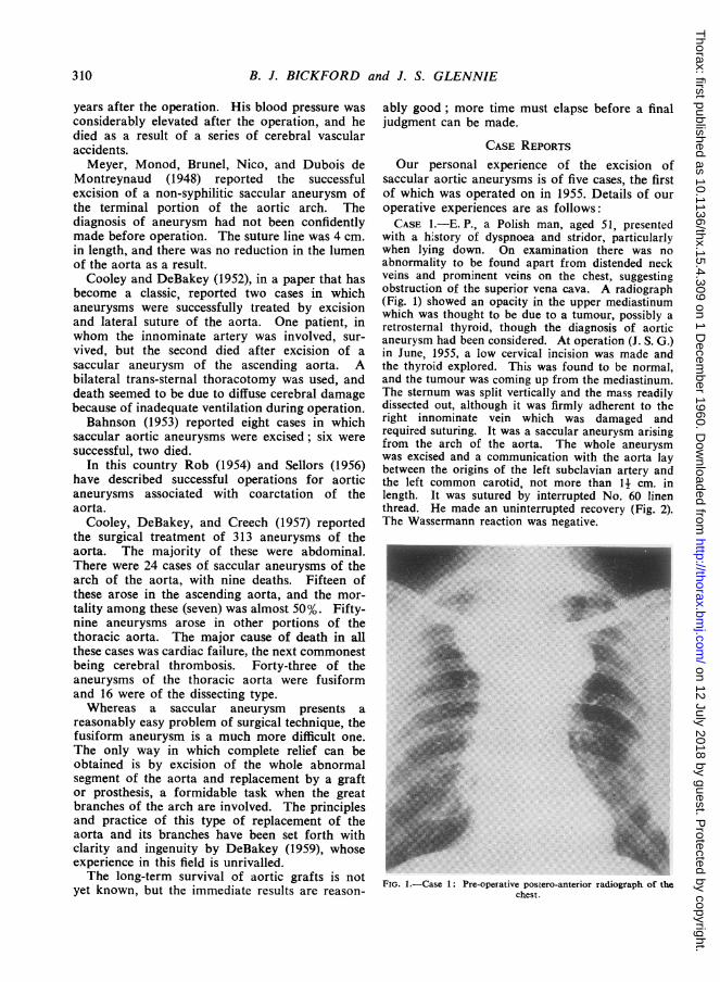
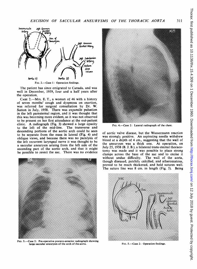
with a history of dyspnoea and stridor, particularlywhen lying down. On examination there was noabnormality to be found apart from distended neckveins and prominent veins on the chest, suggestingobstruction of the superior vena cava. A radiograph(Fig. 1) showed an opacity in the upper mediastinumwhich was thought to be due to a tumour, possibly aretrosternal thyroid, though the diagnosis of aorticaneurysm had been considered. At operation (J. S. G.)in June, 1955, a low cervical incision was made andthe thyroid explored. This was found to be normal,and the tumour was coming up from the mediastinum.The sternum was split vertically and the mass readilydissected out, although it was firmly adherent to theright innominate vein which was damaged andrequired suturing. It was a saccular aneurysm arisingfrom the arch of the aorta. The whole aneurysmwas excised and a communication with the aorta laybetween the origins of the left subclavian artery andthe left common carotid, not more than II cm. inlength. It was sutured by interrupted No. 60 linenthread. He made an uninterrupted recovery (Fig. 2).The Wassermann reaction was negative.
FIG. 1.-Case 1: Pre-operative postero-anterior radiograph of thechest.
310
on 12 July 2018 by guest. Protected by copyright.
http://thorax.bmj.com
/T
horax: first published as 10.1136/thx.15.4.309 on 1 Decem
ber 1960. Dow
nloaded from
EXCISION OF SACCULAR ANEURYSMS OF THE THORACIC AORTA
Aorta () Aorta (1) I
FIG. 2.-Case 1: Operation findings.
The patient has since emigrated to Canada, and waswell in December, 1959, four and a half years afterthe operation.CASE 2.-Mrs. E. T., a woman of 46 with a history
of seven months' cough and dyspnoea on exertion,was referred for surgical consultation by Dr. W.Sutton in July, 1958. There was expansile pulsationin the left parasternal region, and it was thought thatthis was becoming more evident, as it was not observedto be present on her first attendance at the out-patientclinic. A radiograph (Fig. 3) showed a large opacityto the left of the mid-line. The transverse anddescending portions of the aortic arch could be seento be separate from the mass in lateral (Fig. 4) andoblique views, and because there was no paralysis ofthe left recurrent laryngeal nerve it was thought to bea saccular aneurysm arising from the left side of theascending part of the aortic arch, and that it mightbe possible to resect the sac. There was no evidence
Fio. 3.-Case 2: Pre-operative postero-anterior radiograph showinglarge saccular aneurysm of the arch of the aorta.
FIG. 4.-Case 2: Lateral radiograph of the chest.
of aortic valve disease, but the Wassermann reactionwas strongly positive. An aspirating needle withdrewblood at a depth of 4 cm., suggesting that the wall ofthe aneurysm was a thick one. At operation, onJuly 25, 1958 (B. J. B.), a bilateral trans-sternal thoraco-tomy was made and it was possible to place strongclamps across the base of the sac and to excise itwithout undue difficulty. The wall of the aorta,though diseased, patchily calcified, and atheromatous,proved to be much thickened, and held sutures well.The suture line was 8 cm. in length (Fig. 5). Being
FIG. 5.-Case 2: Operation findings.
311
..O..
.42-shm.iwn.
on 12 July 2018 by guest. Protected by copyright.
http://thorax.bmj.com
/T
horax: first published as 10.1136/thx.15.4.309 on 1 Decem
ber 1960. Dow
nloaded from
B. J. BICKFORD and J. S. GLENNIE
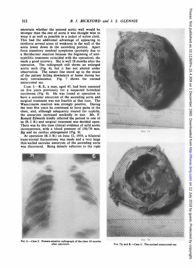
uncertain whether the sutured aortic wall would bestronger than the rest of aorta it was thought wise towrap it as well as possible in a jacket of nylon cloth.This had the additional advantage of appearing toreinforce several areas of weakness in the wall of theaorta lower down in the ascending portion. Apartfrom transitory cerebral symptoms (probably due toa Herxheimer reaction because the beginning of anti-syphilitic treatment coincided with the operation), shemade a good recovery. She is well 18 months after theoperation. The radiograph still shows an enlargedaortic arch (Fig. 6), but it has not altered underobservation. The suture line stood up to the strainof the patient falling downstairs at home during herearly convalescence. Fig. 7 shows the excisedaneurysmal sac.CASE 3.-R. S., a man, aged 45, had been operated
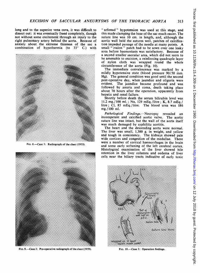
on five years previously for a suspected bronchialcarcinoma (Fig. 8). He was found at operation tohave a saccular aneurysm of the ascending aorta andsurgical treatment was not feasible at that time. TheWassermann reaction was strongly positive. Duringthe next five years he continued to have pains in thechest, and, although adequately treated for syphilis,the aneurysm increased markedly in size. Mr. F.Ronald Edwards kindly referred the patient to one ofus (B. J. B.) and surgical treatment was decided upon.There was by this time clinical evidence of mild aorticincompetence, with a blood pressure of 130/70 mm.flg and no cardiac enlargement (Fig. 9).At operation (B. J. B.) on June 12, 1959, a bilateral
trans-sternal thoracotomy was made and a very largethin-walled saccular aneurysm of the ascending aortawas discovered. Being densely adherent to the right
FIG. 6.-Case 2: Postero-anterior radiograph of the chest 10 monthsafter operation. FIG. 7A and B.-Case 2: The excised aneurysmal sac.
312
...........
on 12 July 2018 by guest. Protected by copyright.
http://thorax.bmj.com
/T
horax: first published as 10.1136/thx.15.4.309 on 1 Decem
ber 1960. Dow
nloaded from
EXCISION OF SACCULAR ANEURYSMS OF THE THORACIC AORTA
lung and to the superior vena cava, it was difficult todissect out: it was eventually freed completely, thoughnot without some excitement through an injury to theright pulmonary artery behind the aorta. Because ofanxiety about the extreme thinness of the sac acombination of hypothermia (to 33° C.) with
FIG. 8.-Case 3: Radiograph of the chest (19.
" arfonad " hypotension was used at this stage, andthis made clamping the base of the sac much easier. Thesuture line was 10 cm. in length, and, although theaortic wall held the sutures well, patches of calcifica-tion impeded passage of the needle at many points. Asmall " ivalon " patch had to be sewn over one leakyarea before haemostasis was satisfactory. Because ofa second smaller saccular area, which did not seem tobe amenable to excision, a reinforcing quadruple layerof nylon cloth was wrapped round the wholecircumference of the aorta (Fig. 10).The immediate convalescence was marked by a
mildly hypotensive state (blood pressure 90/50 mm.Hg). The general condition was good until the secondpost-operative day, when jaundice and oliguria wereevident. The jaundice became profound and wasfollowed by anuria and coma, death taking placeabout 70 hours after the operation, apparently fromhepatic and renal failure.
Shortly before death the serum bilirubin level was11.2 mg./100 ml.; Na, 129 mEq./litre; K, 8.7 mEq./litre; Cl, 85 mEq./litre. The blood urea was 186mg./100 ml.
Pathological Findings.-Necropsy revealed anincompetent and calcified aortic valve. The aorticsuture line was intact, but the wall of the aorta itselfwas much damaged by syphilitic aortitis.The heart and the descending aorta were normal.
The liver was small, 1,300 g. in weight, and yellowand tough in consistency. The kidneys showed palewide cortices and congestion of the medullae. There
53. were a number of cortical haemorrhages in the brain53) and some early softening of the left cerebral cortex.
Histological examination of the liver showed bileretention in the liver columns and oedema of livercells near the biliary tracts indicative of early toxic
'SLCtLyC LUw loons6.
Fir. 9.-Case 3: Pre-operative radiograph ofthe chest (1959).
313
FIG. 10.-Case 3: Operation findings.
on 12 July 2018 by guest. Protected by copyright.
http://thorax.bmj.com
/T
horax: first published as 10.1136/thx.15.4.309 on 1 Decem
ber 1960. Dow
nloaded from
B. J. BICKFORD and J. S. GLENNIE
change. The kidneys showed proteinous non-haemo-globin casts in the second convoluted and in thecollecting tubules with little cellular reaction, and noglomerular or vascular lesions. The changes in theliver indicated that it had recently been exposed to atoxic agent. The renal changes were those of a lowernephron nephrosis, part of a hepato-renal syndrome,and secondary to the liver damage. The histologicalexamination of the excised sac showed that it was forthe most part 2-3 mm. thick but with a thinned areaonly 1 mm. in thickness. Microscopically the wallconsisted of acellular collagen.
This disappointing result cannot be adequatelyexplained. A prominent feature was the failure torecover satisfactory vascular tone and it seems at leastpossible that the prolonged hypotension in associationwith aortic incompetence produced a sufficientlydiminished visceral circulation to make a very smalldose of chlorpromazine (25 mg.) a toxic agent forthe liver. Alternatively it is possible that the liverwas in some way sensitized to this drug. Perhapshypotensive drugs should be avoided in similar caseswhere there is significant aortic incompetence.CASE 4.-Mrs. M. W., aged 61, had had vague
symptoms attributed to anaemia and a raised bloodpressure during the previous three years. She wasreferred by Dr. Donald Leeming for investigation,having for six months had a persistent cough. Duringmost of this time the sputum had been streaked withblood, without a frank haemoptysis. There was somepain anteriorly, in the lower part of the left side ofthe chest, and she was mildly dyspnoeic on exertion.
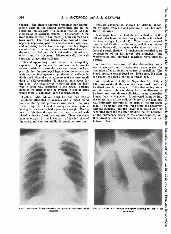
Physical examination showed no definite abnor-mality, apart from a blood pressure of 240/120 mm.Hg in the arms.A radiograph of the chest showed a shadow on the
left side which was at first thought to be a bronchialcarcinoma (Figs. 11 and 12). Closer study revealedunusual calcification in the aorta, and it was impos-sible radiologically to separate the abnormal opacityfrom the aortic shadow. Bronchoscopy revealed somecompression of the left lower lobe bronchus. TheWassermann and Meinicke reactions were stronglypositive.A saccular aneurysm of the descending aorta
was diagnosed, and preparations were made foroperation after an intensive course of penicillin. Theblood pressure was reduced to 120/80 mm. Hg afterthe patient had had a period of rest in bed.At operation (B. J. B.) on September 11, 1959, a
left posterolateral thoracotomy was made and alocalized saccular aneurysm of the descending aortawas discovered. It was about 6 cm. in diameter atits base, and was almost cylindrical, being somewhatlonger than its diameter. It projected laterally intothe upper part of the oblique fissure of the lung andwas intimately adherent to the apex of the left lowerlobe. The upper lobe was freed from the aneurysmwithout difficulty, but the lower lobe could only beseparated from the sac after dividing the two branchesof the pulmonary artery to the apical segment, andthen dividing the lung immediately below the sacbetween clamps.
FiG. 11.-Case 4: Postero-anterior radiograph of the chest before FIG. 12.-Case 4: Oblique tomogram showing the sac of theoperation. aneurysm.
314
on 12 July 2018 by guest. Protected by copyright.
http://thorax.bmj.com
/T
horax: first published as 10.1136/thx.15.4.309 on 1 Decem
ber 1960. Dow
nloaded from
EXCISION OF SACCULAR ANEURYSMS OF THE THORACIC AORTA
whole circumference of the aorta over about 8 cm. ofits length. This was tightly sutured with nylon thread
NI\/(Fig. 13).Recovery was uneventful, and the patient went
home on the 23rd post-operative day. When seenN/} = -X\five months after operation she was continuing to
a4hercvd .;~ siv \make satisfactory progress. A radiograph showedto / ^^ -$ \ Xsome irregular opacity in the operation area, inter-'d"t'07) 4$ t* 1 \preted as non-vascular (Fig. 14).
Pathological Report.-The sac was filled with athick gelatinous type of clot. Histologically the wallshowed syphilitic aortitis with considerable lympho-cytic "cuffing" of the vasa vasorum and a patchyinfiltration of the media with lymphocytes and plasma
s\y S _ ^ ~~~~~~~~cells.CASE 5.-Mrs. M. D., aged 49, was a heavy smoker
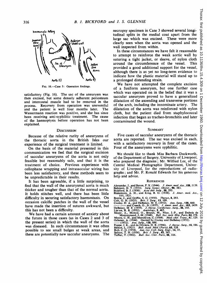
who complained of haemoptysis and rather persistentpain in the right pectoral region. There were noabnormal physical signs, but a radiograph of the chestshowed a rounded opacity to the right of the mid-line,with a wedge-shaped projection anteriorly, which inthe lateral view resembled a collapse of the anteriorsegment of the right upper lobe (Fig. 15). A previous" contact " radiograph in 1957 had been passed as
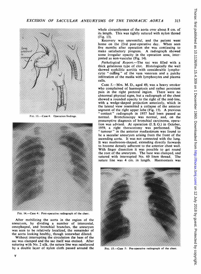
FIG. 13.-Case 4: Operation findings. normal. Bronchoscopy was normal, and, on thepresumptive diagnosis of bronchial carcinoma, opera-tion was advised. At operation (J. S. G.) in October,1959, a right thoracotomy was performed. The"tumour' in the anterior mediastinum was found toh0PW,y."....... be a saccular aneurysm arising from the front of the
«.......ascending aorta. It was not connected with the lung.It was mushroom-shaped, extending directly forwardsto become densely adherent to the anterior chest wall.With finger dissection it was possible to get roundthe root of the aneurysm. The base was clamped, andsutured with interrupted No. 60 linen thread. Thesuture line was 4 cm. in length. Haemostasis was'. .......~~~~~~~~~~~~~~~~~~~~~~~~~~~~~~~~~~~~~~~~~~~~~~~~~~~~...
FiG. 14.-Case 4: Post-operative radiograph of the chest.
After mobilizing the aorta in the region of theaneurysm, by dividing a number of intercostal,oesophageal, and bronchial branches, the aneurysmwas seen to be relatively localized, the remainder ofthe aorta looking healthy, though somewhat dilated.Without interrupting the circulation the base of the
sac was clamped and the sac itself was excised. Aftersuturing w'ith No. 2 silk, the suture line was reinforcedby a double layer of nylon cloth passed around the FiG. 15.-Case 5: Pre-operative radiograph of she chest.
y
315
on 12 July 2018 by guest. Protected by copyright.
http://thorax.bmj.com
/T
horax: first published as 10.1136/thx.15.4.309 on 1 Decem
ber 1960. Dow
nloaded from
B. J. BICKFORD and J. S. GLENNIE
FIG. 16.-Case 5: Operation findings.
satisfactory (Fig. 16). The sac of the aneurysm wasthen excised, but some densely adherent periosteumand intercostal muscle had to be removed in theprocess. Recovery from operation was uneventfuland the patient is well four months later. TheWassermann reaction was positive, and she has sincebeen receiving anti-syphilitic treatment. The causeof the haemoptysis before operation has not beenexplained.
DISCUSSIONBecause of the relative rarity of aneurysms of
the thoracic aorta in the British Isles our
experience of the surgical treatment is limited.On the basis of the material presented in this
communication we feel that the surgical excisionof saccular aneurysms of the aorta is not onlyfeasible but reasonably safe, and that it is thetreatment of choice. Previous experience withcellophane wrapping and intrasaccular wiring hasbeen less satisfactory, and these methods seem tobe unpredictable in their results.
It has been agreeable, if a little surprising, tofind that the wall of the aneurysmal aorta is muchthicker and tougher than that of the normal aorta.It holds stitches well, and there has been littledifficulty in securing satisfactory haemostasis. Onoccasion calcific patches in the wall of the vesselhave made the insertion of sutures awkward, butthis has not been a difficulty.We have had a certain amount of anxiety about
the future in those cases (as in Cases 2 and 3 ofthe present series) in which the wall of the aortawas diseased. In such circumstances it was oftenpossible to see small bulges at weak areas, andthese are potentially new saccular aneurysms. The
necropsy specimen in Case 3 showed several longi-tudinal splits in the medial coat apart from thelarge sac which was excised. These were moreclearly seen when the aorta was opened and thewall inspected from within.
In these circumstances we have felt it reasonableto attempt to reinforce the weak aortic wall bysuturing a tight jacket, or sleeve, of nylon clotharound the circumference of the vessel. Thisprovided a good additional support for the vessel,although there is as yet no long-term evidence toindicate how the plastic material will stand up toa prolonged distending strain.We have not attempted the complete excision
of a fusiform aneurysm, but one further casewhich was operated on in the belief that it was asaccular aneurysm proved to have a generalizeddilatation of the ascending and transverse portionsof the arch, including the innominate artery. Thedilatation of the aorta was reinforced with nyloncloth, but the patient died from staphylococcalinfection that began as tracheo-bronchitis and latercontaminated the wound.
SUMMARYFive cases of saccular aneurysms of the thoracic
aorta are reported. The sac was excised in each,with a satisfactory recovery in four of the cases.Four of the aneurysms were syphilitic.
We should like to thank Miss Barbara Duckworth,of the Department of Surgery, University of Liverpool,who prepared the diagrams; Mr. Wilfred Lee, of theCentral Medical Photographic Department, Univer-sity of Liverpool, for the reproductions of radio-graphs; and Mr. F. Ronald Edwards for his generoushelp and advice.
REFERENCESAlexander, J., and Byron, F. X. (1944). J. Amer. med. Ass., 126, 1139.Bahnson, H. T. (1953). Surg. Gynec. Obstet., 96, 383.Bickford, B. J. (1956). Acta chir. belg., 55, 295.Blakemore, A. H., and King, B. G. (1938). J. Amer. med. Ass.,
111, 1821.Borrie, J.. and Griffin, S. G. (1950). Thorax, 5, 293.Colt, G. H. (1925). Brit. J. Surg., 13, 109.Cooley, D. A., and DeBakey, M. E. (1952). Ann. Surg., 135, 660.-- and Creech, O., Jr. (1957). J. Amer. med. Ass., 163, 1439.DeBakey, M. E (1959). J. thorac. Cardiovasc. Surg., 38, 145.Linson, R. R. (1952). AnRiology, 2, 485.Meyer, A., Monod, O., Brunel, M., Nico, J.-P.. and Dubois de
Montreynaud, J.-M. (1948). Bull. Soc. med. H6p. Paris, 64,278.Moore, C. H., and Murchi;on, C. (1E64). Med-chir. Trans., 4;, 129.Ochsner, A. (1944). Quoted by Cooley and DeBakey (1952).
(1952). A',n. Surg., 135, 686.Poppe, J. K., and de Oliviera, H. R. (1946). J. thorac. Surg., 15, 186.R6non, L. (1921). Bull. Acad. Med. (Paris), 85, 528.Rob, C. G. (1954). Ann. roy. CoIl. Surg. Engl., 14, 35.Sellors, T. H. (1956). Brit. J. Surg., 43, 365.Tuffier, T. (1902). Bull. Soc. Chir. Paris, 28, 326.
316
on 12 July 2018 by guest. Protected by copyright.
http://thorax.bmj.com
/T
horax: first published as 10.1136/thx.15.4.309 on 1 Decem
ber 1960. Dow
nloaded from