repositorio-aberto.up.pt · repositorio-aberto.up.pt ... 16 ... 17 ...

Evolution of the vitamin C biosynthetic pathway and transport mechanism: from the global perspective to a Drosophila melanogaster case study
Pedro Miguel Barros Duque Cell and Molecular Biology Master’s Degree Science Department, FCUP 2018 Supervisor Jorge Manuel de Sousa Basto Vieira Main researcher at IBMC [email protected] Co-supervisor Cristina Alexandra Gonçalves Paula Vieira Auxiliary researcher at IBMC [email protected] Institute Address IBMC – Instituto de Biologia Molecular e Celular I3s building – Rua Alfredo Allen nº 208 4200-135 Porto, Portugal


Todas as correções determinadas pelo júri, e só essas, foram efetuadas.
O Presidente do Júri, Porto, ______/______/_________

Declaração de não-plágio Eu, Pedro Miguel Barros Duque, aluno com o número 201004984, inscrito no mestrado de Biologia Celular e Molecular no presente ano letivo de 2017/2018, declaro por minha honra que sou o autor da totalidade do texto apresentado, não apresento texto plagiado, e tomei conhecimento das consequências de uma situação de plágio. Porto, 30 de Setembro de 2018


Acknowledgements This work was performed in the Phenotypic Evolution group at the I3s facilities, as part of the dissertation thesis of the Cellular and Molecular Biology Master´s degree that belongs to the Faculty of Sciences of the University of Porto. My sincere and great thanks to all the colleagues I had the chance to work with, but especially to Sílvia, Sara, Hugo, Pedro, José, Joel, Rodrigo and Vanessa for the knowledge and friendship provided. Also my thanks to FCUP, I3s and IBMC for allowing the opportunity to perform this work.
I also would like to say a special thank you to Jorge and Cristina Vieira for their patience, great advisement and teaching along this proficuous experience.
Furthermore, I want to thank the support of all my friends, but even more the sacrifice of my family, especially my parents, for allowing me to come back to university while always motivating me to become resilient and hard-working without questioning my capabilities. A thank you is not nearly enough, but I hope in time I get a chance to return their vote of confidence in a similar fashion.


Abstract
Ascorbic acid (vitamin C) is a crucial co-factor for several enzymatic reactions and an indispensable antioxidant agent, playing an important role in normal function and development of eukaryotic cells. Many deuterostomians are capable of synthesizing ascorbic acid, however some species such as Haplorrhini primates, teleost fish and Cavia porcellus (guinea pig) lost this ability due to the loss of the L-gulonolactone oxidase (GULO) gene. Protostomians are often regarded as not having GULO, although there are hints that this may not be the case. We used all available genomic information in GenBank and RefSeq for animal species to clarify this issue. We show that GULO was not lost at the split between the protostomians and deuterostomians, and added supporting evidence that this gene could in fact be present in the ancestral of animals and Fungi. It is consensual that GULO was lost in the Insecta taxonomic group, and our results go in accordance with this finding. Nevertheless, ascorbic acid levels can be detected in one representative species from this group, the model organism Drosophila melanogaster (fly), even in the absence of a dietary source of this vitamin. Given this evidence, it was possible that the fly microbiome could be responsible for the supply of ascorbic acid. Still, we were able to determine that the microbiome is not responsible for ascorbic acid synthesis. Furthermore, we observed that after cold acclimation conditions, D. melanogaster is able to replenish a break in ascorbic acid levels after one day of recovery, which is strong evidence of putative synthesis.
In deuterostomians, specifically in the vertebrates group, ascorbic acid homeostasis is facilitated by the presence of Sodium-dependent Vitamin C Transporters (SVCTs). Four transporters (SVCT1 to 4) have been identified, although only SVCT1 and SVCT2 are known to be correlated with ascorbic acid cellular transport. The evolutionary history of these four transporters remains undiscovered, as well as the ancestral subtract specificity traits, but it is suspected that SVCT1 and SVCT2 genes are derived from a common ancestor and that SVCT3 and SVCT4 are “orphan genes”. Furthermore, one uncharacterized SVCT transporter is also detected in D. melanogaster. Using all available animal genome annotations, we sought out to understand the evolutionary history of the vertebrate transporters and its phylogenetic relationship to the SVCT transporters observed in protostomian species. We uncovered that within vertebrates, the general presence of four transporters is likely the result of two rounds of whole genome duplication that are already reported in literature, from a single ancestral gene. The protostomian SVCT seems to be duplicated independently several times in many distinct taxonomic groups, nevertheless our results also indicate the presence of a single ancestral gene at the base of this taxonomic group. Nevertheless, we were unable to imply the protostomian SVCTs in ascorbic acid transport.
Keywords: GULO, SVCT, ascorbic acid, D. melanogaster, microbiome, synthesis, transport


Resumo O ácido ascórbico (vitamina C) é um cofator crucial em várias reações enzimáticas e um agente antioxidante indispensável, contribuindo de forma notória para o normal funcionamento e desenvolvimento de células eucarióticas. Muitos deuterostómios são capazes de sintetizar ácido ascórbico, no entanto algumas espécies como os primatas Haplorrhini, os peixes teleósteos e Cavia porcellus (porquinho da Índia) não possuem esta capacidade devido há perda do gene L-gulonolactona oxidase (GULO). É considerado que os protostómios não possuem GULO, mas existem pistas que sugerem o contrário. Usámos toda a informação genómica disponível para espécies animais nas bases de dados GenBank e RefSeq para clarificar este assunto. Demonstrámos que o gene GULO não foi perdido na divergência entre protostómios e deuterostómios, e acrescentámos evidência da possível presença deste gene no ancentral dos animais e Fungi. É consensual que o gene GULO foi perdido no grupo taxonómico Insecta, e os nossos resultados estão de acordo com esta premissa. No entanto, níveis de ácido ascórbico podem ser detetados numa espécie representativa deste grupo taxonómico, Drosophila melanogaster, mesmo na ausência de uma fonte desta vitamina na dieta. Dada esta evidência, seria possível que o microbioma fosse responsável pela suplementação de ácido ascórbico. Contudo, fomos capazes de determinar que o microbioma não desempenha esse papel. Observamos ainda que em condições de aclimatação ao frio, D. melanogaster é capaz de normalizar uma descida dos níveis de ácido ascórbico após um dia de recuperação, o que é uma forte evidência de produção putativa.
Em deuterostómios, especificamente no grupo dos vertebrados, a homeostasia do ácido ascórbico é facilitada pela presença de Transportadores de Vitamina C dependentes de Sódio (SVCTs). Quatro transportadores (SVCT1 a 4) foram identificados, apesar de apenas os transportadores SVTC1 e SVCT2 estarem implicados no transporte de ácido ascórbico. A história evolutiva destes transportadores permanece desconhecida, bem como as propriedades funcionais ancestrais dos mesmos, mas é extrapolado que os genes SVCT1 e SVCT2 derivem de um ancestral comum enquanto os genes SVCT3 e SVCT4 sejam “órfãos”. Um transportador SVCT não caracterizado foi também descoberto em D. melanogaster. Usando todas as anotações de genoma disponíveis para espécies animais, procurámos compreender a história evolutiva dos transportadores identificados em vertebrados e a sua relação filogenética com os transportadores observados em protostómios. Descobrimos que dentro dos vertebrados, a presença de quatro transportadores pode ser correlacionada com duas duplicações totais de genoma reportadas na literatura, a partir de um único gene ancestral. O gene SVCT parece estar duplicado independentemente em várias linhagens de protostómios, no entanto os nossos resultados indicam que o mesmo é derivado de uma cópia ancestral única. Todavia, não fomos capazes de implicar os transportadores SVCT de protostómios na possível capacidade de transporte de ácido ascórbico.
Palavras chave: GULO, SVCT, ácido ascórbico, D. melanogaster, microbioma, produção, transporte


Index
I. Introduction ................................................................................................................................ 1
II. Materials and methods ............................................................................................................ 11
II.1. Animal GULO and SVCT CDS phylogenies ................................................................... 11
II.2. GULO CDS annotations .................................................................................................. 15
II.3. Drosophila melanogaster Oregon-R maintenance .......................................................... 16
II.4. Control and cold exposure experimental conditions ....................................................... 17
II.5. Generation of axenic D. melanogaster ............................................................................ 17
II.6. High Performance Liquid Chromatography (HPLC) analysis ........................................ 18
II.7. Determination of L-ascorbate in microbiome cultures expanded ex-vivo....................... 19
III. Results and discussion ........................................................................................................... 21
III.1. GULO gene CDS Bayesian phylogeny .......................................................................... 21
III.2. Non-Bilateria/Protostomia GULO gene annotations and phylogenetic analysis............ 32
III.2.1. GULO sequence annotation process ........................................................................ 32
III.2.2. GULO CDS annotations phylogenetic analysis ...................................................... 39
III.3. Microbiome and HPLC assays ....................................................................................... 50
III.4. SVCT phylogenies .......................................................................................................... 56
IV. Conclusion ............................................................................................................................ 72
V. References .............................................................................................................................. 74
VI. Supplementary Data .............................................................................................................. 89

Index of Figures
Figure 1 – Graphic display of the currently known ascorbic acid synthetic pathways. The final
oxidation step of the distinct aldono-1,4-lactones to ascorbate is performed by an FAD-linked
oxidase or dehydrogenase (GULO, GALDH or ALO). Photosynthetic protists appear to possess
enzymatic components from animal and plant pathways, and due to this characteristic, the current
described pathway for these species likely evolved from a secondary endosymbiosis event
regarding a non photosynthetic ancestor and algae (Wheeler et al. 2015). The figure here
presented and the corresponding description were adapted from Smirnoff (2018). ..................... 2
Figure 2 - Summary of the findings regarding the presence of putative functional GULO genes in
non-bilaterian, protostomian and deuterostomian lineages. The lineages where a possibly
functional GULO has been detected are represented in green, while lineages where the GULO
gene has not been detected are presented in red. Additionally, lineages for which there is
insufficient information to extrapolate a conclusion are represented in blue, with lineages showing
a notable number of species with a functional and non-functional GULO gene represented in
violet. The specific Acari lineage case, in which some species were excluded from the final
phylogeny but may potentially have a functional GULO gene, is represented in orange. The first
three numbers next to the represented lineages indicate species excluded from the dataset because:
i) no sequence with significant homology was found in the initial BLAST; ii) the sequences did
not possess the typical GULO amino acid pattern or showed ambiguous nucleotide positions; iii)
the sequences do not present an ATG start codon, are non-multiple of three, have in frame stop
codons, or have a size difference larger than 10% relative to the reference M. musculus GULO
sequence. The last number indicates the number of species from each lineage present in the final
tree. Numbers in parentheses indicate species that were found taxonomically misplaced in the
final phylogeny, and that do not likely have a GULO gene. Broken lines show uncertain
relationships. Taxonomic relationships are depicted as in the Tree of life web project
(http://www.tolweb.org/tree/) and in Helgen (2011). This cladogram is depicted an in López-
Fernández et al. (2018)................................................................................................................ 22
Figure 3 – Protostomes and non-bilaterians putative L-gulonolactone oxidase (GULO)
annotations phylogeny. Two GULO CDS from Fungi species were used to help rooting the tree.
Six deuterostomian species (representative of the Actinopteri, Amphibia, Euarchontoglires, Aves,
Reptilia and Cephalochordata groups) GULO CDS were used to facilitate the interpretation of the
results. Relevant higher taxonomic classifications are shown next to the species name. ........... 41
Figure 4 – Refined protostomes and non-bilaterians putative GULO annotations phylogeny. Two
GULO CDS from Fungi species were used to help rooting the tree. Six deuterostomian species
(representative of the Actinopteri, Amphibia, Euarchontoglires, Aves, Reptilia and

Cephalochordata groups) GULO CDS were used to facilitate the interpretation of the results.
Relevant higher taxonomic classifications are shown next to the species name. ........................ 43
Figure 5 – Cladogram representation of the findings regarding the annotation of GULO in six
Araneae species. The species in which GULO is likely present are highlighted in green, while the
species where the presence or absence of GULO cannot be inferred are highlighted in blue. The
pink and light blue regions differentiate the Araneae species analyzed into two superorders,
respectively Araneomorphae and Mygalomorphae. The cladogram branches represent the
taxonomic relation between the species analyzed, depicted as in the Tree of life web project
(http://tolweb.org/tree/) and in Wheeler et al. (2015). ................................................................ 44
Figure 6 - Cladogram representation of the findings regarding the annotation of GULO in 16 Acari
species. The species in which GULO is likely present are highlighted in green, the species where
the presence or absence of GULO cannot be inferred are highlighted in blue, the species in which
the annotations may be the result of genome contamination are highlighted in orange and the
species where GULO is probably absent are highlighted in red. The pink and light blue regions
differentiate the Acari species analyzed into two superorders, respectively Parasitiformes and
Acariformes. The cladogram branches represent the taxonomic relationship between the species
analyzed, depicted as in the Tree of life web project (http://tolweb.org/tree/), Black et al. (1997),
Liana and Witaliński (2005), Domes et al. (2007) and Dowling and OConnor (2010). ............. 47
Figure 7 - Cladogram representation of the findings regarding the annotation of GULO in 18
Mollusca species. The species in which GULO is likely present are highlighted in green, the
species where the presence or absence of GULO cannot be inferred are highlighted in blue and
the species where GULO is probably absent are highlighted in red. The pink, light blue and olive
green regions differentiate the Mollusca species analyzed into three classes, respectively
Gastropoda, Bivalvia and Cephalopoda. The cladogram branches represent the taxonomic
relationship between the species analyzed, depicted as in the Tree of life web project
(http://tolweb.org/tree/), Taylor et al. (2007), Plazzi et al. (2011), Zapata et al. (2014) and Liu et
al. (2018). .................................................................................................................................... 48
Figure 8 - Summary of the findings regarding the presence of putative functional GULO genes in
protostomian and non-bilaterian animal lineages. In green, red and blue, are, respectively, the
lineages where a likely functional GULO has been detected, lineages where a functional GULO
gene has not been detected and lineages for which there is indecisive data and thus no firm
conclusions can be made. The new findings concerning the analysis of non-annotated genomes
are marked with an “*”. Broken lines show uncertain relationships. Taxonomic relationships are
depicted as in the Tree of life web project (http://tolweb.org/tree/). ........................................... 49
Figure 9 – Graphical portrayal of the data obtained from the HPLC regarding the female biological
sample used as reference for the elution time and chromatogram peak region of ascorbic acid. The
orange line represents a control homogenate technical replica, the blue dash line represents a

control homogenate technical replica with ascorbate oxidase treatment and the green line
represents a 25 µM ascorbic acid standard solution. ................................................................... 51
Figure 10 – Ascorbic acid levels (µM) of seven-day axenic and control flies. The graph displays
separate columns for male and female individuals, with control virgin flies represented in blue,
control mated flies in light blue and axenic mated flies in orange. No significant statistical
differences were found (*** corresponds to P≤0.001) within the male and female flies groups for
the three evaluated conditions, suggesting that the microbiome does not contribute for the
synthesis of this nutrient. Nevertheless, there are significant statistical differences when
comparing male and female values for the same experimental conditions, with females showing
values around three fold higher than males. ................................................................................ 51
Figure 11 – Seven-day male D. melanogaster Oregon-R flies ascorbic acid levels in control
(green), cold acclimation (blue) and recovery (light blue) conditions. The levels are represented
in percentage relative to the control used (100%). After one day of cold acclimation, the flies
ascorbic acid levels significantly decrease (* corresponds to P≤0.05), returning to control values
after allowing one day of recovery at 25ºC. ................................................................................ 53
Figure 12 - Seven-day male D. melanogaster Oregon-R flies ascorbic acid levels in control
(green) and after 30 minutes, two hours, or 48 hours of cold shock recovery conditions (ranging
from darker to lighter blue, respectively). The levels are presented in percentage relative to the
control used (100%). After exposure to cold shock, the flies ascorbic acid levels significantly
decrease at all the measured recovery stages (* corresponds to P≤0.05 and ** to P≤0.01), when
compared to control values. ........................................................................................................ 54
Figure 13 – SVCTNB and SVCTP evolutionary history cladogram. The non-bilaterian taxonomic
groups are highlighted in purple, while the protostomian groups and deuterostomia split branch
are highlighted in olive green. Lineages where a gene is extrapolated as lost have red branches,
while dashed branches are representative of lineages where gene loss cannot be inferred with the
available data. The “*” marks lineages with possible local duplications and where important gene
loss events may have happened (see text for details). Taxonomic groups duplicated with “1” and
“2” tags are affected by duplication events before speciation. Taxonomic relationships are
depicted as in the Tree of life web project. ................................................................................. 64
Figure 14 – Basal deuterostomian species inferred SVCT evolutionary histories. A) The green
branch represents the ancestral duplication that may have affected all deuterostomian species. In
this scenario, the Echinodermata, Hemichordata, Urochordata and Cephalochordata retained two
SVCT copies in their genomes, while the remaining Chordata species SVCTs evolved from a single
copy (Chordata 2) while the other was lost (Chordata 1, in the branch represented in red). B) In
this scenario, the three green branches represent independent duplications that originated two
copies of SVCT in the Echinodermata/Hemichordata, the Urochordata and the Cephalochordata
groups, while the Chordata species SVCTs derived from the single ancestral SVCT copy.
Taxonomic relationships are depicted as in the Tree of life web project. ................................... 65
Figure 15 – Inferred evolutionary history for the SVCT1, SVCT2 and SVCT3 genes. SVCT1 (green)
is depicted as the phylogenetically closer to SVCT2 (pink), while SVCT3 (yellow) closer to
SVCT4 in a distinct lineage. The two rounds of whole genome duplication known to have occurred
in vertebrates are represented respectively as WGD1 and WGD2. Further lineage-specific
duplications can also be observed in teleosts (TWGD), Salmonidae (SWGD), Cyprinidae
(CWGD) and X. laevis (XWGD). Lineages where either gene is extrapolated as lost have red
branches. The “*” marks lineages with possible local duplications and where important gene loss
events may have happened (see text for details). Taxonomic relationships are depicted as in the
Tree of life web project and in Helgen (2011). ........................................................................... 67
Figure 16A - Inferred evolutionary history for the SVCT4 and SVCT5 genes. SVCT4 (orange) is
depicted as phylogenetically closer to SVCT5 (blue), and is linked to the SVCT3 gene at the top
of the cladogram. The putative ancestral duplication event at the base of the Actinopteri and
Amphibia is represented as AWGD. Further lineage-specific duplications can also be observed in
teleosts (TWGD), Salmonidae (SWGD), Cyprinidae (CWGD) and X. laevis (XWGD). Lineages
where either gene is extrapolated as lost have red branches, while dashed branches are
representative of lineages where gene loss cannot be inferred with the available data. The “*”
marks lineages with possible local duplications (see text for details). Taxonomic relationships are
depicted as in the Tree of life web project and in Helgen (2011). .............................................. 68
Figure 16B - Inferred evolutionary history for the SVCT4, SVCT5 and SVCT6 genes. The SVCT4
gene (orange) is linked to the SVCT3 represented as AcWGD and AmWGD, respectively. Further
lineage-specific duplications can also be observed in teleosts (TWGD), Salmonidae (SWGD),
Cyprinidae (CWGD) and X. laevis (XWGD). SVCT5 (blue) is the the prevalent gene from the
independently duplicated SVCT4 gene in Actinopteri, while SVCT6 (green) is the remaining gene
from the SVCT4 duplication in the Amphibia. Lineages where genes are extrapolated as lost have
red branches, while dashed branches are representative of lineages where gene presence or loss
cannot be inferred with the available data. The “*” marks lineages with possible local duplications
(see text for details). Taxonomic relationships are depicted as in the Tree of life web project and
in Helgen (2011)……………………………………………………………………………….. 69


List of abreviations
2R-WGD – Two rounds of whole genome duplication
ADOPS - Automatic Detection Of Positively Selected Sites
ALO - D-arabino-1,4-lactone oxidase
CDS - Coding nucleotide sequences
DHA - Dehydro-L-ascorbic acid
DTPA - Diethylenetriaminepentaacetic acid
FAD - Flavin adenine dinucleotide
GlcUAR - D-glucuronate reductase
GLDH - L-Galactono-lactone dehydrogenase
GLUT - Sodium-independent facilitative glucose transporters
GTR - General time-reversible model
GULO - L-gulonolactone oxidase
HEPES - Hydroxyethyl piperazineethanesulfonic acid
HPLC - High-Performance Liquid Chromatography
L-galDH - L-galactose dehydrogenase
MDHA - Monodehydroascorbic acid
mRNA - Messenger ribonucleic acid
NAT - Nucleobase-ascorbate transporter
NCBI - National Center for Biotechnology Information database
ORF - Open reading frames
ROS – Reactive Oxigen species
SEDA - Sequence Dataset builder
SVCT- Sodium-dependent Vitamin C Transporter
UDP - Uridine diphosphate glucose
UTR - Untranslated regions
VTC4 - L-galactose-1-phosphate phosphatase
WGD - Whole genome duplication


1
I. Introduction L-ascorbic acid, also known as vitamin C (C6H8O6), is a water-soluble vitamin that can
be found in solution in a reduced ionizable form (L-ascorbic acid), in a one electron oxidized form
(monodehydroascorbic acid (MDHA)) or an oxidized nonionic form (dehydro-L-ascorbic acid
(DHA)) (Muñoz et al. 2015, Smirnoff 2018). It is known that this micronutrient is necessary for
normal cell function, growth and development (Subramanian et al. 2017), acting as an important
cellular antioxidant capable of detoxifying exogenous radical species present in the cell or those
that have arisen due to excess superoxide generation by mitochondrial metabolism (May and
Harrison 2013; Zhang et al. 2014). The fully oxidized version of ascorbic acid originated by the
reduction of radical species, namely DHA, is usually reduced back to ascorbic acid by glutathione
(GSH) directly (Winkler et al. 1994) or by glutaredoxin (Wells et al. 1990), whereas the ascorbic
acid radical form, MDHA, is reduced back to ascorbic acid by the action of for example the
NADH-cytochrome b5 reductase (Borgese et al. 1987) or thioredoxin reductase (Du et al. 2012).
This antioxidant ability is known to be correlated with protection against degenerative diseases
and cancer (Figueroa-Méndez and Rivas-Arancibia 2015). Nevertheless, this vitamin may also
display pro-oxidative behavior in higher doses inside the cell, when in the presence of catalytic
metal ions (Buettner and Jurkiewicz 1996, Podmore et al. 1998, Halliwell 1999, Bahadorani et
al. 2008, Frei and Lawson 2008). In these conditions, for example, ascorbic acid reduces ferric
(Fe3+) to ferrous (Fe2+) iron, becoming an oxidized radical, while the resultant Fe2+ easily reacts
with O2, originating a superoxide radical. This radical can later dismute into H2O2 and O2, and
between the interaction of H2O2 and the Fe2+ ion that can be recycled by the presence of ascorbic
acid, several radical oxygen species can be produced via Fenton reaction (Du et al. 2012). Among
various other key roles, ascorbic acid is also essential in collagen biosynthesis, serving as a
cofactor for collagen stabilization enzymes, namely prolysyl and lysyl hydroxylase, but also by
stimulating lipid peroxidation (Podmore et al. 1998, Traikovich 1999, Szarka and Lőrincz 2014).
Furthermore, the presence of this vitamin is required for proper brain development, as several
reports show that the inadequate levels of ascorbic acid lead to ineffective neuromodulation,
which in turn results in impared cognitive function or even death (Gale et al. 1996, Tveden-
Nyborg and Lykkesfeldt 2009, Tveden-Nyborg et al. 2009, Hansen et al. 2014). Additionally,
He et al. (2015) has shown that ascorbic acid can be used as cofactor in enzymes involved in
DNA or histones demethylation such as TET1 and JMJD3, playing a role as modulator in
epigenetic modifications.
Regarding the synthesis of ascorbic acid, three main biosynthetic pathways are currently
described in the literature: the mammals pathway, the plant pathway and the photosynthetic
protists pathway (Wheeler et al. 2015, Smirnoff 2018). The mammals pathway is characterized

2
by the use of glucose as an initial precursor that is ultimately converted to L-gulonolactone by the
action of several enzymes such as D-glucuronate reductase (GlcUAR) and SMP30/Regucalcin.
This molecule, through an oxidation process catalized by L-gulonolactone oxidase (GULO), leads
to the synthesis of 2-keto-L-gulonolactone, which is spontaneously converted to ascorbic acid
(Linster and Van Schaftingen 2007, Wheeler et al. 2015, Aumailley et al. 2016). The plant
pathway is somewhat different as it uses fructose as the initial precursor. Fructose is gradually
converted through the action of several enzymes, such as L-galactose-1-phosphate phosphatase
(VTC4) and L-galactose dehydrogenase (L-galDH), to L-galactono-lactone, which is oxidized to
ascorbic acid by the enzyme L-Galactono-lactone dehydrogenase (GLDH) (Wheeler et al. 2015).
As for the photosynthetic protists pathway, glucose is again used as a precursor molecule as seen
in the mammals pathway. Nevertheless, in this case, the glucose is converted to L-galactono-
lactone, and this molecule is oxidized by the action of GLDH to ascorbic in the final step of the
metabolic pathway, as observed in plants (Wheeler et al. 2015, Jiang et al. 2018, Smirnoff 2018).
In Fungi, several species are able to synthesize ascorbic acid analogues due to the action of D-
arabino-1,4-lactone oxidase (ALO), such as D-erythroascorbate (Loewus 1999), using the
conversion of D-arabinose as the initial substrate through a pathway considered similar to the one
found in plants (Wheeler et al. 2015). The known ascorbic acid and D-erythroascorbate synthesis
pathways can be observed in Figure 1.
UDP-D-Glucose
UDP-D-Glucuronate
D-Glucuronate 1-P
D-Glucuronate
L-Gulonate
L-Gulono-1,4-lactone
L-ascorbate
UDP-D-Glucose
UDP-D-Glucuronate
UDP-D-Galactouronate
D-Galactouronate 1-P
D-Galactouronate
L-Galactonate
L-Galactono-1,4-lactone
GDP-D-mannose
GDP-L-Galactose
L-Galactose 1-P
L-Galactose
L-Galactono-1,4-lactone
L-ascorbate
D-Arabinose
D-Arabinono-1,4-lactone
D-ErythroascorbateL-ascorbate
UDP-GlcDH
GlcUA reductase
Lactonase (SMP30)
GULO
UDP-GlcDH
GalUA reductase
GALDH
AraDH
ALOGALDH
L-GalDH
GDP-D-Man3',5'-
epimerase
GDP-L-Gal phos (VTC2/5)
Phosphatase (VTC4)
AnimalsPhotosynthetic
protistsPlants and
green algae
Fungi
Figure 1 – Graphic display of the currently known ascorbic acid synthetic pathways. The final oxidation step of the distinct aldono-
1,4-lactones to ascorbate is performed by an FAD-linked oxidase or dehydrogenase (GULO, GALDH or ALO). Photosynthetic protists
3
appear to possess enzymatic components from animal and plant pathways, and due to this characteristic, the current described pathway
for these species likely evolved from a secondary endosymbiosis event regarding a non photosynthetic ancestor and algae (Wheeler
et al. 2015). The figure here presented and the corresponding description were adapted from Smirnoff (2018).
In summary, the three major pathways of ascorbic acid biosynthesis use different routes
and initial substrates to synthetize an aldonolactone precursor (L-gulono-lactone or L-galactono-
lactone), which is converted to ascorbic acid by either GULO (animal pathway) or GLDH (plant
and photosynthetic protists pathways) (Shigeoka et al. 1979, Wheeler et al. 1998, Loewus 1999,
Wheeler et al. 2015, Smirnoff 2018). It is interesting to note that both these aldonolactone
oxidoreductases possess a well-characterized conserved HWXK motif, known to be involved in
FAD-binding at the catalytic domain (Fraaije et al. 1999, Logan et al. 2007, Aboobucker and
Lorence 2016). It is not known how or why the distinct pathways of ascorbic acid biosynthesis
arose in animals, plants and algae. In this context, two evolutionary scenarios should be
considered (Wheeler et al. 2015). In the first scenario, a gene duplication event could have
occurred in the last common ancestral of these taxonomic groups, followed by a differential loss
of either gene in the different lineages. In the second, a lateral gene transfer event of a novel gene
was followed by functional replacement of the ancestral gene (Keeling and Inagaki 2004). In the
currently proposed model, ancestral eukaryotes synthesized ascorbic acid via GULO, and GLDH
appeared later in the Archaeplastida (land plants, green algae, red algae, and glaucophytes)
lineage following endosymbiosis of a cyanobacterium, after the divergence of the glaucophytes
(Wheeler et al. 2015). Nevertheless, it is known that within Fungi, several species are able to
synthesize ascorbic acid analogues (Loewus 1999, Wheeler et al. 2015). Moreover, this protein
also contains the conserved HWXK amino acid motif known to exist in the animal GULO and
plant GLDH (Fraaije et al. 1999, Logan et al. 2007, Aboobucker and Lorence 2016). This
evidence adds further questions regarding the ancestral pathway of ascorbic acid synthesis within
eukaryote species, and as such, further analysis regarding the molecular evolution of the enzymes
participating in this pathway are needed to uncover the probable evolutionary history.
The ability to produce endogenous ascorbic acid is not ubiquitous to all eukaryotic
organisms. In humans (Homo sapiens), vitamin C deficiency caused by a daily lack of ingestion
of this vitamin is often correlated with absence of collagen hydroxylation, leading in extreme
cases to scurvy (Lux-Battistelli and Battistelli 2017). Humans are unable to synthesize ascorbic
acid, making them auxotrophs regarding this molecule (Davey et al. 2002, Montel-Hagen et al.
2008). Like humans, non-human primates, the teleost fishes, some birds, Cavia porcellus (guinea
pig) and various bats have lost the ability to synthesize ascorbic acid, due to the complete or
partial loss of the GULO gene (Drouin et al. 2011). Within the Euteleostomi (bony vertebrates),
it is known that some ancestral actinopterygian fish species, like cartilaginous and non-teleost
(Holostei) bony fishes, are able to synthesize ascorbic acid, placing the probable GULO loss event
in teleost fishes around 200 to 210 million years ago (Dabrowski 1994, Moreau and Dabrowski

4
1998, Moreau and Dabrowski 2005, Cho et al. 2007). Given the time scale of GULO loss in
teleosts, there is no any identifiable remnant gene sequence in these species genomes (Lachapelle
and Drouin 2011). However, in Haplorrhini primates and C. porcellus (guinea pig), evidence for
partial GULO gene sequences has been found (Nishikimi et al. 1992, Nishikimi et al. 1994, Ohta
and Nishikimi 1999). These findings are evidence of much more recent independent GULO gene
loss events, which were calculated to have happened around 61 million years ago in Haplorrhini
primates and 14 million years ago in C. porcellus (guinea pig) (Lachapelle and Drouin 2011).
Nevertheless, in teleosts, Haplorrhini primates and C. porcellus, no gene reactivation events
occurred since the loss of GULO, while in some bats (Cui et al. 2011, Drouin et al. 2011) and
passeriform birds (Drouin et al. 2011) that phenomenon seems to have happened many times
independently. Despite many exhaustive studies regarding the genetics behind the loss of ascorbic
acid production, in several species, the reasons behind the loss of function regarding the GULO
gene across several taxonomic groups are not yet fully understood (Drouin et al. 2011, Fernie and
Tohge 2015, Wheeler et al. 2015, Smirnoff 2018). Several authors believe that the GULO gene is
“predisposed” to pseudogenization when faced against other genes belonging to the animal
ascorbic acid biosynthetic pathway. These authors argue that GULO is only implicated in the
production of ascorbic acid, a compound unnecessary for other metabolic pathways (Linster et al.
2007), whereas proteins encoded by other genes of the pathway, such as Regucalcin, affect many
metabolic traits when absent (in the example given, caprolactam degradation, and the pentose
phosphate pathway, among others) (Moreno et al. 2017). Given that the synthesis of ascorbic acid
catalyzed by GULO is attached with the production of hydrogen peroxide (H2O2), alternatively,
some authors suggest that the loss of GULO is related with the evasion of H2O2-induced oxidative
stress by diminishing the concentration of this molecule in the cell (Smirnoff 2018). Other
hypothesis relies on the evidence that, in humans, the loss of the enzyme responsible for the
oxidation of uric acid, uricase, may be related with the loss of GULO. Ames et al. (1981) showed
that uric acid can act almost with the same efficacy as an antioxidant, when compared to ascorbic
acid. Furthermore, they showed that uric acid plasma levels in human cells were notably higher
than the ascorbic acid levels, a possible indication of the importance this molecule has in the
oxidative stress response. Given these reports, it is possible that with the loss of uricase, the
availability of uric acid in the cells rose, and that phenomenon led to the facultative use of ascorbic
acid as an antioxidant, which ultimately resulted in the loss of GULO (Ames et al. 1981, Smirnoff
2018). Yet another hypothesis relies on the need to detoxify the cellular environment when it is
exposed to the prejudicial substances. The UDP-glucuronate that results from the activity of the
UDP-D-glucose dehydrogenase enzyme in the third step of the animal ascorbic acid biosynthesis
pathway (Wheeler et al. 2015), can be used to remove xenobiotics or endobiotics from the cell
through a process of glucuronidation (Ritter 2000). Given that 30% of the available UDP-
glucuronate is used for ascorbic acid synthesis in rat (Rattus norvegicus) liver (Linster and Van
5
Schaftingen 2007), it is possible that with the loss of ability to synthesize ascorbic acid, the
increased UDP-glucuronate levels in the cell allow for more effective detoxification processes
(Linster and Van Schaftingen 2007, Smirnoff 2018).
While GULO presence or loss in vertebrates have been thoroughly scrutinized (Drouin et
al. 2011, Yang 2013), it has not been carefully attended in the remaining animal taxonomic
groups, especially in the more basal protostomian and non-bilaterian groups. Within the non-
bilaterians little is known, with a single report from Wheeler et al. (2015) indicating that the
GULO gene is present in the poriferan Amphimedon queenslandica, the placozoan Trichoplax
adhaerens and the cnidarian Nematostella vectensis. As for the protostomians, Wheeler et al.
(2015) also reports the detection of GULO in the genome of the annelid Capitella teleta, the acari
tick Ixodes scapularis, the centipede Strigamia maritima and the gastropods Aplysia californica,
Haliotis discus hannai and Lottia gigantea. Nevertheless, no details were provided on how the
data was gathered and the analysis performed, and apart from the conclusion that the ability to
synthesize ascorbic acid via GULO is an ancestral trait of the Ophistokonta (animal and fungi)
taxonomic group that was probably lost in some lineages, there is no discussion on these findings.
Nevertheless, an interesting report with much informative context is available regarding
Caenorhabditis elegans, an invertebrate nematode, where a novel ascorbic acid synthesis pathway
may have been found. Although the final enzyme of either the animal or plant ascorbic acid
synthesis pathway is not present in C. elegans, Patananan et al. (2015) demonstrated the
incorporation of 13C into C. elegans ascorbic acid pool, using 13C-labeled Escherichia coli food.
This information is highly curious as it suggests that ascorbic acid could be synthetized by a
pathway not reliant on either GULO or GLDH to catalyze the final oxidation reaction. However,
in this work no other enzyme was proposed as a potential candidate to do the biological function
of both GULO and GLDH. Furthermore, ascorbic acid levels were detected in several marine
invertebrates without an identifiable GULO gene, but the general consensus regarding these
results implies that these species obtain this vitamin through their diet, and not because of de novo
synthesis (Carr and Neff 1980, Carr et al. 1983, Dabrowski and Hinterleitner 1989, López-
Fernández et al. 2018). It is well described that GULO was lost in the insects lineage (Wheeler et
al. 2015, López-Fernández et al. 2018). Nevertheless, ascorbic acid levels can also be measured
in several species from this taxonomic group, since high levels of ascorbic acid possibly
associated with enhanced tolerance against the ROS-inducing agent tannin, were found in the
moth Orgyia leucostigma (Barbehenn et al. 2001). Moreover, ascorbic acid levels are thought to
aid the enzymatic antioxidant systems in the Callosobrochus maulatus beetle, since the
concentration of this vitamin decreases in a dose-dependent manner in response to the presence
of different ROS-inducing insecticides in this species (Kolawole et al. 2014). However, the initial
ascorbic acid levels detected in these species could again be explained by dietary

6
supplementation, as seen for marine invertebrates. Still, one interesting report was published
concerning Drosophila melanogaster Oregon-R, where not only it is shown that D. melanogaster
adult flies reared on ascorbic acid free food maintain detectable levels of ascorbic acid, but also
that these levels increase when flies are exposed to 4ºC for 10 min (cold shock conditions) (Massie
et al. 1991). Since D. melanogaster does not have the GULO protein that allows for the synthesis
of ascorbic acid and does not obtain this vitamin from the food source, the most logical
explanation for the increased levels detected seems to be endogenous synthesis through an
undescribed pathway as seen in C. elegans. However, although many authors still consider that
prokaryotes do not synthetize or depend on ascorbic acid, several symbiotic bacteria living in
metazoan hosts that are able to produce this vitamin have been identified, namely Mycobacterium
tuberculosis and a particular strain of Pseudomonas aeruginosa (Wolucka and Communi 2006,
Chang et al. 2018). Furthermore, it is known that the human gut commensal bacteria can
synthesize and supply vitamins to the host (LeBlanc et al. 2013), and that Vitamin B1 can be
synthetized by D. melanogaster’s microbiota in sufficient amounts to support the viability of its
offspring (Sannino et al. 2018). With this information, we are able to extrapolate that in addition
to a possible alternative pathway of ascorbic acid synthesis, it is reasonable to consider the
hypothesis that the D. melanogaster microbiome may be synthesizing ascorbic acid.
Independently from the means of obtaining ascorbic acid (synthesis, microbiome or diet),
the ascorbic acid levels in tissues need to be in homeostasis for the optimal function of the
organism (Bürzle et al. 2013). In deuterostomians, one important regulatory mechanism shown
to be involved in ascorbic acid homeostasis relies on transporter proteins to control the
accumulation of this vitamin in several tissues (Savini et al. 2008, Du et al. 2012, Bürzle et al.
2013, Lindblad et al. 2013). Two known classes of transporter proteins with this specific function
are already identified, namely the Sodium-dependent vitamin C transporters (SVCTs) that are
related to the absorption and distribution of ascorbic acid through cells, and the Sodium-
independent facilitative glucose transporters (GLUTs), responsible for the absorption of DHA
(Diliberto et al. 1983, Welch et al. 1993, Vera et al. 1995, Welch et al. 1995, Savini et al. 2008,
Du et al. 2012). However, studies revealed that although GLUTs contribute to DHA absorption,
the contribution of these transporters for ascorbic acid concentrations in the cell is rather small,
and thus, SVCTs have been shown to be the main regulator of ascorbic acid uptake (Tsukaguchi
et al. 1999, Corpe et al. 2010).
SVCTs are surface glycoproteins that belong in the nucleobase-ascorbate transporter
(NAT) protein family (Bürzle et al. 2013). The proteins included in this family are assorted into
three distinct groups given their corresponding substrate specificity: i) xanthine and uric acid, ii)
uracil or iii) ascorbic acid (Bürzle et al. 2013). The ascorbic acid group proteins are known to be
exclusive to vertebrate species and are designated as SVCT1, SVCT2, SVCT3 and SVCT4 (de
7
Koning and Diallinas 2000, Yamamoto et al. 2010). Curiously, from these four proteins, only
SVCT1 and SVCT2, the translated product of the SLC23A1 and SLC23A2 genes (Muñoz et al.
2015), respectively, are involved in ascorbic acid uptake and share a unique and characteristic
conserved amino acid motif (SSSP) (Wang et al. 2000, Corpe et al. 2005, Wilson 2005, Biondi
et al. 2007, Godoy et al. 2007, Luo et al. 2008, Mackenzie et al. 2008, Nualart et al. 2014,
Kourkoulou et al. 2018). In humans, the SVCT1 transporter is mainly expressed in the epithelial
tissues of several organs, such as the intestine, kidney and liver, while SVCT2 is expressed
ubiquitously throughout the body (Rajan et al. 1999, Tsukaguchi et al. 1999, Wang et al. 2000,
Clark et al. 2002, Lee et al. 2006). Furthermore, it is known that the SVCT1 transporter
contributes mainly to ascorbic acid uptake and therefore whole-body ascorbic acid level
regulation, whereas the SVCT2 transporter is linked with specific responses to oxidative stress in
the cells (Bürzle et al. 2013). Moreover, Kuo et al. (2004) showed that the SVCT1 and SVCT2
transporters seem to function and be expressed independently in mice (Mus musculus), since a
lower expression of SVCT2 in heterozygous SVCT2 knockout individuals did not affect the
expression of SVCT1 in the kidney and liver, condition that allowed for normal ascorbic acid
levels in these organs. In addition, Kuo et al. (2004) also showed that the ascorbic acid levels in
SVCT2-predominat organs, such as the brain or spleen, were lower in these mutant mice, possible
evidence that this transporter is essential for the maintenance of ascorbic acid levels in tissues
without notable presence of SVCT1. Further support for this hypothesis is given by an
independent study performed by Sotiriou et al. (2002), in which similar results are obtained.
No function has yet been attributed to the SVCT3 transporter, encoded by the SLC23A3
gene. Nevertheless, this transporter was shown to be mainly expressed in the kidney in both
human and mouse, and in the later organism, likely present in the S3 segment of renal proximal
tubules (Bürzle et al. 2013). This result led Bürzle et al. (2013) to conclude that the SVCT3
transporter might be responsible for the reabsorption of substrates that would otherwise be
excreted in the kidney, although no likely substrate was found. In fact, Bürzle et al. (2013) showed
that SVCT3 does not transport either ascorbic acid or nucleobases. Coincidently, several
phylogenetic analyses indicate that SVCT3 may have diverged early in evolution from the SVCT1
and SVCT2 transporters, and it is probable that this event led to the loss of ascorbic acid transport
capacity, while allowing for specialization in the regulation of the absorption of other substrates
(Bürzle et al. 2013, Kourkoulou et al. 2018). The fact that this transporter has a similar conserved
amino acid motif [SS(FIV)(PAS)] to the one characteristic of the known ascorbic acid transporters
SVCT1 and SVCT2 (SSSP), further supports the hypothesis of a common ancestor between them
(Kourkoulou et al. 2018).
The SVCT4 transporter was initially described in M. musculus (mouse) and is encoded
by the SLC23A4 gene, which was found to be a pseudogene (SLC23A4P) in H. sapiens (humans)

8
(Yamamoto et al. 2010). In M. musculus (mouse), this transporter is known to transport various
nucleobases, such as xanthine, hypoxanthine, guanine, thymine and uracil, but not ascorbic acid
(Yamamoto et al. 2010). SVCT4 appears to be mostly expressed in the apical membrane of the
mouse small intestine, and is likely to have an important role in uracil uptake from the diet
(Yamamoto et al. 2010). Although unconfirmed, it is proposed that the SVCT3 may perform the
functions of this transporter in species were SVCT4 was lost, for example H. sapiens (Yamamoto
et al. 2010, Bürzle et al. 2013). It is possible that evolutionary pressure to suppress the absorption
of nucleobases may have contributed to the loss of function of SVCT4 gene in humans
(Yamamoto et al. 2010). It is also interesting to note that SVCT4 loss in some species appears to
have a correlation with inexistent ascorbic acid synthesis (Kourkoulou et al. 2018).
In terms of molecular evolution, it is accepted that the SVCT1 and SVCT2 genes probably
arose from a duplication of a common ancestral gene about 450 million years ago, before the
divergence of bony fish (Osteichthyes) and tetrapods (Savini et al. 2008, Kourkoulou et al. 2018).
Many evidences support this hypothesis, such as for example, relatively similar-sized open
reading frames (ORFs) between the two genes, the identical exon-intron borders positions with
the exception of the 5’ and 3’ untranslated regions (UTRs) and highly homologous mRNAs across
several species (Savini et al. 2008). Furthermore, it has been shown that the neighboring genes of
SVCT1 and SVCT2 are highly conserved in H. sapiens (humans) and M. musculus (mouse) (Savini
et al. 2008). Nevertheless, both SVCT3 and SVCT4 genes are considered orphan genes (Bürzle et
al. 2013, Nualart et al. 2014) and their evolutionary origin is rather undefined at the moment. It
is known that orphan genes can be the result of duplication events with subsequent fast divergence
(Tautz and Domazet-Lošo 2011). In fact, gene duplications, along with genome rearrangement
events, are very relevant in the origin of new genes and phenotypes, and are thought to have had
a crucial role in the diversification of vertebrates (Tautz and Domazet-Lošo 2011, Cañestro et al.
2013). Duplicated genes can undergo a process of subfunctionalization, in which the ancestral
functions of the ancestral gene are subdivided between the daughter genes, without consequences
regarding loss of function (Wolfe 2001, Glasauer and Neuhauss 2014). Alternatively, they can
also suffer a process of neofunctionalization, in which one of the duplicated genes acquires
mutations that eventually confer a novel function while the other copy retains the ancestral
function without any mutational event (Wolfe 2001, Glasauer and Neuhauss 2014). However, the
most common scenario is the process of non-functionalization, in which one of the duplicated
genes simply accumulates mutations and is eventually lost (Wolfe 2001, Glasauer and Neuhauss
2014). One acknowledged phenomenon that induces higher genome complexity is known as
whole genome duplication (WGD). This event is extremely important regarding the adaptation of
several species to new environmental conditions and usually results in notable genome
diversification. (Kasahara 2013, Moriyama and Koshiba-Takeuchi 2018). Currently, it is

9
proposed that vertebrates underwent two rounds of whole genome duplication, and several
evidences for this hypothesis where gathered. For example, it has been shown that approximately
25% of the H. sapiens (human) genome is covered by four sets of paralogous regions, and that by
comparing this genome to one belonging to the invertebrate Branchiostoma floridae (amphioxus),
there is an evident occurrence of quadruple conserved synteny (gene order on chromosomes)
(Putnam et al. 2008, Kasahara 2013). Moreover, several sets of paralogous genes thought to have
emerged by the two rounds of whole genome duplication (2R-WGD) at the stem of the vertebrates
lineage, are present in cartilaginous fish but not in invertebrate chordates (Putnam et al. 2008).
Given these evidences, it is proposed that 2R-WGD occurred in vertebrates after the separation
from invertebrate chordates. Furthermore, it is considered that the first round of WGD affected
the common ancestor of all vertebrates, while the second concerns the common ancestor of jawed
vertebrates, after the separation from jawless vertebrates (as lampreys and hagfish) (Dehal and
Boore 2005, Kasahara 2013).
Nevertheless, it is thought that a teleost-specific whole genome duplication has taken
place in the common ancestor of all teleosts (Taylor et al. 2003, Glasauer and Neuhauss 2014). It
has long been known that several genes from the tetrapods (four-legged vertebrate) lineage can
be seen duplicated in teleosts (Taylor et al. 2001, Glasauer and Neuhauss 2014). However, only
when the four tetrapod Hox genes clusters (homeotic genes found in all animal groups and thus
conserved genes, essential for the development of organisms) were found duplicated within the
most basal teleost groups Elopomorpha (Guo et al. 2010, Henkel et al. 2012) and
Osteoglossomorpha (Chambers et al. 2009), did this hypothesis gain strength (Glasauer and
Neuhauss 2014). Given that these are the most ancient representatives of the bony fish lineage, it
is proposed that the WGD occurred at the base of the teleost origin, but before these species
radiation (Glasauer and Neuhauss 2014). In fact, this WGD event appears to have taken place
around 320-350 million years ago, according to the analysis performed by Vandepoele et al.
(2004) using the well-established divergence time between bony fish and tetrapods (450 million
years ago) as a reference point for a molecular clock approach. Curiously, within teleosts,
additional lineage-specific whole genome duplications appear to have occurred in salmonids
(Johnson et al. 1987, Alexandrou et al. 2013, Glasauer and Neuhauss 2014) and some cyprinids
(Uyeno and Smith 1972, Ferris and Whitt 1977, David 2003, Wang et al. 2012, Zhang et al. 2013,
Glasauer and Neuhauss 2014).
Within the tetrapod Amphibia taxonomic group, a WGD event that may have affected
specifically Xenopus laevis (african clawed frog) around 21 to 54 million years ago, but not
species of the same genera (as Xenopus tropicalis) has also been proposed (Evans et al. 2005,
Chain and Evans 2006, Pollet and Mazabraud 2006, Sémon and Wolfe 2008). Recently, this
hypothesis has been proven to be correct, since Session et al. (2016) was able to demonstrate that

10
the tetraploid X. laevis has two partitioned subgenomes which likely belong to two distinct diploid
progenitor species.
Although this much knowledge is available for deuterostomian, and most particularly, to
the vertebrates lineage, the knowledge of WGD regarding protostomian lineages is still limited
(Li et al. 2018). Coincidently, although molecular evolution analyses were performed for the
distinct SVCT genes identified in vertebrates, very few reports mention studies in protostomian
species. In fact, a SVCT protein can be found in D. melanogaster (fly) (AAF54519.1), although
so far it has been not characterized regarding ascorbic acid transport ability. Knowing that D.
melanogaster (fly) may be producing ascorbic acid, the presence of a transporter protein with the
capacity to maintain ascorbic acid homeostasis would certainly be indicative of a putative
important role of this vitamin in this species and ultimately, in other protostomians.
In this work, we seek to describe the evolutionary history of the GULO gene within all
animal lineages with available genomes, focusing our attentions on the scarcely analyzed
Protostomia and Non-Bilateria taxonomic groups. Furthermore, we want to evaluate if species
thought to be unable to synthesize ascorbic acid due to the loss of GULO, such as D.
melanogaster, can eventually be using either an alternative biosynthetic pathway, an alternative
GULO-like protein or the ascorbic acid supplied by the microbiome to maintain homeostatic
levels of this vitamin in the organism. Related with ascorbic acid homeostasis, we desire to
elucidate the molecular evolution of the SVCT transporters in non-bilaterian and protostomian
lineages, and uncover if the function traits of the ancestral protein could include the ability to
transport ascorbic acid.

11
II. Materials and methods II.1. Animal GULO and SVCT CDS phylogenies
Coding sequences (CDS) files were downloaded from NCBI
(https://www.ncbi.nlm.nih.gov/assembly/) by typing "Animals" under the "Assembly" search
option. Given the incomplete overlap in the CDS annotations between the GenBank and RefSeq
databases, we downloaded all of the available data in FASTA format from both, seeking to obtain
the maximum information possible. Next, using the SEDA (http://sing-group.org/seda/) software
“NCBI Rename” option, we added a prefix to each file name with information on the species
name, common name, and kingdom to which the species belongs to. This step allowed us to
identify contaminations with badly classified species in the downloaded files, as were the case of
Escherichia coli (bacteria) and Bovine orthopneumovirus (virus). These FASTA files were then
removed from the dataset. Due to the cheer size of the animal complete CDS FASTA files, namely
6.3 and 26.4 GB for GenBank and Refseq, respectively, we then proceeded to narrow the
information for our genes of interest, namely GULO, SVCT1, SVCT2, SVCT3 and SVCT4.
Regarding the GULO gene, a tblastn search was performed using the SEDA software.
The M. musculus GULO protein available at NCBI (NP_848862.1) was used as query against the
GenBank and RefSeq CDS files, separately. The BLAST algorithm version used was 2.7.1+ and
the tblastn parameters selected included a 0.05 expectation value and a limitless number of
BLAST hits to retrieve. These output files were further processed using SEDA's "NCBI rename"
option, to prefix the header of each of the retrieved sequences with the name of the species,
common name, and the family name to which the species belongs to. For both GenBank and
RefSeq data, we used SEDA´s “Merge” option so the files would be merged into a single file.
The GenBank and RefSeq files were then processed for the removal of sequence line breaks using
the "Reformat file" option. Using the "Rename header” option, we altered the sequence headers
and kept only the species name, common name, family name, and protein accession number.
Again using the "Merge” option, the GenBank and RefSeq files were merged into a single file.
This file was refined using the “Pattern filtering” option and sequences with ambiguous
nucleotides ([NRMSHDVYKWB]), as well as those not showing the typical amino acid HWXK
motif, were removed. Next, using the “Remove redundant sequences” option, identical nucleotide
sequences were removed and a list of merged sequence headers was produced. This list was
exported to check if different species had identical nucleotide sequences. None of those cases
were found. After, using the SEDA “Filtering” option, sequences that are non-multiple of three,
that do not have a valid start codon (ATG), and that have in frame stop codons were removed.
The M. musculus GULO CDS was then reallocated to the first sequence position of the file using
the “Reallocate reference sequences” option, and then the “Filtering” option was used to remove

12
sequences with a size difference larger than 10% relative to the M. musculus sequence. This 10%
size difference limitation was imposed to eliminate badly annotated GULO sequences. Using the
MEGA7 software (https://www.megasoftware.net/) we then aligned the sequences in the
processed FASTA file using the "MUSCLE (Codons)" option. Still in MEGA7 and using this
aligned sequence file, we were able to obtain a neighbor-joining phylogeny using the standard
parameters. This phylogeny was used to identify possible CDS isoforms in need of removal. The
identified isoforms were confirmed by protein sequence comparison, using the "Align two or
more sequences" option in a standard protein BLAST available at NCBI
(https://blast.ncbi.nlm.nih.gov/Blast.cgi?PAGE=Proteins). The identification of isoforms was
performed following these criteria: 98% or more similarity between sequences, sequences with
less than 98% similarity but with obvious annotation errors (such as wrong intron locations) or
sequences already identified with an "Isoform" tag by the NCBI database. In the case of 100%
similarity between isoforms, we chose the isoform to remove randomly. In the remaining cases,
isoforms chosen for removal were the least similar to M. musculus GULO between the compared
lot, in terms of size and/or identity. The resulting FASTA file was used to produce the alignment
after adding five Fungi ALO (D-arabinono-1,4-lactone oxidase) CDSs available at NCBI database
(Saccharomyces cerevisiae S288C (NP_013624.1), Sugiyamaella lignohabitans
(XP_018736459.1), Fusarium oxysporum f. sp. lycopersici 4287 (XP_018236955.1), Pochonia
chlamydosporia 170 (XP_018144218.1) and Metarhizium majus ARSEF 297
(XP_014580409.1)). These sequences were used as an outgroup to facilitate the future rooting of
the final Bayesian phylogenetic tree.
The GULO Bayesian phylogenetic tree was produced by the analysis of the final FASTA
format file (Animals GULO CDS plus five Fungi ALO CDS) using the ADOPS (Automatic
Detection Of Positively Selected Sites) pipeline (Reboiro-Jato et al. 2012). In this pipeline,
nucleotide sequences are first translated and aligned using the amino-acid alignment as a guide.
We used the MUSCLE alignment algorithm as implemented in T-Coffee (Notredame et al. 2000).
Only codons with a support value above two were used for phylogenetic reconstruction. We used
MrBayes 3.1.2 (Ronquist et al. 2012) as implemented in the ADOPS pipeline. The general time-
reversible model (GTR) of sequence evolution was implemented in the analysis, allowing for
among-site rate variation and a proportion of invariable sites. Third codon positions were allowed
to have a gamma distribution shape parameter different from that of first and second codon
positions. Two independent runs of 1,000,000 generations with four chains each (one cold and
three heated chains) were performed. The average standard deviation of split frequencies was
always about 0.01 and the potential scale reduction factor for every parameter about 1.00 showing
that convergence has been achieved. Trees were sampled every 100th generation with a defined
burn-in of 25% for the complete analysis (first 2500 samples were discarded). The undiscarded

13
trees were used to compute the Bayesian posterior probability values of each clade of the
consensus tree.
The Nexus format Bayesian trees produced as output by the ADOPS pipeline were
converted to Newick format using the Format Conversion Website
(http://phylogeny.lirmm.fr/phylo_cgi/data_converter.cgi). This Newick formated file was
imported to MEGA7 in order to root the consensus phylogenetic tree using the five Fungi ALO
CDSs. This protocol is based on that described in López-Fernández et al. (2018).
Regarding the SVCT genes, the initial tblastn search was performed three times with
different reference protein sequences, namely H. sapiens SVCT1 (NP_689898.2;
XP_011542067.1), SVCT2 (CAB58120.1; NP_005107.4) and SVCT3 (NP_001138362.1). The
BLAST parameters used were the same as for the GULO protocol. The three resulting files were
separately processed using the protocol already described for GULO, although without a specific
amino acidic pattern filtering and the addition of a Fungi outgroup to the processed final FASTA
files. In the size difference step, the chosen reallocated reference sequence for each file was
respectively the corresponding initial tblastn H. sapiens reference sequence. An alignment file
was then produced for each individual SVCT1, SVCT2 and SVCT3 datasets using the Clustal
Omega software (https://www.ebi.ac.uk/Tools/msa/clustalo/), posteriorly used to obtain a
Neighbor-Joining tree in MEGA7 from which isoforms could be detected and analyzed for future
removal. Due to the presence of two sequences from Manacus vitelinus with the same header and
accession number (XP_008924532.1) but different nucleotide sequences (99% identical), the
initial alignments failed for the datasets. For this reason, we performed the removal of one of the
problematic sequences since it constitutes redundant information for posterior analysis. The
Neighbor-joining trees for SVCT1, SVCT2 and SVCT3 were rooted using a branch belonging to a
basal animal taxonomic group, namely the Placozoa (represented by T. adhaerens). After the
removal of all isoforms from the datasets, we merged the final files for each SVCT gene into a
single one, using the SEDA software. After, also using SEDA, we performed the removal of
redundant sequences since some species SVCT sequences could be represented in more than one
of the merged datasets. After verifying the presence of any remnant isoforms in this dataset using
the methodology already mentioned, we used the finalized dataset to produce a Bayesian
phylogeny.
The Bayesian tree was obtained using MrBayes 3.1.2 as implemented in the ADOPS
pipeline. The general time-reversible model (GTR) of sequence evolution was implemented in
the analyses, allowing for among-site rate variation and a proportion of invariable sites. Third
codon positions were allowed to have a gamma distribution shape parameter different from that
of first and second codon positions. Two independent runs of 5,000,000 generations with four

14
chains each (one cold and three heated chains) were performed. The average standard deviation
of split frequencies was always about 0.01 and the potential scale reduction factor for every
parameter about 1.00 showing that convergence has been achieved. Trees were sampled every
100th generation with a defined burn-in of 25% for the complete analysis (first 12500 samples
were discarded). The remaining trees were used to compute the Bayesian posterior probability
values of each clade of the consensus tree.
Unfortunately, the output phylogeny did not converge in all the model parameters and as
such could not be utilized as a valid representation of phylogenetic relationship between the SVCT
genes. Seeking to overcome this technical limitation, we tried to further refine the dataset by
manually observing the produced MUSCLE alignment file and excluded sequences that
originated several alignment gaps, improving the amount of information gathered by MrBayes
for the inference of phylogenetic relationship and hopefully allowing for the convergence of a
new consensus tree. However, this approach led to same result obtained for the original file. Since
the sequences present in the dataset represent greatly divergent species and belong to different
genes that can have distinct rates of sequence evolution, perhaps our methodology simply did not
allow for highly defined results. As such, we decided to subdivide our dataset into smaller files
representative of all SVCT genes we could detect in our merged file, and create individual ADOPS
runs for each one. For this purpose, we used the refined but unfinished Nexus format Bayesian
tree obtained before as a draft representation to manually identify clusters of sequences that could
belong to different SVCT genes. This tree was converted to Newick format using the Format
Conversion Website and later imported to MEGA7 for the placement of a root (sequences
belonging to cnidarian species from the Anthozoa group). By observing the tree, we were able to
group sequences based on the position they had relative to our initial H. sapiens SVCT reference
sequences. For instance, the sequence of M. musculus present in the same branch of the H. sapiens
SVCT1, was considered an ortholog of SVCT1. Using this approach, we were able to detect
SVCT1, SVCT2, SVCT3, SVCT4 and SVCT sequences belonging to protostomian species, which
we designated SVCTP, and created individual files for each group of sequences. Notably, the
SVCT4 CDS group was validated by the absence of sequences from any primate species, in which
this gene is known to be lost (Yamamoto et al. 2010). In addition, several Echinodermata,
hemichordate, urochordate and cephalochordate sequences, as well as non-bilaterian ones, did not
group with any of the SVCT gene sequences identified. Given the context of our analysis and
knowing that the first round of WGD likely occurred after the divergence of vertebrate and
invertebrate chordates (Kasahara 2013), these sequences were included in the SVCTP file. Using
the SEDA software, we added a tag suffix in the header of the sequences based on the
representative file in which they were included (like “_SVCT1” for the sequences identified as
SVCT1). Five sequences were selected to represent the outgroups in further analysis for the newly

15
created files, namely D. melanogaster SVCTP (AAF54519.1), H. sapiens SVCT1
(XP_011542067.1), H. sapiens SVCT2 (NP_005107.4), H. sapiens SVCT3 (NP_001138362.1)
and M. musculus SVCT4 (XP_006506197.1), each properly placed in the corresponding files.
The SVCT1, SVCT2, SVCT3, SVCT4 and SVCTP files were then analyzed using the
ADOPS pipeline as described above. Convergence was achieved in all cases. After converting
the Nexus format trees to images and placing the appropriate roots, we verified that a group of
sequences in the SVCT4 phylogeny did not actually appear phylogenetically close to any of the
five SVCT genes identified. As such, a new file with the SVCT5 tag was created with these
sequences, the five outgroup sequences already mentioned, nine Actinopteri SVCT3 sequences
and three Amphibia SVCT3 sequences. This file was analyzed using the already described
parameters for the ADOPS pipeline and the resulting Nexus formatted tree treated as previously
described.
II.2. GULO CDS annotations The non-annotated species representative genomes were obtained from NCBI by
querying for various non-bilaterian and protostomian taxonomic groups under the "Assembly"
option. Only genomes represented in the GenBank database were downloaded in FASTA format,
since many of the species of interest did not have a representative genome in the RefSeq database.
Using the SEDA software, a tblastn was performed using M. musculus GULO sequence available
at NCBI (NP_848862.1) as a protein query against the species genomes previously obtained. The
BLAST algorithm version used was 2.7.1+ and the tblastn parameters selected included a 0.05
expectation value, but also a "extract only hit regions" option with a window of 5000 flanking
nucleotides. The tblastn results obtained for each species genome (FASTA format) were further
processed using the "Grow sequences" option included in the SEDA software, with a selected
minimum overlap of 2500 nucleotides. This step was important because the species representative
genomes were available mainly as contigs. As such, this option allowed several contigs and even
scaffolds to be expanded into a larger representative sequence, later simplifying the annotation
process. Next, still using the SEDA software, the possible redundant sequences (originated by
overlapping contigs for instance) were removed using the "Remove redundant sequences option".
The headers of the redundant sequences removed were merged.
Using the NCBI BLAST website (https://blast.ncbi.nlm.nih.gov), a tblastn was performed
using the M. musculus GULO protein sequence (NP_848862.1) as query against individual
processed sequence datasets (FASTA format) originated using SEDA for the various species of
interest. The default BLAST parameters were altered regarding the word size (from 3 to 2) and
the low complexity regions filter was removed. This allowed a better alignment between
sequences representing greatly divergent species. Using the tblastn results, it was possible to

16
identify nucleotide region coordinates of putative introns and exons along some of the previously
processed nucleotide sequences, and consequently, to annotate the putative GULO gene coding
sequences in several non-annotated species genomes.
All of the performed CDS annotations were included in a FASTA format file along with
the GULO CDS of M. musculus (NP_848862.1), X. laevis (OCT81467.1), Priapulus caudatus
(XP_014666894.1), Gallus gallus (XP_015140704.1), Alligator mississippiensis (KYO43973.1),
Lepisosteus oculatus (XP_015207781.1), Branchiostoma belcheri (XP_019645195.1), N.
vectensis (EDO44935.1), S. cerevisiae S288C (NP_013624.1) and M. majus ARSEF 297
(XP_014580409.1), available at NCBI. This file was processed using the ADOPS pipeline. In this
pipeline, nucleotide sequences are first translated and aligned using the amino-acid alignment as
a guide. We used the MUSCLE alignment algorithm as implemented in T-Coffee. Only codons
with a support value above two are used for phylogenetic reconstruction when using this pipeline.
Bayesian trees were obtained using MrBayes 3.1.2 as implemented in the ADOPS
pipeline. The general time-reversible model (GTR) of sequence evolution was implemented in
the analyses, allowing for among-site rate variation and a proportion of invariable sites. Third
codon positions were allowed to have a gamma distribution shape parameter different from that
of first and second codon positions. Two independent runs of 5,000,000 generations with four
chains each (one cold and three heated chains) were performed. The average standard deviation
of split frequencies was always about 0.01 and the potential scale reduction factor for every
parameter about 1.00 showing that convergence has been achieved. Trees were sampled every
100th generation with a defined burn-in of 25% for the complete analysis (first 12500 samples
were discarded). The remaining trees were used to compute the Bayesian posterior probability
values of each clade of the consensus tree.
The Nexus format Bayesian trees produced as output by the ADOPS software were
converted to Newick format using the Format Conversion Website. Then, the Newick format files
were imported to MEGA7. The root of the consensus tree was placed at the split of the Fungi and
the remaining species.
II.3. Drosophila melanogaster Oregon-R maintenance The fly strain used in all experiments is the Oregon-R strain that was obtained from
Drosophila Stock Centre (http://blogs.cornell.edu/drosophila/).
Fly stocks were kept at environmental chambers with a constant temperature of 25ºC and
12h day/night cycles. Flies were reared on cornmeal food supplemented with yeast extract.

17
II.4. Control and cold exposure experimental conditions Six male and six female virgin Oregon-R flies were collected from the stocks to new vials
and reared at 25ºC with 12h day/night cycles. These flies were transferred to new vials every one
to two days up to five times and then discarded. Using this transfer strategy, we were able to
ensure that newborn flies would inhabit the distinct transfer vials during successive days in a
rather large number, optimizing the sampling needed for future experiments. At the day of their
birth, newborn flies were separated according to their gender into different vials, and were then
kept at 25ºC for seven days with a 12h day/night cycle.
As controls, male and female flies were collected separately and snap frozen in liquid
nitrogen in sets of 25 individuals after this seven-day period.
Using seven days male flies, different cold exposure experimental conditions were tested.
In the case of cold acclimation, flies were transferred to 15ºC for one day with a 12h day/night
cycle and then back to 25ºC for one day for recovery. Biological samples with 25 individuals
were snap frozen with liquid nitrogen at two different time points, namely immediately after cold
exposure and after one day of recovery.
Regarding cold shock, flies were transferred from 25ºC to ice containers and kept at 4ºC
for four hours. After the exposure to 4ºC, flies were transferred back to 25ºC and collected after
a recovery time of 30 minutes, two hours and two days, being then snap frozen with liquid
nitrogen in sets of 25 individuals. The frozen samples for all the experimental conditions were
after kept at -80ºC until further analysis.
We did not select flies younger than seven days to avoid possible problems caused by
differences regarding gene expression and gut microbiome population between same age flies,
which may be significant at an early stage (Carlson et al. 2015, Odamaki et al. 2016). These
differences could lead to intrinsic ascorbic acid concentration differences between biological
samples and easily mislead our interpretation of the results.
II.5. Generation of axenic D. melanogaster To obtain axenic flies, Oregon-R flies moved from a stock vial into a fly trap were allowed
to lay eggs for two hours in a plate containing yeast extract media. Next, the media was
resuspended in water to allow the eggs retrieval resorting to a sieve, and the consequent transferal
to a 1.5 ml tube. The eggs were then centrifuged twice at 350G for 2.5 minutes in a sodium
hypochlorite solution (2.7% v/v), seeking to remove the chorion from the samples and, as such,
any microbial population present (as described in Aboobucker and Lorence 2016). From this step
forward, the protocol was performed under sterile conditions. The obtained dechorinated eggs
were after washed three times using a sterile saline triton solution (300 µl.l-1 Triton X-100, 4 g.l-

18
1 NaCl,) and posteriorly placed in UV-sterilized food vials. To verify if the flies microbiome was
effectively removed, three axenic flies and three control flies were separately collected, washed
with sodium hypochlorite solution (2.7% v/v) and homogenized in 200 µl of a sterile 0.9% (w/v)
NaCl solution. One hundred microliters of these homogenates were cultured in plates containing
Luria-Bertoli (LB) solid medium at 25 °C for two days. The homogenates belonging to the axenic
flies did not display bacterial growth.
II.6. High Performance Liquid Chromatography (HPLC) analysis
An extraction buffer containing 1 ml of SIGMA® HEPES sodium salt (Ref.H3784) at
250 mM concentration with pH 7.2 and 1 ml ALDRICH® Diethylenetriaminepentaacetic acid
(DTPA; Ref.D1133) at 2.5 mM in 23 ml tri-distilled water was prepared. Samples of 25 frozen
flies were homogenized by maceration in 500 µl of extraction buffer and then transferred to a 1.5
ml tube. To avoid loss of sample, the material used for maceration was washed with another 500
µl of extraction buffer that was then added to the 1.5 ml tube of the homogenized sample. Every
step was performed in ice. The homogenate was then subjected to a centrifugation of five minutes
at 9000G inside a 4ºC chamber, and 850 µl of the supernatant were transferred to another 1.5 ml
tube present in ice. Next and again at 4ºC, a second centrifugation of 20 minutes at 16000G was
performed for that tube and three aliquots (250 µl) of the supernatant were transferred to three
separate 1.5 ml tubes placed in ice (technical replicas). Finally, at 4ºC, a third centrifugation of
15 minutes at 16000G was applied to the three technical replicas. All the centrifugations were
performed using a Eppendorf® 5415D centrifuge and they allowed the deprivation of any
suspended particles in solution that could influence the HPLC ascorbic acid quantification of the
samples. In parallel, three 1 ml ascorbic acid standard controls with different concentrations were
prepared for each HPLC run for both male (25 µM, 10 µM and 5 µM) and female (100 µM, 25
µM and 10 µM) flies samples, using a stock of SIGMA® L-Ascorbic acid BioXtra (Ref.A5960)
and extraction buffer as a solvent. The ascorbic acid controls were then preserved in ice and
deprived of direct light to avoid degradation. Low temperature centrifugations were used to avoid
possible ascorbic acid degradation along the protocol steps, as well as the conservation of
intermediary samples in ice.
Two hundred microliters of each processed technical replicas and ascorbic acid controls
were loaded into a HPLC 96-well plate, making a total of six samples per run for each condition
tested. A single biological sample (divided by three technical replicas) was used in each HPLC
run to prevent any kind of ascorbic acid degradation that could arise due to the increased waiting
time between sample measurements in a fully loaded plate, in which case a single HPLC run
could take up to days. The buffers used in the HPLC analysis were buffer A, consisting of 20 mM
SIGMA-ALDRICH® Triethylammonium acetate buffer (Ref.69372) in tri-distilled water at pH
6, and buffer B, 20 mM SIGMA-ALDRICH® Triethylammonium acetate buffer (Ref.69372) in

19
40% Acetonitrile, LiChrosolv® Reag. Ph. Eur. (Ref.1.00030.1000) at pH 6. The HPLC run was
performed at 25ºC with an injection volume of 90 µl and a flow rate of 1 ml/min. The six samples
were separated using a LiChrospher® 100 RP-18 (5 µm) LiChroCART® 250-4 reversed-phase
column using the following buffer gradient: constant flow of 100% buffer A for 7 minutes, a 1
min linear gradient from 100% buffer A to 0% buffer A, 5 min at 0% buffer A, a 1 min linear
gradient from 0% buffer A to 100% buffer A, and constant flow for 10 min at 100% buffer A.
Ascorbic acid was detected at a wavelength of 265 nm and eluted between 3 and 3.6 minutes.
This HPLC methodology was adapted from Patananan et al. (2015).
We also used a single biological sample of 25 female control flies (seven days) to
determine the elution time and chromatogram peak region of ascorbic acid during a HPLC run.
The experimental protocol was very similar to the one used for test samples, with an additional
step before the third centrifugation. In this step, we added 10 units (10 µl) of SIGMA® Ascorbate
Oxidase from Cucurbita sp. (Ref.A0157) to one of three 250 µl technical replicas, and incubated
all three replicas at 25ºC for 25 minutes. After that step, we resumed the standard protocol already
described. Using this approach, we sought to use the two unaltered technical replicas as controls
for the detection of ascorbic acid, while using the oxidase treated replica as an ascorbic acid
deprived control, due to the oxidase activity. This essay allowed us to validate our ascorbic acid
peak location in the test samples for the various experimental conditions, avoiding possible false
positives in the measurements.
The ascorbic acid levels in the samples were determined by using an ascorbic acid
calibration curve and normalized by the number of flies in the individual samples (25 in every
case). Differences between treated samples and the appropriate control samples was tested using
an t-test statistic after checking if data points are normally distributed.
II.7. Determination of L-ascorbate in microbiome cultures expanded
ex-vivo The possible microbiome contribution for the production of ascorbic acid was assayed
using microbiome cultures expanded ex-vivo in De Man, Rogosa and Sharpe (MRS) media. For
this purpose, 25 seven day flies were initially collected and washed with sodium hypochlorite
solution (2.7 % v/v), followed by three washes with sterile water. After, the flies were
homogenized in a 0.9 % (w/v) NaCl solution for a total volume suspension of 1 ml, with this
suspension being posteriorly used to inoculate distinct flasks with 100 ml of MRS medium
supplemented, or not, with 2% w/v glucose. These flasks were then incubated at 25 and 30 °C,
and the microbial growth was monitored using a spectrometric approach (600 nm wavelength).
Samples were collected one, two and three days after the inoculation, from both the supernatant
and the pellet of bacterial cells. The measurement of ascorbic acid levels in these samples was

20
performed using a HPLC approach. For the supernatant, 900 µl of the clarified medium were
combined with 100 µl of 10x HEPES/DTPA extraction buffer prior to injection in the HPLC. As
for the pellet assay, 1 ml of 1 % SDS (Sodium dodecyl sulfate), 0.2 M NaCl solution was used to
resuspend the cells, and the obtained homogenate was vortexed for 1 min. This homogenate was
then centrifuged at 18000G for 5 min at 4 °C, and 800 µl of the acquired supernatant were mixed
with 200 µl of 10x HEPES/DTPA extraction buffer before the HPLC injection.

21
III. Results and discussion III.1. GULO gene CDS Bayesian phylogeny The phylogenetic relationship of the 118 animal GULO sequences that we have identified
after the GULO CDS dataset filtering are represented in Supplementary figure 1.
Regarding the presence or loss of the GULO gene, the identification of a GULO protein
with all expected features (similarity with reference sequence, presence of the typical amino acid
pattern, expected size, and expected position in the phylogenetic tree), allows us to extrapolate
that the GULO gene has not been lost in the lineage of the species in which it was detected.
However, we cannot assume that the lack of a detectable GULO protein in a single species means
there was loss of the GULO gene in that species lineage. This is due to technical issues such as
incompleteness of the genome sequence for that particular species, or even failure in the gene
annotation. As such, a minimum of three species of a given lineage in which GULO CDS with all
expected features is not present is needed to sustain the hypothesis that the GULO gene was lost.
Using this approach, we can achieve higher confidence in our conclusions because it is unlikely
that technical issues affect three different species genomes in the same way. Moreover, GULO
CDSs removed from the final phylogenetic analysis due to size difference or in-frame stop codons
can be the result of miss-annotation of a functional GULO gene or a GULO pseudogene,
respectively. Using these criteria, we were able to summarize all of the findings regarding GULO
presence/absence in animal lineages on a cladogram, seen in Figure 2.
The A. queenslandica sequence (XP_003389075.2) that represents the Porifera
taxonomic group in the consensus phylogeny appears misplaced in the taxonomic context, being
an outgroup to the five Fungi ALO CDSs (Supplementary figure 1). In addition, the amino acidic
motif present in the protein encoded by this sequence is HWGK, rather than the HWAK motif
seen in the majority of the remaining sequences. As such, this species does not likely have a
GULO, but another FAD domain protein. Nevertheless, since a single species was present in the
analysis, we cannot assume that GULO was lost in the Porifera group.
The GULO gene seems lost in the non-bilaterian species from the Placozoa group. Still,
since a single species (T. adhaerens) was analyzed concerning this taxonomic group, we cannot
formulate any hypothesis on the subject (Figure 2). Within the non-bilaterian Cnidaria group, we
identified a putative GULO gene sequence in three out of five species of the Anthozoa class.
Although no likely functional GULO gene sequence was found for both Myxozoa and Hydrozoa
classes, a single species of each (Thelohanellus kitauei and Hydra vulgaris, respectively) was
analyzed, and thus no conclusions were taken (Figure 2). This result goes in accordance with
putative GULO gene identification by Wheeler et al. (2015) in the Cnidaria group, although the
results for the Placozoa and Porifera do not.

22
Laurasiatheria (w/o Chiroptera) 0; 2; 4; 36
Strepsirrhini 0; 0; 0; 3Haplorrhini 0; 21; 0; 0
Chiroptera 0; 8; 0; 2
Cavia 0; 0; 1; 0Euarchontoglires (w/o Cavia and Primates) 0; 2; 2; 20
Insects 21; 95; 0; 0Hexapoda (w/o Insects) 0; 1; 1; 0Crustacea 1; 2; 0; 0 (1)
Chelicerata (w/o Arachnida) 0; 0; 1; 0Tardigrada 0; 0; 2; 0
Nematoda 16; 19; 0; 0Mesozoa 1; 0; 0; 0Platyhelminthes 7; 2; 0; 0
Annelida 0; 1; 1; 0
Echinodermata 0; 0; 1; 2Hemichordata 0; 0; 1; 0
Araneae 0; 0; 1; 1Acari 2; 4; 2; 0
Priapulida 0; 0; 0; 1
Gastropoda 0; 1; 0; 2
Bivalvia 1; 3; 0; 0Cephalopoda 0; 1; 0; 0
Myxozoa 0; 1; 0; 0Anthozoa 0; 1; 1; 3Hydrozoa 0; 1; 0; 0
Urochordata 1; 0; 0; 1
Teleostei 0; 44; 0; 0
Elasmobranchii 1; 0; 1; 1
Reptiles 1; 2; 4; 8Amphibians 0; 1; 1; 2
Passeiriformes 0; 8; 4; 2Non-Passeiriformes 0; 11; 21; 21Monotremata 0; 0; 0; 1Metatheria 0; 0; 0; 3
Afrotheria 0; 1; 1; 4
Latimeria 0; 1; 0; 0
Placozoa 0; 0; 1; 0
Brachiopoda 0; 0; 0; 1
Cephalocordata 0; 0; 0; 2
Xenarthra 0; 0; 0; 1
Actinopteri (w/o Teleostei) 0; 0; 0; 1
Non-bilaterian
Protostomians
Deuterostomians
Porifera 0; 0; 0; 0 (1)
Figure 2 - Summary of the findings regarding the presence of putative functional GULO genes in non-bilaterian, protostomian and
deuterostomian lineages. The lineages where a possibly functional GULO has been detected are represented in green, while lineages
where the GULO gene has not been detected are presented in red. Additionally, lineages for which there is insufficient information to
extrapolate a conclusion are represented in blue, with lineages showing a notable number of species with a functional and non-
functional GULO gene represented in violet. The specific Acari lineage case, in which some species were excluded from the final
phylogeny but may potentially have a functional GULO gene, is represented in orange. The first three numbers next to the represented
lineages indicate species excluded from the dataset because: i) no sequence with significant homology was found in the initial BLAST;
ii) the sequences did not possess the typical GULO amino acid pattern or showed ambiguous nucleotide positions; iii) the sequences
do not present an ATG start codon, are non-multiple of three, have in frame stop codons, or have a size difference larger than 10%
relative to the reference M. musculus GULO sequence. The last number indicates the number of species from each lineage present in
the final tree. Numbers in parentheses indicate species that were found taxonomically misplaced in the final phylogeny, and that do
not likely have a GULO gene. Broken lines show uncertain relationships. Taxonomic relationships are depicted as in the Tree of life
web project (http://www.tolweb.org/tree/) and in Helgen (2011). This cladogram is depicted an in López-Fernández et al. (2018).
The bilaterian group is comprised of the Protostomia and Deuterostomia taxonomic
groups. The deuterostomes can be divided into two large taxonomic branches: one leading to the
Echinodermata and Hemichordata sister groups, and the other leading to the Chordata group, in

23
which the vertebrates are included. In the Echinodermata group, we were able to identify putative
GULO genes for two species, namely Acanthaster planci and Strongylocentrotus purpuratus,
with the later species result going in accordance with the findings by Wheeler et al. (2015). As
for the Hemichordata, we could not find any probable GULO gene, although only one species
was analyzed (Saccoglossus kowalevskii). Nevertheless, this was also the expected result
(Wheeler et al. 2015).
Inside the Chordata group, we found putative GULO genes in one Urochordata species
(Ciona intestinalis), already referenced as a GULO containing species by Wheeler et al. (2015).
Still regarding the Chordata group, we detected a potentially functional GULO in two species
belonging to the Cephalochordata subphylum, namely B. belcheri and B. floridae. All of the
remaning deuterostomian species analyzed belong to the Gnathostomata taxonomic group,
included in the Craniata subphylum.
We observed the lack of a putative GULO gene in the teleost (bony) fishes group, in
which 44 species were analyzed. Nevertheless, this was the expected result since reports state that
no teleost fish species is able to synthesize ascorbic acid due to the loss of the GULO gene (Drouin
et al. 2011). Interestingly, we detected a putative GULO gene in a species (L. oculatus) belonging
to a Teleostei sister group inside the Neopterygii subclass, Holostei, represented as “Actinopteri
(w/o Teleostei)” in Figure 2. Furthermore, we could not identify a GULO gene in Latimeria
chalumnae (coelacanth), a fish species more closely related to tetrapods (four-limbed vertebrates)
than to teleost fish. Moreover, we detected a putative functional GULO gene sequence in one
Elasmobranch species (Rhincodon typus).
We also observed the presence of a potentially functional GULO gene in two amphibian
species belonging to the Anura order, namely Nanorana parkeri and X. laevis.
Regarding the birds taxonomic group, we identified a putative GULO in 23 species
representing 15 different orders. We also detected that 25 species were excluded from the analysis
due to either a size difference larger than 10% relative to the reference CDS, absence of a start
codon, sequences non-multiple of three or in frame stop codons, suggesting the presence of a
GULO pseudogene. Furthermore, we found that in the Galliformes, Gruiformes, Pelecaniformes,
and Passeriformes orders there were at least three species removed in this step. As such, for each
of these groups, we performed a tblastn search using the GULO mouse sequence as query
(NP_848862.1) against the genomes of the species excluded, and found that the GULO gene is
not well annotated in the Galliformes and Pelecaniformes groups. In these cases, there were no
in-frame stop codons and all exons could be found, so it is likely that these genomes could harbor
a functional GULO gene. In the case of Gruiformes, the GULO sequences were only missing the
first methionine. However, we observed that the second codon was preceded by the nucleotides
AG, which could indicate a canonical GT-AG splice site at this location, and therefore, the
presence of an intron after the exon containing the first methionine. Given this hypothesis, in the
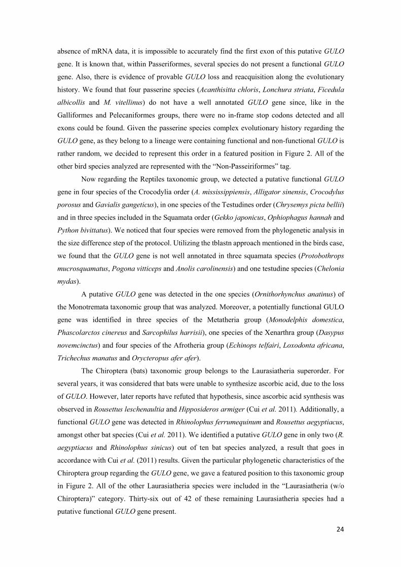
24
absence of mRNA data, it is impossible to accurately find the first exon of this putative GULO
gene. It is known that, within Passeriformes, several species do not present a functional GULO
gene. Also, there is evidence of provable GULO loss and reacquisition along the evolutionary
history. We found that four passerine species (Acanthisitta chloris, Lonchura striata, Ficedula
albicollis and M. vitellinus) do not have a well annotated GULO gene since, like in the
Galliformes and Pelecaniformes groups, there were no in-frame stop codons detected and all
exons could be found. Given the passerine species complex evolutionary history regarding the
GULO gene, as they belong to a lineage were containing functional and non-functional GULO is
rather random, we decided to represent this order in a featured position in Figure 2. All of the
other bird species analyzed are represented with the “Non-Passeiriformes” tag.
Now regarding the Reptiles taxonomic group, we detected a putative functional GULO
gene in four species of the Crocodylia order (A. mississippiensis, Alligator sinensis, Crocodylus
porosus and Gavialis gangeticus), in one species of the Testudines order (Chrysemys picta bellii)
and in three species included in the Squamata order (Gekko japonicus, Ophiophagus hannah and
Python bivittatus). We noticed that four species were removed from the phylogenetic analysis in
the size difference step of the protocol. Utilizing the tblastn approach mentioned in the birds case,
we found that the GULO gene is not well annotated in three squamata species (Protobothrops
mucrosquamatus, Pogona vitticeps and Anolis carolinensis) and one testudine species (Chelonia
mydas).
A putative GULO gene was detected in the one species (Ornithorhynchus anatinus) of
the Monotremata taxonomic group that was analyzed. Moreover, a potentially functional GULO
gene was identified in three species of the Metatheria group (Monodelphis domestica,
Phascolarctos cinereus and Sarcophilus harrisii), one species of the Xenarthra group (Dasypus
novemcinctus) and four species of the Afrotheria group (Echinops telfairi, Loxodonta africana,
Trichechus manatus and Orycteropus afer afer).
The Chiroptera (bats) taxonomic group belongs to the Laurasiatheria superorder. For
several years, it was considered that bats were unable to synthesize ascorbic acid, due to the loss
of GULO. However, later reports have refuted that hypothesis, since ascorbic acid synthesis was
observed in Rousettus leschenaultia and Hipposideros armiger (Cui et al. 2011). Additionally, a
functional GULO gene was detected in Rhinolophus ferrumequinum and Rousettus aegyptiacus,
amongst other bat species (Cui et al. 2011). We identified a putative GULO gene in only two (R.
aegyptiacus and Rhinolophus sinicus) out of ten bat species analyzed, a result that goes in
accordance with Cui et al. (2011) results. Given the particular phylogenetic characteristics of the
Chiroptera group regarding the GULO gene, we gave a featured position to this taxonomic group
in Figure 2. All of the other Laurasiatheria species were included in the “Laurasiatheria (w/o
Chiroptera)” category. Thirty-six out of 42 of these remaining Laurasiatheria species had a
putative functional GULO gene present.

25
The Primates order and the species C. porcellus (guinea pig) are contained in the
Euarchontoglires superorder. The Primates order is comprised by the Haplorhini suborder (which
includes the anthropoid primates) and the Strepsirrhini (prosimians) suborder. Several reports
show that anthropoid primates and guinea pigs have lost the capacity to synthesize ascorbic acid
(Drouin et al. 2011). Nevertheless, their genomes still contain trace sequences similar
to GULO gene sequences (Drouin et al. 2011). In agreement with these reports, although all 21
Haplorhini genomes showed at least one hit when performing the tblastn step of the protocol,
none had the typical HWAK GULO amino acid motif. We were also able to identify a potentially
functional GULO gene in three species (Microcebus murinus, Propithecus coquereli and
Otolemur garnettii) belonging to the Haplorhini sister group, Strepsirrhini. Furthermore,
although a GULO gene is annotated in the guinea pig genome, the CDS is not present in the
phylogenetic tree shown in Supplementary Figure 1. This putative functional gene encodes a
protein that is half the size of the ones seen in other species, and thus it is considered a pseudogene.
This is an example relative to the advantages of using a sequence size removal step, showing how
useful this option can be to avoid misleading information available in many databases, such as
bad annotations. The Haplorhini and Strepsirrhini suborders, such as the guinea pig, were
discriminated in Figure 2. All of the remaining Euarchontoglires species analyzed were included
in the “Euarchontoglires (w/o Cavia and Primates)” category. Regarding this category, we were
able to identify a putative GULO gene in 20 out of the 24 species available in the protocol.
Analyzing the Prostotomian species, we verified that there was a GULO gene loss in the
Pancrustacea taxonomic group. We could not identify any putative GULO in 116 insect species.
The insects are included in the Hexapoda subphylum, and although we could not find a putative
GULO gene in any insect species, the number of remaining Hexapoda species analyzed was not
sufficient to conclude that the gene was lost in this complete lineage. We also could not identify
any potentially functional GULO gene in the analyzed species of the Crustacea subphylum.
Although one crustacean species (Hyalella azteca) GULO CDS is present in the final phylogeny,
the sequence appears misplaced in the tree. In addition, the amino acidic motif of the protein
encoded by this sequence is HWGK, rather than the typically seen HWAK pattern. With this
information, we did not consider this sequence to be representative of the GULO gene.
The Arachnida taxonomic group contains the Araneae order and the Acari subclass, being
integrated the Chelicerata subphylum. We failed to detect a potentially functional GULO gene in
eight species of the Acari subclass. Still, using a tblastn against the two genomes of the species
removed by size (Euroglyphus maynei and I. scapularis), we found that both of them could
contain a functional GULO sequence (HWAK pattern and great alignment coverage), a likely
occurrence for at least I. scapularis given the report by Wheeler et al. (2015). Their exclusion
from the analysis could therefore be the result of wrong gene annotation, as seen various times in
deuterostomes. Regarding the Araneae order, a putative functional GULO sequence was

26
identified in Parasteatoda tepidariorum, while none was found in Stegodyphus mimosarum. We
also could not find a putative GULO gene in the remaining Chelicerata species, namely Limulus
polyphemus. Similarly to the Hexapoda case, we cannot assume that the GULO gene is lost in the
whole Chelicerata (w/o Arachnida) lineage, since only one species was analyzed.
Two Tardigrades species (Hypsibius dujardini and Ramazzottius varieornatus) were
analyzed and no putative GULO gene was found, even after a tblastn search against the species
genomes. Given the number of species analyzed, we cannot assume that GULO gene is lost in
this lineage.
We detected a potentially functional GULO gene in Priapulus caudatus, a member of the
Priapulida phylum. In contrast, when observing the 35 analyzed species belonging to the
Nematoda phylum, we failed to detect a putative GULO gene. We obtained equal results when
observing the nine analyzed species included in the Platyhelminthes phylum, strongly indicating
the loss of GULO in this lineage. In the Mesozoa subphylum, only Intoshia linei was analyzed,
being removed at the initial tblasnt step. Since a single species was observed, we could not draw
any conclusions regarding this lineage. GULO appears present in the Brachiopoda phylum, as we
detected a putative gene in Lingula anatina. We could not formulate any hypothesis regarding the
Annelida phylum, since we could not identify a possibly functional GULO in Helobdella robusta
and C. teleta.
The Bivalvia, Cephalopoda and Gastropoda classes are included in the Mollusca phylum.
Regarding the four analyzed species representative of the Bivalvia class (Mytilus
galloprovincialis, Crassostrea gigas, Crassostrea virginica and Mizuhopecten yessoensis), we
could not detect any putative GULO gene, and thus, it is likely the gene was lost in this lineage.
Moving to the Gastropoda class, we were able to detect a potentially functional GULO in two
species, namely, Aplysia californica and L. gigantea, in accordance with the reports by Wheeler
et al. (2015). This result indicates that the GULO gene was probably maintained in this lineage.
We could not take any conclusions concerning the Cephalopoda class, since we were not able to
identify a putative GULO gene in the only species analyzed (Octopus bimaculoides).
The remaining sequences detected that were not placed in the expected phylogenetic
position, were not analyzed. We observed that these GULO CDSs encoded for proteins that had
the amino acidic motif HWGK (as the Porifera and Crustacea sequences), while all the GULO
sequences had the motif HWAK. This information suggests that the GULO protein always has
the HWAK motif across all animal species, and that proteins with the HWGK motif are not truly
GULO, but rather FAD domain proteins coded by a gene other than GULO. Given this hypothesis,
we propose that the real GULO amino acidic motif is HWAK, rather than the referenced HWXK.
Interestingly, the five outgroup Fungi ALO sequences also seem to encode for proteins that
contain the conserved HWAK motif.

27
Our results suggests that GULO origin predates at least the split of the Cnidaria and
Bilateria taxonomic groups, around 642 million years ago. This is due to findings regarding the
non-bilaterian Anthozoa class, in which we found a putative GULO gene, and to observations
concerning the presence of GULO in 21 bilaterian taxonomic groups. This is a conservative
overview when compared to the hypothesis by Wheeler et al. (2015). Nevertheless, following our
criteria and considering the shortage of information regarding the basal Porifera group, we cannot
assume that GULO was present or absent in the ancestor species from which all of the Metazoa
(animal) taxonomic groups descended. Furthermore, although Fungi ALO synthetizes D-
erythroascorbic acid instead of L-ascorbic acid, it shares several characteristics with animal
GULO and is known to have some substrate specificity to L-Gulonolactone (Smirnoff et al. 2001).
Given this evidence, GULO may be in fact much older than we can estipulate in our results,
possibly being present in the ancestral of the Metazoa and Fungi taxonomic groups, as proposed
by Wheeler et al. (2015).
The GULO gene appears to have been lost independently in many lineages, both in the
Protostomia and Deuterostomia groups (five and three times, respectively). Inside the
protostomians, regarding the Arthropoda phylum, we cannot draw conclusions concerning the
GULO gene in the Hexapoda (w/o Insects) category, given that only one species was analyzed.
Nevertheless, we suspect that this gene was lost after the split between the Pancrustacea and the
Chelicerata taxonomic groups in the former lineage. Failure to detect a putative functional GULO
in the Crustacea subphylum and Insecta class while confirming the presence of GULO in the
Araneae order strongly supports this hypothesis. Regarding the Nematoda, there seems to be a
clear loss of a putative GULO gene. Given the presence of a potentially functional GULO in the
Priapulida and Arthropoda phyla and the absence of this gene in the Nematoda phylum, it seems
evident that the gene was lost independently in the latter. These three phyla belong to the
Ecdysozoa group and, as such, should share a common ancestor carrying a GULO gene. We also
hypothesize that the Lophotrochozoa (comprised of Mollusca, Annelida and Brachiopoda phyla)
ancestral had a potentially functional GULO gene, and that this gene was lost in the Bivalvia after
the radiation of the Conchifera subphylum (which includes the Bivalvia, Cephalopoda and
Gastropoda classes), while it was maintained in the Gastropoda class. The Platyhelminthes
phylum has an uncertain taxonomic position in the Protostomia group. We do know that this
phylum is not included within the analyzed Ecdysozoa and Lophotrochozoa, and thus it is possible
that the GULO gene was also lost independently in this unplaced group. Observing the wide
context regarding the detection of GULO sequences in Protostomian species, it is surprising that
although we failed to detect GULO in three large Protostomian data sets (117 Insects, 25
Nematoda, and 10 Platyhelminthes genomes), we could identify a putative GULO in five rather
small datasets (five Anthozoa, two Araneae, one Priapulida, one Brachipoda and three Gastropoda
genomes). Furthermore, these putative GULO sequences do not seem the result of contamination,
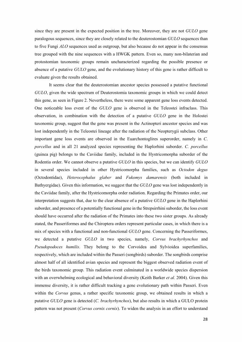
28
since they are present in the expected position in the tree. Moreover, they are not GULO gene
paralogous sequences, since they are closely related to the deuterostomian GULO sequences than
to five Fungi ALO sequences used as outgroup, but also because do not appear in the consensus
tree grouped with the nine sequences with a HWGK pattern. Even so, many non-bilaterian and
protostomian taxonomic groups remain uncharacterized regarding the possible presence or
absence of a putative GULO gene, and the evolutionary history of this gene is rather difficult to
evaluate given the results obtained.
It seems clear that the deuterostomian ancestor species possessed a putative functional
GULO, given the wide spectrum of Deuterostomia taxonomic groups in which we could detect
this gene, as seen in Figure 2. Nevertheless, there were some apparent gene loss events detected.
One noticeable loss event of the GULO gene is observed in the Teleostei infraclass. This
observation, in combination with the detection of a putative GULO gene in the Holostei
taxonomic group, suggest that the gene was present in the Actinopteri ancestor species and was
lost independently in the Teleostei lineage after the radiation of the Neopterygii subclass. Other
important gene loss events are observed in the Euarchontoglires superorder, namely in C.
porcellus and in all 21 analyzed species representing the Haplorhini suborder. C. porcellus
(guinea pig) belongs to the Caviidae family, included in the Hystricomorpha suborder of the
Rodentia order. We cannot observe a putative GULO in this species, but we can identify GULO
in several species included in other Hystricomorpha families, such as Octodon degus
(Octodontidae), Heterocephalus glaber and Fukomys damarensis (both included in
Bathyergidae). Given this information, we suggest that the GULO gene was lost independently in
the Caviidae family, after the Hystricomorpha order radiation. Regarding the Primates order, our
interpretation suggests that, due to the clear absence of a putative GULO gene in the Haplorhini
suborder, and presence of a potentially functional gene in the Strepsirrhini suborder, the loss event
should have occurred after the radiation of the Primates into these two sister groups. As already
stated, the Passeriformes and the Chiroptera orders represent particular cases, in which there is a
mix of species with a functional and non-functional GULO gene. Concerning the Passeriformes,
we detected a putative GULO in two species, namely, Corvus brachyrhynchos and
Pseudopodoces humilis. They belong to the Corvoidea and Sylvioidea superfamilies,
respectively, which are included within the Passeri (songbirds) suborder. The songbirds comprise
almost half of all identified avian species and represent the biggest observed radiation event of
the birds taxonomic group. This radiation event culminated in a worldwide species dispersion
with an overwhelming ecological and behavioral diversity (Keith Barker et al. 2004). Given this
immense diversity, it is rather difficult tracking a gene evolutionary path within Passeri. Even
within the Corvus genus, a rather specific taxonomic group, we obtained results in which a
putative GULO gene is detected (C. brachyrhynchos), but also results in which a GULO protein
pattern was not present (Corvus cornix cornix). To widen the analysis in an effort to understand
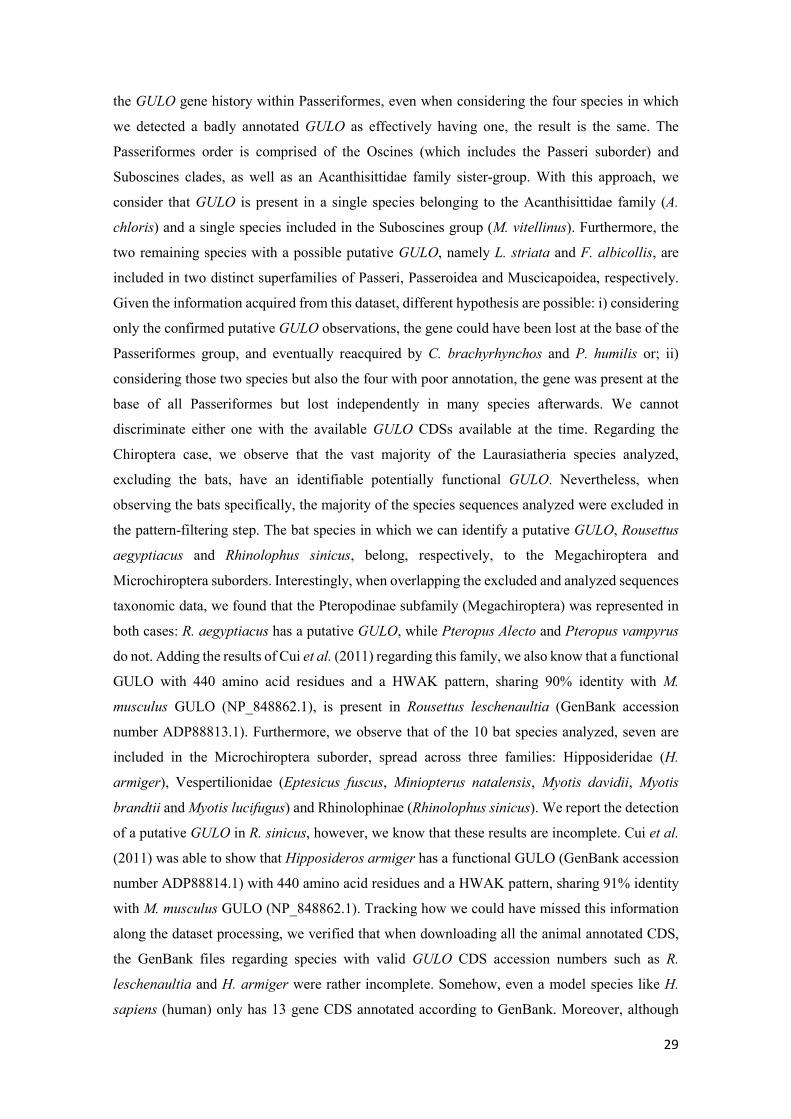
29
the GULO gene history within Passeriformes, even when considering the four species in which
we detected a badly annotated GULO as effectively having one, the result is the same. The
Passeriformes order is comprised of the Oscines (which includes the Passeri suborder) and
Suboscines clades, as well as an Acanthisittidae family sister-group. With this approach, we
consider that GULO is present in a single species belonging to the Acanthisittidae family (A.
chloris) and a single species included in the Suboscines group (M. vitellinus). Furthermore, the
two remaining species with a possible putative GULO, namely L. striata and F. albicollis, are
included in two distinct superfamilies of Passeri, Passeroidea and Muscicapoidea, respectively.
Given the information acquired from this dataset, different hypothesis are possible: i) considering
only the confirmed putative GULO observations, the gene could have been lost at the base of the
Passeriformes group, and eventually reacquired by C. brachyrhynchos and P. humilis or; ii)
considering those two species but also the four with poor annotation, the gene was present at the
base of all Passeriformes but lost independently in many species afterwards. We cannot
discriminate either one with the available GULO CDSs available at the time. Regarding the
Chiroptera case, we observe that the vast majority of the Laurasiatheria species analyzed,
excluding the bats, have an identifiable potentially functional GULO. Nevertheless, when
observing the bats specifically, the majority of the species sequences analyzed were excluded in
the pattern-filtering step. The bat species in which we can identify a putative GULO, Rousettus
aegyptiacus and Rhinolophus sinicus, belong, respectively, to the Megachiroptera and
Microchiroptera suborders. Interestingly, when overlapping the excluded and analyzed sequences
taxonomic data, we found that the Pteropodinae subfamily (Megachiroptera) was represented in
both cases: R. aegyptiacus has a putative GULO, while Pteropus Alecto and Pteropus vampyrus
do not. Adding the results of Cui et al. (2011) regarding this family, we also know that a functional
GULO with 440 amino acid residues and a HWAK pattern, sharing 90% identity with M.
musculus GULO (NP_848862.1), is present in Rousettus leschenaultia (GenBank accession
number ADP88813.1). Furthermore, we observe that of the 10 bat species analyzed, seven are
included in the Microchiroptera suborder, spread across three families: Hipposideridae (H.
armiger), Vespertilionidae (Eptesicus fuscus, Miniopterus natalensis, Myotis davidii, Myotis
brandtii and Myotis lucifugus) and Rhinolophinae (Rhinolophus sinicus). We report the detection
of a putative GULO in R. sinicus, however, we know that these results are incomplete. Cui et al.
(2011) was able to show that Hipposideros armiger has a functional GULO (GenBank accession
number ADP88814.1) with 440 amino acid residues and a HWAK pattern, sharing 91% identity
with M. musculus GULO (NP_848862.1). Tracking how we could have missed this information
along the dataset processing, we verified that when downloading all the animal annotated CDS,
the GenBank files regarding species with valid GULO CDS accession numbers such as R.
leschenaultia and H. armiger were rather incomplete. Somehow, even a model species like H.
sapiens (human) only has 13 gene CDS annotated according to GenBank. Moreover, although

30
four putative GULO CDSs are identified in the initial tblastn regarding the RefSeq file for H.
armiger, only one is a predicted as being a real GULO (XP_019491656.1). Neverthless, even this
one is badly annotated and does not even contain the HWXK amino acid pattern. This is a peculiar
case in which, even when overlapping the GenBank and RefSeq data to obtain the most
informative FASTA file possible, the file ends up incomplete and thus misleading when analyzed.
This does not represent a methodological problem, but rather a limitation of the databases. Even
so, this is a great example that illustrates the need for caution when formulating hypotheses based
on obtained phylogenetic results. Returning to the results, we observe that Galeopterus
variegatus, the only species belonging to the Dermoptera order in this dataset, is excluded in the
GULO HWXK pattern filtering step. When only one species is analyzed, our criteria indicates
that we cannot make conclusions regarding a possible loss of the GULO gene in that given lineage.
In spite of that, it is curious that the Chiroptera and Dermoptera orders are sister groups, although
they belong to distinct superorders, Laurasiatheria and Euarchontoglires, respectively. Also
within the Euarchontoglires, a putative GULO is observed within the Primates order, but also in
the Scadentia sister group (represented by Tupaia chinensis). Collecting all this evidence, we
suggest two possible hypotheses regarding the GULO gene in bats. The first implies that GULO
could have been lost after the Boreoeutheria group (comprised of Laurasiatheria and
Euarchontoglires) radiation, in the lineage that leads to the Chiroptera/Dermoptera phylogenetic
branch, while it was maintained in the Primate and Scadentia lineage. As such, the current
taxonomic knowledge regarding the placement of the Chiroptera and Dermoptera orders in
distinct superorders does not correctly apply in this specific case. The second hypothesis suggests
an independent gene loss event in both Chiroptera and Dermoptera orders that occurred after the
Laurasiatheria and Euarchontoglires radiations, respectively. The second option seems more
likely with the information we have, while the first needs some complementary work concerning
the number of species analyzed in the Dermoptera order. Nevertheless, in both alternatives there
is a common assumption: given the bat phylogeny presented by Teeling et al. (2005) already
adapted by Cui et al. (2011) and also our results, it seems plausible that GULO was lost at the
base of all Chiroptera, and that the inactive gene was reactivated during evolution in some species.
Even so, and as Cui et al. (2011) states, we cannot assume that GULO may not evolve to become
a pseudogene eventually, since it is clear that a large number of bat species do not require this
functional gene.
The consensus phylogeny obtained (Supplementary figure 1) also shows that GULO is a
single copy gene in all animal species analyzed, with the exception of A. planci. Therefore, it
seems that GULO duplicates were not retained after the two rounds of closely spaced auto-
tetraploidization events (commonly known as 1R and 2R) that occurred early in vertebrate
evolution (Dehal and Boore 2005; Putnam et al. 2008). Some reports suggest that they may have
occurred during chordate evolution, after the split of the urochordate and cephalochordate

31
lineages but before the radiation of gnathostomes. It is possible that GULO had already been lost
in the common ancestor of all Teleosti fish, and thus the whole-genome duplication event that
occurred at that time (Taylor 2003) is not relevant regarding the gene evolutionary history within
this group. Moreover, a single GULO gene is found even in a recent allotetraploid species such
as Xenopus leavis (Session et al. 2016).
Our results imply that GULO was not lost a single time in the Protostomia group, but
multiple times independently, after the split of the Protostomia and the Deuterostomia lineages,
which complement the hypothesis formulated by Wheeler et al. (2015).

32
III.2. Non-Bilateria/Protostomia GULO gene annotations and
phylogenetic analysis
III.2.1. GULO sequence annotation process The results concerning the phylogenetic analysis of available annotated animal GULO
CDS implied the unreported presence of a putative functional GULO gene in three Protostomia
lineages, namely the Araneae family and the Priapulida and Brachiopoda phyla. Nevertheless,
many protostomian and non-bilaterian lineages remained uncategorized regarding the presence or
absence of a potential GULO gene. The main reason for these inconclusive results was the number
of species analyzed with an already annotated genome, since we could not assert the absence of
GULO with any less than three representative species CDS within each taxa, and in many cases,
only one or two species were available in the final processed dataset. To overcome this lack of
information, we performed annotations for the representative GenBank genomes of all available
species included in the Protostomia and non-bilaterian groups highlighted in Figure 2.
Within the Non-Bilateria, we analyzed two species included in the Porifera phylum,
namely A. queenslandica and Aplysina aerophoba. In A. queenslandica, the tblastn results
obtained showed a high alignment coverage between M. musculus GULO and the used genome,
dispersed across several scaffolds. Moreover, we detected a putative exonic sequence that
encoded for the HWGK amino acid pattern. We cannot assume that the scaffolds in which we
detected potential GULO exons are successional portions of the genome, since they are not
assembled and can even be present in different chromosomes. As such, we failed to annotate the
GULO gene in this species. As for A. aerophoba, we could not detect a probable GULO exon that
encodes for the conserved HWXK GULO amino acid motif, and so the annotation was not
performed. We were able to perform an annotation for T. adhaerens, the only available species
belonging to the Placozoa phylum. According to our annotation, we were able to identify 13
introns for this potential gene, which encodes a protein with a HWAK amino acid motif.
Regarding the Myxozoa phylum, we tried to perform annotations using the genomes of four
species, namely T. kitauei, Kudoa iwatai, Sphaeromyxa zaharoni and Enteromyxum leei, but were
only able to annotate a putative GULO in K. iwatai. Our annotation suggests the presence of 13
intronic regions within this K. iwatai potential gene, which may code for a putative protein with
a HWAK amino acid motif. T. kitauei tblastn result presented a complete sequence coverage with
a possibly encoded HWGK amino acid motif in the alignment. Even so, we verified the presence
of in-frame stop codons within the alignment, and as such, could not annotate a putative gene.
For S. zaharoni and E. leei, we observed a partial alignment coverage in the tblastn results, in
both cases with exons that may code for a HWAK amino acid motif. Given the alignment gaps
observed in these results, we were not able to perform an annotation for these species. We did not
analyze any Anthozoa species since we considered that the results obtained for our GULO CDS

33
dataset and the reports by Wheeler et al. (2015) represented strong enough evidence that the gene
may be present within this taxonomic group. As for the Hydrozoa phylum, we analyzed only one
genome belonging to Hydra magnipapillata. In this species, we could not identify a single
genomic sequence with homology with the M. musculus GULO that could code for the conserved
HWXK amino acid pattern, and so an annotation was not performed.
Concerning the Protostomia, we analyzed four Hexapoda (excluding insects) species
genomes, namely Catajapyx aquilonaris, Folsomia candida, H. duospinosa and Orchesella
cincta. We observed a low coverage sequence alignment with no putative encoded HWXK amino
acid motif for C. aquilonaris, and therefore did not perform an annotation for this species. In O.
cincta, we were able to annotate a putative GULO gene with three introns, which may code for a
protein with an HWGK amino acid pattern. Furthermore, we identified a possibly duplicated
GULO gene in F. candida and H. duospinosa. Regarding F. candida, two gene annotations were
performed, which we designated as Folsomia candida 1 and 2, respectively. The first annotated
putative GULO sequence had four introns, whereas the second had two. Nevertheless, both
sequences may code for proteins with an HWGK amino acid pattern. In H. duospinosa, the two
performed sequence annotations were entitled Holacanthella duospinosa 1 and 2, and both
presented a single intron. In addition, we were able to observe that both potential GULO
sequences could possibly code for a protein with an HWGK amino acid pattern. Given the results
obtained for the Insecta class using the previously described GULO CDS dataset approach, in
which a putative GULO was not detected in 116 species, we considered that further analyzing this
taxonomic group would lead to the same conclusion, and did not try to perform annotations in
this case. As for the Crustacea, we felt that the previous results, where we did not detect a GULO
in four species, were on the brink of our three species criteria for hypothesizing a gene loss within
a lineage. Although we were confident in our results, to strengthen our conclusion, we further
analyzed this taxonomic group by performing a direct tblastn in the NCBI database, using as
query M. musculus GULO against only the Crustacea class species representatives. Using this
assay, we were able to determine that only two species had a tblastn alignment hit, namely
Eurytemora affinis and Hyalella azteca. Furthermore, by observing the sequence alignment
results, we uncovered that the only species which could code for a protein with a conserved
HWXK pattern was H. azteca. Given this result, we proceeded to download this species genome
for a more detailed analysis. Using the H. azteca genome, we were able to annotate two distinct
putative GULO sequences with a single intron each, both probably encoding for a potential
HWGK pattern containing protein. These sequences were labeled as Hyalella azteca 1 and 2.
Concerning the Araneae family, included in the Chelicerata subphylum, we were able to
refine the annotation already available for GULO in Parasteatoda tepidariorum
(XP_015913395.1) that was displayed in the GULO CDS protocol consensus phylogeny. Since

34
we already had an annotation for this species, we did not perform a new annotation using the
complete genome. Instead, we performed a blastp to identify the regions of homology between
the P. tepidariorum and the M. musculus GULO. Verifying the blastp results, we observed that
the GULO protein of P. tepidariorum was 26 amino acids bigger in the N-terminus region when
compared with M. musculus GULO, given that the first methionine of M. musculus GULO aligned
with the methionine at the amino acidic position 27 of P. tepidariorum GULO. Given this
evidence, we assumed an alternative putative ATG start for the available CDS and shortened it in
78 nucleotides at the beginning of the sequence, leaving the rest of the sequence unchanged. For
the remaining five Araneae species analyzed, we tried to perform a de novo annotation using the
available genomes information. We failed to annotate a potential GULO gene in three of these
species, namely L. reclusa, S. mimosarum and Acanthoscurria geniculata. In L. reclusa, the
tblastn genomic hit region obtained contained many ambiguous nucleotide sites, which partially
overlapped putative exons and did not allow a complete annotation. As for S. mimosarum and A.
geniculata, we obtained a partial sequence alignment coverage spread across several scaffolds in
the tblastn results, and therefore, an annotation was not performed. Nevertheless, in both species,
genomic regions that potentially code for the GULO HWAK conserved amino acid motif were
identified. We were able to perform an annotation for the two remaining Araneae species
analyzed, Latrodectus hesperus and Nephila clavipes. In L. hesperus we detected a possible
GULO gene with nine putative introns, while in N. clavipes we observed a potential GULO with
11 introns. Furthermore, we found that both annotations possibly code for proteins with an
HWAK amino acid pattern. Still within the Chelicerata, and now regarding the Acari subclass,
we used 16 species genomes to execute our annotation analysis. No putative GULO sequence
could be annotated in eight of these species genomes, belonging to Galendromus occidentalis,
Varroa destructor, Varroa jacobsoni, Sarcoptes scabiei, Tropilaelaps mercedesae, Tetranychus
urticae, Ixodes ricinus and Dermatophagoides pteronyssinus. In the first five mentioned species,
the results obtained from the tblastn showed no similarities between any genomic scaffold and
the M. musculus GULO used as query. In T. urticae, we were not able to identify any genomic
region in the tblastn alignment that could code for a conserved HWXK amino acid motif, and thus
it is not likely that the sequences identified in the tblastn results belong to GULO gene. As for I.
ricinus, the nucleotide sequences that share identity with M. musculus GULO are scattered across
different scaffolds, which may or may not be successive in the genomic context. Although we did
not carry out an annotation for a potential GULO in this species, we verified that one of the
sequences present in the alignment could code for a protein region with a HWAK amino acid
pattern. This conserved amino acid pattern could also be encoded by a sequence retrieved from
the D. pteronyssinus genome after the tblastn search. Even so, an annotation was not performed
for this species due to the presence of a single in-frame stop codon preceding the third potential
exon of a putative GULO gene. We performed partial gene annotations for I. scapularis,

35
Steganacarus magnus and E. maynei. In I. scapularis, we failed to annotate the putative GULO
last exon, due to the presence of ambiguous nucleotide positions in this genome region. For S.
magnus, we annotated a truncated putative GULO sequence that shared identity with the M.
musculus GULO only after amino acidic position 109. The premature end of the scaffold that we
used to perform the annotation at the 5’ extremity did not allow a further annotation. As for E.
maynei, we annotated a truncated putative GULO sequence at the 5’ end, but also missing the
final putative exon. This sequence shared identity with the M. musculus GULO after amino acidic
positions 134 until 398, and could not be further annotated due to the premature end of the
analyzed genomic scaffold at both extremities. Although these three sequences are all partial
annotations, they have high Blast alignment scores (>=200) and can potentially code for a protein
with a HWAK conserved motif. We were able to annotate a potential GULO in the remaining six
species genomes analyzed, belonging to Dermatophagoides farinae, Achipteria coleoptrata,
Hypochthonius rufulus, Platynothrus peltifer and Rhipicephalus microplus, respectively. These
annotated sequences enabled us to infer five probable introns within the putative GULO gene for
D. farinae, A. coleoptrata and H. rufulus, three introns for P. peltifer and one for R. microplus.
Furthermore, all of these annotated sequences might code for a protein with a conserved HWAK
amino acid motif. We also tried to perform annotations for three Chelicerata species that were not
included in the Araneae and Acari taxonomic groups, namely Limulus polyphemus, Mesobuthus
martensii and Centruroides sculpturatus. No annotation resulted from the analysis of the genomes
for each of these species, since the sequences that had similarities with the M.musculus GULO
obtained after the tblastn belonged to different and possibly non-successive genomic scaffolds.
Nevertheless, all these species had a putative exonic sequence that could code for a protein region
with a conserved HWAK amino acid motif. Regarding the Tardigrada phylum, we analyzed the
available genomes for R. varieornatus and H. dujardini. We failed to annotate a probable GULO
gene in these species, since the alignment coverage of the sequences retrieved from the tblastn
initial results was rather incomplete in both cases. Even so, by observing these low coverage
sequence alignments, we detected possible genomic sequences that could code for a protein with
a HWGK amino acid pattern, for both species. The only representative genome available for the
Priapulida species in the GenBank database belongs to P. caudatus. Since we already obtained a
representative GULO sequence for this species in the previous animal GULO CDS protocol
analysis (XP_014666894.1), we did not perform a new annotation using the genome information.
Alternatively, we performed a blastp to identify the regions of homology between the P. caudatus
and the M. musculus GULO to verify if the available annotation could be further refined, similarly
to the case of P. tepidariorum. The blastp alignment had full coverage between the query and
target protein sequences, except for the first four amino acids, with only one observable gap out
of 437 amino acid positions analyzed. Moreover, the P. caudatus GULO CDS encodes for a
protein that has only four more amino acids than the M. musculus GULO. These evidences are

36
indicative of a good annotation process, and as such, we did not perform additional adjustments
to the P. caudatus available CDS. The GULO gene in P. caudatus has 11 potential introns and
may code for a protein with a HWAK amino acid pattern. For the Nematoda and Platyhelminthes,
the results obtained in the previous animal CDS analysis suggested the absence of a GULO gene
in 35 and nine species, respectively. Even so, we sought out to strengthen these results using a
tblastn in the NCBI database, using as query M. musculus GULO against the Nematoda and
Platyhelminthes phyla species representatives. Using this approach, we were able to determine
that only one Nematoda species (Brugia timori) had a tblastn alignment hit. However, the
genomic sequence with similarities to M. musculus GULO did not code for a HWXK amino acid
pattern and had poor alignment coverage with a expect value of 0.002. With these results, we
considered unnecessary any attempt to perform a putative GULO gene annotation in the species
belonging to these taxonomic groups, given the clear evidence for the lack of GULO seen within
the nematodes and Platyhelminthes genomes analyzed so far. As for the Mesozoa, we analyzed
the only available species genome, belonging to I. linei. We could not perform a gene annotation
for these species since the tblastn output did not contain any sequences with homology with M.
musculus GULO. Regarding the Brachiopoda group, we already detected a putative GULO
sequence in Lingula anatina (XP_013393535.1), using the animal available CDS protocol. Like
in P. tepidariorum and P. caudatus, we verified if this annotation could be further refined using
a blastp approach. We verified that the obtained alignment between L. anatina GULO and the M.
musculus GULO had full coverage, with the exception of the first three amino acids. Furthermore,
the alignment had only two observable gaps out of 437 amino acid positions analyzed, and the
GULO protein encoded by the available L. anatina GULO CDS had merely five more amino acids
than the reference M. musculus GULO. By observing these parameters, we did not find evidence
for a bad annotation process, and as such, did not perform additional adjustments to the L. anatina
annotated GULO CDS. The GULO gene in L. anatina has 11 potential introns and may code for
a protein with a HWAK amino acid pattern. Still regarding the Brachipoda phylum, we analyzed
the available genome for Phoronis australis, but could not perform a gene annotation given that
no nucleotide sequences with homology with M. musculus GULO were detected through the tblast
search. We also tried to annotate a putative GULO gene in six Annelida species genomes, namely
Amynthas corticis A, Amynthas corticis C, C. teleta, Eisenia fetida, H. robusta and Hydroides
elegans. For H. robusta and H. elegans, we did not obtain sequences with homology to M.
musculus GULO from the output of the tblastn, and thus no annotation was performed. Moreover,
we were not able to annotate a probable GULO sequence in A. corticis A and A. corticis C. In A.
corticis A, the nucleotide sequences that shared homology with M. musculus GULO were
scattered across several genomic scaffolds. Even so, we observed that one of the sequences could
code for a protein with a HWAK amino acid. In A. corticis C, we found no identifiable
homologous nucleotide sequence that could code for a HWXK amino acid pattern. For C. teleta

37
and E. fetida, we were able to annotate potential GULO gene sequences that may code for proteins
with a conserved HWAK and HWGK amino acid motifs, respectively. However, the obtained E.
fetida sequence is missing nucleotide information that may code for the first 58 amino acid
positons of a putative GULO protein, due to the precocious ending of the genomic scaffold we
used to perform the annotation at the 5’ terminus. We were able to infer 11 possible introns within
the annotated C. teleta GULO, while for E. fetida we could not make an estimate.
Within the Gastropoda group, we evaluated if the available GULO annotations for A.
californica (XP_005103891.1) and Lottia gigantea (ESO97787.1) could be further refined. With
a blastp search using M. musculus GULO as query, we were able to determine that the obtained
sequence alignments had full coverage for both species, with the exception of the first three amino
acids. In addition, the alignments had only two observable gaps out of 437 amino acid positions
analyzed for A. californica, and four distinguishable gaps out of 439 amino acid positions
analyzed for L. gigantea. Moreover, the putative GULO protein encoded by the A. californica
CDS had fifteen more amino acids than the reference M. musculus GULO, while the protein
possibly encoded by the L. gigantea CDS had sixteen more. With this information, we considered
the available CDSs for these species rather well annotated, and as such, did not perform additional
annotation adjustments. The GULO gene in A. californica has 10 potential introns and may code
for a protein with a HWAK amino acid pattern, as also seen in L. gigantea. We also tried to
perform our own GULO gene annotations using the genomes of Biomphalaria glabrata,
Colubraria reticulata, Conus tribblei, Lymnaea stagnalis and Radix auricularia. We failed to
annotate a putative gene in B. glabrata and C. tribblei. In B. glabrata, the annotation process led
to an incomplete CDS with in-frame stop codons. Furthermore, the only potential exonic sequence
that could code for a protein region with a HWAK amino acid motif was in a different translation
frame from the one seen in the rest of the putative gene. As for C. tribblei, the tblastn results
revealed a poor sequence alignment coverage between the M. musculus GULO and the used
genome, with the detected homologous sequences dispersed across several scaffolds. It was also
possible to observe that, for this species, two distinct exonic sequences that could code for HWAK
and HWGK amino acid patterns were identified. We were able to obtain annotations for C.
reticulata, L. stagnalis and R. auricularia. The putative GULO annotations for L. stagnalis and
R. auricularia allowed us to infer the presence of 11 possible introns in the gene sequence. As for
C. reticulata, the tblastn results allowed us to annotate one full-coverage putative GULO sequence
and another truncated at both extremities due to premature scaffold ending, which we labeled as
Colubraria reticulata 1 and 2, respectively. We deduced the presence of one intron in the full-
coverage annotation, while for the truncated sequence we were unable to estimate an intron
number due to lack of information regarding the first 171 amino acidic positions in the tblastn
alignment. Neverthless, all the annotated sequences for these three species may code for proteins

38
that contain a conserved HWAK amino acid pattern. Concerning the Bivalvia taxonomic group,
we sought to annotate a putative GULO gene in ten species genomes, belonging to Bankia
setacea, Bathymodiolus platifrons, Modiolus philippinarum, M. galloprovincialis, C. virginica,
Pinctada martensii, C. gigas, Corbicula fluminea, M. yessoensis and Dreissena polymorpha. The
tblastn search with M. musculus GULO as query only found homologous nucleotide sequences in
the B. setacea, B. platifrons, M. philippinarum and M. galloprovincialis species genomes, while
in the remaining genomes none was retrieved. However, the identified homologous sequences did
not present any putative exonic regions that could code for the typical GULO HWXK amino acid
pattern, and thus, an annotation was not performed. Regarding the Cephalopoda group, we tried
to annotate the GULO gene in the only available representative genome for this class, belonging
to Octopus bimaculoides. However, we could not detect any nucleotide sequence with homology
to M. musculus GULO through the tblastn search, and as such, did not perform an annotation.
In all our de novo gene annotations, we were not able to detect the potential ATG start
codon that encodes for the first translated protein amino acid, methionine. Trying to overcome
this problem, we performed several tblastn searchs using M. musculus GULO as query against
our annotations plus their extended 5’ flanking genomic region, whenever possible. Using this
approach, we hoped to detect a putative first exon for our gene annotations that could contain the
ATG start of the coding sequence. Even so, this methodology did not work. Still, the passerine
coding sequences analyzed in the previous dataset gave information regarding a potential ATG
site preceded by the AG nucleotides, which could indicate a canonical GT-AG splice site at that
location. Given this evidence, it is possible to speculate that our annotations may present the same
features as these passerine sequences, and that without gene expression analysis, we cannot
simply infer the ATG start codon using only a tblastn search. Even so, our annotations have high
sequence alignment coverage with expect values remarkably close to zero when queried against
the M. musculus GULO protein, while also not presenting any in-frame stop codons. Therefore,
it is rather unlikely that these gene annotations could represent a pseudogene using only the
absence of an identifiable ATG start codon as evidence, and as such, we are confident that we are
identifying functional genes. As such, we adjusted the number of observed putative introns (by
adding one intron) in our gene annotations, to follow the hypothesis in which the first exon
containing the ATG start codon is not found, but expected to exist preceding our annotations in
the analyzed genomes. The number of identified introns for each annotated putative GULO
sequence presented until now already considers this correction.

39
III.2.2. GULO CDS annotations phylogenetic analysis The consensus phylogeny obtained from the ADOPS pipeline output concerning our
annotations is presented in Figure 3. Given the results obtained for the animal GULO CDS
protocol, we suspected that sequences that code for a protein with an HWGK amino acid motif
likely do not constitute a real GULO. By performing our own annotations, we were able to obtain
eight sequences that may encode putative proteins with this HWGK pattern, namely for three
Hexapoda species from the Collembola class (O. cincta, H. duospinosa and F. candida), for one
species from the Crustacea group (H. Azteca) and for one Annelida species from the Clitellata
group (E. fetida). The Bayesian phylogeny reveals that these eight sequences are represented as
an external tree branch to our selected Fungi ALO outgroup. This result further reinforces the
evidence found in the previous protocol, and suggests that these annotations do not represent a
real GULO, but rather another gene that could possibly code for a distinct FAD-domain protein.
Interestingly, the annotations belonging to K. iwatai and R. microplus also appear misplaced in
the phylogeny, even though they may code for proteins with an HWAK amino acid motif. To
identify the possible cause for this unexpected result, we started by performing blastp searches
using the translated K. iwatai and R. microplus annotations against the complete NCBI database
to check for possible genome contamination or horizontal gene transfer events that could
influence the phylogeny outcome. For R. microplus, we observed that the translated annotation is
remarkably similar to a FAD-linked oxidoreductase from the Alphaproteobacteria Sphingomonas
sp. 67-36 (OJV32873.1), sharing 88% identity across a 420 amino acid alignment with the mutual
presence of a conserved HWAK pattern. Moreover, the alignment result had a 0.0 expect value,
strongly suggesting that no protein sequences are expected to match as well or better than the hit
we obtained, and so it is very unlikely that this result constitutes a false positive. Furthermore, the
annotated sequence for R. microplus derived from a single continuous exon. Given these
observations, it seems likely that a process of genome contamination with exogenous bacterial
genetic material may have occurred, which in turn, can explain the abnormal position of the
putative R. microplus GULO sequence in the consensus phylogeny. Coincidentally, this also
seems to be the case for the E. fetida annotation grouped with the R. microplus sequence. Through
a blastp search, we observe that the putative protein encoded by the clitellate E. fetida gene
sequence is 99% identical to a FAD-binding protein from the Candidatus lumbricidophila
eiseniae actinobacteria (PDQ34519.1) across the 355 amino acid alignment, with a 0.0 expect
value. In addition, this annotation was obtained from a single continuous putative exon within the
E. fetida genome, further suggesting a contamination process. Regarding the K. iwatai annotation,
we did not find evidence for any of these events, and the relatively low 71% posterior credibility

40
value suggests that this sequence may in fact be placed in another position of the phylogeny with
a considerable probability.
Within the green highlighted sequences that may represent a real GULO (Figure 3), we
observe that the partial annotation for the parasitiform tick Ixodes scapularis is abnormaly placed
in the phylogeny. Given that I. scapularis is a protostome included within the Acari subclass,
theoretically, this sequence should be grouped with the Acari Sarcoptiformes taxonomic group
comprised of the A. coleoptrata, P. peltifer, H. rufulus, S. magnus, D. farinae and E. maynei
species, in the consensus tree. Instead, this sequence is grouped with five GULO sequences
representative of the deuterostome Reptiles (A. mississippiensis), Aves (G. gallus),
Euarchontoglires (M. musculus), Amphibia (Xenopus laevis) and Actinopteri (L. oculatus)
taxonomic groups.
It is know that ticks such as I. scapularis are blood-feeding ectoparasites with a broad
host range, ranging from small vertebrates to humans (Gulia-Nuss et al. 2016). Our annotation
protocol for I. scapularis allowed us to identify 11 putative introns within a possible GULO gene,
the same intron number identified in the G. gallus, M. musculus, X. laevis and L. oculatus GULO.
However, annotations performed for the remaining Acari species from the Sarcoptiformes
taxonomic group allowed us to account for only three (P. peltifer) to five (A. coleoptrata, D.
farinae and Hypochthonius rufulus) introns within the putative gene. Given these considerations,
it would seem plausible that the probable GULO annotated in I. scapularis resulted from technical
issues, due to contamination of the sequenced genome with genetic material from some
deuterostome host species. Nevertheless, this does not appear to be the case, since by analyzing
our I. scapularis translated annotation using a blastp search against all annotated species NCBI
database, we did not obtain hit results with values of identity over 53%, in alignments with 98 to
100% query coverage. Alternatively, as considered for K. iwatai, the 77% posterior credibility
value given to the I. scapularis sequence branch suggests that the sequence can be placed
somewhere else in the consensus tree. Still regarding the taxonomic context, observing the non-
bilaterian N. vectensis species adjacent to the deuterostomian cephalochordate B. belcheri in a
branch with 100% posterior credibility value is surprising. Moreover, the ancestral species
belonging to the Placozoa (T. adhaerens) phyla is represented as a sister group of the Acari
(Sarcoptiformes) and Araneae (Theridiidae and Nephilidae), in a branch with a 88% posterior

41
credibility value. OKOKOKOKOKOKOKOKOKOKOKOKOKO Folsomia candida 1
Folsomia candida 2
Orchesella cincta
Holacanthella duospinosa 1
Holacanthella duospinosa 2
Eisenia fetida
Rhipicephalus microplus
Hyalella azteca 1
Hyalella azteca 2
Kudoa iwatai
Saccharomyces cerevisiae S288C NP_013624.1
Metarhizium majus ARSEF 297 XP_014580409.1
Priapulus caudatus XP_014666894.1
Lingula anatina XP 013393535.1
Capitella teleta
Radix auricularia
Lymnaea stagnalis
Aplysia californica (California sea hare) XP_005103891.1
Lottia gigantea (owl limpet) ESO97787.1
Colubraria reticulata 1
Colubraria reticulata 2
Branchiostoma belcheri (Belchers lancelet) XP_019645195.1
Nematostella vectensis (starlet sea anemone) EDO44935.1
Parasteatoda tepidariorum (common house spider)
Latrodectus hesperus
Nephila clavipes
Achipteria coleoptrata
Platynothrus peltifer
Hypochthonius rufulus
Steganacarus magnus
Dermatophagoides farinae
Euroglyphus maynei
Trichoplax adhaerens
Ixodes scapularis
Lepisosteus oculatus (spotted gar) XP_015207781.1
Xenopus laevis (african clawed frog) OCT81467.1
Mus musculus (house mouse) Muridae NP_848862.1
Gallus gallus (chicken) XP_015140704.1
Alligator mississippiensis (american alligator) KYO43973.1
52
10099
100
100
100
65
100
100
6372
100
100
99
100
100
71
66
89 100100
100
98
100
100
100
97
88
77
100
97
100100
0.20
Collembola
Clitellata
Parasitiformes
Crustacea
Myxozoa
Saccharomycetaceae
Clavicipitaceae
Priapulidae
Brachiopoda
Polychaeta
Gastropoda
Cephalochordata
Anthozoa
Araneae
Sarcoptiformes
Placozoa
Parasitiformes
Actinopteri
Amphibia
Euarchontoglires
Aves
Reptilia
L-gulonolactone oxidase Fungi
Figure 3 – Protostomes and non-bilaterians putative L-gulonolactone oxidase (GULO) annotations phylogeny. Two
GULO CDS from Fungi species were used to help rooting the tree. Six deuterostomian species (representative of the
Actinopteri, Amphibia, Euarchontoglires, Aves, Reptilia and Cephalochordata groups) GULO CDS were used to
facilitate the interpretation of the results. Relevant higher taxonomic classifications are shown next to the species name.

42
It is rather common to find conflicting information when analyzing real datasets and the
events leading to these disaccords are well described. Possible horizontal gene transfer incidents,
lack of synapomorphy due to multiple nucleotide substitutions or stochastic errors can lead to
erratic phylogenies. Nucleotide substitutions may also randomly create sequence similarities that,
in turn, may cluster distantly related lineages in a phylogeny (Wägele and Mayer 2007). It is
known that substitution models can sometimes correct a few of these effects (Arenas 2015).
However, the sequences displayed in the consensus phylogeny represent species with an
evolutionary divergence time up to 656 million years (Peterson et al. 2004), which in turn
originates a very complex dataset for phylogenetic analysis. Given this time scale, it was expected
that even using the most likely and complete phylogenetic relations inference model for our data,
some annotations would be misplaced in the phylogeny due to, for example, greatly distinct rates
of sequence evolution. Nevertheless, the obtained phylogeny seems to follow a correct taxonomic
context in general. Furthermore, apart from the K. iwatai annotation, even the particular sequence
placements mentioned above are suggestive of GULO, since they share an internal position
relative to the Fungi outgroup.
Hopping to generate a more defined phylogeny from which we could draw conclusions
regarding the K. iwatai putative GULO sequence placement, we produced a new FASTA file
without the annotations that may code for a protein with an amino acid HWGK conserved pattern
and the R. microplus sequence we think derives from bacterial contamination. This file was
analyzed with the same parameters as before using the ADOPS pipeline, and the phylogeny output
can be observed in Figure 4. In the new consensus tree, it is possible to observe that all the
Gastropoda species annotations are clustered with 100% posterior credibility value, with the
Polychaeta and Brachiopoda groups (represented by the C. teleta and L. anatina annotations,
respectively) placed as sister groups, as expected. Additionally, GULO appears to be duplicated
in the gastropod C. reticulata. Contrary to the remaining Gastropoda annotations, these two
putative gene sequences resulted from different single continuous exons. Even so, they are
included in the correct taxonomic position in the phylogeny and should likely constitute a real
GULO. The three Araneae species annotations are placed together with 100% posterior credibility
value, seen as a sister group to the six Sarcoptiformes species putative GULO sequences branch.
Furthermore, we can also see that the K. iwatai annotation is now placed internally relative to the
Fungi outgroup. This result suggests that the K. iwatai sequence appears in fact closely related to
the remaining annotated putative GULO sequences than to the Fungi GULO, strongly
corroborating our hypothesis of technical issues regarding the previous phylogeny representation.
No changes are seen regarding the non-bilaterian N. vectensis and cephalochordate B. belcheri
species annotations, remaining grouped in a branch with 99% posterior credibility value derived
from the polytomy. Additionally, the placement of the placozoan T. adhaerens annotation is still

43
adjacent to the Acari (Sarcoptiformes) and Araneae (Theridiidae and Nephilidae) sequences, in a
branch with an even higher 99% posterior credibility value, when compared to the previous
phylogeny. The P. caudatus sequence is now unexpectedly grouped with five deuterostomians
(A. mississippiensis, G. gallus, M. musculus, X. laevis and L. oculatus) and the parasitiform I.
scapularis GULO annotations. However, the very low 60% posterior credibility value suggests
that this annotation can be placed almost randomly in the phylogeny, and could likely be inserted
in the correct taxonomic context (near the Araneae and Sarcoptiformes annotations). The I.
scapularis annotation maintains the same surprising placement in the phylogeny when compared
to the previous consensus tree, with a higher posterior credibility value for the corresponding
branch (rise from 77 to 82%).
L-gulonolactone oxidase Fungi
Saccharomyces cerevisiae S288C NP_013624.1
Metarhizium majus ARSEF 297 XP_014580409.1
Kudoa iwatai
Lingula anatina XP_013393535.1
Capitella teleta
Radix auricularia
Lymnaea stagnalis
Aplysia californica (California sea hare) XP_005103891.1
Lottia gigantea (owl limpet) ESO97787.1
Colubraria reticulata 1
Colubraria reticulata 2
Branchiostoma belcheri (Belchers lancelet) XP_019645195.1
Nematostella vectensis (starlet sea anemone) EDO44935.1
Parasteatoda tepidariorum (common house spider)
Latrodectus hesperus
Nephila clavipes
Achipteria coleoptrata
Platynothrus peltifer
Hypochthonius rufulus
Steganacarus magnus
Dermatophagoides farinae
Euroglyphus maynei
Trichoplax adhaerens
Priapulus caudatus XP_014666894.1
Ixodes scapularis
Lepisosteus oculatus (spotted gar) XP_015207781.1
Xenopus laevis (african clawed frog) OCT81467.1
Mus musculus (house mouse) Muridae NP_848862.1
Gallus gallus (chicken) XP_015140704.1
Alligator mississippiensis (american alligator) KYO43973.1
90
100100
99
100
100
100
99
100
99
100100
100
96
100
100
100
93
99
60
82
100
85
100100
0.20
Saccharomycetaceae
Clavicipitaceae
Myxozoa
Brachiopoda
Polychaeta
Gastropoda
Cephalochordata
Anthozoa
Araneae
Sarcoptiformes
Placozoa
Priapulidae
Parasitiformes
Actinopteri
Amphibia
Euarchontoglires
Aves
Reptilia
Figure 4 – Refined protostomes and non-bilaterians putative GULO annotations phylogeny. Two GULO CDS from Fungi species
were used to help rooting the tree. Six deuterostomian species (representative of the Actinopteri, Amphibia, Euarchontoglires, Aves,
Reptilia and Cephalochordata groups) GULO CDS were used to facilitate the interpretation of the results. Relevant higher taxonomic
classifications are shown next to the species name.

44
In conclusion, we can summarize the findings concerning the Araneae, Acari and
Mollusca taxonomic groups and hypothesize the GULO evolutionary history within these
lineages. Starting with the Araneae order, the six species analyzed are included in two
superorders: Araneomorphae (L. hesperus, P. tepidariorum, N. clavipes, S. mimosarum and L.
reclusa) and Mygalomorphae (A. geniculata). Inside the Araneomorphae, we have one species
representing the Haplogynae (L. reclusa) and four species representing the known sister group,
Entelegynae (L. hesperus, P. tepidariorum, N. clavipes and S. mimosarum). Concerning the
Entelegynae, we analyzed three species from the Orbiculariae group (L. hesperus, P. tepidariorum
and N. clavipes) and one from the Eresoidea group (S. mimosarum). We were able to annotate a
putative GULO in L. hesperus and N. clavipes, but also to refine the annotation already available
for P. tepidariorum, coincidently the three representative species for the Orbiculariae group in
this work. As such, we can assume that the GULO gene is maintained at least in the Orbiculariae
species lineage, specifically in the Theridiidae (L. hesperus and P. tepidariorum) and Nephilidae
(N. clavipes) families. Although we did not perform annotations for S. mimosarum, L. reclusa
and A. geniculata, we cannot rule out the presence of a GULO gene in these species genomes. For
S. mimosarum and A. geniculata, we were able to find potential exonic sequences dispersed across
several scaffolds, including some that may code for a protein with the typical HWAK amino acid
pattern, while for L. reclusa we could not obtain a reliable tblastn alignment due to the bad quality
of the genome. According to these evidences, it is likely that with better genome sequencing and
assembly we can in fact perform a complete putative GULO annotation for these species. The
summarized results for the Araneae group analysis can be observed in Figure 5.
Figure 5 – Cladogram representation of the findings regarding the annotation of GULO in six Araneae species. The species in which
GULO is likely present are highlighted in green, while the species where the presence or absence of GULO cannot be inferred are
highlighted in blue. The pink and light blue regions differentiate the Araneae species analyzed into two superorders, respectively
Araneomorphae and Mygalomorphae. The cladogram branches represent the taxonomic relation between the species analyzed,
depicted as in the Tree of life web project (http://tolweb.org/tree/) and in Wheeler et al. (2015).
The Acari species analyzed are representative of two Acari superorders, namely
Acariformes, and Parasitiformes. In the NCBI database, we were able to obtain genomes from
seven Parasitiformes species (I. scapularis, I. ricinus, R. microplus, G. occidentalis, V. destructor,
Latrodectus hesperus
Parasteatoda tepidariorum
Nephila clavipes
Stegodyphus mimosarum
Loxosceles reclusa
Acanthoscurria geniculata
Araneomorphae
Mygalomorphae

45
V. jacobsoni and T. mercedesae) and nine Acariformes species (A. coleoptrata, P. peltifer, S.
magnus, H. rufulus, D. farinae, D. pteronyssinus, E. maynei, S. scabiei and T. urticae). Within
the Parasitiformes, we were able to annotate a putative GULO gene in two species belonging to
the Ixodidae family included in the Ixodida order (I. scapularis and R. microplus), although with
further phylogenetic analysis we uncovered that the R. microplus annotation obtained may in fact
be the result of genome contamination. Curiously, the I. scapularis annotation also shows
irregular placement in the obtained phylogenies for this dataset, when observing the taxonomic
context. This is probably due to the complexity of our dataset regarding the greatly divergent
species analyzed rather than a problem with our annotation, and as such, we consider GULO to
be present in this species. This consideration is supported by the results of Wheeler et al. (2015),
where I. scapularis was presented as a species with a identifiable GULO gene, although no details
are given by these authors. Additionally, the I. ricinus species (also included in the Ixodida order)
genome had features of a possible GULO gene presence, however the tblastn alignment was
scattered across many genomic scaffolds. Using our criteria, we cannot infer the presence or
absence of GULO in I. ricinus, but given the results obtained for I. scapularis, it is possible to
extrapolate that a GULO gene may be present in the Ixodida order, specifically in the Ixodinae
subfamily in which these two species are included. We could not annotate a putative GULO gene
in the four remaining Parasitiformes species (G. occidentalis, V. destructor, V. jacobsoni and T.
mercedesae) belonging to the Mesostigmata order (Gamasina infraorder), due to the lack of
sequence homology in the performed tblastn search. This result presents strong evidence
regarding the possible loss of GULO in the Mesostigmata lineage. Concerning the Acariformes,
the nine analyzed species can be included in two sister orders, namely Trombidiformes (T.
urticae) and Sarcoptiformes (A. coleoptrata, P. peltifer, S. magnus, H. rufulus, D. farinae, D.
pteronyssinus, E. maynei and S. scabiei). Regarding the Trombidiformes, we did not annotate a
putative GULO for T. urticae since no exonic sequence coded for a conserved HWXK amino acid
motif. Nevertheless, since only one species was analyzed, we cannot exclude the presence of
GULO in this lineage. As for the Sarcoptiformes, the eight species analyzed are representative of
the Astigmata (D. farinae, D. pteronyssinus, E. maynei and S. scabiei) and Oribatida (A.
coleoptrata, P. peltifer, S. magnus and H. rufulus) suborders. Within the Astigmata, we were able
to annotate a putative GULO for D. farinae and E. maynei, species that belong to the
Pyroglyphidae family. However, we were not able to obtain an annotation for another species of
this family (D. pteronyssinus), due to the presence of a single in-frame stop codon in the expected
CDS. It is surprising to find species of the same genus with opposite annotation results. Even so,
several reports show that although D. farinae and D. pteronyssinus (house dust mites) have a
worldwide geographical distribution, and sometimes even share the same microenvironment (e.g.
carpets), they have distinct optimal growth conditions (Thomas 2010). It is rather common to see
an alternation between the most predominant Dermatophagoides species present across several

46
geographical locations, and conventionally D. farinae is known as the American house dust mite
and D. pteronyssinus as the European house dust mite due to the effective population size of each
species in these regions (Thomas 2010, Liu et al. 2018). Perhaps the observed species
regionalization pattern, which seems to derive from different growth conditions needs, can
account for the results obtained. Since distinct selective pressures are present in the preferable
environments of each of these species, we can extrapolate that they may condition the putative
GULO gene evolutionary path by enhancing a purifying selection mechanism for D. farinae
(leading to GULO conservation), while for D. pteronyssinus they do not exert an effect and allow
a GULO pseudogeneization event. As for S. scabiei, the absence of a valid putative GULO
annotation is surprising. We could not obtain any homologous sequence hit through the tblastn
search against this species genome, even though we found potential GULO sequences in the
remaining seven Sarcoptiformes species. This is unexpected since even if GULO was lost in the
S. scabiei lineage, we should theoretically observe partial remnants of the gene undergoing
pseudogenization using a tblastn approach, given that putative GULO sequences were easily
found in closely related species and usually the accumulation of genomic mutations leading to
loss-of-function is a rather slow process. As such, considering these observations, we contemplate
that an annotation could not be performed for S. scabiei likely due to a technical issue that did not
allow the genomic region in which GULO is contained to be sequenced. Alternatively, the gene
could have been physically removed because of unequal crossing over during meiosis or even due
to the mobilization of a transposable element. Given these results, we cannot extrapolate the
presence or absence of GULO in this organism. Regarding the Oribatida group, we were able to
perform annotations for the four analyzed species, which appear in the correct phylogenetic
context in the consensus phylogeny. The summarized results for the Acari group analysis can be
observed in Figure 6.
Based on the cladogram, we can infer that GULO is present in the Acariformes and
Parasitiformes lineages. Within the Parasitiformes, GULO appears to have been lost in the
Mesostigmata order and maintained in the Ixodida order, while in the Acariformes, GULO seems
to be present in the Sarcoptiformes order and may be present or absent in the Trombidiformes
order.

47
Figure 6 - Cladogram representation of the findings regarding the annotation of GULO in 16 Acari species. The species in which
GULO is likely present are highlighted in green, the species where the presence or absence of GULO cannot be inferred are highlighted
in blue, the species in which the annotations may be the result of genome contamination are highlighted in orange and the species
where GULO is probably absent are highlighted in red. The pink and light blue regions differentiate the Acari species analyzed into
two superorders, respectively Parasitiformes and Acariformes. The cladogram branches represent the taxonomic relationship between
the species analyzed, depicted as in the Tree of life web project (http://tolweb.org/tree/), Black et al. (1997), Liana and Witaliński
(2005), Domes et al. (2007) and Dowling and OConnor (2010).
Regarding the Mollusca phylum, we analyzed 18 species that represent three separate
taxonomic classes, namely the Gastropoda, the Bivalvia and the Cephalopoda. Remarkably, we
could not annotate a single putative GULO gene in any of the ten bivalve species genomes
downloaded (B. setacea, B. platifrons, M. philippinarum, M. galloprovincialis, C. virginica, P.
martensii, C. gigas, C. fluminea, M. yessoensis and D. polymorpha), due to lack of homology or
identifiable HWXK amino acid motif encoded by the potential exons found. This is also the case
for the only Cephalopoda species evaluated in this work (O. bimaculoides), since there were no
homologous sequences to the M. musculus GULO found when performing the tblastn search. In
addition, we were able to annotate a putative GULO in three Gastropoda species genomes (C.
reticulata, L. stagnalis and R. auricularia) and confirm the already available GULO CDS for A.
californica and L. gigantea. For the remaining two Gastropoda species, C. tribblei and B.
glabrata, no annotations were performed due to the spread of putative exonic sequences across
Ixodes scapularis
Ixodes ricinus
Rhipicephalus microplus
Varroa jacobsoni
Varroa destructor
Tropilaelaps mercedesae
Galendromus occidentalis
Achipteria coleoptrata
Platynothrus peltifer
Steganacarus magnus
Hypochthonius rufulus
Dermatophagoides pteronyssinus
Dermatophagoides farinae
Euroglyphus maynei
Sarcoptes scabiei
Tetranychus urticae
Parasitiformes
Acariformes

48
several scaffolds or in-frame stop codons in the CDS, respectively. The results for the Molusca
group are summarized in Figure 7.
Figure 7 - Cladogram representation of the findings regarding the annotation of GULO in 18 Mollusca species. The species in which
GULO is likely present are highlighted in green, the species where the presence or absence of GULO cannot be inferred are highlighted
in blue and the species where GULO is probably absent are highlighted in red. The pink, light blue and olive green regions differentiate
the Mollusca species analyzed into three classes, respectively Gastropoda, Bivalvia and Cephalopoda. The cladogram branches
represent the taxonomic relationship between the species analyzed, depicted as in the Tree of life web project (http://tolweb.org/tree/),
Taylor et al. (2007), Plazzi et al. (2011), Zapata et al. (2014) and Liu et al. (2018).
It is likely that GULO was lost in the Bivalvia class but was present in the common
ancestral of both Bivalvia and Gastropoda taxonomic groups, given that this gene is maintained
in the majority of the gastropods analyzed. Inside the Gastropoda group, GULO may have been
recently lost in B. glabrata, a member of the Planorboidea superfamily included in one of the
Gastropoda subclasses, Heterobranchia, since it appears present in two closely related species, L.
stagnalis and R. auricularia. Regarding the Cephalopoda group, we cannot infer the presence or
absence of GULO since only one representative genome was available for inquiry, belonging to
O. bimaculoides. Nevertheless, GULO seems to not be present at least in this species.
Radix auricularia
Lymnaea stagnalis
Biomphalaria glabrata
Aplysia californica
Colubraria retiulata
Conus tribblei
Lottia gigantea
Bathymodiolus platifrons
Modiolus philippinarum
Mytilus galloprovincialis
Crassostrea virginica
Crassostrea gigas
Pinctada martensii
Mizuhopecten yessoensis
Dreissena polymorpha
Corbicula fluminea
Bankia setacea
Octopus bimaculoides
Gastropoda
Bivalvia
Cephalopoda

49
Using non-annotated genomes, we were able to infer the presence of GULO in the
Placozoa, Myxozoa, Acari and Annelida taxonomic groups and extrapolate the absence of GULO
in the Hexapoda (w/o Insects) rank, going in accordance and complementing some results of
Wheeler et al. (2015). Moreover, we also reinforced the evidence of GULO presence in the
Gastropoda and Araneae groups. In general, it seems that the presence of a GULO gene is actually
common across the protostomian and non-bilaterian species. Nevertheless, GULO appears to have
been lost independently in four relatively large taxonomic groups, namely the Pancrustacea,
Nematoda, Platyhelminthes and Bivalvia. The refined cladogram that considers the new findings
regarding the analysis of non-annotated genomes is represented in Figure 8.
Figure 8 - Summary of the findings regarding the presence of putative functional GULO genes in protostomian and non-bilaterian
animal lineages. In green, red and blue, are, respectively, the lineages where a likely functional GULO has been detected, lineages
where a functional GULO gene has not been detected and lineages for which there is indecisive data and thus no firm conclusions can
be made. The new findings concerning the analysis of non-annotated genomes are marked with an “*”. Broken lines show uncertain
relationships. Taxonomic relationships are depicted as in the Tree of life web project (http://tolweb.org/tree/).
Porifera
Placozoa*
Myxozoa*
Anthozoa
Hydrozoa
Insects
Hexapoda (w/o Insects)*
Crustacea
Araneae
Acari*
Chelicerata (w/o Araneae and Acari)
Tardigrada
Priapulida
Nematoda
Mesozoa
Platyhelminthes
Brachiopoda
Annelida*
Gastropoda
Cephalopoda
Bivalvia
Non-bilaterian
Protostomians

50
III.3. Microbiome and HPLC assays Ascorbic acid can be detected in a great number of insects, including D. melanogaster,
and the current consensus regarding the source of this nutrient in these species lies on direct
absorption from their available diet. This hypothesis goes in accordance with several results that
suggest that GULO was lost in the insect lineage (Massie et al. 1991, Barbehenn et al. 2001,
López-Fernández et al. 2018). Nevertheless, we know that D. melanogaster can be reared in an
ascorbic acid depleted medium for many generations. Therefore, if D. melanogaster obtains
ascorbic acid from the available diet, we should not be able to detect this vitamin in flies reared
under standard conditions. However, our results demonstrate that this is not the case (Figures 9,
10, 11 and 12). Moreover, an interesting report suggests that the invertebrate C. elegans is able to
synthesize ascorbic acid even in the absence of a functional GULO gene (Patananan et al. 2015).
Given these evidences, we were able to formulate two distinct hypotheses: i) the D.
melanogaster’s gut microbiome may be fully or at least partially responsible for the ascorbic acid
synthesis, or ii) D. melanogaster may inherently be able to synthetize this vitamin either using a
GULO-alternative final step enzyme or a completely alternative metabolic pathway.
To address the first hypothesis, we need to compare ascorbic acid levels in flies without
gut microbiome (axenic) and control flies. However, contrary to control flies, male and female
axenic flies could not be separated after hatch to ensure that they were kept under sterile
conditions until collection. This represented a relevant technical issue, given that reports show
that mated female flies commonly alter their diet regime toward specific nutrients, such as amino
acids, and this could lead to biased results (Ribeiro and Dickson 2010, Uchizono et al. 2017). To
overcome this problem, we also performed ascorbic acid measurements using control male and
female flies kept together during a seven-day period seeking to verify if these conditions led to
different results. Furthermore, to ensure that the axenic flies used were depleted of bacterial
contamination from exogenous sources that could influence the results, flies were collected in
parallel of the HPLC assay, homogenized and plated in standard LB (Luria-Bertani) solid
medium. 7-day flies with microbiome were also used in the same conditions to ensure a positive
growth control.

51
Figure 9 – Graphical portrayal of the data obtained from the HPLC regarding the female biological sample used as reference for the
elution time and chromatogram peak region of ascorbic acid. The orange line represents a control homogenate technical replica, the
blue dash line represents a control homogenate technical replica with ascorbate oxidase treatment and the green line represents a 25
µM ascorbic acid standard solution.
The measurement results of ascorbic acid levels in seven-day axenic flies (mated), seven-
day control flies (virgin) and seven-day control flies (mated) are shown in Figure 10.
0
10
20
30
40
50
60
70
80
Males Females
Asco
rbic
aci
d (µ
M)
Microbiome vs axenic flies assay
Microbiome (virgin)
Microbiome (mated)
Axenic (mated)
******
***
Figure 10 – Ascorbic acid levels (µM) of seven-day axenic and control flies. The graph displays separate columns for male and female
individuals, with control virgin flies represented in blue, control mated flies in light blue and axenic mated flies in orange. No
significant statistical differences were found (*** corresponds to P≤0.001) within the male and female flies groups for the three
evaluated conditions, suggesting that the microbiome does not contribute for the synthesis of this nutrient. Nevertheless, there are
significant statistical differences when comparing male and female values for the same experimental conditions, with females showing
values around three fold higher than males.
No significantly different ascorbic acid levels were seen between matted, virgin or axenic
individuals within the same gender, strongly suggesting that the microbiome has minimal or null
influence in the synthesis of this vitamin. Moreover, we performed a microbiome ex-vivo assay,
0
0,1
0,2
0,3
0,4
0,5
0,6
0,7
0 1 2 3 4 5 6
Abso
rban
ce 2
65 n
m
Time (min)
Female control ascorbate oxidase treatment
Homogenate
Homogenate + 10 Units AO
L-ascorbate 25 µM

52
using a homogenate obtained from 25 whole individuals without environmental microbial
contaminants to inoculate MRS liquid medium. The MRS media is known to allow the
propagation of several bacteria genera that are likely involved in ascorbic acid synthesis, such as
Gluconacetobacter, Gluconobacter and Acetobacter, and as such was an ideal choice for the
experimental approach (Bremus et al 2006, Newell et al. 2014, Simhadri et al. 2017).
Nevertheless, no ascorbic acid was detected in the supernatant or the bacterial pellet after one,
two and three days.
Another possible observation regarding the results shown in Figure 10 concerns the
unexpected difference in ascorbic acid levels (µM) exhibited by male (16.0 ± 1.8) and female
(48.6 ± 4.4) flies. It is known that female D. melanogaster individuals are normally bigger than
their male counterparts, a consideration confirmed by our own measurements, in which we
determined that the body weight (wet weight) of our female flies is approximately 50% higher
than the males body weight. Nevertheless, that weight difference alone cannot explain the 3-fold
higher ascorbic acid levels measured in female flies when compared to males. This evidence
seems to reinforce the hypothesis that D. melanogaster might be able to synthesise ascorbic acid,
since flies with the same microbiome and sharing the same environmental conditions should not
likely produce this vitamin in such distinct amounts, suggesting possible gender-specific
differences regarding molecular regulatory mechanisms or physiological processes. Interestingly,
in M. musculus females, the plasma ascorbic acid levels were also found to be higher when
compared to males. This difference was primarily attributed to discrepant ascorbate excretion
rates due to sexual dimorphism of the renal tubule. As such, it seems plausible that the different
ascorbic acid levels in male and female flies could for example be related with gender-specific
traits regarding ascorbic acid excretion rates across the D. melanogaster Malpighian tubule
system.
Seeking to reinforce the intrinsic D. melanogaster ascorbic acid synthesis hypothesis, we
decided to follow up on the evidence found by Massie et al. (1991), in which ascorbic acid levels
of male D. melanogaster Oregon-R flies were shown to increase after a brief cold shock of 10
min at 4 °C. For this purpose, we performed two distinct cold exposure protocols, namely cold
shock and cold acclimation, to check if an equal response occurred. The cold acclimation protocol
consisted in measuring ascorbic acid levels in 7-days-old male virgin flies (control) and 7-days-
old male virgin flies subjected to 15 °C for one day with or without one day of post-recovery at
25ºC, and the obtained results are presented in Figure 11.

53
0
20
40
60
80
100
120
140
Control (25°C) Cold acclimation(1d, 15°C)
Recovery (1d, 25°C)
Asco
rbic
acid
(%)
Cold acclimation assay
**
Figure 11 – Seven-day male D. melanogaster Oregon-R flies ascorbic acid levels in control (green), cold acclimation (blue) and
recovery (light blue) conditions. The levels are represented in percentage relative to the control used (100%). After one day of cold
acclimation, the flies ascorbic acid levels significantly decrease (* corresponds to P≤0.05), returning to control values after allowing
one day of recovery at 25ºC.
In these conditions, the ascorbic acid levels decreased 23 % in flies exposed one day to
15ºC when compared to control flies, although the level recovers to control values after one day
at 25ºC. These results allow us to infer putative ascorbic acid synthesis, or alternatively, the
reduction of dehydroascorbic acid back to ascorbic acid due to enzymatic recycling pathways.
Knowing that ascorbic acid levels can be easily detected in D. melanogaster, but also that the
microbiome is not likely involved in ascorbic acid synthesis and that this vitamin is not obtained
through the provided food source, the evidence seems to indicate probable de novo ascorbic acid
production in the flies because no alternative source is evident. As such, it seems more
parsimonious to assume that D. melanogaster is primarily producing ascorbic acid in the recovery
phase after the cold acclimation stress.
In the cold shock protocol, 7-days-old male virgin flies were subjected to 4 °C for four
hours and collected after 30 minutes, two hours, and 48 hours of recovery at 25ºC for ascorbic
acid level measurements. The corresponding experimental results can be seen in Figure 12.

54
0
20
40
60
80
100
120
Control (25°C)
30 min Recovery
(25°C)
2h Recovery
(25°C)
48h Recovery
(25°C)
Asco
rbic
aci
d (%
)
Cold shock Assay
**
**
Figure 12 - Seven-day male D. melanogaster Oregon-R flies ascorbic acid levels in control (green) and after 30 minutes, two hours,
or 48 hours of cold shock recovery conditions (ranging from darker to lighter blue, respectively). The levels are presented in percentage
relative to the control used (100%). After exposure to cold shock, the flies ascorbic acid levels significantly decrease at all the measured
recovery stages (* corresponds to P≤0.05 and ** to P≤0.01), when compared to control values.
In these conditions, the ascorbic acid levels decreased 9 % in flies with 30 min of recovery
when compared to the control. When we extended the recovery period to two and 48 hours, the
ascorbic acid levels decreased even more to values of 18 and 19 %, respectively, when compared
to the control.
It is known that cold shock stress and cold acclimation stress conditions originate two
distinct phenotypic reactions in the cell. The first condition is the most aggressive to the cell and
usually leads to mechanical membrane damage due to ice crystals formation, accumulation of
reactive oxygen species that contributes to protein/DNA denaturation, and drastic variations of
osmotic potential, all possible inductors of cell death (Panoff et al. 1997, Amato and Christner
2009). Transcriptional and translational alterations that allow for the expression of isozymes and
cold shock proteins, which in turn stabilize affected nucleic acids and proteins, commonly
characterize the cold acclimation stress response (Phadtare et al. 1999, Hoyoux et al. 2004). It
was previously observed that the activity of enzymes from the oxidative pathway was increased
in response to cold acclimation in ectotherm species (Guderley 2004). Since mitochondrial
activity is known to potentiate reactive oxygen species production, the antioxidant activity of
ascorbic acid emerges as an important non-enzymatic scavenger mechanism of these molecules
(Sastre et al. 2000). In fact, previous studies have correlated the reduced expression of the human
ascorbic acid transporter SVCT2 with a reduced ability of neurons and platelets to cope with
oxidative stress, implicating vitamin C in the direct protection against this stress (Qiu et al. 2007,
Savini et al. 2007). With increased mitochondrial activity, we can deduce that the adenosine

55
triphosphate (ATP) levels should also rise in the cell. In fact, metabolomics analysis of cold-
acclimated flies showed an increase of adenosine triphosphate (ATP) levels, and other studies
reported that cold stress leads to the increase of ATP levels in cricket muscles and beetle larvae
(Colinet 2011, MacMillan et al. 2012, MacMillan et al. 2016). Given these reports, it was
suggested that insects could increase ATP levels during cold acclimation as a defence mechanism
to handle a possible forthcoming more acute stress, since the extra ATP produced could later be
allocated to the recovery process. From our point of view, this mechanism can explain the results
obtained for our cold acclimated flies. Following this interpretation and knowing that cold shock
and cold acclimation correspond to different stress stimuli, the contrasting results for our cold
shock experiment can be related to very distinct physiological responses of the cell. Coincidently,
studies performed in the flesh fly (Sarcophaga crassipalpis) showed that ATP levels of flies
maintained at 0 °C for 20 days are significantly lower than the ATP levels detected in flies that
received a 24 hour warming pulse (15ºC or 20ºC) at the 10th day of cold exposure (Dollo et al.
2010). This report suggests that the regeneration of ATP reserves needed for the reduction of
chilling injuries and subsequent promotion of organism survival is allowed through brief warming
pulses during the cold exposure (Dollo et al. 2010). This evidence suggests that, contrary to the
cold acclimation conditions, the cold shock exposure does not inherently allow for the creation of
ATP reserves needed for an effective recovery within our defined 30 min, two hour and two day
time periods, which could be related to the absence of ascorbic acid replenishment detected.

56
III.4. SVCT phylogenies In the previous chapters, we show possible synthesis of ascorbic acid in D. melanogaster
even in the absence of GULO. Knowing that in deuterostomians ascorbic acid homeostasis is
maintained by transporter proteins designated as SVCTs (Lindblad et al. 2013), we sought out to
understand if the SVCT transporter known to exist in protostomians could eventually play the
same role. Our phylogenetic approach regarding these transporters sought out to uncover any
correlation between the evolutionary paths of the SVCT1, SVCT2, SVCT3, SVCT4 genes and the
protostomian SVCT, which we designated as SVCTP. With this information, we expected to
deduce substrate specificity characteristics of these transporters and extrapolate if ascorbic acid
transport could be an ancestral trait or a product of gene neofunctionalization after events of WGD
in vertebrates.
The SVCT1 consensus tree (Supplementary figure 2) shows that the H. sapiens SVCT2
sequence (NP_005107.4) is phylogenetically closer to the animal SVCT1 sequences out of all the
used outgroups. Regarding the Chondrichthyes class, we are able to detect a SVCT1 gene in both
species analyzed, belonging to the Elasmobranchii (R. typus) and Holocephali (Callorhinchus
milii) subclasses. Concerning the Actinopteri class, the SVCT1 gene was not found in Fundulus
heteroclitus, Labrus bergylta, L. oculatus, Nothobranchius furzeri and Notothenia coriiceps. F.
heteroclitus, L. bergylta, N. furzeri and N. coriiceps are the only representatives of the Fundulidae,
Labridae, Nothobranchiidae and Nototheniidae families (all Teleostei), while L. oculatus is the
only representative of the Lepisosteidae family (Holostei), respectively. Since these five families
are represented by only one species each, we cannot assume that the SVCT1 gene was lost in these
lineages, because there is a high chance that incomplete genome sequencing/annotation or wrong
annotation of the gene could lead to the result we observed. This conclusion is further reinforced
by the detection of the SVCT1 gene in the remaining 40 Actinopteri species studied, since it is
highly unlikely that five independent gene losses occurred when the vast majority of species still
maintain a putative functional transporter. Interestingly, we also observed that SVCT1 is
duplicated in Salmo salar and Cyprinus carpio, belonging to the Salmonidae and Cyprinidae
families, respectively. This duplication event can be correlated with the WGD events known to
affect specifically the salmonid and some cyprinid lineages (Glasauer and Neuhauss 2014). We
observed a SVCT1 copy in L. chalumnae. Now regarding the four Amphibia species analyzed, we
were able to detect the SVCT1 gene in two members of the Pipidae family (X. laevis and X.
tropicalis), but were unable to find this gene in the single representatives of the Dicroglossidae
and Ranidae familes (N. parkeri and Rana catesbeiana, respectively). Within the Reptilia group,
we were able to detect the SVCT1 gene in 11 species out of 15, but not in Thamnophis sirtalis, O.
hannah, G. japonicus and Pelodiscus sinensis. These species are the single representatives of four
reptile families, respectively Colubridae, Elapidae, Gekkonidae and Trionychidae, and as such,

57
we cannot extrapolate if SVCT1 was lost in these lineages, similarly to the Actinopteri cases
mentioned. When observing the species representative of the Aves class, we verify that the SVCT1
gene was only detected in 17 out of 67 genomes. From the 50 species in which we do not detect
the SVCT1 gene, only in Aquila chrysaetos canadensis, Haliaeetus albicilla and Haliaeetus
leucocephalus we found enough evidence of possible gene loss, since they belong to the same
taxonomic family (Accipitridae) and it is highly unlikely that a genomic information gap could
be present consistently in the three genomes. Concerning the Mammalia class, we were able to
detect the SVCT1 gene in 99 species out of the 112 analyzed. Of the 13 excluded species, only
four were solo representatives of their corresponding family, namely Balaenoptera acutorostrata
scammoni (Balaenopteridae), Castor canadensis (Castoridae), Nomascus leucogeny
(Hylobatidae) and Carlito syrichta (Tarssiidae). As such, as seen for the Actinopteri and Reptilia,
although we were not able to find a SVCT1 gene in these species, we cannot infer if the gene was
lost in their lineages with the limited information gathered. Nevertheless, given the overwhelming
number of species in which we detected a putative functional SVCT1, this appears to be the result
of bad gene annotation or incomplete genome sequencing and not a gene loss event.
The SVCT2 consensus tree (Supplementary figure 3) shows that the H. sapiens SVCT1
sequence (XP_011542068.1) is phylogenetically closer to the animal SVCT2 sequences out of all
the used outgroups, as expected from the previous SVCT1 results. No SVCT2 gene was found in
the two analyzed species from the Chondrichthyes class, namely R. typus and C. milii, but since
they are the only representatives of the Elasmobranchii and Holocephali subclasses, respectively,
no conclusions regarding possible gene loss were performed. Within Actinopteri, eleven species
that were shown to contain the SVCT1 gene do not seem to possess the SVCT2 gene. They are
almost exclusively single representatives of their corresponding taxonomic families, such as
Seriola dumerili (Carangidae), Lates calcarifer (Centropomidae), Cynoglossus semilaevis
(Cynoglossidae), Cyprinodon variegatus (Cyprinodontidae), Kryptolebias marmoratus
(Rivulidae) and Larimichthys crocea (Sciaenidae). Inside Poeciliidae, two (Xiphophorus
maculatus and Poecilia mexicana) out of the five species that contained the SVCT1 gene, may
have lost the SVCT2 gene. The opposite scenario is observed for Sinocyclocheilus anshuiensis
(Cyprinidae), L. oculatus (Lepisosteidae), N. coriiceps (Nototheniidae), H. burtoni and N.
brichardi (both contained in Cichlidae), species in which no SVCT1 gene was identified but that
seem to contain SVCT2. Interestingly, F. heteroclitus (Fundulidae), L. bergylta (Labridae) and N.
furzeri (Nothobranchiidae) do not seem to possess any of the SVCT1 and SVCT2 genes associated
with the ability to transport ascorbic acid. Several evidences for SVCT2 gene duplications can be
observed within the Actinopteri, specifically in the Salmonidae (Oncorhynchus kisutch,
Oncorhynchus mykiss and S. salar), Cyprinidae (Sinocyclocheilus anshuiens and
Sinocyclocheilus rhinocerous) and Osteoglossidae (Scleropages formosus) families. The

58
duplications in the Salmonidae and Cyprinidae species can again be explained by the WGD events
that occurred in these specific family lineages. There is also an identifiable SVCT2 duplication in
S. formosus, that seems to be an independent local duplication. We observed a SVCT2 copy in L.
chalumnae. Now regarding the Amphibia group, we were able to identify the SVCT2 gene in X.
laevis, X. tropicalis and N. parkeri, but not in R. catesbeiana. The first two species had the SVCT1
gene, whereas N. parkeri and R. catesbeiana did not seem to possess it. A duplication event seems
to have occurred in X. laevis but not in X. tropicalis, a result that can be explained by the known
specific X. laevis WGD that did not affect X. tropicalis (Sémon and Wolfe 2008). Within the
Reptilia, we could observe that three species in which we did not identify the SVCT1 gene have
the SVCT2 gene, namely O. hannah, G. japonicus and P. sinensis. Contrary to these cases,
although we could detect the SVCT1 gene in A. carolinensis, we were not able to observe the
SVCT2 gene in this species. Interestingly, T. sirtalis does not appear to have both the SVCT1 and
SVCT2 genes. Concerning the Aves taxonomic group, the SVCT2 is detected in 47 out of 67
species, a remarkably higher value than the one seen for the SVCT1 gene. It is interesting to note
that the SVCT2 gene can be found in the species representing the single family for which we found
evidence of possible SVCT1 loss, A. chrysaetos canadensis, H. albicilla and H. leucocephalus
(Accipitridae). Moreover, there seems to be species that may not have both the SVCT1 or SVCT2
genes, namely Colius striatus (Coliidae), Merops nubicus (Meropidae), Mesitornis unicolor
(Mesitornithidae), Zonotrichia albicollis (Passerellidae), Phaethon lepturus (Phaethontidae),
Nestor notabilis, Amazona aestiva (both Psittacidae), Pterocles gutturalis (Pteroclidae) and
Geospiza fortis (Thraupidae). We were able to detect the SVCT2 gene in 105 out of the 112
Mammalia species analyzed, verifying that only two mammal families (Manidae and
Dasypodidae) are missing due to the exclusion of their only representatives, Manis javanica and
Dasypus novemcinctus, respectively. Although we cannot assert possible SVCT2 absence or
presence in these specific lineages, it is very likely that the gene may not be well annotated in
these species, or alternatively, contained in a genome region that could not be sequenced or
assembled, since the vast majority of mammal species have this gene.
The SVCT3 consensus tree (Supplementary figure 4) shows that this gene does not appear
to be closely related with the SVCT1, SVCT2 and SVCT4 genes. This somewhat distant
phylogenetic relation with SVCT1 and SVCT2 was expected, since Bürzle et al. (2013) has shown
that SVCT3 appears to have diverged from these genes at an early evolutionary stage.
Nevertheless, knowing that the SVCT3 transporter is proposed as the replacement of SVCT4
function in species were SVCT4 is pseudogenized (Yamamoto et al. 2010, Bürzle et al. 2013), it
was expected that these genes would be phylogenetically closer than what is shown in our results.
Even so, in our consensus phylogeny, the SVCT3 representative sequences have remarkably big
branch lengths, which are indicative of great genetic divergence between the SVCT3 gene and the

59
used outgroups. These results go in accordance with what is reported by Kourkoulou et al. (2018),
where even using several phylogenetic prediction models, the SVCT3 sequences appear to be
unstable in the consensus tree. It is known that genes can have distinct rates of evolution correlated
with, for example, different expression levels in the organism (Wolf et al. 2009). This can possibly
explain the observed results for the SVCT3 gene, since an accelerated rate of evolution by
comparison to the other SVCT representatives could result in a greater accumulation of nucleotide
substitutions, and therefore, higher divergence levels. Although we cannot extrapolate a
reasonable explanation for this phenomenon, this hypothesis is compatible with the idea in which
SVCT3 and SVCT4 derive from a single ancestral lineage, although they are considered to be
orphan genes (Bürzle et al. 2013, Nualart et al. 2014). Contrary to the results observed for the
SVCT1 and SVCT2 genes, the SVCT3 gene does not seem to be duplicated in any taxonomic
group. In the Chondrichthyes class, we are able to detect the SVCT3 gene in C. milii, while for R.
typus no sequence is represented. Remarkably, many Actinopteri species seem to have lost the
SVCT3 gene, since only eight species out of the 45 analyzed are present in the final consensus
phylogeny. These results support the hypothesis of Kourkoulou et al. (2018), in which he
extrapolates that a notable SVCT3 loss event happened in the very disparate Acanthomorpha
taxonomic group. The most representative cases of possible SVCT3 loss are seen within the
Cichlidae and Salmonidae families, since five Cichlidae species (H. burtoni, N. brichardi,
Maylandia zebra, Oreochromis niloticus and Pundamilia nyererei) and three Salmonidae species
(O. kisutch, O. mykiss and S. salar) do not seem to possess this gene. Furthermore, the SVCT3
gene was not detected in F. heteroclitus (Fundulidae), L. bergylta (Labridae) and N. furzeri
(Nothobranchiidae), species that already did not seem to possess SVCT1 and SVCT2. We observed
a SVCT3 copy in L. chalumnae. Regarding the Amphibia class, we were able to detect the SVCT3
gene in X. laevis, X. tropicalis and N. parkeri, while for R. catesbeiana no sequence is presented
in the final dataset. Inside the Reptilia group, we observed that of the 15 species analyzed,
Chelonia mydas (Cheloniidae), Crocodylus porosus (Crocodylidae), Anolis carolinensis
(Dactyloidae), O. hannah (Elapidae), Python bivittatus (Pythonidae) and P. sinensis
(Trionychidae) may not have the SVCT3 gene. Notably, the SVCT3 gene appears to be present in
T. sirtalis (Colubridae), while the SVCT1 and SVCT2 are not. Within the Aves, we detected the
SVCT3 gene in 36 out of the analyzed 67 species. We can observe that the two representative
species from the Estrildidae family (Lonchura striata domestica and Taeniopygia guttata) possess
the SVCT1 and SVCT2 genes, but do not seem to have the SVCT3 gene. In addition, six species
without identifiable SVCT1 and SVCT2 genes appear to possess the SVCT3 gene, namely M.
nubicus (Meropidae), M. unicolor (Mesitornithidae), Z. albicollis (Passerellidae), N. notabilis
(Psittacidae), P. gutturalis (Pteroclidae) and G. fortis (Thraupidae). Nevertheless, none of the
SVCT1, SVCT2 or SVCT3 genes was detected in C. striatus (Coliidae), P. lepturus
(Phaethontidae) and A. aestiva (Psittacidae). Concerning the Mammalia class, we were able to

60
detect the SVCT3 gene in 92 of the total 112 species analyzed. Although they possess the SVCT1
and SVCT2 genes, Chrysochloris asiatica (Chrysochloridae), Galeopterus variegatus
(Cynocephalidae), Jaculus jaculus (Dipodidae), Loxodonta africana (Elephantidae),
Hipposideros armiger (Hipposideridae), Oryctolagus cuniculus (Leporidae), Ochotona princeps
(Ochotonidae), Sorex araneus (Soricidae), Sus scrofa (Suidae) and Tupaia chinensis (Tupaiidae)
do not have any representative sequence for the SVCT3 gene in the consensus phylogeny.
Coincidentally, D. novemcinctus (Dasypodidae) appears to have only the SVCT3 gene.
The SVCT4 consensus tree (Supplementary Figure 5) indicates that this gene is more
closely related to SVCT1 and SVCT2 than to the remaining outgroup genes used. Furthermore, the
SVCT4 gene appears to have been duplicated several times, mainly within the Actinopteri group.
Curiously, there is also evidence for duplication in the mammal Ceratotherium simum simum
(Rhinocerotidae). This duplication is unexpected, since in the Mammalia group there are no other
identifiable duplications across a wide number of species. However, it may be the result of
independent local gene duplication in this species. Indeed, a report by Lund and Sherman (1998)
demonstrates that the LGHbeta gene is duplicated in C. simum simum, although it was only
believed to be duplicated in primates at the time. To our knowledge, no descriptive articles are
available regarding the genome of C. simum simum, but with these findings, it is possible that
further studies concerning this species will reveal evidence of duplication events undisclosed until
now. In Chondrichthyes class, we are able to detect the SVCT4 gene in R. typus and C. milii.
Regarding the Actinopteri group, the majority of species analyzed appear to possess the SVCT4
gene, since only the Synbranchidae family out of 28 is excluded from the final phylogeny, due to
the possible absence of SVCT4 in Monopterus albus. Moreover, the SVCT4 gene does not seem
duplicated in P. mexicana (Poeciliidae), Haplochromis burtoni (Cichlidae), L. calcarifer
(Centropomidae), Paralichthys olivaceus (Paralichthyidae), Boleophthalmus pectinirostris
(Gobiidae), Poecilia latipinna (Poeciliidae), Esox lucius (Esocidae), Sinocyclocheilus grahami,
Danio rerio (both Cyprinidae), Pygocentrus nattereri (Serrasalmidae), Astyanax mexicanus
(Characidae), Ictalurus punctatus (Ictaluridae), Clupea harengus (Clupeidae) and L. oculatus
(Lepisosteidae). In addition, it seems duplicated two times in Takifugu rubripes (Tetraodontidae),
while for the remaining presented species, only once. We did not observe a SVCT4 copy in L.
chalumnae. Within the Amphibia, the SVCT4 is only present in X. laevis and N. parkeri, whereas
in X. tropicalis and R. catesbeiana it seems to be missing. As for the Reptilia class, three single
representative species do not seem to possess SVCT4, namely C. mydas (Cheloniidae), T. sirtalis
(Colubridae) and O. hannah (Elapidae), whereas the remaining reptile families are represented by
at least one species. Forty-five Aves species within 37 taxonomic families have the SVCT4 gene,
while the single species representatives of ten families, namely Acanthisittidae, Ardeidae,
Bucerotidae, Cariamidae, Eurypygidae, Otididae, Thraupidae, Tinamidae, Trogonidae and

61
Tytonidae are not represented in the final phylogeny. Concerning the Mammalia class, as
expected and in agreement with results from Yamamoto et al. (2010), the majority of primates
have lost the SVCT4 gene. However, C. syrichta (Tarsiidae), Propithecus coquereli (Indriidae)
and Microcebus murinus (Cheirogaleidae) still maintain the SVCT4 gene. Although C. syrichta is
present in the same suborder as for example H. sapiens and Gorilla gorilla, where SVCT4 is
known to be pseudogenized, it belongs to a distinct infraorder from these excluded primates,
namely Tarsii. Given this evidence, it is likely that the SVCT4 gene may have been lost at the split
of the Tarsiiformes from the other Haplorrhini infraorder, Simiiformes. Curiously, five non-
primate species who displayed identifiable SVCT1, SVCT2 and SVCT3 genes, respectively Canis
lupus (Canidae), Dipodomys ordii (Heteromyidae), Octodon degus (Octodontidae) and
Nannospalax galili (Spalacidae), do not appear to possess the SVCT4 gene.
Following the refinement of the SVCT4 dataset, the SVCT5 file was created with 30
Actinopteri/Amphibia CDSs that did not appear phylogenetically close to any of the SVCT genes
identified. The Bayesian consensus phylogeny obtained for this dataset can be observed in
Supplementary Figure 6. From the species represented in the SVCT5 phylogeny, only two
Actinopteri (M. albus and Neolamprologus brichardi) and one Amphibia (X. tropicalis) species
did not appear to possess the SVCT4 gene, but it is possible that the gene may be present but not
correctly annotated, or alternatively, contained in a genome information gap. Considering the
premise above, one possible origin for these putative SVCT5 gene sequences could be related to
an ancestral duplication event that preceded the split of the Actinopteri and Amphibia taxonomic
groups. Alternatively, two local and independent SVCT4 duplication events at the base of the
Actinopteri and Amphibia taxonomic groups could originate two distinct genes similar to SVCT4,
designated by us as SVCT5 and SVCT6. Unfortunately, with the abcense of the only representative
species that could provide evidence of one or another hypothesis given the intermediate
taxonomic position relative to both groups (the coelacanth L. chalumnae), we cannot determine
the most likely scenario. Nevertheless, regardless of the evolutionary origin, these sequences
certainly do not represent any of the known SVCT genes so far characterized. Further studies are
needed for the characterization of this putative gene or genes.
In the SVCTP consensus phylogeny (Supplementary Figure 7), we observed great branch
polytomy, probably due to the high sequence divergence between the representative non-
bilaterian, protostomian and basal deuterostomian species analyzed. Given the polytomy, the
relationship between the SVCTNB, SVCTP and SVCT gene sequences and the corresponding
SVCT1, SVCT2, SVCT3 and SVCT4 outgroups is not well defined, although it is possible to verify
that the first, second and fourth outgroups are clustered together, while the SVCT3 gene is
represented alone with an abnormal branch length. Concerning the Non-Bilateria, we can observe
that the SVCTNB gene (not refered as SVCTP since it does not follow the taxonomic

62
nomenclature) is not present in the only Porifera species analyzed (A. queenslandica) and is
duplicated in T. adhaerens (Placozoa). As for the non-bilaterian Anthozoa, we were able to
observe SVCTNB duplications in Stylophora pistillata, Orbicella faveolata and N. vectensis, while
no evidence for SVCTNB was found in Exaiptasia pallida and Acropora digitifera. In T.
adhaerens, the local duplications happened after speciation, while for the S. pistillata, O.
faveolata and N. vectensis, there is evidence of one duplication event in the common ancestor but
also of duplication after speciation. We were also able to observe that the only representative of
the Hydrozoa phylum (H. vulgaris) appears to have a single SVCTNB copy and the only
representative of the Myxozoa phylum (T. kitauei) is not represented in the final phylogeny.
Within the Protostomia, specifically in the Insecta class, the SVCTP gene is present as a single
copy in 90 species, including D. melanogaster. This is particularly interesting, since this gene
appears to be frequently duplicated in many of the remaining protostomian taxonomic groups
represented. Excluding the insect species, but within the Hexapoda phylum, we were able to detect
a single SVCTP copy in O. cincta (springtail) and several duplications in F. candida (springtail),
one putatively present in the ancestor of this species and several local duplications after
speciation. In the Crustacea, one local SVCTP duplication seems to have occurred in the H. azteca
genome, but not in D. pulex since only one sequence is represented. As for D. magna, no SVCTP
sequence was observed. Regarding the Araneae, only one SVCTP sequence was detected in P.
tepidariorum, while for S. mimosarum no sequence is represented in the final dataset. In the Acari,
there is evidence of an ancient SVCTP duplication event before speciation for V. destructor, V.
jacobsoni and G. occidentalis, while for T. mercedesae (bee mite) and T. urticae (two-spotted
spider mite) only a single SVCT copy was identified. For E. maynei and I. scapularis no SVCTP
sequences were identified. In the remaining Chelicerata species (L. polyphemus), there is evidence
of two SVCTP duplications. Concerning the Tardigrada, none of the two species analyzed (H.
dujardini and R. varieornatus) seem to contain the SVCTP gene. This result is also observed for
the single Priapulida species represented in the initial dataset (P. caudatus) since no sequence is
included in the consensus phylogeny. In the Nematoda taxonomic group, the SVCTP gene is
duplicated in Ancylostoma ceylanicum and Caenorhabditis remanei, is a single copy gene in
Haemonchus contortus, Caenorhabditis nigoni, Caenorhabditis briggsae and Diploscapter
pachys and is not detected in the remaining 29 species analyzed. This gene appears to be lost in
the Trichinellida order, given the twelve Trichinella species excluded from the final phylogeny.
In the Mesozoa, we were not able to detect a SVCTP sequence in the only analyzed species I.
linei. Out of the nine Platyhleminthes species analyzed, we were only able to identify one SVCTP
sequence in Macrostomum lignano. Within the Brachiopoda, we were able to detect SVCTP
duplications before and after speciation in L. anatina, while in the Annelida, we were able to
detect several local duplications in C. teleta but no SVCTP sequence representative for H. robusta.
Regarding the Mollusca phylum, we were able to find SVCTP duplications in two Gastropoda

63
species (L. gigantea and A. californica) and only one representative SVCTP sequence in the
gastropod B. glabrata. In the Bivalvia class, the SVCTP appears to be duplicated a remarkable
number of times compared to the remaining protostomians in C. gigas, C. virginica and M.
yessoensis, since in the consensus phylogeny 20 SVCTP copies represent each of these species.
Nevertheless, the remaining Bivalvia species analyzed (M. galloprovincialis) does not appear to
have the SVCTP gene. As for the Cephalopoda, we were not able to detect the SVCTP gene in O.
bimaculoides.
Within the deuterostome Echinodermata taxonomic group, the SVCT gene (not refered as
SVCTP since it does not follow the taxonomic nomenclature) can be found duplicated one time
in A. planci, two times in S. purpuratus and as a single copy in Apostichopus japonicus. Within
the Hemichordata, the SVCT gene can be seen duplicated in S. kowalevskii, and in the Urochordata
the same scenario is seen for O. dioica and C. intestinalis. Finally, regarding the Cephalochordata
group, the SVCT gene is duplicated in B. floridae and B. belcheri. Seeing duplications in the four
most basal deuterostomian groups implicates a possible ancestral duplication event at the base of
all the deuterostomian species.
It is interesting to note that the vertebrate SVCT3 gene with unknown function emerges
as the one closely related to SVCTP. However, the phylogenetic relations displayed for the SVCT3
gene relative to the other genes analyzed may be biased, since this gene in particular seems to
present signs of accelerated rate of evolution, as can be seen by the particularly long branches
representative of the SVCT3 sequences in the consensus phylogenies (Supplementary figures 2-
7). This is a known technical issue regarding the Bayesian phylogenetic relations inference
(Wägele and Mayer 2007), and it may be the reason why this gene is not represented closely
related to the SVCT4 instead of the SVCTP. Considering this hypothesis, we were able to
summarize our findings regarding the possible evolutionary history of the SVCT genes in
cladogram format, represented in Figures 13 to 16B from the most basal animal taxonomic groups
to the more recently diverged.

64
Figure 13 – SVCTNB and SVCTP evolutionary history cladogram. The non-bilaterian taxonomic groups are highlighted in purple,
while the protostomian groups and deuterostomia split branch are highlighted in olive green. Lineages where a gene is extrapolated
as lost have red branches, while dashed branches are representative of lineages where gene loss cannot be inferred with the available
data. The “*” marks lineages with possible local duplications and where important gene loss events may have happened (see text for
details). Taxonomic groups duplicated with “1” and “2” tags are affected by duplication events before speciation. Taxonomic
relationships are depicted as in the Tree of life web project.
Figure 13 indicates the most likely scenario regarding the non-bilaterian and protostomian
species. However, when considering the taxonomic transition to basal deuterostomians and
subsequence deuterostomian radiation, there are two scenarios to consider: i) the SVCT1, SVCT2,
SVCT3 and SVCT4 genes in chordates arose from an ancestral basal deuterostome duplicated
SVCT while the other copy was lost (Figure 14A); or ii) the SVCT1, SVCT2, SVCT3 and SVCT4
genes in chordates arose from an ancestral basal deuterostome SVCT gene and the Echinodermata/
Hemichordata, Urochordata and Cephalochordata SVCT gene was independently duplicated three
times (Figure 14B).
SVCTP
Bivalvia
Cephalopoda
Gastropoda
Annelida
Brachiopoda
Platyhelminthes
Mesozoa
Nematoda (w/o Trichinellida)
Priapulida
Tardigrada
Chelicerata (w/o Aranea and Acari)
Gamasina 2
Gamasina 1
Acari (w/o Gamasina)
Araneae
Crustacea
Hexapoda 2
Hexapoda 1
Insects
Hydrozoa
Anthozoa 2
Anthozoa 1
Myxozoa
Placozoa
Porifera SVCTNB
Deuterostomia
*Possible local duplications
*Possible local duplications
*Possible local duplications
Trichinellida
*Possible local duplications
*Possible local duplications, present in 21% of the species
*Possible local duplications
*Possible local duplications
*Possible local duplications
*Possible local duplications
*Possible local duplications
*Present in 11% of the species

65
Figure 14 – Basal deuterostomian species inferred SVCT evolutionary histories. A) The green branch represents the ancestral
duplication that may have affected all deuterostomian species. In this scenario, the Echinodermata, Hemichordata, Urochordata and
Cephalochordata retained two SVCT copies in their genomes, while the remaining Chordata species SVCTs evolved from a single
copy (Chordata 2) while the other was lost (Chordata 1, in the branch represented in red). B) In this scenario, the three green branches
represent independent duplications that originated two copies of SVCT in the Echinodermata/Hemichordata, the Urochordata and the
Cephalochordata groups, while the Chordata species SVCTs derived from the single ancestral SVCT copy. Taxonomic relationships
are depicted as in the Tree of life web project.
Although we cannot extrapolate the most likely situation at the base of deuterostomians,
we can infer the phylogenetic history of the SVCT1, SVCT2, SVCT3 and SVCT4 genes within the
Chordata. Given the more complex history regarding the possible duplication of the SVCT4 gene
in Actinopteri and Amphibia species, we first summarize our results for the SVCT1, SVCT2 and
SVCT3 genes in Figure 15.
Urochordata 2
Hemichordata 2
Echinodermata 2
Cephalochordata 2
Chordata 2
Urochordata 1
Hemichordata 1
Echinodermata 1
Cephalochordata 1
Chordata 1
A
Chordata
Cephalochordata 2
Cephalochordata 1
Urochordata 2
Urochordata 1
Hemichordata 2
Echinodermata 2
Hemichordata 1
Echinodermata 1
Protostomia
Protostomia
B

66
SVCT2
SVCT1
Mammalia (w/o primates)
Simiiformes
Tarsiiformes
Strepsirrhini
Aves
Reptilia
Xenopus laevis 1'
Xenopus laevis 1
Amphibia (w/o Xenopus laevis)
Latimeria chalumnae
XWGD
*Present in only 25% of the species
Cyprinidae 2'
Cyprinidae 2
Salmonidae 2'
Salmonidae 2
Teleostei 2 (w/o Cyprinidae, Salmonidae and Acanthomorpha)
Acanthomorpha 2
SWGD
CWGD
Teleostei 2 (w/o Cyprinidae, Salmonidae and Acanthomorpha)
Teleostei 2 (w/o Cyprinidae, Salmonidae and Acanthomorpha)
Cyprinidae 1'
Cyprinidae 1
Salmonidae 1'
Salmonidae 1
Teleostei 1 (w/o Cyprinidae, Salmonidae and Acanthomorpha)
Acanthomorpha 1
SWGD
CWGD
Teleostei 1 (w/o Cyprinidae, Salmonidae and Acanthomorpha)
Teleostei 1 (w/o Cyprinidae, Salmonidae and Acanthomorpha)TWGD
Actinopteri (w/o Teleostei)
Chondrichthyes
Mammalia (w/o primates)
Tarsiiformes
Strepsirrhini
Aves
Reptilia
Xenopus laevis 1'
Xenopus laevis 1
Amphibia (w/o Xenopus laevis)
Latimeria chalumnae
XWGD
*Present in 70% of the species
Cyprinidae 2'
Cyprinidae 2
Salmonidae 2'
Salmonidae 2
Teleostei 2 (w/o Cyprinidae, Salmonidae and Acanthomorpha)
Acanthomorpha 2
SWGD
CWGD
Teleostei 2 (w/o Cyprinidae, Salmonidae and Acanthomorpha)
Teleostei 2 (w/o Cyprinidae, Salmonidae and Acanthomorpha)
Cyprinidae 1'
Cyprinidae 1
Salmonidae 1'
Salmonidae 1
Teleostei 1 (w/o Cyprinidae, Salmonidae and Acanthomorpha)
Acanthomorpha 1
SWGD
CWGD
Teleostei 1 (w/o Cyprinidae, Salmonidae and Acanthomorpha)
Teleostei 1 (w/o Cyprinidae, Salmonidae and Acanthomorpha)TWGD
Actinopteri (w/o Teleostei)
Chondrichthyes
SimiiformesContinues next page
Non-bilateria, Protostomia and basal Deuterostomia
WGD2
WGD1
*Possible local duplications
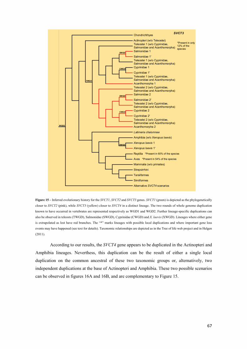
67
SVCT3
Mammalia (w/o primates)
Tarsiiformes
Strepsirrhini
Aves
Reptilia
Xenopus laevis 1'
Xenopus laevis 1
Amphibia (w/o Xenopus laevis)
Latimeria chalumnae
XWGD
*Present in 54% of the species
Cyprinidae 2'
Cyprinidae 2
Salmonidae 2'
Salmonidae 2
Teleostei 2 (w/o Cyprinidae, Salmonidae and Acanthomorpha)
Acanthomorpha 2
SWGD
CWGD
Teleostei 2 (w/o Cyprinidae, Salmonidae and Acanthomorpha)
Teleostei 2 (w/o Cyprinidae, Salmonidae and Acanthomorpha)
Cyprinidae 1'
Cyprinidae 1
Salmonidae 1'
Salmonidae 1
Teleostei 1 (w/o Cyprinidae, Salmonidae and Acanthomorpha)
Acanthomorpha 1
SWGD
CWGD
Teleostei 1 (w/o Cyprinidae, Salmonidae and Acanthomorpha)
Teleostei 1 (w/o Cyprinidae, Salmonidae and Acanthomorpha)TWGD
Actinopteri (w/o Teleostei)
Chondrichthyes
Alternative SVCT4 scenarios
WGD2
*Present in 60% of the species
Simiiformes
*Present in only 12% of the species
Figure 15 – Inferred evolutionary history for the SVCT1, SVCT2 and SVCT3 genes. SVCT1 (green) is depicted as the phylogenetically
closer to SVCT2 (pink), while SVCT3 (yellow) closer to SVCT4 in a distinct lineage. The two rounds of whole genome duplication
known to have occurred in vertebrates are represented respectively as WGD1 and WGD2. Further lineage-specific duplications can
also be observed in teleosts (TWGD), Salmonidae (SWGD), Cyprinidae (CWGD) and X. laevis (XWGD). Lineages where either gene
is extrapolated as lost have red branches. The “*” marks lineages with possible local duplications and where important gene loss
events may have happened (see text for details). Taxonomic relationships are depicted as in the Tree of life web project and in Helgen
(2011).
According to our results, the SVCT4 gene appears to be duplicated in the Actinopteri and
Amphibia lineages. Neverthess, this duplication can be the result of either a single local
duplication on the common ancestral of these two taxonomic groups or, alternatively, two
independent duplications at the base of Actinopteri and Amphibia. These two possible scenarios
can be observed in figures 16A and 16B, and are complementary to Figure 15.

68
Figure 16A - Inferred evolutionary history for the SVCT4 and SVCT5 genes. SVCT4 (orange) is depicted as phylogenetically closer
to SVCT5 (blue), and is linked to the SVCT3 gene at the top of the cladogram. The putative ancestral duplication event at the base of
SVCT5
SVCT4
Mammalia (w/o primates)
Tarsiiformes
Strepsirrhini
Aves
Reptilia
Xenopus laevis 1'
Xenopus laevis 1
Amphibia (w/o Xenopus laevis)
Latimeria chalumnae
XWGD
Cyprinidae 2'
Cyprinidae 2
Salmonidae 2'
Salmonidae 2
Teleostei 2 (w/o Cyprinidae, Salmonidae and Acanthomorpha)
Acanthomorpha 2
SWGD
CWGD
Teleostei 2 (w/o Cyprinidae, Salmonidae and Acanthomorpha)
Teleostei 2 (w/o Cyprinidae, Salmonidae and Acanthomorpha)
Cyprinidae 1'
Cyprinidae 1
Salmonidae 1'
Salmonidae 1
Teleostei 1 (w/o Cyprinidae, Salmonidae and Acanthomorpha)
Acanthomorpha 1
SWGD
CWGD
Teleostei 1 (w/o Cyprinidae, Salmonidae and Acanthomorpha)
Teleostei 1 (w/o Cyprinidae, Salmonidae and Acanthomorpha)TWGD
Actinopteri (w/o Teleostei)
Chondrichthyes
Simiiformes
Cyprinidae 3'
Cyprinidae 3
Salmonidae 3'
Salmonidae 3
Teleostei 3 (w/o Cyprinidae, Salmonidae and Acanthomorpha)
Acanthomorpha 3
SWGD
CWGD
Teleostei 3 (w/o Cyprinidae, Salmonidae and Acanthomorpha)
Teleostei 3 (w/o Cyprinidae, Salmonidae and Acanthomorpha)
Actinopteri (w/o Teleostei)
Teleostei 4
Latimeria chalumnae
Amphibia (w/o Xenopus laevis)
Xenopus laevis 2
Xenopus laevis 2'Sarcopteriigy (w/o L. chalumnae and Amphibia)
XWGD
AWGD *Possible local duplication
*Possible local duplication
SVCT3
TWGD

69
the Actinopteri and Amphibia is represented as AWGD. Further lineage-specific duplications can also be observed in teleosts
(TWGD), Salmonidae (SWGD), Cyprinidae (CWGD) and X. laevis (XWGD). Lineages where either gene is extrapolated as lost have
red branches, while dashed branches are representative of lineages where gene loss cannot be inferred with the available data. The “*”
marks lineages with possible local duplications (see text for details). Taxonomic relationships are depicted as in the Tree of life web
project and in Helgen (2011).
SVCT4
SVCT5
Mammalia (w/o primates)
Simiiformes
Tarsiiformes
Strepsirrhini
Aves
Reptilia
Xenopus laevis 2
Amphibia 2 (w/o Xenopus laevis)
Latimeria chalumnae
Xenopus laevis 1
Amphibia 1 (w/o Xenopus laevis)
Xenopus laevis 1'
Xenopus laevis 2'
SVCT4
SVCT6
SVCT4
XWGD
XWGD
AmWGD
Cyprinidae 2'
Cyprinidae 2
Salmonidae 2'
Salmonidae 2
Teleostei 2 (w/o Cyprinidae, Salmonidae and Acanthomorpha)
Acanthomorpha 2
SWGD
CWGD
Teleostei 2 (w/o Cyprinidae, Salmonidae and Acanthomorpha)
Teleostei 2 (w/o Cyprinidae, Salmonidae and Acanthomorpha)
Cyprinidae 1'
Cyprinidae 1
Salmonidae 1'
Salmonidae 1
Teleostei 1 (w/o Cyprinidae, Salmonidae and Acanthomorpha)
Acanthomorpha 1
SWGD
CWGD
Teleostei 1 (w/o Cyprinidae, Salmonidae and Acanthomorpha)
Teleostei 1 (w/o Cyprinidae, Salmonidae and Acanthomorpha)TWGD
Actinopteri 1 (w/o Teleostei)*Possible local duplication
Teleostei 2'
Cyprinidae 1'’
Cyprinidae 1'
Salmonidae 1'’
Salmonidae 1'
Teleostei 1' (w/o Cyprinidae, Salmonidae and Acanthomorpha)
Acanthomorpha 1
SWGD
CWGD
Teleostei 1' (w/o Cyprinidae, Salmonidae and Acanthomorpha)
Teleostei 1 (w/o Cyprinidae, Salmonidae and Acanthomorpha)
TWGD
Actinopteri 1' (w/o Teleostei)
Chondrichthyes
SVCT3
AcWGD
*Possible local duplication
Figure 16B - Inferred evolutionary history for the SVCT4, SVCT5 and SVCT6 genes. The SVCT4 gene (orange) is linked to the SVCT3

70
represented as AcWGD and AmWGD, respectively. Further lineage-specific duplications can also be observed in teleosts (TWGD),
Salmonidae (SWGD), Cyprinidae (CWGD) and X. laevis (XWGD). SVCT5 (blue) is the the prevalent gene from the independently
duplicated SVCT4 gene in Actinopteri, while SVCT6 (green) is the remaining gene from the SVCT4 duplication in the Amphibia.
Lineages where genes are extrapolated as lost have red branches, while dashed branches are representative of lineages where gene
presence or loss cannot be inferred with the available data. The “*” marks lineages with possible local duplications (see text for
details). Taxonomic relationships are depicted as in the Tree of life web project and in Helgen (2011).
Given the intermediate taxonomic position of L. chalumnae, the presence of either one or
two SVCT4 copies in this species could provide support to the hypotheses of Figure 16A or Figure
16B, respectively. However, in the absence of this information, none of the scenarios seems more
likely.
In summary, the SVCT genes are generally ubiquous in all animal taxonomic groups.
Nevertheless, several considerable gene loss events may have happened in specific and rather
large lineages. In protostomians, the SVCTP gene appears to have been lost in the Trichinellida
group, as well as several Platyhelminthes lineages. In deuterostomians, the SVCT1 gene may have
been lost in many Aves lineages ancestrally, such as the SVCT3. Furthermore, the SVCT3 gene
also seems to be lost in many Actinopteri lineages, such as the Acanthomorpha. The other loss
event already reported in the literature (Yamamoto et al. 2010) is also worth mentioning,
regarding the SVCT4 gene in the Simiiformes group. However, the SVCT4 gene also seems
duplicated specifically in the Actinopteri and Amphibia lineages, a rather unexpected result.
Although a possible SVCT molecular evolution history can be extrapolated, what
happened many basal animal lineages is quite difficult to desbribe. The fact that, in humans, the
SVCT1 transporter is actively involved in ascorbic acid absorption through the gastrointestinal
tract and the SVCT2 transporter in direct ascorbic acid uptake from specific tissues such as the
brain (May 2011, Savini et al. 2008), suggests that processes of neofunctionalization may have
been important during SVCT evolution, contributing to gene fixation. Nevertheless, we cannot tell
if the ancestor gene of the SVCT1/SVCT2 and SVCT3/SVCT4 lineages underwent a process of
subfunctionalization after a duplication event in vertebrates, which led the SVCT1/SVCT2 lineage
to acquire the capacity to transport ascorbic acid, or alternatively, if neofunctionalization occurred
after a duplication event. As such, it is also not possible to infer whether protostomians SVCTP is
able to transport vitamin C. Therefore, in the absence of convincing phylogenetic results, we
turned to conserved protein motifs to possibly find answers. By comparison of aligned protein
sequences, we were able to find a mutual eight amino acid conserved protein motif for SVCT1
and SVCT2 (QHYLTCFS) for all the animal species present in the final datasets, with the
exception of a single sequence belonging to Aotus nancymaae (XP_012329492.1). Furthermore,
Kourkoulou et al. (2018) was also able to discover a conserved SSSP amino acid motif for these
transporters. Since the ascorbic acid transport capacity is largely attributed to these proteins, it is
possible that the found protein motifs can be related with this functional characteristic. Following

71
this hypothesis, if the conserved QHYLTCFS pattern was identified in the SVCTP proteins, that
could be indicative of similar substrate specificity for ascorbic acid. Nevertheless, we uncovered
that none of the conserved patterns are found in the SVCTP proteins, but also that both conserved
patterns are unique of these transporters, and thus not found in the SVCT3, SVCT4 and SVCTP
proteins. These results corroborate and add information to the evidences presented by Kourkoulou
et al. (2018), but do not allow to assume that the ancestral transporter protein could in fact be
transporting ascorbic acid.

72
IV. Conclusion Using a molecular evolution approach, we were able to determine that the GULO gene is
present in several lineages within Protostomia, adding support to the hints given by Wheeler et
al. (2015). Furthermore, we were able to describe several GULO loss events within several animal
lineages, and verify that in general, this gene is not duplicated in deuterostomians, protostomians
and non-bilaterians. Furthermore, with the evidence provided by our phylogenetic analysis, we
were able to trace a putative GULO to the ancestral of animal and Fungi taxonomic groups, in
accordance with the hypothesis of Wheeler et al. (2015). Furthermore, we provide evidence that
the conserved amino acid motif in the GULO protein may not be the HWXK described in the
literature, but a more specific HWAK amino acid pattern found in animal GULO and Fungi ALO.
Moreover, we were also able to extrapolate putative ascorbic acid synthesis in a model organism
(D. melanogaster) without an identifiable GULO, given that our results show that the dietary
source and microbiome do not influence the ascorbic acid levels detected in this species. In
addition, to reinforce this idea, and following the reports by Massie et al. (1991), we were able to
determine that after cold acclimation conditions, D. melanogaster is able to compensate loss in
ascorbic acid levels after being allowed to recover for one day at 25ºC. Considering our results,
we propose that either a novel pathway may exist for ascorbic acid synthesis in D. melanogaster,
or alternatively, that a single enzyme may be performing the same function as GULO in this
species. Either way, further studies are needed to uncover candidate genes that can be involved in
ascorbic acid synthesis in GULO-absent species.
The four SVCT genes found in most vertebrates seem to follow two whole genome
duplications from a single ancestral gene. Specifically, the SVCT4 gene appears to have been
duplicated in two specific lineages (Amphibia and Actinopteri), either by two independent local
duplications or a single ancestral duplication that affected the ancestral of these two taxonomic
groups. However, with the available data, it is impossible to infer the most likely scenario. We
were also able to reinforce the evidence of SVCT3 loss in many teleost lineages but also SVCT4
loss in the Simiiformes, where H. sapiens is included. Moreover, the Aves taxonomic group
appears to follow a complex gene loss scenario for the SVCT1 and SVCT3 genes. Although in the
Chordata the duplications observed can be attributed to a single ancestral gene, duplications
observed in Echinodermata, Hemichordata, Urochordata and Cephalochordata indicate that basal
deuterostomians may have been affected by three independent genome duplications or an ancient
duplication event. Within protostomians and non-bilaterians, the SVCT gene appears to be
independently duplicated many times, and the more ancient duplications detected regard the
Anthozoa, Hexapoda and Gamasina taxonomic groups. Nevertheless, notable gene loss appears
to have affected the Nematoda lineages (essencially the Trichinellida) and the Platyhelminthes.

73
Curiously, the Insecta species have always only one SVCT copy. With the molecular evolution
findings regarding the SVCT gene, we could not uncover putative ascorbic acid transport traits in
the ancient transporter. Trying to overcome this issue, we used conserved protein motifs to
establish a correlation between the ascorbic acid transporters SVCT1 and SVCT2 and the
remaining proteins analyzed, but no conclusion that supports ancestral ascorbic acid transport
traits was taken. Further analyses are therefore needed to further describe the SVCTs functional
characteristics, and establish a correlation between the presence of SVCTs in protostomians and
ascorbic acid homeostasis.
74
V. References Aboobucker, S. I. and A. Lorence (2016). "Recent Progress on the Characterization of
Aldonolactone Oxidoreductases." Plant physiology and biochemistry”. PPB / Societe francaise
de physiologie vegetale 98: 171-185.
Alexandrou, M. A., B. A. Swartz, N. J. Matzke and T. H. Oakley (2013). "Genome duplication
and multiple evolutionary origins of complex migratory behavior in Salmonidae." Mol
Phylogenet Evol 69(3): 514-523.
Amato, P. and B. C. Christner (2009). "Energy metabolism response to low-temperature and
frozen conditions in Psychrobacter cryohalolentis." Appl Environ Microbiol 75(3): 711-718.
Ames, B. N., R. Cathcart, E. Schwiers and P. Hochstein (1981). "Uric acid provides an antioxidant
defense in humans against oxidant- and radical-caused aging and cancer: a hypothesis."
Proceedings of the National Academy of Sciences 78(11): 6858.
Arenas, M. (2015). "Trends in substitution models of molecular evolution." Frontiers in Genetics
6: 319.
Aumailley, L., A. Warren, C. Garand, M. J. Dubois, E. R. Paquet, D. G. Le Couteur, A. Marette,
V. C. Cogger and M. Lebel (2016). "Vitamin C modulates the metabolic and cytokine profiles,
alleviates hepatic endoplasmic reticulum stress, and increases the life span of Gulo-/- mice."
Aging (Albany NY) 8(3): 458-483.
Bahadorani, S., P. Bahadorani, J. P. Phillips and A. J. Hilliker (2008). "The Effects of Vitamin
Supplementation on Drosophila Life Span Under Normoxia and Under Oxidative Stress." The
Journals of Gerontology: Series A 63(1): 35-42.
Barbehenn, R. V., S. L. Bumgarner, E. F. Roosen and M. M. Martin (2001). "Antioxidant defenses
in caterpillars: role of the ascorbate-recycling system in the midgut lumen." J Insect Physiol 47(4-
5): 349-357.
Barker, F. K., A. Cibois, P. Schikler, J. Feinstein and J. Cracraft (2004). "Phylogeny and
diversification of the largest avian radiation." Proceedings of the National Academy of Sciences
of the United States of America 101(30): 11040-11045.
Biondi, C., B. Pavan, A. Dalpiaz, S. Medici, L. Lunghi and F. Vesce (2007). "Expression and
characterization of vitamin C transporter in the human trophoblast cell line HTR-8/SVneo: effect
of steroids, flavonoids and NSAIDs." Mol Hum Reprod 13(1): 77-83.
75
Black, W. C. t., J. S. Klompen and J. E. Keirans (1997). "Phylogenetic relationships among tick
subfamilies (Ixodida: Ixodidae: Argasidae) based on the 18S nuclear rDNA gene." Mol
Phylogenet Evol 7(1): 129-144.
Borgese, N., G. Pietrini and S. Gaetani (1987). "Concentration of NADH-cytochrome b5
reductase in erythrocytes of normal and methemoglobinemic individuals measured with a
quantitative radioimmunoblotting assay." Journal of Clinical Investigation 80(5): 1296-1302.
Bremus, C., U. Herrmann, S. Bringer-Meyer and H. Sahm (2006). "The use of microorganisms
in L-ascorbic acid production." J Biotechnol 124(1): 196-205.
Buettner, G. R. and B. A. Jurkiewicz (1996). "Catalytic metals, ascorbate and free radicals:
combinations to avoid." Radiat Res 145(5): 532-541.
Burzle, M., Y. Suzuki, D. Ackermann, H. Miyazaki, N. Maeda, B. Clemencon, R. Burrier and M.
A. Hediger (2013). "The sodium-dependent ascorbic acid transporter family SLC23." Mol
Aspects Med 34(2-3): 436-454.
Cañestro, C., R. Albalat, M. Irimia and J. Garcia-Fernàndez (2013). "Impact of gene gains, losses
and duplication modes on the origin and diversification of vertebrates." Seminars in Cell &
Developmental Biology 24(2): 83-94.
Carlson, K. A., K. Gardner, A. Pashaj, D. J. Carlson, F. Yu, J. D. Eudy, C. Zhang and L. G.
Harshman (2015). "Genome-Wide Gene Expression in relation to Age in Large Laboratory
Cohorts of Drosophila melanogaster." Genetics Research International 2015: 19.
Carr, R. S., M. B. Bally, P. Thomas and J. M. Neff (1983). "Comparison of methods for
determination of ascorbic acid in animal tissues." Anal Chem 55(8): 1229-1232.
Carr, R. S. and J. M. Neff (1980). "Determination of ascorbic acid in tissues of marine animals
by liquid chromatography with electrochemical detection." Anal Chem 52(14): 2428-2430.
Chain, F. J. and B. J. Evans (2006). "Multiple mechanisms promote the retained expression of
gene duplicates in the tetraploid frog Xenopus laevis." PLoS Genet 2(4): e56.
Chambers, K. E., R. McDaniell, J. D. Raincrow, M. Deshmukh, P. F. Stadler and C. H. Chiu
(2009). "Hox cluster duplication in the basal teleost Hiodon alosoides (Osteoglossomorpha)."
Theory Biosci 128(2): 109-120.
Chang, Y. L., M. Rossetti, H. Vlamakis, D. Casero, G. Sunga, N. Harre, S. Miller, R. Humphries,
T. Stappenbeck, K. W. Simpson, R. B. Sartor, G. Wu, J. Lewis, F. Bushman, D. P. B. McGovern,
N. Salzman, J. Borneman, R. Xavier, C. Huttenhower and J. Braun (2018). "A screen of Crohn's

76
disease-associated microbial metabolites identifies ascorbate as a novel metabolic inhibitor of
activated human T cells." Mucosal Immunol.
Cho, Y. S., S. E. Douglas, J. W. Gallant, K. Y. Kim, D. S. Kim and Y. K. Nam (2007). "Isolation
and characterization of cDNA sequences of L-gulono-gamma-lactone oxidase, a key enzyme for
biosynthesis of ascorbic acid, from extant primitive fish groups." Comp Biochem Physiol B
Biochem Mol Biol 147(2): 178-190.
Clark, A. G., A. L. Rohrbaugh, I. Otterness and V. B. Kraus (2002). "The effects of ascorbic acid
on cartilage metabolism in guinea pig articular cartilage explants." Matrix Biol 21(2): 175-184.
Colinet, H. (2011). "Disruption of ATP homeostasis during chronic cold stress and recovery in
the chill susceptible beetle (Alphitobius diaperinus)." Comp Biochem Physiol A Mol Integr
Physiol 160(1): 63-67.
Corpe, C. P., J. H. Lee, O. Kwon, P. Eck, J. Narayanan, K. L. Kirk and M. Levine (2005). "6-
Bromo-6-deoxy-L-ascorbic acid: an ascorbate analog specific for Na+-dependent vitamin C
transporter but not glucose transporter pathways." J Biol Chem 280(7): 5211-5220.
Corpe, C. P., H. Tu, P. Eck, J. Wang, R. Faulhaber-Walter, J. Schnermann, S. Margolis, S.
Padayatty, H. Sun, Y. Wang, R. L. Nussbaum, M. G. Espey and M. Levine (2010). "Vitamin C
transporter Slc23a1 links renal reabsorption, vitamin C tissue accumulation, and perinatal survival
in mice." J Clin Invest 120(4): 1069-1083.
Cui, J., Y. H. Pan, Y. Zhang, G. Jones and S. Zhang (2011). "Progressive pseudogenization:
vitamin C synthesis and its loss in bats." Mol Biol Evol 28(2): 1025-1031.
Dabrowski, K. (1994). "Primitive actimoterigian fishes can synthesize ascorbic acid." Experientia
50(8): 745-748.
Dabrowski, K. and S. Hinterleitner (1989). "Applications of a simultaneous assay of ascorbic
acid, dehydroascorbic acid and ascorbic sulphate in biological materials." Analyst 114(1): 83-87.
Davey, M. W., M. V. Montagu, D. Inzé, M. Sanmartin, A. Kanellis, N. Smirnoff, I. J. J. Benzie,
J. J. Strain, D. Favell and J. Fletcher (2002). "Plant L-ascorbic acid: chemistry, function,
metabolism, bioavailability and effects of processing." Journal of the Science of Food and
Agriculture 80(7): 825-860.
David, L., S. Blum, M. W. Feldman, U. Lavi and J. Hillel (2003). "Recent duplication of the
common carp (Cyprinus carpio L.) genome as revealed by analyses of microsatellite loci." Mol
Biol Evol 20(9): 1425-1434.

77
de Koning, H. and G. Diallinas (2000). "Nucleobase transporters (review)." Mol Membr Biol
17(2): 75-94.
Dehal, P. and J. L. Boore (2005). "Two Rounds of Whole Genome Duplication in the Ancestral
Vertebrate." PLOS Biology 3(10): e314.
Diliberto, E. J., Jr., G. D. Heckman and A. J. Daniels (1983). "Characterization of ascorbic acid
transport by adrenomedullary chromaffin cells. Evidence for Na+-dependent co-transport." J Biol
Chem 258(21): 12886-12894.
Dollo, V. H., S. X. Yi and R. E. Lee, Jr. (2010). "High temperature pulses decrease indirect
chilling injury and elevate ATP levels in the flesh fly, Sarcophaga crassipalpis." Cryobiology
60(3): 351-353.
Domes, K., M. Althammer, R. A. Norton, S. Scheu and M. Maraun (2007). "The phylogenetic
relationship between Astigmata and Oribatida (Acari) as indicated by molecular markers." Exp
Appl Acarol 42(3): 159-171.
Dowling, A. P. G. and B. M. Oconnor (2010). "Phylogenetic relationships within the suborder
Dermanyssina (Acari: Parasitiformes) and a test of dermanyssoid monophyly." International
Journal of Acarology 36(4): 299-312.
Drouin, G., J.-R. Godin and B. Pagé (2011). "The Genetics of Vitamin C Loss in Vertebrates."
Current Genomics 12(5): 371-378.
Du, J., J. J. Cullen and G. R. Buettner (2012). "Ascorbic acid: Chemistry, biology and the
treatment of cancer." Biochimica et biophysica acta 1826(2): 443-457.
Evans, B. J., D. B. Kelley, D. J. Melnick and D. C. Cannatella (2005). "Evolution of RAG-1 in
polyploid clawed frogs." Mol Biol Evol 22(5): 1193-1207.
Fernie, A. R. and T. Tohge (2015). "A cross-kingdom history." eLife 4: e07527.
Ferris, S. D. and G. S. Whitt (1977). "Duplicate gene expression in diploid and tetraploid loaches
(Cypriniformes, Cobitidae)." Biochem Genet 15(11-12): 1097-1112.
Figueroa-Méndez, R. and S. Rivas-Arancibia (2015). "Vitamin C in Health and Disease: Its Role
in the Metabolism of Cells and Redox State in the Brain." Frontiers in Physiology 6: 397.
Fraaije, M. W., R. H. van den Heuvel, W. J. van Berkel and A. Mattevi (1999). "Covalent
flavinylation is essential for efficient redox catalysis in vanillyl-alcohol oxidase." J Biol Chem
274(50): 35514-35520.

78
Frei, B. and S. Lawson (2008). "Vitamin C and cancer revisited." Proc Natl Acad Sci U S A
105(32): 11037-11038.
Gale, C. R., C. N. Martyn and C. Cooper (1996). "Cognitive impairment and mortality in a cohort
of elderly people." Bmj 312(7031): 608-611.
Glasauer, S. M. and S. C. Neuhauss (2014). "Whole-genome duplication in teleost fishes and its
evolutionary consequences." Mol Genet Genomics 289(6): 1045-1060.
Godoy, A., V. Ormazabal, G. Moraga-Cid, F. A. Zuniga, P. Sotomayor, V. Barra, O. Vasquez, V.
Montecinos, L. Mardones, C. Guzman, M. Villagran, L. G. Aguayo, S. A. Onate, A. M. Reyes, J.
G. Carcamo, C. I. Rivas and J. C. Vera (2007). "Mechanistic insights and functional determinants
of the transport cycle of the ascorbic acid transporter SVCT2. Activation by sodium and absolute
dependence on bivalent cations." J Biol Chem 282(1): 615-624.
Guderley, H. (2004). "Metabolic responses to low temperature in fish muscle." Biol Rev Camb
Philos Soc 79(2): 409-427.
Gulia-Nuss, M., A. B. Nuss, J. M. Meyer, D. E. Sonenshine, R. M. Roe, R. M. Waterhouse, D. B.
Sattelle, J. de la Fuente, J. M. Ribeiro, K. Megy, J. Thimmapuram, J. R. Miller, B. P. Walenz, S.
Koren, J. B. Hostetler, M. Thiagarajan, V. S. Joardar, L. I. Hannick, S. Bidwell, M. P. Hammond,
S. Young, Q. Zeng, J. L. Abrudan, F. C. Almeida, N. Ayllón, K. Bhide, B. W. Bissinger, E.
Bonzon-Kulichenko, S. D. Buckingham, D. R. Caffrey, M. J. Caimano, V. Croset, T. Driscoll, D.
Gilbert, J. J. Gillespie, G. I. Giraldo-Calderón, J. M. Grabowski, D. Jiang, S. M. S. Khalil, D.
Kim, K. M. Kocan, J. Koči, R. J. Kuhn, T. J. Kurtti, K. Lees, E. G. Lang, R. C. Kennedy, H.
Kwon, R. Perera, Y. Qi, J. D. Radolf, J. M. Sakamoto, A. Sánchez-Gracia, M. S. Severo, N.
Silverman, L. Šimo, M. Tojo, C. Tornador, J. P. Van Zee, J. Vázquez, F. G. Vieira, M. Villar, A.
R. Wespiser, Y. Yang, J. Zhu, P. Arensburger, P. V. Pietrantonio, S. C. Barker, R. Shao, E. M.
Zdobnov, F. Hauser, C. J. P. Grimmelikhuijzen, Y. Park, J. Rozas, R. Benton, J. H. F. Pedra, D.
R. Nelson, M. F. Unger, J. M. C. Tubio, Z. Tu, H. M. Robertson, M. Shumway, G. Sutton, J. R.
Wortman, D. Lawson, S. K. Wikel, V. M. Nene, C. M. Fraser, F. H. Collins, B. Birren, K. E.
Nelson, E. Caler and C. A. Hill (2016). "Genomic insights into the Ixodes scapularis tick vector
of Lyme disease." Nature Communications 7: 10507.
Guo, B., X. Gan and S. He (2010). "Hox genes of the Japanese eel Anguilla japonica and Hox
cluster evolution in teleosts." J Exp Zool B Mol Dev Evol 314(2): 135-147.
Halliwell, B. (1999). "Vitamin C: poison, prophylactic or panacea?" Trends Biochem Sci 24(7):
255-259.
79
Hansen, S. N., P. Tveden-Nyborg and J. Lykkesfeldt (2014). "Does Vitamin C Deficiency Affect
Cognitive Development and Function?" Nutrients 6(9): 3818-3846.
He, X. B., M. Kim, S. Y. Kim, S. H. Yi, Y. H. Rhee, T. Kim, E. H. Lee, C. H. Park, S. Dixit, F.
E. Harrison and S. H. Lee (2015). "Vitamin C facilitates dopamine neuron differentiation in fetal
midbrain through TET1- and JMJD3-dependent epigenetic control manner." Stem Cells 33(4):
1320-1332.
Helgen, K. M. (2011). "The Mammal Family Tree." Science 334(6055): 458-459.
Henkel, C. V., E. Burgerhout, D. L. de Wijze, R. P. Dirks, Y. Minegishi, H. J. Jansen, H. P.
Spaink, S. Dufour, F. A. Weltzien, K. Tsukamoto and G. E. van den Thillart (2012). "Primitive
duplicate Hox clusters in the European eel's genome." PLoS One 7(2): e32231.
Hoyoux, A., V. Blaise, T. Collins, S. D'Amico, E. Gratia, A. L. Huston, J. C. Marx, G. Sonan, Y.
Zeng, G. Feller and C. Gerday (2004). "Extreme catalysts from low-temperature environments."
J Biosci Bioeng 98(5): 317-330.
Jiang, Z. Y., Y. Zhong, J. Zheng, M. Ali, G. D. Liu and X. L. Zheng (2018). "L-ascorbic acid
metabolism in an ascorbate-rich kiwifruit (Actinidia. Eriantha Benth.) cv. 'White' during
postharvest." Plant Physiol Biochem 124: 20-28.
Johnson, K. R., J. E. Wright, Jr. and B. May (1987). "Linkage relationships reflecting ancestral
tetraploidy in salmonid fish." Genetics 116(4): 579-591.
Kasahara, M. (2013). "Impact of whole-genome duplication on vertebrate development and
evolution." Semin Cell Dev Biol 24(2): 81-82.
Keeling, P. J. and Y. Inagaki (2004). "A class of eukaryotic GTPase with a punctate distribution
suggesting multiple functional replacements of translation elongation factor 1α." Proceedings of
the National Academy of Sciences of the United States of America 101(43): 15380-15385.
Kolawole, A. O., F. M. Olajuyigbe, J. O. Ajele and C. O. Adedire (2014). "Activity of the
Antioxidant Defense System in a Typical Bioinsecticide-and Synthetic Insecticide-treated
Cowpea Storage Beetle Callosobrochus maculatus F. (Coleoptera: Chrysomelidae)." International
Journal of Insect Science 6: IJIS.S19434.
Kourkoulou, A., A. A. Pittis and G. Diallinas (2018). "Evolution of substrate specificity in the
Nucleobase-Ascorbate Transporter (NAT) protein family." Microbial Cell 5(6): 280-292.
Kuo, S. M., M. E. MacLean, K. McCormick and J. X. Wilson (2004). "Gender and sodium-
ascorbate transporter isoforms determine ascorbate concentrations in mice." J Nutr 134(9): 2216-
2221.

80
Lachapelle, M. Y. and G. Drouin (2011). "Inactivation dates of the human and guinea pig vitamin
C genes." Genetica 139(2): 199-207.
LeBlanc, J. G., C. Milani, G. S. de Giori, F. Sesma, D. van Sinderen and M. Ventura (2013).
"Bacteria as vitamin suppliers to their host: a gut microbiota perspective." Curr Opin Biotechnol
24(2): 160-168.
Lee, J. H., C. S. Oh, G. H. Mun, J. H. Kim, Y. H. Chung, Y. I. Hwang, D. H. Shin and W. J. Lee
(2006). "Immunohistochemical localization of sodium-dependent L-ascorbic acid transporter 1
protein in rat kidney." Histochem Cell Biol 126(4): 491-494.
Li, Z., G. P. Tiley, S. R. Galuska, C. R. Reardon, T. I. Kidder, R. J. Rundell and M. S. Barker
(2018). "Multiple large-scale gene and genome duplications during the evolution of hexapods."
Proc Natl Acad Sci U S A 115(18): 4713-4718.
Liana, M. and W. Witalinski (2005). "Sperm structure and phylogeny of Astigmata." J Morphol
265(3): 318-324.
Lindblad, M., P. Tveden-Nyborg and J. Lykkesfeldt (2013). "Regulation of Vitamin C
Homeostasis during Deficiency." Nutrients 5(8): 2860-2879.
Linster, C. L. and E. Van Schaftingen (2007). "Vitamin C. Biosynthesis, recycling and
degradation in mammals." Febs j 274(1): 1-22.
Liu, J., H. Liu and H. Zhang (2018). "Phylogeny and evolutionary radiation of the marine mussels
(Bivalvia: Mytilidae) based on mitochondrial and nuclear genes." Mol Phylogenet Evol 126: 233-
240.
Liu, X.-Y., K. Y. Yang, M.-Q. Wang, J. S.-L. Kwok, X. Zeng, Z. Yang, X.-J. Xiao, C. P.-Y. Lau,
Y. Li, Z.-m. Huang, J.-g. Ba, A. K.-Y. Yim, C.-Y. Ouyang, S.-M. Ngai, T.-F. Chan, E. L.-H.
Leung, L. Liu, Z.-G. Liu and S. K.-W. Tsui (2018). "High-quality assembly of Dermatophagoides
pteronyssinus genome and transcriptome reveals a wide range of novel allergens." Journal of
Allergy and Clinical Immunology 141(6): 2268-2271.e2268.
Loewus, F. A. (1999). "Biosynthesis and metabolism of ascorbic acid in plants and of analogs of
ascorbic acid in fungi." Phytochemistry 52(2): 193-210.
Logan, F. J., M. C. Taylor, S. R. Wilkinson, H. Kaur and J. M. Kelly (2007). "The terminal step
in vitamin C biosynthesis in Trypanosoma cruzi is mediated by a FMN-dependent
galactonolactone oxidase." Biochem J 407(3): 419-426.
López-Fernández, H., P. Duque, S. Henriques, N. Vázquez, F. Fdez-Riverola, C. P. Vieira, M.
Reboiro-Jato and J. Vieira (2018). A Bioinformatics Protocol for Quickly Creating Large-Scale

81
Phylogenetic Trees. Practical Applications of Computational Biology and Bioinformatics, 12th
International Conference, Cham, Springer International Publishing.
Lund, L. A. and G. B. Sherman (1998). "Duplication of the southern white rhinoceros
(Ceratotherium simum simum) luteinizing hormone beta subunit gene." J Mol Endocrinol 21(1):
19-30.
Luo, S., Z. Wang, V. Kansara, D. Pal and A. K. Mitra (2008). "Activity of a sodium-dependent
vitamin C transporter (SVCT) in MDCK-MDR1 cells and mechanism of ascorbate uptake."
International journal of pharmaceutics 358(1-2): 168-176.
Lux-Battistelli, C. and D. Battistelli (2017). "Latent scurvy with tiredness and leg pain in
alcoholics: An underestimated disease three case reports." Medicine 96(47): e8861.
Mackenzie, B., A. C. Illing and M. A. Hediger (2008). "Transport model of the human Na+-
coupled L-ascorbic acid (vitamin C) transporter SVCT1." Am J Physiol Cell Physiol 294(2):
C451-459.
MacMillan, H. A., J. M. Knee, A. B. Dennis, H. Udaka, K. E. Marshall, T. J. S. Merritt and B. J.
Sinclair (2016). "Cold acclimation wholly reorganizes the Drosophila melanogaster transcriptome
and metabolome." Scientific Reports 6: 28999.
Macmillan, H. A., C. M. Williams, J. F. Staples and B. J. Sinclair (2012). "Metabolism and energy
supply below the critical thermal minimum of a chill-susceptible insect." J Exp Biol 215(Pt 8):
1366-1372.
Massie, H. R., M. E. Shumway, S. J. Whitney, S. M. Sternick and V. R. Aiello (1991). "Ascorbic
acid in Drosophila and changes during aging." Exp Gerontol 26(5): 487-494.
May, J. M. (2011). "The SLC23 family of ascorbate transporters: ensuring that you get and keep
your daily dose of vitamin C." Br J Pharmacol 164(7): 1793-1801.
May, J. M. and F. E. Harrison (2013). "Role of Vitamin C in the Function of the Vascular
Endothelium." Antioxidants & Redox Signaling 19(17): 2068-2083.
Montel-Hagen, A., S. Kinet, N. Manel, C. Mongellaz, R. Prohaska, J.-L. Battini, J. Delaunay, M.
Sitbon and N. Taylor (2008). "Erythrocyte Glut1 Triggers Dehydroascorbic Acid Uptake in
Mammals Unable to Synthesize Vitamin C." Cell 132(6): 1039-1048.
Moreau, R. and K. Dabrowski (1998). "Body pool and synthesis of ascorbic acid in adult sea
lamprey (Petromyzon marinus): An agnathan fish with gulonolactone oxidase activity."
Proceedings of the National Academy of Sciences of the United States of America 95(17): 10279-
10282.

82
Moreau, R. and K. Dabrowski (2005). "Biosynthesis of ascorbic acid by extant actinopterygians."
Journal of Fish Biology 57(3): 733-745.
Moreno, K. X., C. E. Harrison, M. E. Merritt, Z. Kovacs, C. R. Malloy and A. D. Sherry (2017).
"Hyperpolarized delta-[1-(13) C] gluconolactone as a probe of the pentose phosphate pathway."
NMR Biomed 30(6).
Moriyama, Y. and K. Koshiba-Takeuchi (2018). "Significance of whole-genome duplications on
the emergence of evolutionary novelties." Briefings in Functional Genomics: ely007-ely007.
Muñoz, A., M. Villagrán, P. Guzmán, C. Solíz, M. Gatica, C. Aylwin, K. Sweet, M. Maldonado,
E. Escobar, A. M. Reyes, J. R. Toledo, O. Sánchez, S. A. Oñate, J. Carlos Vera and C. I. Rivas
(2015). "Cis-regulatory elements involved in species-specific transcriptional regulation of the
SVCT1 gene in rat and human hepatoma cells." Free radical biology & medicine 85: 183-196.
Newell, P. D., J. M. Chaston, Y. Wang, N. J. Winans, D. R. Sannino, A. C. Wong, A. J. Dobson,
J. Kagle and A. E. Douglas (2014). "In vivo function and comparative genomic analyses of the
Drosophila gut microbiota identify candidate symbiosis factors." Front Microbiol 5: 576.
Nishikimi, M., R. Fukuyama, S. Minoshima, N. Shimizu and K. Yagi (1994). "Cloning and
chromosomal mapping of the human nonfunctional gene for L-gulono-gamma-lactone oxidase,
the enzyme for L-ascorbic acid biosynthesis missing in man." J Biol Chem 269(18): 13685-13688.
Nishikimi, M., T. Kawai and K. Yagi (1992). "Guinea pigs possess a highly mutated gene for L-
gulono-gamma-lactone oxidase, the key enzyme for L-ascorbic acid biosynthesis missing in this
species." J Biol Chem 267(30): 21967-21972.
Notredame, C., D. G. Higgins and J. Heringa (2000). "T-Coffee: A novel method for fast and
accurate multiple sequence alignment." J Mol Biol 302(1): 205-217.
Nualart, F., L. Mack, A. García, P. Cisternas, E. R. Bongarzone, M. Heitzer, N. Jara, F. Martínez,
L. Ferrada, F. Espinoza, V. Baeza and K. Salazar (2014). "Vitamin C Transporters, Recycling and
the Bystander Effect in the Nervous System: SVCT2 versus Gluts." Journal of stem cell research
& therapy 4(5): 209.
Odamaki, T., K. Kato, H. Sugahara, N. Hashikura, S. Takahashi, J.-z. Xiao, F. Abe and R. Osawa
(2016). "Age-related changes in gut microbiota composition from newborn to centenarian: a
cross-sectional study." BMC Microbiology 16: 90.
Ohta, Y. and M. Nishikimi (1999). "Random nucleotide substitutions in primate nonfunctional
gene for L-gulono-gamma-lactone oxidase, the missing enzyme in L-ascorbic acid biosynthesis."
Biochim Biophys Acta 1472(1-2): 408-411.

83
Panoff, J. M., D. Corroler, B. Thammavongs and P. Boutibonnes (1997). "Differentiation between
cold shock proteins and cold acclimation proteins in a mesophilic gram-positive bacterium,
Enterococcus faecalis JH2-2." Journal of Bacteriology 179(13): 4451-4454.
Patananan, A. N., L. M. Budenholzer, M. E. Pedraza, E. R. Torres, L. N. Adler and S. G. Clarke
(2015). "The invertebrate Caenorhabditis elegans biosynthesizes ascorbate." Arch Biochem
Biophys 569: 32-44.
Peterson, K. J., J. B. Lyons, K. S. Nowak, C. M. Takacs, M. J. Wargo and M. A. McPeek (2004).
"Estimating metazoan divergence times with a molecular clock." Proceedings of the National
Academy of Sciences of the United States of America 101(17): 6536.
Phadtare, S., J. Alsina and M. Inouye (1999). "Cold-shock response and cold-shock proteins."
Curr Opin Microbiol 2(2): 175-180.
Plazzi, F., A. Ceregato, M. Taviani and M. Passamonti (2011). "A Molecular Phylogeny of
Bivalve Mollusks: Ancient Radiations and Divergences as Revealed by Mitochondrial Genes."
PLOS ONE 6(11): e27147.
Podmore, I. D., H. R. Griffiths, K. E. Herbert, N. Mistry, P. Mistry and J. Lunec (1998). "Vitamin
C exhibits pro-oxidant properties." Nature 392: 559.
Pollet, N. and A. Mazabraud (2006). "Insights from Xenopus genomes." Genome Dyn 2: 138-
153.
Putnam, N. H., T. Butts, D. E. Ferrier, R. F. Furlong, U. Hellsten, T. Kawashima, M. Robinson-
Rechavi, E. Shoguchi, A. Terry, J. K. Yu, E. L. Benito-Gutierrez, I. Dubchak, J. Garcia-
Fernandez, J. J. Gibson-Brown, I. V. Grigoriev, A. C. Horton, P. J. de Jong, J. Jurka, V. V.
Kapitonov, Y. Kohara, Y. Kuroki, E. Lindquist, S. Lucas, K. Osoegawa, L. A. Pennacchio, A. A.
Salamov, Y. Satou, T. Sauka-Spengler, J. Schmutz, I. T. Shin, A. Toyoda, M. Bronner-Fraser, A.
Fujiyama, L. Z. Holland, P. W. Holland, N. Satoh and D. S. Rokhsar (2008). "The amphioxus
genome and the evolution of the chordate karyotype." Nature 453(7198): 1064-1071.
Qiu, S., L. Li, E. J. Weeber and J. M. May (2007). "Ascorbate transport by primary cultured
neurons and its role in neuronal function and protection against excitotoxicity." J Neurosci Res
85(5): 1046-1056.
Rajan, D. P., W. Huang, B. Dutta, L. D. Devoe, F. H. Leibach, V. Ganapathy and P. D. Prasad
(1999). "Human placental sodium-dependent vitamin C transporter (SVCT2): molecular cloning
and transport function." Biochem Biophys Res Commun 262(3): 762-768.

84
Reboiro-Jato, D., M. Reboiro-Jato, F. Fdez-Riverola, C. P. Vieira, N. A. Fonseca and J. Vieira
(2012). "ADOPS--Automatic Detection Of Positively Selected Sites." J Integr Bioinform 9(3):
200.
Ribeiro, C. and B. J. Dickson (2010). "Sex Peptide Receptor and Neuronal TOR/S6K Signaling
Modulate Nutrient Balancing in Drosophila." Current Biology 20(11): 1000-1005.
Ritter, J. K. (2000). "Roles of glucuronidation and UDP-glucuronosyltransferases in xenobiotic
bioactivation reactions." Chem Biol Interact 129(1-2): 171-193.
Ronquist, F., M. Teslenko, P. van der Mark, D. L. Ayres, A. Darling, S. Höhna, B. Larget, L. Liu,
M. A. Suchard and J. P. Huelsenbeck (2012). "MrBayes 3.2: Efficient Bayesian Phylogenetic
Inference and Model Choice Across a Large Model Space." Systematic Biology 61(3): 539-542.
Sannino, D. R., A. J. Dobson, K. Edwards, E. R. Angert and N. Buchon (2018). "The Drosophila
melanogaster Gut Microbiota Provisions Thiamine to Its Host." MBio 9(2).
Sastre, J., F. V. Pallardo, J. Garcia de la Asuncion and J. Vina (2000). "Mitochondria, oxidative
stress and aging." Free Radic Res 32(3): 189-198.
Savini, I., M. V. Catani, R. Arnone, A. Rossi, G. Frega, D. Del Principe and L. Avigliano (2007).
"Translational control of the ascorbic acid transporter SVCT2 in human platelets." Free Radic
Biol Med 42(5): 608-616.
Savini, I., A. Rossi, C. Pierro, L. Avigliano and M. V. Catani (2008). "SVCT1 and SVCT2: key
proteins for vitamin C uptake." Amino Acids 34(3): 347-355.
Sémon, M. and K. H. Wolfe (2008). "Preferential subfunctionalization of slow-evolving genes
after allopolyploidization in Xenopus laevis." Proceedings of the National Academy of Sciences
of the United States of America 105(24): 8333-8338.
Session, A. M., Y. Uno, T. Kwon, J. A. Chapman, A. Toyoda, S. Takahashi, A. Fukui, A.
Hikosaka, A. Suzuki, M. Kondo, S. J. van Heeringen, I. Quigley, S. Heinz, H. Ogino, H. Ochi, U.
Hellsten, J. B. Lyons, O. Simakov, N. Putnam, J. Stites, Y. Kuroki, T. Tanaka, T. Michiue, M.
Watanabe, O. Bogdanovic, R. Lister, G. Georgiou, S. S. Paranjpe, I. van Kruijsbergen, S. Shu, J.
Carlson, T. Kinoshita, Y. Ohta, S. Mawaribuchi, J. Jenkins, J. Grimwood, J. Schmutz, T. Mitros,
S. Mozaffari, Y. Suzuki, Y. Haramoto, T. S. Yamamoto, C. Takagi, R. Heald, K. Miller, C.
Haudenschild, J. Kitzman, T. Nakayama, Y. Izutsu, J. Robert, J. Fortriede, K. Burns, V. Lotay,
K. Karimi, Y. Yasuoka, D. S. Dichmann, M. F. Flajnik, D. W. Houston, J. Shendure, L.
DuPasquier, P. D. Vize, A. M. Zorn, M. Ito, E. Marcotte, J. B. Wallingford, Y. Ito, M. Asashima,
N. Ueno, Y. Matsuda, G. J. C. Veenstra, A. Fujiyama, R. M. Harland, M. Taira and D. S. Rokhsar

85
(2016). "Genome evolution in the allotetraploid frog Xenopus laevis." Nature 538(7625): 336-
343.
Shigeoka, S., Y. Nakano and S. Kitaoka (1979). "The biosynthetic pathway of L-ascorbic acid in
Euglena gracilis." Journal of Nutritional Science and Vitaminology 25(4): 299-307.
Simhadri, R. K., E. M. Fast, R. Guo, M. J. Schultz, N. Vaisman, L. Ortiz, J. Bybee, B. E. Slatko
and H. M. Frydman (2017). "The Gut Commensal Microbiome of Drosophila melanogaster Is
Modified by the Endosymbiont Wolbachia” mSphere 2(5).
Smirnoff, N. (2001). "L-ascorbic acid biosynthesis." Vitam Horm 61: 241-266.
Smirnoff, N. (2018). "Ascorbic acid metabolism and functions: A comparison of plants and
mammals." Free Radical Biology and Medicine.
Sotiriou, S., S. Gispert, J. Cheng, Y. Wang, A. Chen, S. Hoogstraten-Miller, G. F. Miller, O.
Kwon, M. Levine, S. H. Guttentag and R. L. Nussbaum (2002). "Ascorbic-acid transporter
Slc23a1 is essential for vitamin C transport into the brain and for perinatal survival." Nat Med
8(5): 514-517.
Subramanian, V. S., P. Srinivasan, A. J. Wildman, J. S. Marchant and H. M. Said (2017).
"Molecular mechanism(s) involved in differential expression of vitamin C transporters along the
intestinal tract." Am J Physiol Gastrointest Liver Physiol 312(4): G340-g347.
Szarka, A. and T. Lőrincz (2014). "The role of ascorbate in protein folding." Protoplasma 251(3):
489-497.
Tautz, D. and T. Domazet-Lošo (2011). "The evolutionary origin of orphan genes." Nature
Reviews Genetics 12: 692.
Taylor, J. D., S. T. Williams, E. A. Glover and P. Dyal (2007). "A molecular phylogeny of
heterodont bivalves (Mollusca: Bivalvia: Heterodonta): new analyses of 18S and 28S rRNA
genes." Zoologica Scripta 36(6): 587-606.
Taylor, J. S., I. Braasch, T. Frickey, A. Meyer and Y. Van de Peer (2003). "Genome Duplication,
a Trait Shared by 22,000 Species of Ray-Finned Fish." Genome Research 13(3): 382-390.
Taylor, J. S., Y. Van de Peer, I. Braasch and A. Meyer (2001). "Comparative genomics provides
evidence for an ancient genome duplication event in fish." Philos Trans R Soc Lond B Biol Sci
356(1414): 1661-1679.
Teeling, E. C., M. S. Springer, O. Madsen, P. Bates, J. O'Brien S and W. J. Murphy (2005). "A
molecular phylogeny for bats illuminates biogeography and the fossil record." Science 307(5709):
580-584.

86
Thomas, W. R. (2010). "Geography of house dust mite allergens." Asian Pac J Allergy Immunol
28(4): 211-224.
Traikovich, S. S. (1999). "Use of topical ascorbic acid and its effects on photodamaged skin
topography." Archives of Otolaryngology–Head & Neck Surgery 125(10): 1091-1098.
Tsukaguchi, H., T. Tokui, B. Mackenzie, U. V. Berger, X. Z. Chen, Y. Wang, R. F. Brubaker and
M. A. Hediger (1999). "A family of mammalian Na+-dependent L-ascorbic acid transporters."
Nature 399(6731): 70-75.
Tveden-Nyborg, P., L. K. Johansen, Z. Raida, C. K. Villumsen, J. O. Larsen and J. Lykkesfeldt
(2009). "Vitamin C deficiency in early postnatal life impairs spatial memory and reduces the
number of hippocampal neurons in guinea pigs." Am J Clin Nutr 90(3): 540-546.
Tveden-Nyborg, P. and J. Lykkesfeldt (2009). "Does vitamin C deficiency result in impaired brain
development in infants?" Redox Rep 14(1): 2-6.
Uchizono, S., Y. Tabuki, N. Kawaguchi, T. Tanimura and T. Q. Itoh (2017). "Mated Drosophila
melanogaster females consume more amino acids during the dark phase." PLOS ONE 12(2):
e0172886.
Uyeno, T. and G. R. Smith (1972). "Tetraploid origin of the karyotype of catostomid fishes."
Science 175(4022): 644-646.
Vandepoele, K., W. De Vos, J. S. Taylor, A. Meyer and Y. Van de Peer (2004). "Major events in
the genome evolution of vertebrates: paranome age and size differ considerably between ray-
finned fishes and land vertebrates." Proc Natl Acad Sci U S A 101(6): 1638-1643.
Vera, J. C., C. I. Rivas, F. V. Velasquez, R. H. Zhang, Concha, II and D. W. Golde (1995).
"Resolution of the facilitated transport of dehydroascorbic acid from its intracellular accumulation
as ascorbic acid." J Biol Chem 270(40): 23706-23712.
Wägele, J. W. and C. Mayer (2007). "Visualizing differences in phylogenetic information content
of alignments and distinction of three classes of long-branch effects." BMC Evolutionary Biology
7: 147-147.
Wang, J.-T., J.-T. Li, X.-F. Zhang and X.-W. Sun (2012). "Transcriptome analysis reveals the
time of the fourth round of genome duplication in common carp (Cyprinus carpio)." BMC
Genomics 13(1): 96.
Wang, Y., B. Mackenzie, H. Tsukaguchi, S. Weremowicz, C. C. Morton and M. A. Hediger
(2000). "Human vitamin C (L-ascorbic acid) transporter SVCT1." Biochem Biophys Res
Commun 267(2): 488-494.

87
Welch, R. W., P. Bergsten, J. D. Butler and M. Levine (1993). "Ascorbic acid accumulation and
transport in human fibroblasts." Biochem J 294 ( Pt 2): 505-510.
Welch, R. W., Y. Wang, A. Crossman, Jr., J. B. Park, K. L. Kirk and M. Levine (1995).
"Accumulation of vitamin C (ascorbate) and its oxidized metabolite dehydroascorbic acid occurs
by separate mechanisms." J Biol Chem 270(21): 12584-12592.
Wells, W. W., D. P. Xu, Y. F. Yang and P. A. Rocque (1990). "Mammalian thioltransferase
(glutaredoxin) and protein disulfide isomerase have dehydroascorbate reductase activity." J Biol
Chem 265(26): 15361-15364.
Wheeler, G., T. Ishikawa, V. Pornsaksit and N. Smirnoff (2015). "Evolution of alternative
biosynthetic pathways for vitamin C following plastid acquisition in photosynthetic eukaryotes."
eLife 4: e06369.
Wheeler, G. L., M. A. Jones and N. Smirnoff (1998). "The biosynthetic pathway of vitamin C in
higher plants." Nature 393: 365.
Wilson, J. X. (2005). "Regulation of vitamin C transport." Annu Rev Nutr 25: 105-125.
Winkler, B. S., S. M. Orselli and T. S. Rex (1994). "The redox couple between glutathione and
ascorbic acid: a chemical and physiological perspective." Free Radic Biol Med 17(4): 333-349.
Wolf, Y. I., P. S. Novichkov, G. P. Karev, E. V. Koonin and D. J. Lipman (2009). "The universal
distribution of evolutionary rates of genes and distinct characteristics of eukaryotic genes of
different apparent ages." Proceedings of the National Academy of Sciences of the United States
of America 106(18): 7273-7280.
Wolfe, K. H. (2001). "Yesterday's polyploids and the mystery of diploidization." Nat Rev Genet
2(5): 333-341.
Wolucka, B. A. and D. Communi (2006). "Mycobacterium tuberculosis possesses a functional
enzyme for the synthesis of vitamin C, L-gulono-1,4-lactone dehydrogenase." Febs j 273(19):
4435-4445.
Yamamoto, S., K. Inoue, T. Murata, S. Kamigaso, T. Yasujima, J. Y. Maeda, Y. Yoshida, K. Y.
Ohta and H. Yuasa (2010). "Identification and functional characterization of the first nucleobase
transporter in mammals: implication in the species difference in the intestinal absorption
mechanism of nucleobases and their analogs between higher primates and other mammals." J Biol
Chem 285(9): 6522-6531.
Yang, H. (2013). "Conserved or Lost: Molecular Evolution of the Key Gene GULO in Vertebrate
Vitamin C Biosynthesis." Biochemical Genetics 51(5): 413-425.

88
Zapata, F., N. G. Wilson, M. Howison, S. C. Andrade, K. M. Jorger, M. Schrodl, F. E. Goetz, G.
Giribet and C. W. Dunn (2014). "Phylogenomic analyses of deep gastropod relationships reject
Orthogastropoda." Proc Biol Sci 281(1794): 20141739.
Zhang, P. Y., X. Xu and X. C. Li (2014). "Cardiovascular diseases: oxidative damage and
antioxidant protection." Eur Rev Med Pharmacol Sci 18(20): 3091-3096.
Zhang, X., Y. Zhang, X. Zheng, Y. Kuang, Z. Zhao, L. Zhao, C. Li, L. Jiang, D. Cao, C. Lu, P.
Xu and X. Sun (2013). "A consensus linkage map provides insights on genome character and
evolution in common carp (Cyprinus carpio L.)." Mar Biotechnol (NY) 15(3): 275-312.

89
VI. Supplementary Data Acanthaster planci crown-of-thorns starfish Acanthasteridae XP_022096513.1
Acanthaster planci crown-of-thorns starfish Acanthasteridae XP_022110602.1
Strongylocentrotus purpuratus purple sea urchin Strongylocentrotidae XP_011663397.1
Acinonyx jubatus cheetah Felidae XP_014925651.1
Felis catus domestic cat Felidae XP_019684152.1
Panthera pardus leopard Felidae XP_019296394.1
Panthera tigris altaica Amur tiger Felidae XP_015399705.1
Ailuropoda melanoleuca giant panda Ursidae XP_011216970.2
Enhydra lutris kenyoni sea otter Mustelidae XP_022372216.1
Mustela putorius furo domestic ferret Mustelidae XP_004775164.1
Odobenus rosmarus divergens Pacific walrus Odobenidae XP_004411725.1
Canis lupus familiaris dog Canidae XP_005635730.1
Balaenoptera acutorostrata scammoni minke whale Balaenopteridae XP_007192798.1
Delphinapterus leucas beluga whale Monodontidae XP_022450701.1
Orcinus orca killer whale Delphinidae XP_004270744.1
Lipotes vexillifer Yangtze River dolphin Lipotidae XP_007466224.1
Physeter catodon sperm whale Physeteridae XP_007127934.1
Bison bison bison American bison Bovidae XP_010838440.1
Bos mutus wild yak Bovidae XP_005903071.1
Bos taurus cattle Bovidae DAA26668.1
Bubalus bubalis water buffalo Bovidae XP_006057367.1
Capra hircus goat Bovidae XP_005684101.1
Ovis aries sheep Bovidae XP_004004204.1
Ovis aries musimon mouflon Bovidae XP_011997423.1
Pantholops hodgsonii chiru Bovidae XP_005967668.1
Odocoileus virginianus texanus white-tailed deer Cervidae XP_020754301.1
Sus scrofa pig Suidae NP_001123420.1
Camelus bactrianus Bactrian camel Camelidae XP_010946865.1
Camelus ferus Wild Bactrian camel Camelidae XP_006194067.1
Camelus dromedarius Arabian camel Camelidae XP_010993808.1
Vicugna pacos alpaca Camelidae XP_006203265.1
Ceratotherium simum simum southern white rhinoceros Rhinocerotidae XP_014645409.1
Equus asinus ass Equidae XP_014698791.1
Equus caballus horse Equidae XP_005607755.1
Equus przewalskii Przewalskis horse Equidae XP_008517520.1
Manis javanica Malayan pangolin Manidae XP_017511311.1
Rhinolophus sinicus Chinese rufous horseshoe bat Rhinolophidae XP_019588647.1
Rousettus aegyptiacus Egyptian rousette Pteropodidae XP_016000110.1
Condylura cristata star-nosed mole Talpidae XP_004682583.1
Erinaceus europaeus western European hedgehog Erinaceidae XP_016041119.1
Sorex araneus European shrew Soricidae XP_004614792.1
Microcebus murinus gray mouse lemur Cheirogaleidae XP_012630826.1
Propithecus coquereli Coquerels sifaka Indriidae XP_012518430.1
Otolemur garnettii small-eared galago Galagidae XP_012663175.1
100
85
94
100
100
79
61
100
100
10099
92
100
5390
50
100
61
100
100
100
98
99
100
100
100
70
99100
99
97
63
92
92
100
100
60
86Acanthasteridae
Strongylocentrotidae
Felidae
Ursidae
Mustelidae
Odobenidae
Canidae
Balaenopteridae
Monodontidae
Delphinidae
Lipotidae
Physeteridae
Bovidae
Cervidae
Suidae
Camelidae
Rhinocerotidae
Equidae
Manidae
Rhinolophidae
Pteropodidae
Talpidae
Erinaceidae
Soricidae
Cheirogaleidae
Indriidae
Galagidae
L-gulonolactone oxidase
Continues next page

90
Cricetulus griseus Chinese hamster Cricetidae XP_003505213.1
Mesocricetus auratus golden hamster Cricetidae XP_012972737.1
Microtus ochrogaster prairie vole Cricetidae XP_005355607.1
Peromyscus maniculatus bairdii prairie deer mouse Cricetidae XP_006988833.1
Mus caroli Ryukyu mouse Muridae XP_021036878.1
Mus musculus house mouse Muridae NP_848862.1
Mus pahari shrew mouse Muridae XP_021059328.1
Rattus norvegicus Norway rat Muridae EDL85374.1
Meriones unguiculatus Mongolian gerbil Muridae XP_021510212.1
Nannospalax galili Upper Galilee mountains blind mole rat Spalacidae XP_017652394.1
Jaculus jaculus lesser Egyptian jerboa Dipodidae XP_012805930.1
Ictidomys tridecemlineatus thirteen-lined ground squirrel Sciuridae XP_021590207.1
Marmota marmota marmota Alpine marmot Sciuridae XP_015354934.1
Dipodomys ordii Ords kangaroo rat Heteromyidae XP_012879644.1
Fukomys damarensis Damara mole-rat Bathyergidae XP_010621731.1
Heterocephalus glaber naked mole-rat Bathyergidae XP_004848449.1
Octodon degus degu Octodontidae XP_012370088.1
Ochotona princeps American pika Ochotonidae XP_012786868.1
Oryctolagus cuniculus rabbit Leporidae XP_008247496.1
Tupaia chinensis Chinese tree shrew Tupaiidae XP_006153286.1
Dasypus novemcinctus nine-banded armadillo Dasypodidae XP_004454215.1
Echinops telfairi small Madagascar hedgehog Tenrecidae XP_012861353.1
Loxodonta africana African savanna elephant Elephantidae XP_003412410.1
Trichechus manatus latirostris Florida manatee Trichechidae XP_012412689.1
Orycteropus afer afer aardvark Orycteropodidae XP_007936968.1
Monodelphis domestica gray short-tailed opossum Didelphidae XP_007475977.1
Phascolarctos cinereus koala Phascolarctidae XP_020839639.1
Sarcophilus harrisii Tasmanian devil Dasyuridae XP_012396172.1
Alligator mississippiensis American alligator Alligatoridae KYO43973.1
Alligator sinensis Chinese alligator Alligatoridae XP_014382044.1
Crocodylus porosus Australian saltwater crocodile Crocodylidae XP_019392806.1
Gavialis gangeticus Gharial Gavialidae XP_019376412.1
Chrysemys picta bellii western painted turtle Emydidae XP_008172130.1
Gekko japonicus lizards Gekkonidae XP_015266653.1
Ophiophagus hannah king cobra Elapidae ETE68825.1
Python bivittatus Burmese python Pythonidae XP_007438840.1
Anas platyrhynchos mallard Anatidae XP_021135031.1
Anser cygnoides domesticus swan goose Anatidae XP_013057319.1
Aptenodytes forsteri emperor penguin Spheniscidae XP_009273398.1
Pygoscelis adeliae Adelie penguin Spheniscidae XP_009323258.1
Aquila chrysaetos canadensis golden eagle Accipitridae XP_011589194.1
Haliaeetus leucocephalus bald eagle Accipitridae XP_010581514.1
Calypte anna Annas hummingbird Trochilidae XP_008503861.1
Chaetura pelagica chimney swift Apodidae XP_010000521.1
Cuculus canorus common cuckoo Cuculidae XP_009562090.1
Melopsittacus undulatus budgerigar Psittaculidae XP_005146526.1
Nestor notabilis Kea Psittacidae XP_010012223.1
Picoides pubescens Downy woodpecker Picidae XP_009906689.1
Colius striatus speckled mousebird Coliidae XP_010206806.1
Falco cherrug Saker falcon Falconidae XP_005443605.1
Fulmarus glacialis Northern fulmar Procellariidae XP_009571134.1
Egretta garzetta little egret Ardeidae XP_009643567.1
Columba livia rock pigeon Columbidae XP_021135540.1
Calidris pugnax ruff Scolopacidae XP_014818284.1
Coturnix japonica Japanese quail Phasianidae XP_015715329.1
Gallus gallus chicken Phasianidae XP_015140704.1
Numida meleagris helmeted guineafowl Numididae XP_021247600.1
Corvus brachyrhynchos American crow Corvidae XP_017597802.1
Pseudopodoces humilis Tibetan ground-tit Paridae XP_005534261.1
10096
100
100100
100
94
100
99
100
100
98
100
100
100
100
97
94
58
85
99
98
100
57
97
89100
97
99
97
100
100100
88
100
100
100
100
100
100786560
54
96
50
100
59
100
100
100
100
100
98
55
97
97
97
97
68
81
Cricetidae
Muridae
Spalacidae
Dipodidae
Sciuridae
Heteromyidae
Bathyergidae
Octodontidae
Ochotonidae
Leporidae
Tupaiidae
Dasypodidae
Tenrecidae
Elephantidae
Trichechidae
Orycteropodidae
Didelphidae
Phascolarctidae
Dasyuridae
Alligatoridae
Crocodylidae
Gavialidae
Emydidae
Gekkonidae
Elapidae
Pythonidae
Anatidae
Spheniscidae
Accipitridae
Trochilidae
Apodidae
Cuculidae
Psittaculidae
Psittacidae
Picidae
Coliidae
Falconidae
Procellariidae
Ardeidae
Columbidae
Scolopacidae
Phasianidae
Numididae
Corvidae
ParidaeContinues next
page
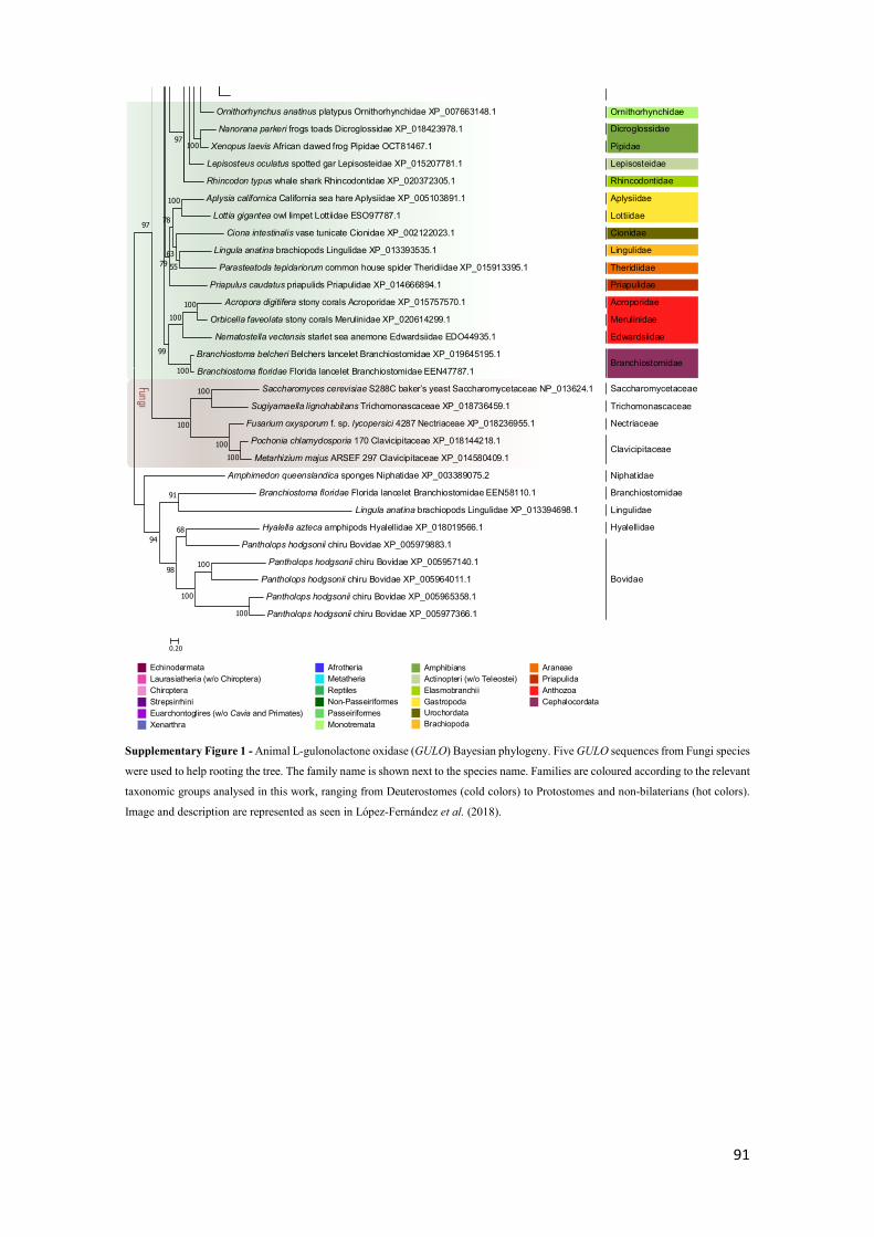
91
Ornithorhynchus anatinus platypus Ornithorhynchidae XP_007663148.1
Nanorana parkeri frogs toads Dicroglossidae XP_018423978.1
Xenopus laevis African clawed frog Pipidae OCT81467.1
Lepisosteus oculatus spotted gar Lepisosteidae XP_015207781.1
Rhincodon typus whale shark Rhincodontidae XP_020372305.1
Aplysia californica California sea hare Aplysiidae XP_005103891.1
Lottia gigantea owl limpet Lottiidae ESO97787.1
Ciona intestinalis vase tunicate Cionidae XP_002122023.1
Lingula anatina brachiopods Lingulidae XP_013393535.1
Parasteatoda tepidariorum common house spider Theridiidae XP_015913395.1
Priapulus caudatus priapulids Priapulidae XP_014666894.1
Acropora digitifera stony corals Acroporidae XP_015757570.1
Orbicella faveolata stony corals Merulinidae XP_020614299.1
Nematostella vectensis starlet sea anemone Edwardsiidae EDO44935.1
Branchiostoma belcheri Belchers lancelet Branchiostomidae XP_019645195.1
Branchiostoma floridae Florida lancelet Branchiostomidae EEN47787.1
Saccharomyces cerevisiae S288C baker’s yeast Saccharomycetaceae NP_013624.1
Sugiyamaella lignohabitans Trichomonascaceae XP_018736459.1
Fusarium oxysporum f. sp. lycopersici 4287 Nectriaceae XP_018236955.1
Pochonia chlamydosporia 170 Clavicipitaceae XP_018144218.1
Metarhizium majus ARSEF 297 Clavicipitaceae XP_014580409.1
Amphimedon queenslandica sponges Niphatidae XP_003389075.2
Branchiostoma floridae Florida lancelet Branchiostomidae EEN58110.1
Lingula anatina brachiopods Lingulidae XP_013394698.1
Hyalella azteca amphipods Hyalellidae XP_018019566.1
Pantholops hodgsonii chiru Bovidae XP_005979883.1
Pantholops hodgsonii chiru Bovidae XP_005957140.1
Pantholops hodgsonii chiru Bovidae XP_005964011.1
Pantholops hodgsonii chiru Bovidae XP_005965358.1
Pantholops hodgsonii chiru Bovidae XP_005977366.1
10097
100100
100
99
91
68
100
100
100
98
94
100
100100
100
97
100
5563
78
79
0.20
Ornithorhynchidae
Dicroglossidae
Pipidae
Lepisosteidae
Rhincodontidae
Aplysiidae
Lottiidae
Cionidae
Lingulidae
Theridiidae
Priapulidae
Acroporidae
Merulinidae
Edwardsiidae
Branchiostomidae
Saccharomycetaceae
Trichomonascaceae
Nectriaceae
Clavicipitaceae
Niphatidae
Branchiostomidae
Lingulidae
Hyalellidae
Bovidae
EchinodermataLaurasiatheria (w/o Chiroptera)ChiropteraStrepsirrhiniEuarchontoglires (w/o Cavia and Primates)Xenarthra
AfrotheriaMetatheriaReptilesNon-PasseiriformesPasseiriformesMonotremata
AmphibiansActinopteri (w/o Teleostei)ElasmobranchiiGastropoda UrochordataBrachiopoda
AraneaePriapulidaAnthozoaCephalocordata
Fungi
Supplementary Figure 1 - Animal L-gulonolactone oxidase (GULO) Bayesian phylogeny. Five GULO sequences from Fungi species
were used to help rooting the tree. The family name is shown next to the species name. Families are coloured according to the relevant
taxonomic groups analysed in this work, ranging from Deuterostomes (cold colors) to Protostomes and non-bilaterians (hot colors).
Image and description are represented as seen in López-Fernández et al. (2018).
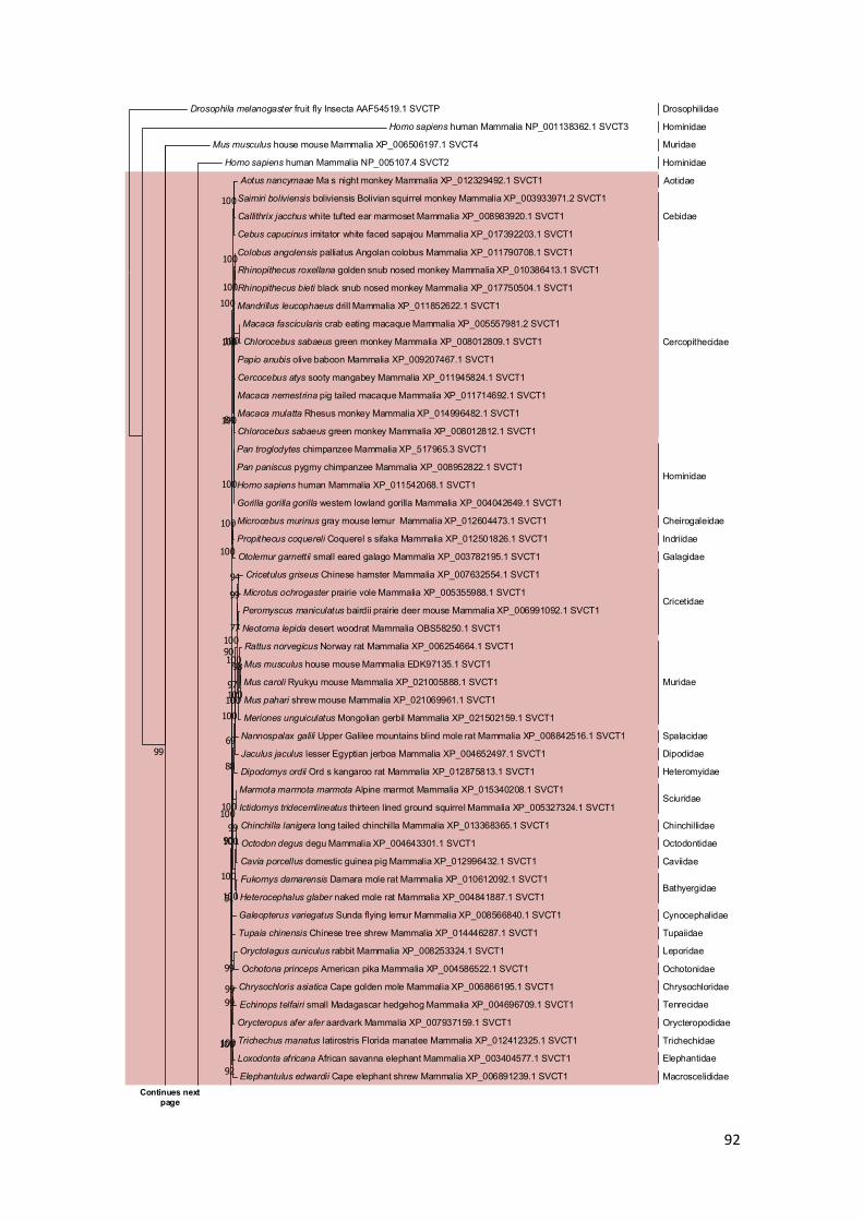
92
Drosophila melanogaster fruit fly Insecta AAF54519.1 SVCTP
Homo sapiens human Mammalia NP_001138362.1 SVCT3
Mus musculus house mouse Mammalia XP_006506197.1 SVCT4
Homo sapiens human Mammalia NP_005107.4 SVCT2
Aotus nancymaae Ma s night monkey Mammalia XP_012329492.1 SVCT1
Saimiri boliviensis boliviensis Bolivian squirrel monkey Mammalia XP_003933971.2 SVCT1
Callithrix jacchus white tufted ear marmoset Mammalia XP_008983920.1 SVCT1
Cebus capucinus imitator white faced sapajou Mammalia XP_017392203.1 SVCT1
Colobus angolensis palliatus Angolan colobus Mammalia XP_011790708.1 SVCT1
Rhinopithecus roxellana golden snub nosed monkey Mammalia XP_010386413.1 SVCT1
Rhinopithecus bieti black snub nosed monkey Mammalia XP_017750504.1 SVCT1
Mandrillus leucophaeus drill Mammalia XP_011852622.1 SVCT1
Macaca fascicularis crab eating macaque Mammalia XP_005557981.2 SVCT1
Chlorocebus sabaeus green monkey Mammalia XP_008012809.1 SVCT1
Papio anubis olive baboon Mammalia XP_009207467.1 SVCT1
Cercocebus atys sooty mangabey Mammalia XP_011945824.1 SVCT1
Macaca nemestrina pig tailed macaque Mammalia XP_011714692.1 SVCT1
Macaca mulatta Rhesus monkey Mammalia XP_014996482.1 SVCT1
Chlorocebus sabaeus green monkey Mammalia XP_008012812.1 SVCT1
Pan troglodytes chimpanzee Mammalia XP_517965.3 SVCT1
Pan paniscus pygmy chimpanzee Mammalia XP_008952822.1 SVCT1
Homo sapiens human Mammalia XP_011542068.1 SVCT1
Gorilla gorilla gorilla western lowland gorilla Mammalia XP_004042649.1 SVCT1
Microcebus murinus gray mouse lemur Mammalia XP_012604473.1 SVCT1
Propithecus coquereli Coquerel s sifaka Mammalia XP_012501826.1 SVCT1
Otolemur garnettii small eared galago Mammalia XP_003782195.1 SVCT1
Cricetulus griseus Chinese hamster Mammalia XP_007632554.1 SVCT1
Microtus ochrogaster prairie vole Mammalia XP_005355988.1 SVCT1
Peromyscus maniculatus bairdii prairie deer mouse Mammalia XP_006991092.1 SVCT1
Neotoma lepida desert woodrat Mammalia OBS58250.1 SVCT1
Rattus norvegicus Norway rat Mammalia XP_006254664.1 SVCT1
Mus musculus house mouse Mammalia EDK97135.1 SVCT1
Mus caroli Ryukyu mouse Mammalia XP_021005888.1 SVCT1
Mus pahari shrew mouse Mammalia XP_021069961.1 SVCT1
Meriones unguiculatus Mongolian gerbil Mammalia XP_021502159.1 SVCT1
Nannospalax galili Upper Galilee mountains blind mole rat Mammalia XP_008842516.1 SVCT1
Jaculus jaculus lesser Egyptian jerboa Mammalia XP_004652497.1 SVCT1
Dipodomys ordii Ord s kangaroo rat Mammalia XP_012875813.1 SVCT1
Marmota marmota marmota Alpine marmot Mammalia XP_015340208.1 SVCT1
Ictidomys tridecemlineatus thirteen lined ground squirrel Mammalia XP_005327324.1 SVCT1
Chinchilla lanigera long tailed chinchilla Mammalia XP_013368365.1 SVCT1
Octodon degus degu Mammalia XP_004643301.1 SVCT1
Cavia porcellus domestic guinea pig Mammalia XP_012996432.1 SVCT1
Fukomys damarensis Damara mole rat Mammalia XP_010612092.1 SVCT1
Heterocephalus glaber naked mole rat Mammalia XP_004841887.1 SVCT1
Galeopterus variegatus Sunda flying lemur Mammalia XP_008566840.1 SVCT1
Tupaia chinensis Chinese tree shrew Mammalia XP_014446287.1 SVCT1
Oryctolagus cuniculus rabbit Mammalia XP_008253324.1 SVCT1
Ochotona princeps American pika Mammalia XP_004586522.1 SVCT1
Chrysochloris asiatica Cape golden mole Mammalia XP_006866195.1 SVCT1
Echinops telfairi small Madagascar hedgehog Mammalia XP_004696709.1 SVCT1
Orycteropus afer afer aardvark Mammalia XP_007937159.1 SVCT1
Trichechus manatus latirostris Florida manatee Mammalia XP_012412325.1 SVCT1
Loxodonta africana African savanna elephant Mammalia XP_003404577.1 SVCT1
Elephantulus edwardii Cape elephant shrew Mammalia XP_006891239.1 SVCT1
99
100
100
100
100100
100
100
100
100
100
84
94
77
99
98
100
100
100
100
97
100
69
100
88
99100
100
100
100
99
51
90
9999
100
92
100
90
Continues next page
Drosophilidae
Hominidae
Muridae
Hominidae
Aotidae
Cebidae
Cercopithecidae
Hominidae
Cheirogaleidae
Indriidae
Galagidae
Cricetidae
Muridae
Spalacidae
Dipodidae
Heteromyidae
Sciuridae
Chinchillidae
Octodontidae
Caviidae
Bathyergidae
Cynocephalidae
Tupaiidae
Leporidae
Ochotonidae
Chrysochloridae
Tenrecidae
Orycteropodidae
Trichechidae
Elephantidae
Macroscelididae

93
Ovis aries musimon mouflon Mammalia XP_012006065.1 SVCT1
Capra hircus goat Mammalia XP_013821052.1 SVCT1
Pantholops hodgsonii chiru Mammalia XP_005971894.1 SVCT1
Bubalus bubalis water buffalo Mammalia XP_006070880.1 SVCT1
Bos indicus zebu cattle Mammalia XP_019820579.1 SVCT1
Bos taurus cattle Mammalia XP_010805674.1 SVCT1
Bison bison bison American bison Mammalia XP_010848620.1 SVCT1
Bos mutus wild yak Mammalia XP_005900233.1 SVCT1
Odocoileus virginianus texanus white tailed deer Mammalia XP_020764076.1 SVCT1
Cervus elaphus hippelaphus red deer Mammalia OWK12011.1 SVCT1
Delphinapterus leucas beluga whale Mammalia XP_022444629.1 SVCT1
Orcinus orca killer whale Mammalia XP_012392355.1 SVCT1
Lipotes vexillifer Yangtze River dolphin Mammalia XP_007461498.1 SVCT1
Physeter catodon sperm whale Mammalia XP_007114756.1 SVCT1
Camelus bactrianus Bactrian camel Mammalia XP_010970008.1 SVCT1
Vicugna pacos alpaca Mammalia XP_015094998.1 SVCT1
Sus scrofa pig Mammalia XP_020940529.1 SVCT1
Pteropus alecto black flying fox Mammalia ELK03036.1 SVCT1
Rousettus aegyptiacus Egyptian rousette Mammalia XP_015992369.1 SVCT1
Rhinolophus sinicus Chinese rufous horseshoe bat Mammalia XP_019595871.1 SVCT1
Hipposideros armiger great roundleaf bat Mammalia XP_019517698.1 SVCT1
Myotis davidii bats Mammalia XP_006772508.1 SVCT1
Myotis brandtii Brandt s bat Mammalia XP_005881093.1 SVCT1
Myotis lucifugus little brown bat Mammalia XP_014309332.1 SVCT1
Eptesicus fuscus big brown bat Mammalia XP_008139883.1 SVCT1
Miniopterus natalensis bats Mammalia XP_016059226.1 SVCT1
Odobenus rosmarus divergens Pacific walrus Mammalia XP_012416958.1 SVCT1
Neomonachus schauinslandi Hawaiian monk seal Mammalia XP_021557863.1 SVCT1
Leptonychotes weddellii Weddell seal Mammalia XP_006730772.1 SVCT1
Ailuropoda melanoleuca giant panda Mammalia XP_019651402.1 SVCT1
Mustela putorius furo domestic ferret Mammalia XP_012907894.1 SVCT1
Enhydra lutris kenyoni sea otter Mammalia XP_022348931.1 SVCT1
Canis lupus familiaris dog Mammalia XP_005617293.2 SVCT1
Felis catus domestic cat Mammalia XP_019688431.1 SVCT1
Acinonyx jubatus cheetah Mammalia XP_014920331.1 SVCT1
Panthera pardus leopard Mammalia XP_019279879.1 SVCT1
Panthera tigris altaica Amur tiger Mammalia XP_015391068.1 SVCT1
Manis javanica Malayan pangolin Mammalia XP_017528579.1 SVCT1
Equus przewalskii Przewalski s horse Mammalia XP_008513761.1 SVCT1
Equus caballus horse Mammalia XP_014586127.1 SVCT1
Equus asinus ass Mammalia XP_014709330.1 SVCT1
Ceratotherium simum simum southern white rhinoceros Mammalia XP_014651016.1 SVCT1
Sorex araneus European shrew Mammalia XP_004609971.1 SVCT1
Erinaceus europaeus western European hedgehog Mammalia XP_007517601.1 SVCT1
Condylura cristata star nosed mole Mammalia XP_004686844.1 SVCT1
Sarcophilus harrisii Tasmanian devil Mammalia XP_003756669.2 SVCT1
Phascolarctos cinereus koala Mammalia XP_020860697.1 SVCT1
Monodelphis domestica gray short tailed opossum Mammalia XP_016278848.1 SVCT1
Chelonia mydas green sea turtle EMP36361.1 SVCT1
Chrysemys picta bellii western painted turtle XP_005280952.1 SVCT1
Alligator mississippiensis American alligator KYO39214.1 SVCT1
Alligator sinensis Chinese alligator XP_014376327.1 SVCT1
Crocodylus porosus Australian saltwater crocodile XP_019390370.1 SVCT1
Gavialis gangeticus Gharial XP_019362108.1 SVCT1
Pogona vitticeps central bearded dragon XP_020643656.1 SVCT1
Python bivittatus Burmese python XP_007429776.1 SVCT1
Protobothrops mucrosquamatus snakes XP_015674188.1 SVCT1
Anolis carolinensis green anole XP_008113629.1 SVCT1
100
99100
100
100
100
100
100
9999
100
100
100
95
100
100
100
100
99
100
100
100
100
100
100
62
100
100
100
100
100
100
75
69
100100
100
90
99
61
100
99
100
100
100
100
100
71
100
96
68
56
Continues next page
Bovidae
Cervidae
Monodontidae
Delphinidae
Lipotidae
Physeteridae
Camelidae
Suidae
Pteropodidae
Hipposideridae
Rhinolophidae
Vespertilionidae
Odobenidae
Phocidae
Ursidae
Mustelidae
Canidae
Felidae
Manidae
Equidae
Rhinocerotidae
Soricidae
Erinaceidae
Talpidae
Dasyuridae
Phascolarctidae
Didelphidae
Cheloniidae
Emydidae
Alligatoridae
Crocodylidae
Gavialidae
Agamidae
Pythonidae
Viperidae
Dactyloidae
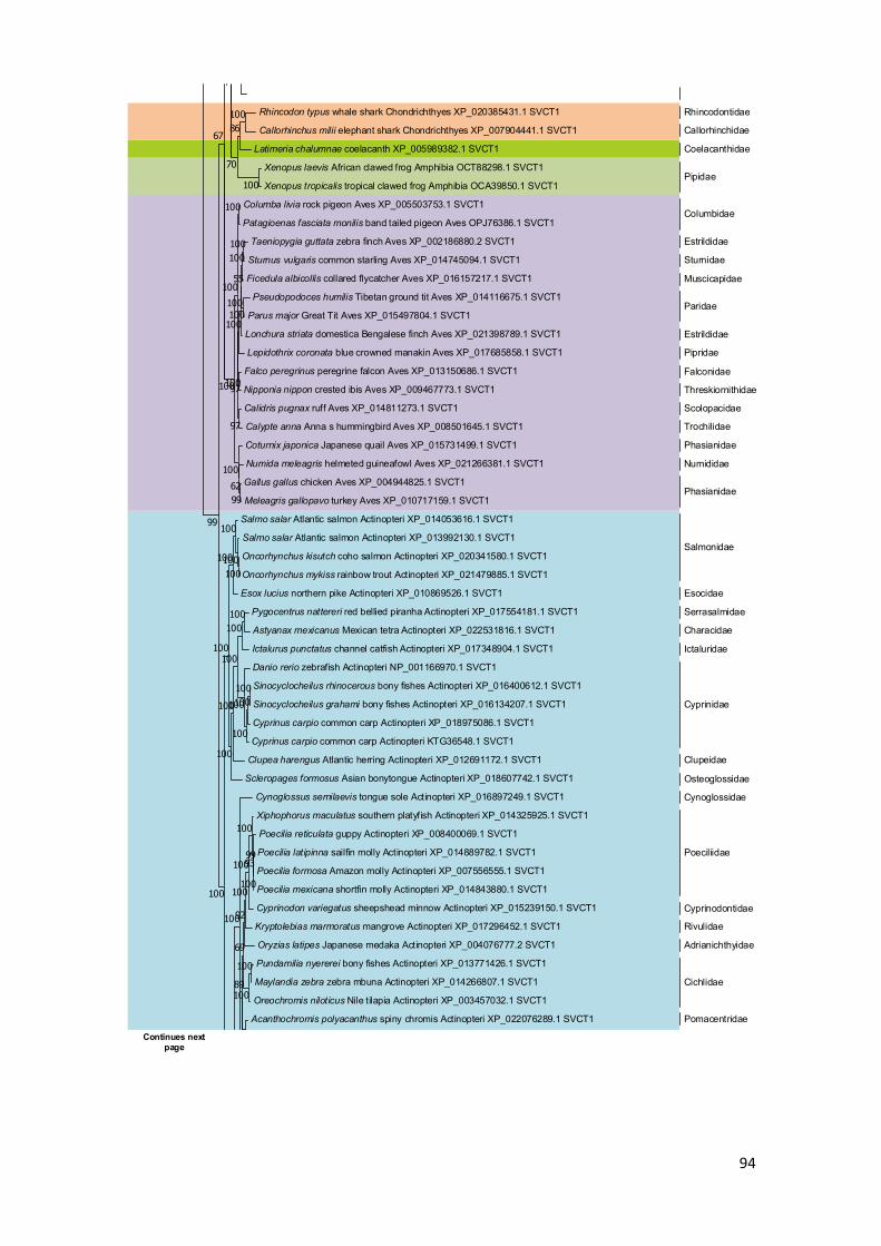
94
Rhincodon typus whale shark Chondrichthyes XP_020385431.1 SVCT1
Callorhinchus milii elephant shark Chondrichthyes XP_007904441.1 SVCT1
Latimeria chalumnae coelacanth XP_005989382.1 SVCT1
Xenopus laevis African clawed frog Amphibia OCT88298.1 SVCT1
Xenopus tropicalis tropical clawed frog Amphibia OCA39850.1 SVCT1
Columba livia rock pigeon Aves XP_005503753.1 SVCT1
Patagioenas fasciata monilis band tailed pigeon Aves OPJ76386.1 SVCT1
Taeniopygia guttata zebra finch Aves XP_002186880.2 SVCT1
Sturnus vulgaris common starling Aves XP_014745094.1 SVCT1
Ficedula albicollis collared flycatcher Aves XP_016157217.1 SVCT1
Pseudopodoces humilis Tibetan ground tit Aves XP_014116675.1 SVCT1
Parus major Great Tit Aves XP_015497804.1 SVCT1
Lonchura striata domestica Bengalese finch Aves XP_021398789.1 SVCT1
Lepidothrix coronata blue crowned manakin Aves XP_017685858.1 SVCT1
Falco peregrinus peregrine falcon Aves XP_013150686.1 SVCT1
Nipponia nippon crested ibis Aves XP_009467773.1 SVCT1
Calidris pugnax ruff Aves XP_014811273.1 SVCT1
Calypte anna Anna s hummingbird Aves XP_008501645.1 SVCT1
Coturnix japonica Japanese quail Aves XP_015731499.1 SVCT1
Numida meleagris helmeted guineafowl Aves XP_021266381.1 SVCT1
Gallus gallus chicken Aves XP_004944825.1 SVCT1
Meleagris gallopavo turkey Aves XP_010717159.1 SVCT1
Salmo salar Atlantic salmon Actinopteri XP_014053616.1 SVCT1
Salmo salar Atlantic salmon Actinopteri XP_013992130.1 SVCT1
Oncorhynchus kisutch coho salmon Actinopteri XP_020341580.1 SVCT1
Oncorhynchus mykiss rainbow trout Actinopteri XP_021479885.1 SVCT1
Esox lucius northern pike Actinopteri XP_010869526.1 SVCT1
Pygocentrus nattereri red bellied piranha Actinopteri XP_017554181.1 SVCT1
Astyanax mexicanus Mexican tetra Actinopteri XP_022531816.1 SVCT1
Ictalurus punctatus channel catfish Actinopteri XP_017348904.1 SVCT1
Danio rerio zebrafish Actinopteri NP_001166970.1 SVCT1
Sinocyclocheilus rhinocerous bony fishes Actinopteri XP_016400612.1 SVCT1
Sinocyclocheilus grahami bony fishes Actinopteri XP_016134207.1 SVCT1
Cyprinus carpio common carp Actinopteri XP_018975086.1 SVCT1
Cyprinus carpio common carp Actinopteri KTG36548.1 SVCT1
Clupea harengus Atlantic herring Actinopteri XP_012691172.1 SVCT1
Scleropages formosus Asian bonytongue Actinopteri XP_018607742.1 SVCT1
Cynoglossus semilaevis tongue sole Actinopteri XP_016897249.1 SVCT1
Xiphophorus maculatus southern platyfish Actinopteri XP_014325925.1 SVCT1
Poecilia reticulata guppy Actinopteri XP_008400069.1 SVCT1
Poecilia latipinna sailfin molly Actinopteri XP_014889782.1 SVCT1
Poecilia formosa Amazon molly Actinopteri XP_007556555.1 SVCT1
Poecilia mexicana shortfin molly Actinopteri XP_014843880.1 SVCT1
Cyprinodon variegatus sheepshead minnow Actinopteri XP_015239150.1 SVCT1
Kryptolebias marmoratus mangrove Actinopteri XP_017296452.1 SVCT1
Oryzias latipes Japanese medaka Actinopteri XP_004076777.2 SVCT1
Pundamilia nyererei bony fishes Actinopteri XP_013771426.1 SVCT1
Maylandia zebra zebra mbuna Actinopteri XP_014266807.1 SVCT1
Oreochromis niloticus Nile tilapia Actinopteri XP_003457032.1 SVCT1
Acanthochromis polyacanthus spiny chromis Actinopteri XP_022076289.1 SVCT1
10086
100
70
100
100100
100
55
100
100
97
97
100
100
9962
100
100
67
100100
100
100
100100
100100
100
100
100
100
100
100
99
100
93
100
100
100
92
100
100
69
89
100
100
99
Continues next page
Rhincodontidae
Callorhinchidae
Coelacanthidae
Pipidae
Columbidae
Estrildidae
Sturnidae
Muscicapidae
Paridae
Estrildidae
Pipridae
Falconidae
Threskiornithidae
Scolopacidae
Trochilidae
Phasianidae
Numididae
Phasianidae
Salmonidae
Esocidae
Serrasalmidae
Characidae
Ictaluridae
Cyprinidae
Clupeidae
Osteoglossidae
Cynoglossidae
Poeciliidae
Cyprinodontidae
Rivulidae
Adrianichthyidae
Cichlidae
Pomacentridae

95
Stegastes partitus bicolor damselfish Actinopteri XP_008302232.1 SVCT1
Seriola dumerili greater amberjack Actinopteri XP_022625107.1 SVCT1
Lates calcarifer barramundi perch Actinopteri XP_018517481.1 SVCT1
Paralichthys olivaceus Japanese flounder Actinopteri XP_019964798.1 SVCT1
Monopterus albus swamp eel Actinopteri XP_020446500.1 SVCT1
Larimichthys crocea large yellow croaker Actinopteri XP_010746004.2 SVCT1
Takifugu rubripes torafugu Actinopteri XP_011609478.1 SVCT1
Boleophthalmus pectinirostris great blue spotted mudskipper Actinopteri XP_020796795.1 SVCT1
Hippocampus comes tiger tail seahorse Actinopteri XP_019747490.1 SVCT1
100
999998
100
96
92
92
100
0.50
Actinopteri
Mammalia Aves
Reptilia Amphibia
Chondrichthyes Coelacanthidae
Pomacentridae
Carangidae
Centropomidae
Paralichthyidae
Synbranchidae
Sciaenidae
Tetraodontidae
Gobiidae
Syngnathidae
Supplementary Figure 2 - Sodium-dependent Vitamin C transporter 1 (SVCT1) Bayesian phylogeny. The species family names are
represented next to the corresponding sequences. Different taxonomic groups are highlighted with distinct colours: Mammalia in
light red, Actinopteri in light blue, Aves in purple, Reptilia in yellow, Chondrichthyes in orange, Amphibia in light green and
Coelacanthidae in green. The four outgroup sequences are not highlighted and can be seen at the top of the phylogeny.
Homo sapiens human Mammalia NP_001138362.1 SVCT3
Mus musculus house mouse Mammalia XP_006506197.1 SVCT4
Homo sapiens human Mammalia XP_011542068.1 SVCT1
Notothenia coriiceps black rockcod Actinopteri XP_010776878.1 SVCT2
Paralichthys olivaceus Japanese flounder Actinopteri XP_019954852.1 SVCT2
Oryzias latipes Japanese medaka Actinopteri XP_011477209.1 SVCT2
Poecilia latipinna sailfin molly Actinopteri XP_014889852.1 SVCT2
Poecilia formosa Amazon molly Actinopteri XP_016532600.1 SVCT2
Poecilia reticulata guppy Actinopteri XP_017162356.1 SVCT2
Neolamprologus brichardi lyretail cichlid Actinopteri XP_006789985.1 SVCT2
Haplochromis burtoni Burton s mouthbrooder Actinopteri XP_005924415.1 SVCT2
Maylandia zebra zebra mbuna Actinopteri XP_004547105.1 SVCT2
Oreochromis niloticus Nile tilapia Actinopteri XP_005473562.1 SVCT2
Monopterus albus swamp eel Actinopteri XP_020449394.1 SVCT2
Takifugu rubripes torafugu Actinopteri XP_003974648.1 SVCT2
Hippocampus comes tiger tail seahorse Actinopteri XP_019742687.1 SVCT2
Boleophthalmus pectinirostris great blue spotted mudskipper Actinopteri XP_020787392.1 SVCT2
Oncorhynchus mykiss rainbow trout Actinopteri XP_021477510.1 SVCT2
Oncorhynchus kisutch coho salmon Actinopteri XP_020345835.1 SVCT2
Salmo salar Atlantic salmon Actinopteri XP_014016985.1 SVCT2
Salmo salar Atlantic salmon Actinopteri XP_014026315.1 SVCT2
Oncorhynchus kisutch coho salmon Actinopteri XP_020329247.1 SVCT2
Oncorhynchus mykiss rainbow trout Actinopteri XP_021461945.1 SVCT2
Esox lucius northern pike Actinopteri XP_010874838.1 SVCT2
Sinocyclocheilus rhinocerous bony fishes Actinopteri XP_016424729.1 SVCT2
Sinocyclocheilus anshuiensis bony fishes Actinopteri XP_016336260.1 SVCT2
Cyprinus carpio common carp Actinopteri KTF95704.1 SVCT2
Sinocyclocheilus rhinocerous bony fishes Actinopteri XP_016411263.1 SVCT2
Sinocyclocheilus anshuiensis bony fishes Actinopteri XP_016340939.1 SVCT2
Sinocyclocheilus grahami bony fishes Actinopteri XP_016091639.1 SVCT2
Danio rerio zebrafish Actinopteri XP_005169157.1 SVCT2
Ictalurus punctatus channel catfish Actinopteri XP_017323629.1 SVCT2
Astyanax mexicanus Mexican tetra Actinopteri XP_022526239.1 SVCT2
Pygocentrus nattereri red bellied piranha Actinopteri XP_017575422.1 SVCT2
Clupea harengus Atlantic herring Actinopteri XP_012676547.1 SVCT2
Scleropages formosus Asian bonytongue Actinopteri XP_018618047.1 SVCT2
Scleropages formosus Asian bonytongue Actinopteri XP_018605189.1 SVCT2
Lepisosteus oculatus spotted gar Actinopteri XP_015197805.1 SVCT2
72
100
100
100
99
100
100
100
99
99
100
100
10066
100100
100
100
100
99
53
53
53
100
100
100100
100
100
99
99
99
99
94
Hominidae
Muridae
Hominidae
Nototheniidae
Paralichthyidae
Adrianichthyidae
Poeciliidae
Cichlidae
Synbranchidae
Tetraodontidae
Syngnathidae
Gobiidae
Salmonidae
Esocidae
Cyprinidae
Ictaluridae
Characidae
Serrasalmidae
Clupeidae
Osteoglossidae
Lepisosteidae
Continues next page

96
Xenopus tropicalis tropical clawed frog Amphibia OCA42243.1 SVCT2
Xenopus laevis African clawed frog Amphibia OCT89946.1 SVCT2
Xenopus laevis African clawed frog Amphibia OCT86650.1 SVCT2
Nanorana parkeri frogs toads Amphibia XP_018424226.1 SVCT2
Latimeria chalumnae coelacanth XP_014352307.1 SVCT2
Apaloderma vittatum bar tailed trogon Aves XP_009866533.1 SVCT2
Egretta garzetta little egret Aves XP_009641685.1 SVCT2
Charadrius vociferus killdeer Aves XP_009878395.1 SVCT2
Aptenodytes forsteri emperor penguin Aves XP_009272071.1 SVCT2
Pygoscelis adeliae Adelie penguin Aves XP_009325462.1 SVCT2
Fulmarus glacialis Northern fulmar Aves XP_009573022.1 SVCT2
Eurypyga helias sunbittern Aves XP_010157544.1 SVCT2
Cariama cristata red legged seriema Aves XP_009703985.1 SVCT2
Falco peregrinus peregrine falcon Aves XP_005234289.1 SVCT2
Falco cherrug Saker falcon Aves XP_005438175.1 SVCT2
Antrostomus carolinensis chuck will s widow Aves XP_010169879.1 SVCT2
Cuculus canorus common cuckoo Aves XP_009566136.1 SVCT2
Buceros rhinoceros silvestris Rhinoceros hornbill Aves XP_010137149.1 SVCT2
Tauraco erythrolophus red crested turaco Aves XP_009991446.1 SVCT2
Aquila chrysaetos canadensis golden eagle Aves XP_011573605.1 SVCT2
Haliaeetus albicilla white tailed eagle Aves XP_009917297.1 SVCT2
Haliaeetus leucocephalus bald eagle Aves XP_010561171.1 SVCT2
Opisthocomus hoazin hoatzin Aves XP_009934865.1 SVCT2
Chlamydotis macqueenii Macqueen s bustard Aves XP_010117140.1 SVCT2
Calidris pugnax ruff Aves XP_014800961.1 SVCT2
Balearica regulorum gibbericeps East African grey crowned crane Aves XP_010305595.1 SVCT2
Tyto alba barn owl Aves XP_009970902.1 SVCT2
Picoides pubescens Downy woodpecker Aves XP_009896108.1 SVCT2
Corvus cornix cornix hooded crow Aves XP_010389019.1 SVCT2
Corvus brachyrhynchos American crow Aves XP_008639803.1 SVCT2
Ficedula albicollis collared flycatcher Aves XP_005058012.1 SVCT2
Sturnus vulgaris common starling Aves XP_014747591.1 SVCT2
Pseudopodoces humilis Tibetan ground tit Aves XP_005533026.1 SVCT2
Taeniopygia guttata zebra finch Aves XP_002196737.2 SVCT2
Lonchura striata domestica Bengalese finch Aves XP_021403611.1 SVCT2
Serinus canaria common canary Aves XP_018778148.1 SVCT2
Manacus vitellinus golden collared manakin Aves XP_008924532.1 SVCT2
Lepidothrix coronata blue crowned manakin Aves XP_017694747.1 SVCT2
Acanthisitta chloris rifleman Aves XP_009072655.1 SVCT2
Chaetura pelagica chimney swift Aves XP_010002228.1 SVCT2
Calypte anna Anna s hummingbird Aves XP_008498950.1 SVCT2
Melopsittacus undulatus budgerigar Aves XP_005141513.1 SVCT2
Anser cygnoides domesticus swan goose Aves XP_013054687.1 SVCT2
Anas platyrhynchos mallard Aves XP_005013037.1 SVCT2
Coturnix japonica Japanese quail Aves XP_015738385.1 SVCT2
Gallus gallus chicken Aves XP_015152797.1 SVCT2
Meleagris gallopavo turkey Aves XP_019478161.1 SVCT2
Colinus virginianus northern bobwhite Aves OXB74818.1 SVCT2
Callipepla squamata scaled quail Aves OXB56939.1 SVCT2
Numida meleagris helmeted guineafowl Aves XP_021230761.1 SVCT2
Tinamus guttatus white throated tinamou Aves XP_010213642.1 SVCT2
Struthio camelus australis African ostrich Aves XP_009667668.1 SVCT2
Gavialis gangeticus Gharial XP_019369885.1 SVCT2
Crocodylus porosus Australian saltwater crocodile XP_019401495.1 SVCT2
Alligator sinensis Chinese alligator XP_006035210.1 SVCT2
Alligator mississippiensis American alligator KYO44422.1 SVCT2
Pelodiscus sinensis Chinese soft shelled turtle XP_006110847.1 SVCT2
Chrysemys picta bellii western painted turtle XP_005285247.1 SVCT2
Chelonia mydas green sea turtle XP_007065554.1 SVCT2
Pogona vitticeps central bearded dragon XP_020635827.1 SVCT2
Python bivittatus Burmese python XP_007427161.1 SVCT2
Protobothrops mucrosquamatus snakes XP_015666992.1 SVCT2
Ophiophagus hannah king cobra ETE68111.1 SVCT2
Gekko japonicus lizards XP_015276598.1 SVCT2
82
100
100
94
100
100
100
100100
51
81
100
100
100
94
94
94
94
100
94
94
90
66
66
67
100
100
100
64
100
100
67
71
94
100
99
100
93
100100
94
100100
89
100
94
99
9699
99
Continues next page
Pipidae
Dicroglossidae
Coelacanthidae
Trogonidae
Ardeidae
Charadriidae
Spheniscidae
Procellariidae
Eurypygidae
Cariamidae
Falconidae
Caprimulgidae
Cuculidae
Bucerotidae
Musophagidae
Accipitridae
Opisthocomidae
Otididae
Scolopacidae
Gruidae
Tytonidae
Picidae
Corvidae
Muscicapidae
Sturnidae
Paridae
Estrildidae
Fringillidae
Pipridae
Acanthisittidae
Apodidae
Trochilidae
Psittaculidae
Anatidae
Phasianidae
Odontophoridae
Numididae
Tinamidae
Struthionidae
Gavialidae
Crocodylidae
Alligatoridae
Trionychidae
Emydidae
Cheloniidae
Agamidae
Pythonidae
Viperidae
Elapidae
Gekkonidae

97
Monodelphis domestica gray short tailed opossum Mammalia XP_007476346.1 SVCT2
Phascolarctos cinereus koala Mammalia XP_020841019.1 SVCT2
Sarcophilus harrisii Tasmanian devil Mammalia XP_003757995.1 SVCT2
Propithecus coquereli Coquerel s sifaka Mammalia XP_012494698.1 SVCT2
Microcebus murinus gray mouse lemur Mammalia XP_012610425.1 SVCT2
Otolemur garnettii small eared galago Mammalia XP_003788081.1 SVCT2
Aotus nancymaae Ma s night monkey Mammalia XP_012310818.1 SVCT2
Callithrix jacchus white tufted ear marmoset Mammalia XP_017827433.1 SVCT2
Cebus capucinus imitator white faced sapajou Mammalia XP_017379496.1 SVCT2
Saimiri boliviensis boliviensis Bolivian squirrel monkey Mammalia XP_003941146.1 SVCT2
Colobus angolensis palliatus Angolan colobus Mammalia XP_011796421.1 SVCT2
Rhinopithecus roxellana golden snub nosed monkey Mammalia XP_010377240.1 SVCT2
Rhinopithecus bieti black snub nosed monkey Mammalia XP_017733451.1 SVCT2
Chlorocebus sabaeus green monkey Mammalia XP_008017157.1 SVCT2
Mandrillus leucophaeus drill Mammalia XP_011830667.1 SVCT2
Cercocebus atys sooty mangabey Mammalia XP_011907540.1 SVCT2
Papio anubis olive baboon Mammalia XP_021776900.1 SVCT2
Macaca fascicularis crab eating macaque Mammalia EHH65524.1 SVCT2
Macaca nemestrina pig tailed macaque Mammalia XP_011739517.1 SVCT2
Macaca mulatta Rhesus monkey Mammalia EHH19898.1 SVCT2
Gorilla gorilla gorilla western lowland gorilla Mammalia XP_018872874.1 SVCT2
Homo sapiens human Mammalia NP_005107.4 SVCT2
Pan troglodytes chimpanzee Mammalia XP_016792888.1 SVCT2
Pan paniscus pygmy chimpanzee Mammalia XP_008973054.1 SVCT2
Pongo abelii Sumatran orangutan Mammalia XP_002830108.1 SVCT2
Nomascus leucogenys northern white cheeked gibbon Mammalia XP_003278035.2 SVCT2
Carlito syrichta Philippine tarsier Mammalia XP_008067109.1 SVCT2
Heterocephalus glaber naked mole rat Mammalia EHB04320.1 SVCT2
Fukomys damarensis Damara mole rat Mammalia XP_010605835.1 SVCT2
Octodon degus degu Mammalia XP_004634334.1 SVCT2
Chinchilla lanigera long tailed chinchilla Mammalia XP_005380880.1 SVCT2
Cavia porcellus domestic guinea pig Mammalia XP_013015230.1 SVCT2
Dipodomys ordii Ord s kangaroo rat Mammalia XP_012866485.1 SVCT2
Castor canadensis American beaver Mammalia XP_020039621.1 SVCT2
Nannospalax galili Upper Galilee mountains blind mole rat Mammalia XP_008834119.1 SVCT2
Mesocricetus auratus golden hamster Mammalia XP_005068720.1 SVCT2
Cricetulus griseus Chinese hamster Mammalia XP_016828992.1 SVCT2
Peromyscus maniculatus bairdii prairie deer mouse Mammalia XP_006984051.1 SVCT2
Microtus ochrogaster prairie vole Mammalia XP_005365622.1 SVCT2
Mus musculus house mouse Mammalia EDL28341.1 SVCT2
Mus caroli Ryukyu mouse Mammalia XP_021039752.1 SVCT2
Mus pahari shrew mouse Mammalia XP_021048207.1 SVCT2
Rattus norvegicus Norway rat Mammalia EDL80256.1 SVCT2
Jaculus jaculus lesser Egyptian jerboa Mammalia XP_004661550.1 SVCT2
Marmota marmota marmota Alpine marmot Mammalia XP_015338131.1 SVCT2
Ictidomys tridecemlineatus thirteen lined ground squirrel Mammalia XP_021578859.1 SVCT2
Ochotona princeps American pika Mammalia XP_004593397.1 SVCT2
Oryctolagus cuniculus rabbit Mammalia XP_002710883.1 SVCT2
Tupaia chinensis Chinese tree shrew Mammalia XP_006164043.1 SVCT2
Galeopterus variegatus Sunda flying lemur Mammalia XP_008577173.1 SVCT2
Pteropus vampyrus large flying fox Mammalia XP_011368277.1 SVCT2
Pteropus alecto black flying fox Mammalia XP_006921682.1 SVCT2
Rousettus aegyptiacus Egyptian rousette Mammalia XP_015989795.1 SVCT2
97
99
100100
100
100
100
100
100
100
100
100
100
99
100
100
95
100
100
99
100
72
100
100100
100
100
100
100
100
59
100
100
99
69
70
71
80
9999
50
Continues next page
Didelphidae
Phascolarctidae
Dasyuridae
Indriidae
Cheirogaleidae
Galagidae
Aotidae
Cebidae
Cercopithecidae
Hominidae
Hylobatidae
Tarsiidae
Bathyergidae
Octodontidae
Chinchillidae
Caviidae
Heteromyidae
Castoridae
Spalacidae
Cricetidae
Muridae
Dipodidae
Sciuridae
Ochotonidae
Leporidae
Tupaiidae
Cynocephalidae
Pteropodidae

98
Miniopterus natalensis bats Mammalia XP_016067542.1 SVCT2
Myotis davidii bats Mammalia XP_006758125.1 SVCT2
Myotis brandtii Brandt s bat Mammalia XP_005874736.1 SVCT2
Myotis lucifugus little brown bat Mammalia XP_006096492.1 SVCT2
Eptesicus fuscus big brown bat Mammalia XP_008139284.1 SVCT2
Rhinolophus sinicus Chinese rufous horseshoe bat Mammalia XP_019589318.1 SVCT2
Hipposideros armiger great roundleaf bat Mammalia XP_019500165.1 SVCT2
Ceratotherium simum simum southern white rhinoceros Mammalia XP_014645584.1 SVCT2
Equus caballus horse Mammalia XP_014590528.1 SVCT2
Equus przewalskii Przewalski s horse Mammalia XP_008518016.1 SVCT2
Equus asinus ass Mammalia XP_014684197.1 SVCT2
Panthera pardus leopard Mammalia XP_019318143.1 SVCT2
Panthera tigris altaica Amur tiger Mammalia XP_007073293.1 SVCT2
Felis catus domestic cat Mammalia XP_019682358.1 SVCT2
Acinonyx jubatus cheetah Mammalia XP_014933640.1 SVCT2
Canis lupus familiaris dog Mammalia NP_001300742.1 SVCT2
Enhydra lutris kenyoni sea otter Mammalia XP_022349621.1 SVCT2
Mustela putorius furo domestic ferret Mammalia XP_004772880.1 SVCT2
Neomonachus schauinslandi Hawaiian monk seal Mammalia XP_021544579.1 SVCT2
Odobenus rosmarus divergens Pacific walrus Mammalia XP_004398260.1 SVCT2
Ailuropoda melanoleuca giant panda Mammalia XP_011230125.1 SVCT2
Ursus maritimus polar bear Mammalia XP_008701537.1 SVCT2
Sus scrofa pig Mammalia XP_020932938.1 SVCT2
Vicugna pacos alpaca Mammalia XP_015097334.1 SVCT2
Camelus dromedarius Arabian camel Mammalia XP_010992925.1 SVCT2
Camelus bactrianus Bactrian camel Mammalia XP_010971033.1 SVCT2
Camelus ferus Wild Bactrian camel Mammalia EPY79165.1 SVCT2
Bubalus bubalis water buffalo Mammalia XP_006047814.1 SVCT2
Bison bison bison American bison Mammalia XP_010834734.1 SVCT2
Bos taurus cattle Mammalia DAA23455.1 SVCT2
Bos indicus zebu cattle Mammalia XP_019835257.1 SVCT2
Bos mutus wild yak Mammalia ELR48512.1 SVCT2
Capra hircus goat Mammalia XP_017912852.1 SVCT2
Ovis aries musimon mouflon Mammalia XP_011995463.1 SVCT2
Ovis aries sheep Mammalia XP_014955204.1 SVCT2
Pantholops hodgsonii chiru Mammalia XP_005974369.1 SVCT2
Odocoileus virginianus texanus white tailed deer Mammalia XP_020739307.1 SVCT2
Balaenoptera acutorostrata scammoni minke whale Mammalia XP_007191767.1 SVCT2
Lipotes vexillifer Yangtze River dolphin Mammalia XP_007464035.1 SVCT2
Orcinus orca killer whale Mammalia XP_004276490.1 SVCT2
Tursiops truncatus bottlenose dolphin Mammalia XP_019797033.1 SVCT2
Delphinapterus leucas beluga whale Mammalia XP_022412162.1 SVCT2
Physeter catodon sperm whale Mammalia XP_007124808.1 SVCT2
Sorex araneus European shrew Mammalia XP_004610999.1 SVCT2
Condylura cristata star nosed mole Mammalia XP_004687251.1 SVCT2
Erinaceus europaeus western European hedgehog Mammalia XP_007539622.1 SVCT2
Chrysochloris asiatica Cape golden mole Mammalia XP_006870473.1 SVCT2
Echinops telfairi small Madagascar hedgehog Mammalia XP_004697908.1 SVCT2
Elephantulus edwardii Cape elephant shrew Mammalia XP_006894287.1 SVCT2
Trichechus manatus latirostris Florida manatee Mammalia XP_004383098.1 SVCT2
Loxodonta africana African savanna elephant Mammalia XP_003411478.1 SVCT2
Orycteropus afer afer aardvark Mammalia XP_007955405.1 SVCT2
Drosophila melanogaster fruit fly Insecta AAF54519.1 SVCTP
95
100
99
99
99
97
100
100
98
96
100
100
100
100
67
100
99
82
83
72
98
96
100
100
100
85
100
100
10099
99
89
64
76
69
51
54
53
97
99
1
Actinopteri
Mammalia Aves
Reptilia Amphibia
Coelacanthidae
Vespertilionidae
Rhinolophidae
Hipposideridae
Rhinocerotidae
Equidae
Felidae
Canidae
Mustelidae
Phocidae
Odobenidae
Ursidae
Suidae
Camelidae
Bovidae
Cervidae
Balaenopteridae
Lipotidae
Delphinidae
Monodontidae
Physeteridae
Soricidae
Talpidae
Erinaceidae
Chrysochloridae
Tenrecidae
Macroscelididae
Trichechidae
Elephantidae
Orycteropodidae
Drosophilidae

99
Supplementary Figure 3 - Sodium-dependent Vitamin C transporter 2 (SVCT2) Bayesian phylogeny. The species family names are
represented next to the corresponding sequences. Different taxonomic groups are highlighted with distinct colours: Mammalia in
light red, Actinopteri in light blue, Aves in purple, Reptilia in yellow, Amphibia in light green and Coelacanthidae in green. The four
outgroup sequences are not highlighted, and can be seen at the top (H. sapiens SVCT1, H. sapiens SVCT3 and M. musculus SVCT4)
and bottom of the phylogeny (D. melanogaster SVCTP).
Mus musculus house mouse Mammalia XP_006506197.1 SVCT4
Homo sapiens human Mammalia XP_011542068.1 SVCT1
Homo sapiens human Mammalia NP_005107.4 SVCT2
Nanorana parkeri frogs toads Amphibia XP_018410410.1 SVCT3
Xenopus tropicalis tropical clawed frog Amphibia XP_012826889.1 SVCT3
Xenopus laevis African clawed frog Amphibia OCT61003.1 SVCT3
Latimeria chalumnae coelacanth XP_014343771.1 SVCT3
Pan paniscus pygmy chimpanzee Mammalia XP_003818682.2 SVCT3
Pan troglodytes chimpanzee Mammalia XP_016806037.1 SVCT3
Homo sapiens human Mammalia NP_001138362.1 SVCT3
Gorilla gorilla gorilla western lowland gorilla Mammalia XP_004033289.1 SVCT3
Pongo abelii Sumatran orangutan Mammalia XP_002812917.2 SVCT3
Nomascus leucogenys northern white cheeked gibbon Mammalia XP_003272435.1 SVCT3
Macaca nemestrina pig tailed macaque Mammalia XP_011738993.1 SVCT3
Macaca fascicularis crab eating macaque Cercopithecidae Mammalia XP_015288309.1 SVCT3
Macaca mulatta Rhesus monkey Mammalia EHH21687.1 SVCT3
Cercocebus atys sooty mangabey Mammalia XP_011918256.1 SVCT3
Mandrillus leucophaeus drill Mammalia XP_011820018.1 SVCT3
Papio anubis olive baboon Mammalia XP_009181374.1 SVCT3
Rhinopithecus bieti black snub nosed monkey Mammalia XP_017712260.1 SVCT3
Rhinopithecus roxellana golden snub nosed monkey Mammalia XP_010358568.1 SVCT3
Colobus angolensis palliatus Angolan colobus Mammalia XP_011787621.1 SVCT3
Callithrix jacchus white tufted ear marmoset Mammalia XP_008997762.1 SVCT3
Saimiri boliviensis boliviensis Bolivian squirrel monkey Mammalia XP_010334602.1 SVCT3
Aotus nancymaae Ma s night monkey Mammalia XP_012301313.1 SVCT3
Carlito syrichta Philippine tarsier Mammalia XP_008057055.1 SVCT3
Microcebus murinus gray mouse lemur Mammalia XP_012595664.1 SVCT3
Propithecus coquereli Coquerel s sifaka Mammalia XP_012498015.1 SVCT3
Otolemur garnettii small eared galago Mammalia XP_003784986.1 SVCT3
Nannospalax galili Upper Galilee mountains blind mole rat Mammalia XP_008823966.1 SVCT3
Cricetulus griseus Chinese hamster Mammalia XP_003507646.1 SVCT3
Mesocricetus auratus golden hamster Mammalia XP_005070537.1 SVCT3
Microtus ochrogaster prairie vole Mammalia XP_005361665.1 SVCT3
Peromyscus maniculatus bairdii prairie deer mouse Mammalia XP_006972234.1 SVCT3
Mus caroli Ryukyu mouse Mammalia XP_021023522.1 SVCT3
Mus musculus house mouse Mammalia XP_006496542.1 SVCT3
Mus pahari shrew mouse Mammalia XP_021054643.1 SVCT3
Rattus norvegicus Norway rat Mammalia EDL75382.1 SVCT3
Meriones unguiculatus Mongolian gerbil Mammalia XP_021520372.1 SVCT3
Dipodomys ordii Ord s kangaroo rat Mammalia XP_012866902.1 SVCT3
Heterocephalus glaber naked mole rat Mammalia EHA97568.1 SVCT3
Fukomys damarensis Damara mole rat Mammalia XP_010610166.1 SVCT3
Chinchilla lanigera long tailed chinchilla Mammalia XP_005395646.1 SVCT3
Cavia porcellus domestic guinea pig Mammalia XP_013001625.1 SVCT3
Octodon degus degu Mammalia XP_004638299.1 SVCT3
Ictidomys tridecemlineatus thirteen lined ground squirrel Mammalia XP_021585683.1 SVCT3
Marmota marmota marmota Alpine marmot Mammalia XP_015336893.1 SVCT3
Mustela putorius furo domestic ferret Mammalia XP_004762859.2 SVCT3
Ursus maritimus polar bear Mammalia XP_008688679.1 SVCT3
Ailuropoda melanoleuca giant panda Mammalia XP_019662789.1 SVCT3
Odobenus rosmarus divergens Pacific walrus Mammalia XP_004402830.1 SVCT3
Neomonachus schauinslandi Hawaiian monk seal Mammalia XP_021558653.1 SVCT3
Canis lupus familiaris dog Mammalia XP_003640230.1 SVCT3
Panthera tigris altaica Amur tiger Mammalia XP_015394744.1 SVCT3
Panthera pardus leopard Mammalia XP_019301447.1 SVCT3
Acinonyx jubatus cheetah Mammalia XP_014922139.1 SVCT3
Felis catus domestic cat Mammalia XP_019694259.1 SVCT3
100
100
100
100
52
96
100
99
100
100
99
100
100
100
100
100
100
63
100
100
100
100
100
100
10051
100
100100
100
100
100
100
100
100
62
100
100
75
100
100
100
100
86
100
100
100
10054
100
100
77
91Continues next
page
Muridae
Hominidae
Dicroglossidae
Pipidae
Coelacanthidae
Hominidae
Hylobatidae
Cercopithecidae
Cebidae
Aotidae
Tarsiidae
Cheirogaleidae
Indriidae
Galagidae
Spalacidae
Cricetidae
Muridae
Heteromyidae
Bathyergidae
Chinchillidae
Caviidae
Octodontidae
Sciuridae
Mustelidae
Ursidae
Odobenidae
Phocidae
Canidae
Felidae

100
Manis javanica Malayan pangolin Mammalia XP_017510021.1 SVCT3
Equus przewalskii Przewalski s horse Mammalia XP_008528176.1 SVCT3
Equus asinus ass Mammalia XP_014711591.1 SVCT3
Ceratotherium simum simum southern white rhinoceros Mammalia XP_004427631.1 SVCT3
Balaenoptera acutorostrata scammoni minke whale Mammalia XP_007188009.1 SVCT3
Tursiops truncatus bottlenose dolphin Mammalia XP_019794477.1 SVCT3
Orcinus orca killer whale Mammalia XP_004262746.1 SVCT3
Delphinapterus leucas beluga whale Mammalia XP_022424022.1 SVCT3
Lipotes vexillifer Yangtze River dolphin Mammalia XP_007445893.1 SVCT3
Physeter catodon sperm whale Mammalia XP_007122523.1 SVCT3
Bubalus bubalis water buffalo Mammalia XP_006059068.1 SVCT3
Bos mutus wild yak Mammalia ELR60768.1 SVCT3
Bison bison bison American bison Mammalia XP_010859522.1 SVCT3
Bos taurus cattle Mammalia DAA32439.1 SVCT3
Pantholops hodgsonii chiru Mammalia XP_005985192.1 SVCT3
Ovis aries musimon mouflon Mammalia XP_011969058.1 SVCT3
Ovis aries sheep Mammalia XP_004004988.1 SVCT3
Capra hircus goat Mammalia XP_005676614.1 SVCT3
Odocoileus virginianus texanus white tailed deer Mammalia XP_020760366.1 SVCT3
Vicugna pacos alpaca Camelidae Mammalia XP_006207355.1 SVCT3
Camelus dromedarius Arabian camel Mammalia XP_010986654.1 SVCT3
Camelus bactrianus Bactrian camel Mammalia XP_010947099.1 SVCT3
Camelus ferus Wild Bactrian camel Mammalia XP_006186977.1 SVCT3
Condylura cristata star nosed mole Mammalia XP_012581319.1 SVCT3
Erinaceus europaeus western European hedgehog Mammalia XP_016042478.1 SVCT3
Myotis brandtii Brandt s bat Mammalia EPQ18447.1 SVCT3
Myotis lucifugus little brown bat Mammalia XP_006082597.1 SVCT3
Myotis davidii bats Mammalia XP_006763420.1 SVCT3
Eptesicus fuscus big brown bat Mammalia XP_008143372.1 SVCT3
Miniopterus natalensis bats Mammalia XP_016052393.1 SVCT3
Rousettus aegyptiacus Egyptian rousette Mammalia XP_016012195.1 SVCT3
Pteropus vampyrus large flying fox Mammalia XP_011356869.1 SVCT3
Pteropus alecto black flying fox Mammalia XP_006906768.1 SVCT3
Rhinolophus sinicus Chinese rufous horseshoe bat Mammalia XP_019597383.1 SVCT3
Trichechus manatus latirostris Florida manatee Mammalia XP_004376384.1 SVCT3
Elephantulus edwardii Cape elephant shrew Mammalia XP_006890366.1 SVCT3
Orycteropus afer afer aardvark Mammalia XP_007933890.1 SVCT3
Echinops telfairi small Madagascar hedgehog Mammalia XP_004701671.1 SVCT3
Dasypus novemcinctus nine banded armadillo Mammalia XP_004467792.1 SVCT3
Phascolarctos cinereus koala Mammalia XP_020858502.1 SVCT3
Monodelphis domestica gray short tailed opossum Mammalia XP_007501758.1 SVCT3
Sarcophilus harrisii Tasmanian devil Mammalia XP_003765969.1 SVCT3
Nestor notabilis Kea Aves XP_010022026.1 SVCT3
Melopsittacus undulatus budgerigar Aves XP_005154146.1 SVCT3
Falco cherrug Saker falcon Aves XP_005442889.1 SVCT3
Falco peregrinus peregrine falcon Aves XP_005234377.1 SVCT3
Haliaeetus leucocephalus bald eagle Aves XP_010567959.1 SVCT3
Aquila chrysaetos canadensis golden eagle Aves XP_011578894.1 SVCT3
Parus major Great Tit Aves XP_018863083.1 SVCT3
Pseudopodoces humilis Tibetan ground tit Aves XP_014105018.1 SVCT3
Sturnus vulgaris common starling Aves XP_014743569.1 SVCT3
Ficedula albicollis collared flycatcher Aves XP_005049552.1 SVCT3
Serinus canaria common canary Aves XP_009086150.1 SVCT3
Zonotrichia albicollis white throated sparrow Aves XP_005485272.1 SVCT3
Geospiza fortis medium ground finch Aves XP_005415853.1 SVCT3
Corvus brachyrhynchos American crow Aves XP_008643148.1 SVCT3
Manacus vitellinus golden collared manakin Aves XP_008921143.1 SVCT3
Lepidothrix coronata blue crowned manakin Aves XP_017680774.1 SVCT3
Acanthisitta chloris rifleman Aves XP_009073311.1 SVCT3
Merops nubicus carmine bee eater Aves XP_008946694.1 SVCT3
Picoides pubescens Downy woodpecker Aves XP_009909463.1 SVCT3
100
100
100100100
52
100
100
100
100
100
98
100
99
100
100
100100
100
100
70
100
59
100100
100100
100
100
69
100
100
5192
100
96
100
99
100
100
100
100
100
92
100
100
76
100100
100
100
100
99
100
56
99
99
97
98
Continues next page
Manidae
Equidae
Rhinocerotidae
Balaenopteridae
Delphinidae
Monodontidae
Lipotidae
Physeteridae
Bovidae
Cervidae
Camelidae
Talpidae
Erinaceidae
Vespertilionidae
Pteropodidae
Rhinolophidae
Trichechidae
Orycteropodidae
Tenrecidae
Dasypodidae
Phascolarctidae
Didelphidae
Dasyuridae
Psittacidae
Psittaculidae
Falconidae
Accipitridae
Paridae
Sturnidae
Muscicapidae
Fringillidae
Passerellidae
Thraupidae
Corvidae
Pipridae
Acanthisittidae
Meropidae
Picidae
Macroscelididae

101
Eurypyga helias sunbittern Aves XP_010156824.1 SVCT3
Calidris pugnax ruff Aves XP_014809739.1 SVCT3
Mesitornis unicolor brown roatelo Aves XP_010180948.1 SVCT3
Cuculus canorus common cuckoo Aves XP_009556131.1 SVCT3
Pterocles gutturalis yellow throated sandgrouse Aves XP_010074277.1 SVCT3
Charadrius vociferus killdeer Aves XP_009882365.1 SVCT3
Egretta garzetta little egret Aves XP_009643974.1 SVCT3
Pygoscelis adeliae Adelie penguin Aves XP_009323126.1 SVCT3
Aptenodytes forsteri emperor penguin Aves XP_009273208.1 SVCT3
Nipponia nippon crested ibis Aves XP_009460409.1 SVCT3
Opisthocomus hoazin hoatzin Aves XP_009943611.1 SVCT3
Columba livia rock pigeon Aves XP_005498651.1 SVCT3
Calypte anna Anna s hummingbird Aves XP_008495256.1 SVCT3
Numida meleagris helmeted guineafowl Aves XP_021254339.1 SVCT3
Gallus gallus chicken Aves XP_015145564.1 SVCT3
Meleagris gallopavo turkey Aves XP_019472971.1 SVCT3
Colinus virginianus northern bobwhite Aves OXB77103.1 SVCT3
Pogona vitticeps central bearded dragon XP_020647659.1 SVCT3
Protobothrops mucrosquamatus snakes XP_015675981.1 SVCT3
Thamnophis sirtalis snakes XP_013918560.1 SVCT3
Gekko japonicus lizards XP_015270850.1 SVCT3
Alligator mississippiensis American alligator KYO34298.1 SVCT3
Alligator sinensis Chinese alligator XP_014381029.1 SVCT3
Chrysemys picta bellii western painted turtle XP_008166063.1 SVCT3
Callorhinchus milii elephant shark Chondrichthyes XP_007909554.1 SVCT3
Clupea harengus Atlantic herring Actinopteri XP_012686153.1 SVCT3
Esox lucius northern pike Actinopteri XP_010897633.1 SVCT3
Astyanax mexicanus Mexican tetra Actinopteri XP_007260007.2 SVCT3
Pygocentrus nattereri red bellied piranha Actinopteri XP_017547152.1 SVCT3
Ictalurus punctatus channel catfish Actinopteri XP_017325892.1 SVCT3
Sinocyclocheilus rhinocerous bony fishes Actinopteri XP_016378793.1 SVCT3
Sinocyclocheilus anshuiensis bony fishes Actinopteri XP_016342013.1 SVCT3
Danio rerio zebrafish Actinopteri XP_017213243.2 SVCT3
Drosophila melanogaster fruit fly Insecta AAF54519.1 SVCTP
7663
89
99
71
100
70
80
75
88
99
100
100
84
100
100
100
100
100
10055
100
100100
100
100
100
98
100
100
0.50
Actinopteri
Mammalia Aves
Reptilia Amphibia
Chondrichthyes Coelacanthidae
Eurypigidae
Scolopacidae
Mesitornithidae
Cuculidae
Pteroclidae
Charadriidae
Ardeidae
Spheniscidae
Threskiornithidae
Opisthocomidae
Columbidae
Trochilidae
Numididae
Phasianidae
Odontophoridae
Agamidae
Viperidae
Colubridae
Gekkonidae
Alligatoridae
Emydidae
Callorhinchidae
Clupeidae
Esocidae
Characidae
Serrasalmidae
Ictaluridae
Cyprinidae
Drosophilidae
Supplementary Figure 4 - Sodium-dependent Vitamin C transporter 3 (SVCT3) Bayesian phylogeny. The species family names are
represented next to the corresponding sequences. Different taxonomic groups are highlighted with distinct colours: Mammalia in
light red, Actinopteri in light blue, Aves in purple, Reptilia in yellow, Chondrichthyes in orange, Amphibia in light green and
Coelacanthidae in green. The four outgroup sequences are not highlighted, and can be seen at the top (H. sapiens SVCT1, H. sapiens
SVCT2 and M. musculus SVCT4) and bottom of the phylogeny (D. melanogaster SVCTP).

102
Drosophila melanogaster fruit fly Insecta AAF54519.1 SVCTP
Homo sapiens human Mammalia NP_001138362.1 SVCT3
Homo sapiens human Mammalia XP_011542068.1 SVCT1
Homo sapiens human Mammalia NP_005107.4 SVCT2
Nanorana parkeri frogs toads Amphibia XP_018423391.1 SVCT4
Xenopus laevis African clawed frog Amphibia OCT58149.1 SVCT4
Equus asinus ass Mammalia XP_014694853.1 SVCT4
Equus caballus horse Mammalia XP_014594784.1 SVCT4
Equus przewalskii Przewalski s horse Mammalia XP_008506874.1 SVCT4
Ceratotherium simum simum southern white rhinoceros Mammalia XP_004430889.1 SVCT4
Ceratotherium simum simum southern white rhinoceros Mammalia XP_004430789.1 SVCT4
Mustela putorius furo domestic ferret Mammalia XP_012905620.1 SVCT4
Ailuropoda melanoleuca giant panda Mammalia XP_011233546.1 SVCT4
Ursus maritimus polar bear Mammalia XP_008705821.1 SVCT4
Neomonachus schauinslandi Hawaiian monk seal Mammalia XP_021548515.1 SVCT4
Odobenus rosmarus divergens Pacific walrus Mammalia XP_012419931.1 SVCT4
Felis catus domestic cat Mammalia XP_019681419.1 SVCT4
Acinonyx jubatus cheetah Mammalia XP_014928545.1 SVCT4
Panthera tigris altaica Amur tiger Mammalia XP_015390389.1 SVCT4
Manis javanica Malayan pangolin Mammalia XP_017534204.1 SVCT4
Ovis aries sheep Mammalia XP_014950603.1 SVCT4
Ovis aries musimon mouflon Mammalia XP_011975591.1 SVCT4
Capra hircus goat Mammalia XP_005679654.2 SVCT4
Pantholops hodgsonii chiru Mammalia XP_005959032.1 SVCT4
Bos taurus cattle Mammalia XP_005205834.1 SVCT4
Odocoileus virginianus texanus white tailed deer Mammalia XP_020735724.1 SVCT4
Balaenoptera acutorostrata scammoni minke whale Mammalia XP_007177334.1 SVCT4
Physeter catodon sperm whale Mammalia XP_007111956.1 SVCT4
Delphinapterus leucas beluga whale Mammalia XP_022445125.1 SVCT4
Orcinus orca killer whale Mammalia XP_004272299.1 SVCT4
Lipotes vexillifer Yangtze River dolphin Mammalia XP_007466719.1 SVCT4
Camelus bactrianus Bactrian camel Mammalia XP_010945807.1 SVCT4
Vicugna pacos alpaca Mammalia XP_015102397.1 SVCT4
Sus scrofa pig Mammalia XP_020934433.1 SVCT4
Carlito syrichta Philippine tarsier Mammalia XP_021566409.1 SVCT4
Propithecus coquereli Coquerel s sifaka Mammalia XP_012498226.1 SVCT4
Microcebus murinus gray mouse lemur Mammalia XP_012600204.1 SVCT4
Tupaia chinensis Chinese tree shrew Mammalia XP_006145069.1 SVCT4
Cavia porcellus domestic guinea pig Mammalia XP_013013991.1 SVCT4
Chinchilla lanigera long tailed chinchilla Mammalia XP_013360395.1 SVCT4
Heterocephalus glaber naked mole rat Mammalia XP_012929891.1 SVCT4
Fukomys damarensis Damara mole rat Mammalia XP_010614481.1 SVCT4
Oryctolagus cuniculus rabbit Mammalia XP_008256371.1 SVCT4
Rattus norvegicus Norway rat Mammalia XP_006236326.1 SVCT4
Mus musculus house mouse Mammalia XP_006506197.1 SVCT4
Peromyscus maniculatus bairdii prairie deer mouse Mammalia XP_015864166.1 SVCT4
Mesocricetus auratus golden hamster Mammalia XP_012977712.1 SVCT4
Cricetulus griseus Chinese hamster Mammalia ERE90339.1 SVCT4
Ictidomys tridecemlineatus thirteen lined ground squirrel Mammalia XP_021586822.1 SVCT4
Loxodonta africana African savanna elephant Mammalia XP_003420321.1 SVCT4
Trichechus manatus latirostris Florida manatee Mammalia XP_012412818.1 SVCT4
Elephantulus edwardii Cape elephant shrew Mammalia XP_006887269.1 SVCT4
Orycteropus afer afer aardvark Mammalia XP_007951109.1 SVCT4
Chrysochloris asiatica Cape golden mole Mammalia XP_006861297.1 SVCT4
Echinops telfairi small Madagascar hedgehog Mammalia XP_004707955.1 SVCT4
Erinaceus europaeus western European hedgehog Mammalia XP_007526630.1 SVCT4
Condylura cristata star nosed mole Mammalia XP_004677256.1 SVCT4
100
100
100100
100
100
100
99
100
100
100
100
99
64
10057
100100
100
100
100
100
100
100
100
97
100
65
100
100
60
100
100
100
81
100
100100
100
98
68
99
100
59
98
100
53
100
52
52
100
100
Continues next page
Drosophilidae
Hominidae
Dicroglossidae
Pipidae
Equidae
Rhinocerotidae
Mustelidae
Ursidae
Phocidae
Odobenidae
Felidae
Manidae
Bovidae
Cervidae
Balaenopteridae
Physeteridae
Monodontidae
Delphinidae
Lipotidae
Camelidae
Suidae
Tarsiidae
Indriidae
Cheirogaleidae
Tupaiidae
Caviidae
Chinchillidae
Bathyergidae
Leporidae
Muridae
Cricetidae
Sciuridae
Elephantidae
Trichechidae
Macroscelididae
Orycteropodidae
Chrysochloridae
Tenrecidae
Erinaceidae
Talpidae

103
Pteropus alecto black flying fox Mammalia XP_006910713.1 SVCT4
Myotis brandtii Brandt s bat Mammalia EPQ04246.1 SVCT4
Myotis lucifugus little brown bat Mammalia XP_006088451.1 SVCT4
Myotis davidii bats Mammalia ELK35565.1 SVCT4
Eptesicus fuscus big brown bat Mammalia XP_008148762.1 SVCT4
Miniopterus natalensis bats Mammalia XP_016077920.1 SVCT4
Rhinolophus sinicus Chinese rufous horseshoe bat Mammalia XP_019606344.1 SVCT4
Phascolarctos cinereus koala Mammalia XP_020857962.1 SVCT4
Sarcophilus harrisii Tasmanian devil Mammalia XP_012406506.1 SVCT4
Monodelphis domestica gray short tailed opossum Mammalia XP_007504437.1 SVCT4
Pelodiscus sinensis Chinese soft shelled turtle XP_006125979.2 SVCT4
Chrysemys picta bellii western painted turtle XP_005305352.1 SVCT4
Crocodylus porosus Australian saltwater crocodile XP_019400699.1 SVCT4
Gavialis gangeticus Gharial XP_019370519.1 SVCT4
Alligator mississippiensis American alligator KYO17514.1 SVCT4
Protobothrops mucrosquamatus snakes XP_015669936.1 SVCT4
Python bivittatus Burmese python XP_007434366.1 SVCT4
Pogona vitticeps central bearded dragon XP_020667374.1 SVCT4
Anolis carolinensis green anole XP_016849222.1 SVCT4
Gekko japonicus lizards XP_015276774.1 SVCT4
Serinus canaria common canary Aves XP_018765600.1 SVCT4
Zonotrichia albicollis white throated sparrow Aves XP_005480850.1 SVCT4
Lonchura striata domestica Bengalese finch Aves OWK59349.1 SVCT4
Sturnus vulgaris common starling Aves XP_014745329.1 SVCT4
Ficedula albicollis collared flycatcher Aves XP_016161402.1 SVCT4
Parus major Great Tit Aves XP_015483204.1 SVCT4
Pseudopodoces humilis Tibetan ground tit Aves XP_005519525.1 SVCT4
Corvus cornix cornix hooded crow Aves XP_010409241.2 SVCT4
Lepidothrix coronata blue crowned manakin Aves XP_017659724.1 SVCT4
Colius striatus speckled mousebird Aves XP_010205953.1 SVCT4
Pterocles gutturalis yellow throated sandgrouse Aves XP_010081923.1 SVCT4
Haliaeetus leucocephalus bald eagle Aves XP_010560250.1 SVCT4
Aquila chrysaetos canadensis golden eagle Aves XP_011573141.1 SVCT4
Falco cherrug Saker falcon Aves XP_005441382.1 SVCT4
Falco peregrinus peregrine falcon Aves XP_005237413.1 SVCT4
Nestor notabilis Kea Aves XP_010008058.1 SVCT4
Melopsittacus undulatus budgerigar Aves XP_005144406.1 SVCT4
Mesitornis unicolor brown roatelo Aves XP_010187960.1 SVCT4
Phaethon lepturus white tailed tropicbird Aves XP_010291316.1 SVCT4
Opisthocomus hoazin hoatzin Aves XP_009938436.1 SVCT4
Merops nubicus carmine bee eater Aves XP_008936635.1 SVCT4
Picoides pubescens Downy woodpecker Aves XP_009896796.1 SVCT4
Calidris pugnax ruff Aves XP_014818940.1 SVCT4
Charadrius vociferus killdeer Aves XP_009892692.1 SVCT4
Pygoscelis adeliae Adelie penguin Aves XP_009319560.1 SVCT4
Aptenodytes forsteri emperor penguin Aves XP_009282317.1 SVCT4
Nipponia nippon crested ibis Aves XP_009462234.1 SVCT4
Fulmarus glacialis Northern fulmar Aves XP_009572187.1 SVCT4
Tauraco erythrolophus red crested turaco Aves XP_009986655.1 SVCT4
Chaetura pelagica chimney swift Aves XP_009997319.1 SVCT4
Calypte anna Anna s hummingbird Aves XP_008494945.1 SVCT4
Antrostomus carolinensis chuck will s widow Aves XP_010165006.1 SVCT4
Cuculus canorus common cuckoo Aves XP_009564375.1 SVCT4
Columba livia rock pigeon Aves EMC82232.1 SVCT4
Patagioenas fasciata monilis band tailed pigeon Aves OPJ69074.1 SVCT4
Balearica regulorum gibbericeps East African grey crowned crane Aves XP_010311743.1 SVCT4
100100
100
100
93
100
100
100
90
100
98
100
100
100
100
100
100100
100
100
97
100
100
100
71
100
100
100
95
89
77
100
100
100
100
59
10081
99
100
100
100
99
Continues next page
Didelphidae
Rhinolophidae
Pteropodidae
Vespertilionidae
Phascolarctidae
Dasyuridae
Trionychidae
Emydidae
Crocodylidae
Gavialidae
Alligatoridae
Viperidae
Pythonidae
Agamidae
Dactyloidae
Gekkonidae
Fringillidae
Passerellidae
Estrildidae
Sturnidae
Muscicapidae
Paridae
Corvidae
Pipridae
Coliidae
Pteroclidae
Accipitridae
Falconidae
Psittacidae
Mesitornithidae
Phaethontidae
Opisthocomidae
Meropidae
Picidae
Scolopacidae
Charadriidae
Spheniscidae
Threskiornithidae
Procellariidae
Musophagidae
Apodidae
Trochilidae
Caprimulgidae
Cuculidae
Columbidae
Gruidae

104
Coturnix japonica Japanese quail Aves XP_015714861.1 SVCT4
Meleagris gallopavo turkey Aves XP_010707944.1 SVCT4
Gallus gallus chicken Aves XP_416178.2 SVCT4
Callipepla squamata scaled quail Aves OXB65166.1 SVCT4
Colinus virginianus northern bobwhite Aves OXB75396.1 SVCT4
Numida meleagris helmeted guineafowl Aves XP_021269381.1 SVCT4
Anser cygnoides domesticus swan goose Aves XP_013037980.1 SVCT4
Anas platyrhynchos mallard Aves XP_005024610.1 SVCT4
Struthio camelus australis African ostrich Aves XP_009675872.1 SVCT4
Fundulus heteroclitus mummichog Actinopteri XP_012718391.2 SVCT4
Poecilia formosa Amazon molly Actinopteri XP_007553298.2 SVCT4
Poecilia mexicana shortfin molly Actinopteri XP_014829229.1 SVCT4
Poecilia reticulata guppy Actinopteri XP_008434738.1 SVCT4
Xiphophorus maculatus southern platyfish Actinopteri XP_005795919.1 SVCT4
Cyprinodon variegatus sheepshead minnow Actinopteri XP_015250813.1 SVCT4
Kryptolebias marmoratus mangrove Actinopteri XP_017274330.1 SVCT4
Nothobranchius furzeri turquoise killifish Actinopteri XP_015820952.1 SVCT4
Oryzias latipes Japanese medaka Actinopteri XP_004069901.2 SVCT4
Stegastes partitus bicolor damselfish Actinopteri XP_008291637.1 SVCT4
Acanthochromis polyacanthus spiny chromis Actinopteri XP_022058796.1 SVCT4
Oreochromis niloticus Nile tilapia Actinopteri XP_005471159.1 SVCT4
Maylandia zebra zebra mbuna Actinopteri XP_004568316.1 SVCT4
Pundamilia nyererei bony fishes Actinopteri XP_013766771.1 SVCT4
Haplochromis burtoni Burton s mouthbrooder Actinopteri XP_005930419.1 SVCT4
Seriola dumerili greater amberjack Actinopteri XP_022605637.1 SVCT4
Lates calcarifer barramundi perch Actinopteri XP_018559000.1 SVCT4
Paralichthys olivaceus Japanese flounder Actinopteri XP_019959142.1 SVCT4
Cynoglossus semilaevis tongue sole Actinopteri XP_008310066.1 SVCT4
Larimichthys crocea large yellow croaker Actinopteri XP_019123248.1 SVCT4
Takifugu rubripes torafugu Actinopteri XP_011605313.1 SVCT4
Labrus bergylta ballan wrasse Actinopteri XP_020495449.1 SVCT4
Notothenia coriiceps black rockcod Actinopteri XP_010768134.1 SVCT4
Hippocampus comes tiger tail seahorse Actinopteri XP_019749482.1 SVCT4
Boleophthalmus pectinirostris great blue spotted mudskipper Actinopteri XP_020790725.1 SVCT4
Hippocampus comes tiger tail seahorse Actinopteri XP_019712018.1 SVCT4
Kryptolebias marmoratus mangrove Actinopteri XP_017264525.1 SVCT4
Nothobranchius furzeri turquoise killifish Actinopteri XP_015803642.1 SVCT4
Poecilia latipinna sailfin molly Actinopteri XP_014886239.1 SVCT4
Poecilia formosa Amazon molly Actinopteri XP_016534421.1 SVCT4
Poecilia reticulata guppy Actinopteri XP_017157564.1 SVCT4
Xiphophorus maculatus southern platyfish Actinopteri XP_005805703.1 SVCT4
Fundulus heteroclitus mummichog Actinopteri XP_021174251.1 SVCT4
Cyprinodon variegatus sheepshead minnow Actinopteri XP_015250104.1 SVCT4
Oryzias latipes Japanese medaka Actinopteri XP_020570372.1 SVCT4
Acanthochromis polyacanthus spiny chromis Actinopteri XP_022065039.1 SVCT4
Stegastes partitus bicolor damselfish Actinopteri XP_008286187.1 SVCT4
Maylandia zebra zebra mbuna Actinopteri XP_004542218.1 SVCT4
Haplochromis burtoni Burton s mouthbrooder Actinopteri XP_005917842.2 SVCT4
Pundamilia nyererei bony fishes Actinopteri XP_005725127.1 SVCT4
Oreochromis niloticus Nile tilapia Actinopteri XP_003440849.2 SVCT4
Seriola dumerili greater amberjack Actinopteri XP_022621430.1 SVCT4
Cynoglossus semilaevis tongue sole Actinopteri XP_008311447.1 SVCT4
Paralichthys olivaceus Japanese flounder Actinopteri XP_019947186.1 SVCT4
Labrus bergylta ballan wrasse Actinopteri XP_020488113.1 SVCT4
Larimichthys crocea large yellow croaker Actinopteri KKF21233.1 SVCT4
Takifugu rubripes torafugu Actinopteri XP_011610308.1 SVCT4
Takifugu rubripes torafugu Actinopteri XP_011610220.1 SVCT4
Notothenia coriiceps black rockcod Actinopteri XP_010776801.1 SVCT4
9289
100
100
100
100
100
100
100100
100
64
100
100
94
100
100
62
99
100
100
99
87
99
99
99
9981
83
99
99
99
100
85100
100
97
100
100
100
100
100
100
100
97
100
70
96
96
95
100
88
88
99
100
99
91
99
Continues next page
Gobiidae
Phasianidae
Odontophoridae
Numididae
Anatidae
Struthionidae
Fundulidae
Poeciliidae
Cyprinodontidae
Rivulidae
Nothobranchiidae
Adrianichthyidae
Pomacentridae
Cichlidae
Carangidae
Centropomidae
Paralichthyidae
Cynoglossidae
Sciaenidae
Tetraodontidae
Labridae
Nototheniidae
Syngnathidae
Syngnathidae
Rivulidae
Nothobranchiidae
Poeciliidae
Fundulidae
Cyprinodontidae
Adrianichthyidae
Pomacentridae
Cichlidae
Carangidae
Cynoglossidae
Paralichthyidae
Labridae
Sciaenidae
Tetraodontidae
Nototheniidae

105
Salmo salar Atlantic salmon Actinopteri XP_013980743.1 SVCT4
Oncorhynchus kisutch coho salmon Actinopteri XP_020311357.1 SVCT4
Oncorhynchus mykiss rainbow trout Salmonidae Actinopteri XP_021435785.1 SVCT4
Salmo salar Atlantic salmon Actinopteri XP_014003410.1 SVCT4
Oncorhynchus mykiss rainbow trout Actinopteri XP_021457550.1 SVCT4
Oncorhynchus kisutch coho salmon Actinopteri XP_020336642.1 SVCT4
Esox lucius northern pike Actinopteri XP_012994206.1 SVCT4
Cyprinus carpio common carp Actinopteri KTF95302.1 SVCT4
Sinocyclocheilus anshuiensis bony fishes Actinopteri XP_016318425.1 SVCT4
Sinocyclocheilus grahami bony fishes Actinopteri XP_016130142.1 SVCT4
Sinocyclocheilus rhinocerous bony fishes Actinopteri XP_016405687.1 SVCT4
Sinocyclocheilus anshuiensis bony fishes Actinopteri XP_016322105.1 SVCT4
Sinocyclocheilus rhinocerous bony fishes Actinopteri XP_016386305.1 SVCT4
Cyprinus carpio common carp Actinopteri KTF83925.1 SVCT4
Danio rerio zebrafish Actinopteri NP_001013353.1 SVCT4
Pygocentrus nattereri red bellied piranha Actinopteri XP_017542068.1 SVCT4
Astyanax mexicanus Mexican tetra Actinopteri XP_022535334.1 SVCT4
Ictalurus punctatus channel catfish Actinopteri XP_017322270.1 SVCT4
Clupea harengus Atlantic herring Actinopteri XP_012688616.1 SVCT4
Scleropages formosus Asian bonytongue Actinopteri XP_018585450.1 SVCT4
Scleropages formosus Asian bonytongue Actinopteri XP_018602686.1 SVCT4
Lepisosteus oculatus spotted gar Actinopteri XP_006633863.2 SVCT4
Callorhinchus milii elephant shark Chondrichthyes XP_007903282.1 SVCT4
Rhincodon typus whale shark Chondrichthyes XP_020391140.1 SVCT4
100
100
100100
99
100
94
100
100
100
100
100
100
98
100
100
100
99
100
99
99
100
0.50
Actinopteri
Mammalia Aves
Reptilia Amphibia
Chondrichthyes
Salmonidae
Esocidae
Cyprinidae
Serrasalmidae
Characidae
Ictaluridae
Clupeidae
Osteoglossidae
Lepisosteidae
Callorhinchidae
Rhincodontidae
Supplementary Figure 5 - Sodium-dependent Vitamin C transporter 4 (SVCT4) Bayesian phylogeny. The species family names are
represented next to the corresponding sequences. Different taxonomic groups are highlighted with distinct colours: Mammalia in
light red, Actinopteri in light blue, Aves in purple, Reptilia in yellow, Chondrichthyes in orange and Amphibia in light green. The
four outgroup sequences are not highlighted and can be seen at the top of the phylogeny.

106
Drosophila melanogaster fruit fly Insecta AAF54519.1 SVCTP
Homo sapiens human Mammalia NP_001138362.1 SVCT3
Nanorana parkeri frogs toads Amphibia XP_018410410.1 SVCT3
Xenopus tropicalis tropical clawed frog Amphibia XP_012826889.1 SVCT3
Xenopus laevis African clawed frog Amphibia OCT61003.1 SVCT3
Clupea harengus Atlantic herring Actinopteri XP_012686153.1 SVCT3
Astyanax mexicanus Mexican tetra Actinopteri XP_007260007.2 SVCT3
Pygocentrus nattereri red bellied piranha Actinopteri XP_017547152.1 SVCT3
Ictalurus punctatus channel catfish Actinopteri XP_017325892.1 SVCT3
Sinocyclocheilus rhinocerous bony fishes Actinopteri XP_016378793.1 SVCT3
Sinocyclocheilus anshuiensis bony fishes Actinopteri XP_016342013.1 SVCT3
Danio rerio zebrafish Actinopteri XP_017213243.2 SVCT3
Esox lucius northern pike Actinopteri XP_010897633.1 SVCT3
Homo sapiens human Mammalia NP_005107.4 SVCT2
Homo sapiens human Mammalia XP_011542068.1 SVCT1
Mus musculus house mouse Mammalia XP_006506197.1 SVCT4
Seriola dumerili greater amberjack Actinopteri XP_022601938.1 SVCT5
Cynoglossus semilaevis tongue sole Actinopteri XP_016888339.1 SVCT5
Hippocampus comes tiger tail seahorse Actinopteri XP_019750396.1 SVCT5
Boleophthalmus pectinirostris great blue spotted mudskipper Actinopteri XP_020774843.1 SVCT5
Lates calcarifer barramundi perch Actinopteri XP_018548144.1 SVCT5
Larimichthys crocea large yellow croaker Actinopteri KKF10420.1 SVCT5
Labrus bergylta ballan wrasse Actinopteri XP_020492851.1 SVCT5
Takifugu rubripes torafugu Actinopteri XP_003967776.1 SVCT5
Monopterus albus swamp eel Actinopteri XP_020448997.1 SVCT5
Stegastes partitus bicolor damselfish Actinopteri XP_008289900.1 SVCT5
Oreochromis niloticus Nile tilapia Actinopteri XP_005454237.1 SVCT5
Neolamprologus brichardi lyretail cichlid Actinopteri XP_006782389.1 SVCT5
Maylandia zebra zebra mbuna Actinopteri XP_014264577.1 SVCT5
Nothobranchius furzeri turquoise killifish Actinopteri XP_015819289.1 SVCT5
Poecilia formosa Amazon molly Actinopteri XP_016536322.1 SVCT5
Poecilia mexicana shortfin molly Actinopteri XP_014841460.1 SVCT5
Oryzias latipes Japanese medaka Actinopteri XP_004086320.2 SVCT5
Oncorhynchus kisutch coho salmon Actinopteri XP_020312531.1 SVCT5
Oncorhynchus mykiss rainbow trout Actinopteri XP_021432371.1 SVCT5
Salmo salar Atlantic salmon Actinopteri XP_013981136.1 SVCT5
Cyprinus carpio common carp Actinopteri KTG45545.1 SVCT5
Sinocyclocheilus rhinocerous bony fishes Actinopteri XP_016366936.1 SVCT5
Danio rerio zebrafish Actinopteri XP_021329513.1 SVCT5
Ictalurus punctatus channel catfish Actinopteri XP_017328967.1 SVCT5
Pygocentrus nattereri red bellied piranha Actinopteri XP_017573200.1 SVCT5
Astyanax mexicanus Mexican tetra Actinopteri XP_022539290.1 SVCT5
Lepisosteus oculatus spotted gar Actinopteri XP_015208243.1 SVCT5
Nanorana parkeri frogs toads Amphibia XP_018412235.1 SVCT5
Xenopus tropicalis tropical clawed frog Amphibia XP_002937113.2 SVCT5
Xenopus laevis African clawed frog Amphibia XP_018106884.1 SVCT5
100100
97
100100
100
100
100
79
100
100
100100
77
100
10099
96
100
10010078
99
100
100
99
100
100
100
100
100100
99100
100
100
100
100100
100
99
100
99
0.50
Actinopteri
Amphibia
Drosophilidae
Hominidae
Dicroglossidae
Pipidae
Clupeidae
Characidae
Serrasalmidae
Ictaluridae
Cyprinidae
Esocidae
Hominidae
Muridae
Carangidae
Cynoglossidae
Syngnathidae
Gobiidae
Centropomidae
Sciaenidae
Labridae
Tetraodontidae
Synbranchidae
Pomacentridae
Cichlidae
Nothobranchiidae
Poeciliidae
Adrianichthyidae
Salmonidae
Cyprinidae
Ictaluridae
Serrasalmidae
Characidae
Lepisosteidae
Dicroglossidae
Pipidae
Supplementary Figure 6 - Putative Sodium-dependent Vitamin C transporter 5 (SVCT5) Bayesian phylogeny. The species family
names are represented next to the corresponding sequences. Different taxonomic groups are highlighted with distinct colours, namely
Actinopteri in light blue and Amphibia in light green. The outgroup sequences used are not highlighted and can be seen at the top of
the phylogeny.
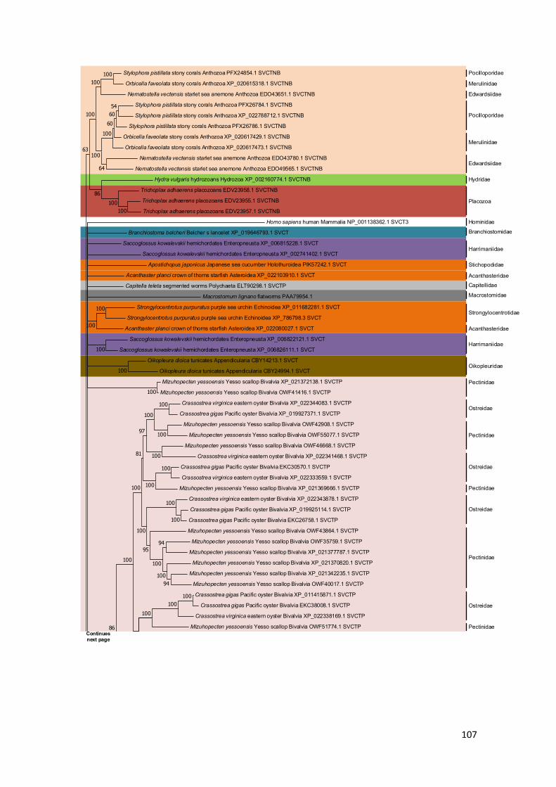
107
Stylophora pistillata stony corals Anthozoa PFX24854.1 SVCTNB
Orbicella faveolata stony corals Anthozoa XP_020615318.1 SVCTNB
Nematostella vectensis starlet sea anemone Anthozoa EDO43651.1 SVCTNB
Stylophora pistillata stony corals Anthozoa PFX26784.1 SVCTNB
Stylophora pistillata stony corals Anthozoa XP_022788712.1 SVCTNB
Stylophora pistillata stony corals Anthozoa PFX26786.1 SVCTNB
Orbicella faveolata stony corals Anthozoa XP_020617429.1 SVCTNB
Orbicella faveolata stony corals Anthozoa XP_020617473.1 SVCTNB
Nematostella vectensis starlet sea anemone Anthozoa EDO43780.1 SVCTNB
Nematostella vectensis starlet sea anemone Anthozoa EDO49565.1 SVCTNB
Hydra vulgaris hydrozoans Hydrozoa XP_002160774.1 SVCTNB
Trichoplax adhaerens placozoans EDV23958.1 SVCTNB
Trichoplax adhaerens placozoans EDV23955.1 SVCTNB
Trichoplax adhaerens placozoans EDV23957.1 SVCTNB
Homo sapiens human Mammalia NP_001138362.1 SVCT3
Branchiostoma belcheri Belcher s lancelet XP_019646793.1 SVCT
Saccoglossus kowalevskii hemichordates Enteropneusta XP_006815228.1 SVCT
Saccoglossus kowalevskii hemichordates Enteropneusta XP_002741402.1 SVCT
Apostichopus japonicus Japanese sea cucumber Holothuroidea PIK57242.1 SVCT
Acanthaster planci crown of thorns starfish Asteroidea XP_022103910.1 SVCT
Capitella teleta segmented worms Polychaeta ELT90298.1 SVCTP
Macrostomum lignano flatworms PAA79954.1
Strongylocentrotus purpuratus purple sea urchin Echinoidea XP_011682281.1 SVCT
Strongylocentrotus purpuratus purple sea urchin Echinoidea XP_786798.3 SVCT
Acanthaster planci crown of thorns starfish Asteroidea XP_022080027.1 SVCT
Saccoglossus kowalevskii hemichordates Enteropneusta XP_006822121.1 SVCT
Saccoglossus kowalevskii hemichordates Enteropneusta XP_006826111.1 SVCT
Oikopleura dioica tunicates Appendicularia CBY14213.1 SVCT
Oikopleura dioica tunicates Appendicularia CBY24994.1 SVCT
Mizuhopecten yessoensis Yesso scallop Bivalvia XP_021372138.1 SVCTP
Mizuhopecten yessoensis Yesso scallop Bivalvia OWF41416.1 SVCTP
Crassostrea virginica eastern oyster Bivalvia XP_022344083.1 SVCTP
Crassostrea gigas Pacific oyster Bivalvia XP_019927371.1 SVCTP
Mizuhopecten yessoensis Yesso scallop Bivalvia OWF42908.1 SVCTP
Mizuhopecten yessoensis Yesso scallop Bivalvia OWF55077.1 SVCTP
Mizuhopecten yessoensis Yesso scallop Bivalvia OWF46668.1 SVCTP
Crassostrea virginica eastern oyster Bivalvia XP_022341468.1 SVCTP
Crassostrea gigas Pacific oyster Bivalvia EKC30570.1 SVCTP
Crassostrea virginica eastern oyster Bivalvia XP_022333559.1 SVCTP
Mizuhopecten yessoensis Yesso scallop Bivalvia XP_021369666.1 SVCTP
Crassostrea virginica eastern oyster Bivalvia XP_022343878.1 SVCTP
Crassostrea gigas Pacific oyster Bivalvia XP_019925114.1 SVCTP
Crassostrea gigas Pacific oyster Bivalvia EKC26758.1 SVCTP
Mizuhopecten yessoensis Yesso scallop Bivalvia OWF43864.1 SVCTP
Mizuhopecten yessoensis Yesso scallop Bivalvia OWF35759.1 SVCTP
Mizuhopecten yessoensis Yesso scallop Bivalvia XP_021377787.1 SVCTP
Mizuhopecten yessoensis Yesso scallop Bivalvia XP_021370820.1 SVCTP
Mizuhopecten yessoensis Yesso scallop Bivalvia XP_021342235.1 SVCTP
Mizuhopecten yessoensis Yesso scallop Bivalvia OWF40017.1 SVCTP
Crassostrea gigas Pacific oyster Bivalvia XP_011415871.1 SVCTP
Crassostrea gigas Pacific oyster Bivalvia EKC38008.1 SVCTP
Crassostrea virginica eastern oyster Bivalvia XP_022338169.1 SVCTP
Mizuhopecten yessoensis Yesso scallop Bivalvia OWF51774.1 SVCTP
100
100
100
100
100
100
100
100
100
97
100
100
81
100
100
94
94100
100
95
100
100
100100
100
100
86
100100
5460
60100
64
100
100
100100
86
63
Pocilloporidae
Merulinidae
Edwardsiidae
Pocilloporidae
Merulinidae
Edwardsiidae
Hydridae
Placozoa
Hominidae
Branchiostomidae
Harrimaniidae
Stichopodidae
AcanthasteridaeCapitellidae
Macrostomidae
Strongylocentrotidae
Harrimaniidae
Oikopleuridae
Pectinidae
Ostreidae
Ostreidae
Acanthasteridae
Pectinidae
Pectinidae
Ostreidae
Pectinidae
Ostreidae
PectinidaeContinues next page

108
Aplysia californica California sea hare Gastropoda XP_012935012.1 SVCTP
Biomphalaria glabrata bloodfluke planorb Gastropoda XP_013067717.1 SVCTP
Lottia gigantea owl limpet Gastropoda ESO95053.1 SVCTP
Lottia gigantea owl limpet Gastropoda ESO93987.1 SVCTP
Lottia gigantea owl limpet Gastropoda ESP03077.1 SVCTP
Mizuhopecten yessoensis Yesso scallop Bivalvia XP_021342820.1 SVCTP
Mizuhopecten yessoensis Yesso scallop Bivalvia OWF47664.1 SVCTP
Mizuhopecten yessoensis Yesso scallop Bivalvia OWF47666.1 SVCTP
Mizuhopecten yessoensis Yesso scallop Bivalvia XP_021359107.1 SVCTP
Haemonchus contortus barber pole worm Chromadorea CDJ84030.1 SVCTP
Ancylostoma ceylanicum nematodes Chromadorea EPB73497.1 SVCTP
Caenorhabditis remanei nematodes Chromadorea OZF97234.1 SVCTP
Ancylostoma ceylanicum nematodes Chromadorea EYC36609.1 SVCTP
Caenorhabditis nigoni nematodes Chromadorea PIC35881.1 SVCTP
Caenorhabditis remanei nematodes Chromadorea EFO85513.1 SVCTP
Caenorhabditis briggsae nematodes Chromadorea CAP22886.2 SVCTP
Ancylostoma ceylanicum nematodes Chromadorea EPB71953.1 SVCTP
Diploscapter pachys nematodes Chromadorea PAV61860.1 SVCTP
Lingula anatina brachiopods Lingulata XP_013379942.1 SVCTP
Lingula anatina brachiopods Lingulata XP_013385284.1 SVCTP
Lingula anatina brachiopods Lingulata XP_013383508.1 SVCTP
Crassostrea gigas Pacific oyster Bivalvia EKC36680.1 SVCTP
Crassostrea virginica eastern oyster Bivalvia XP_022311362.1 SVCTP
Crassostrea gigas Pacific oyster Bivalvia EKC33091.1 SVCTP
Crassostrea virginica eastern oyster Bivalvia XP_022290104.1 SVCTP
Crassostrea virginica eastern oyster Bivalvia XP_022290097.1 SVCTP
Crassostrea virginica eastern oyster Bivalvia XP_022290099.1 SVCTP
Crassostrea virginica eastern oyster Bivalvia XP_022340457.1 SVCTP
Mizuhopecten yessoensis Yesso scallop Bivalvia OWF49908.1
Aplysia californica California sea hare Gastropoda XP_005093309.1 SVCTP
Branchiostoma belcheri Belcher s lancelet XP_019630437.1 SVCT
Branchiostoma floridae Florida lancelet EEN56722.1 SVCT
Ciona intestinalis vase tunicate Ascidiacea XP_002124483.1 SVCT
Ciona intestinalis vase tunicate Ascidiacea XP_002124192.1 SVCT
Ciona intestinalis vase tunicate Ascidiacea XP_018669965.1 SVCT
Ciona intestinalis vase tunicate Ascidiacea XP_002124198.1 SVCT
Ciona intestinalis vase tunicate Ascidiacea XP_002124908.1 SVCT
Ciona intestinalis vase tunicate Ascidiacea XP_002126425.1 SVCT
Ciona intestinalis vase tunicate Ascidiacea XP_002126663.1 SVCT
Hyalella azteca amphipods Malacostraca XP_018026912.1 SVCTP
Hyalella azteca amphipods Malacostraca XP_018009971.1 SVCTP
Capitella teleta segmented worms Polychaeta ELU12246.1 SVCTP
Capitella teleta segmented worms Polychaeta ELT95871.1 SVCTP
Lingula anatina brachiopods Lingulata XP_013391978.1 SVCTP
Capitella teleta segmented worms Polychaeta ELU14337.1 SVCTP
Capitella teleta segmented worms Polychaeta ELU01493.1 SVCTP
Capitella teleta segmented worms Polychaeta ELU13612.1 SVCTP
Capitella teleta segmented worms Polychaeta ELT94865.1 SVCTP
Fopius arisanus wasps c. nsecta XP_011308853.1 SVCTP
Diachasma alloeum wasps c. Insecta XP_015121949.1 SVCTP
Microplitis demolitor wasps c. Insecta XP_008557159.1 SVCTP
Polistes canadensis wasps c. Insecta XP_014609203.1 SVCTP
Polistes dominula European paper wasp Insecta XP_015174395.1 SVCTP
Apis cerana cerana Asiatic honeybee Insecta PBC31908.1 SVCTP
Apis mellifera honey bee Insecta XP_016766931.1 SVCTP
Apis dorsata giant honeybee Insecta XP_006609764.1 SVCTP
Apis florea little honeybee Insecta XP_003698756.1 SVCTP
Bombus terrestris buff tailed bumblebee Insecta XP_003394195.1 SVCTP
Bombus impatiens common eastern bumble bee Insecta XP_003489442.1 SVCTP
Eufriesea mexicana bees Insecta OAD53739.1 SVCTP
Ceratina calcarata bees insecta XP_017888609.1 SVCTP
Megachile rotundata alfalfa leafcutting bee Insecta XP_003705800.1 SVCTP
Dufourea novaeangliae bees Insecta XP_015431505.1 SVCTP
100
100100
100
65
100100
100
100100
100100
92
100
100
100
100
100
100
89
100
100
100
100
100
98
97
100
100
100
100
100100
100
100
100
100
9799
100
99
100100
100
86
94
100
100
98
100
100
91
100100
94
Aplysiidae
Planorbidae
Lotiidae
Pectinidae
Rhabditidae
Lingulidae
Ostreidae
Aplysiidae
Branchiostomidae
Cionidae
Hyalellidae
Capitellidae
Capitellidae
Lingulidae
Haemonchidae
Ancylostomatidae
Rhabditidae
Ancylostomatidae
Ancylostomatidae
Diploscapteridae
Pectinidae
Braconidae
Vespidae
Apidae
Megachilidae
FormicidaeContinues next page
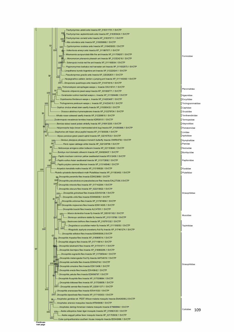
109
Trachymyrmex zeteki ants Insecta XP_018311751.1 SVCTP
Trachymyrmex septentrionalis ants Insecta XP_018355424.1 SVCTP
Trachymyrmex cornetzi ants Insecta XP_018370711.1 SVCTP
Atta colombica ants Insecta XP_018056966.1 SVCTP
Cyphomyrmex costatus ants Insecta XP_018405005.1 SVCTP
Vollenhovia emeryi ants Insecta XP_011867071.1 SVCTP
Wasmannia auropunctata little fire ant Insecta XP_011708207.1 SVCTP
Monomorium pharaonis pharaoh ant Insecta XP_012524218.1 SVCTP
Solenopsis invicta red fire ant Insecta XP_011168554.1 SVCTP
Pogonomyrmex barbatus red harvester ant Insecta XP_011632573.1 SVCTP
Linepithema humile Argentine ant Insecta XP_012220241.1 SVCTP
Pseudomyrmex gracilis ants Insecta XP_020292831.1 SVCTP
Harpegnathos saltator Jerdon s jumping ant Insecta XP_011140392.1 SVCTP
Dinoponera quadriceps ants Insecta XP_014474516.1 SVCTP
Trichomalopsis sarcophagae wasps c. Insecta OXU18741.1 SVCTP
Nasonia vitripennis jewel wasp Insecta XP_001606771.1 SVCTP
Ceratosolen solmsi marchali wasps c. Insecta XP_011506489.1 SVCTP
Copidosoma floridanum wasps c. Insecta XP_014203487.1 SVCTP
Trichogramma pretiosum wasps c. Insecta XP_014234315.1 SVCTP
Cephus cinctus wheat stem sawfly Insecta XP_015595433.1 SVCTP
Orussus abietinus hymenopterans Insecta XP_012278724.1 SVCTP
Athalia rosae coleseed sawfly Insecta XP_012269610.1 SVCTP
Zootermopsis nevadensis termites Insecta KDR08101.1 SVCTP
Bemisia tabaci sweet potato whitefly Insecta XP_018913205.1 SVCTP
Halyomorpha halys brown marmorated stink bug Insecta XP_014283866.1 SVCTP
Diaphorina citri Asian citrus psyllid Insecta XP_017305006.1 SVCTP
Myzus persicae green peach aphid Insecta XP_022167533.1 SVCTP
Danaus plexippus plexippus monarch butterfly Insecta OWR54793.1 SVCTP
Pieris rapae cabbage white Insecta XP_022128799.1 SVCTP
Helicoverpa armigera cotton bollworm Insecta XP_021193845.1 SVCTP
Bombyx mori domestic silkworm Insecta XP_004926407.1 SVCTP
Papilio machaon common yellow swallowtail Insecta KPJ12409.1 SVCTP
Papilio xuthus Asian swallowtail Insecta XP_013172692.1 SVCTP
Papilio polytes common Mormon Insecta XP_013146946.1 SVCTP
Amyelois transitella moths Insecta XP_013199526.1 SVCTP
Plutella xylostella diamondback moth Plutellidae Insecta XP_011553452.1 SVCTP
Drosophila persimilis flies Insecta EDW23868.1 SVCTP
Drosophila pseudoobscura pseudoobscura flies Insecta EAL27338.3 SVCTP
Drosophila miranda flies Insecta XP_017142264.1 SVCTP
Drosophila obscura flies Insecta XP_022210822.1 SVCTP
Drosophila grimshawi flies Insecta EDV93106.1 SVCTP
Drosophila virilis flies Insecta EDW66559.1 SVCTP
Drosophila arizonae flies Insecta XP_017874894.1 SVCTP
Drosophila mojavensis flies Insecta EDW14005.1 SVCTP
Drosophila busckii flies Insecta ALC47051.1 SVCTP
Musca domestica house fly Insecta XP_005181163.1 SVCTP
Stomoxys calcitrans stable fly Insecta XP_013115788.1 SVCTP
Bactrocera latifrons flies Insecta XP_018791332.1 SVCTP
Zeugodacus cucurbitae melon fly Insecta XP_011195055.1 SVCTP
Rhagoletis zephyria snowberry fruit fly Insecta XP_017461274.1 SVCTP
Drosophila willistoni flies Insecta EDW85009.2 SVCTP
Drosophila rhopaloa flies Insecta XP_016969514.1 SVCTP
Drosophila elegans flies Insecta XP_017118814.1 SVCTP
Drosophila takahashii flies Insecta XP_017010711.1 SVCTP
Drosophila biarmipes flies Insecta XP_016960295.1 SVCTP
Drosophila eugracilis flies Insecta XP_017085444.1 SVCTP
Drosophila melanogaster fruit fly Insecta AAF54519.1 SVCTP
Drosophila sechellia flies Insecta EDW42743.1 SVCTP
Drosophila simulans flies Insecta EDX13498.1 SVCTP
Drosophila erecta flies Insecta EDV49642.1 SVCTP
Drosophila yakuba flies Insecta EDW96797.1 SVCTP
Drosophila ficusphila flies Insecta XP_017039884.1 SVCTP
Drosophila kikkawai flies Insecta XP_017036859.1 SVCTP
Drosophila serrata flies Insecta XP_020811371.1 SVCTP
Drosophila ananassae flies Insecta EDV41533.1 SVCTP
Drosophila bipectinata flies Insecta XP_017100303.1 SVCTP
Anopheles gambiae str. PEST African malaria mosquito Insecta EAA08390.2 SVCTP
Anopheles sinensis mosquitos Insecta KFB48488.1 SVCTP
Anopheles darlingi American malaria mosquito Insecta ETN66964.1 SVCTP
Aedes albopictus Asian tiger mosquito Insecta XP_019563125.1 SVCTP
Aedes aegypti yellow fever mosquito Insecta XP_021705930.1 SVCTP
Culex quinquefasciatus southern house mosquito Insecta EDS42686.1 SVCTP
73
100
100
100
95
100
98
100
100
100
100
100
100
100
10091
100
100
93
58
100
100
100
97
100
98
99
10099
88
100
100
92
67
100
98100
100
100
100
100
88
100
100
100
100
100
100
81
100
100
100
100
99
100
100
100
100
99
100
99
63
100
100
98100
100
100
100
100
100
100
97
Formicidae
Culicidae
Tephritidae
Muscidae
Pteromalidae
Agaonidae
Encyrtidae
Trichogrammatidae
Cephidae
Orussidae
Tentheredinidae
Termopsidae
Aleyrodidae
Pentatomidae
LiviidaeAphididae
NymphalidaePieridae
Noctuidae
Bombycidae
Papilionidae
Pyralidae
Plutellidae
Drosophilidae
Drosophilidae
Continues next page

110
Daphnia pulex common water flea Branchiopoda EFX82255.1 SVCTP
Crassostrea gigas Pacific oyster Bivalvia XP_011413077.1 SVCTP
Crassostrea gigas Pacific oyster Bivalvia EKC41034.1 SVCTP
Mizuhopecten yessoensis Yesso scallop Bivalvia OWF56540.1 SVCTP
Mizuhopecten yessoensis Yesso scallop Bivalvia XP_021359049.1 SVCTP
Aplysia californica California sea hare Gastropoda XP_005098105.1 SVCTP
Lottia gigantea owl limpet Gastropoda ESO84592.1 SVCTP
Branchiostoma belcheri Belcher s lancelet XP_019633776.1 SVCT
Branchiostoma floridae Florida lancelet EEN53318.1 SVCT
Branchiostoma belcheri Belcher s lancelet XP_019636191.1 SVCT
Branchiostoma floridae Florida lancelet EEN51097.1 SVCT
Folsomia candida springtails Collembola OXA61626.1 SVCTP
Folsomia candida springtails Collembola OXA58214.1 SVCTP
Folsomia candida springtails Collembola OXA62597.1 SVCTP
Folsomia candida springtails Collembola XP_021961788.1 SVCTP
Folsomia candida springtails Collembola OXA57747.1 SVCTP
Orchesella cincta springtails Collembola ODN05933.1 SVCTP
Folsomia candida springtails Collembola XP_021955363.1 SVCTP
Mus musculus house mouse Mammalia XP_006506197.1 SVCT4
Homo sapiens human Mammalia XP_011542068.1 SVCT1
Homo sapiens human Mammalia NP_005107.4 SVCT2
Strongylocentrotus purpuratus purple sea urchin Echinoidea XP_783160.3 SVCT
Strongylocentrotus purpuratus purple sea urchin Echinoidea XP_011676247.1 SVCT
Varroa jacobsoni mites ticks Arachnida XP_022696844.1 SVCTP
Varroa destructor honeybee mite Arachnida XP_022645394.1 SVCTP
Galendromus occidentalis western predatory mite Arachnida XP_003738546.1 SVCTP
Varroa destructor honeybee mite Arachnida XP_022653396.1 SVCTP
Varroa jacobsoni mites ticks Arachnida XP_022699346.1 SVCTP
Tropilaelaps mercedesae mites ticks Arachnida OQR77963.1 SVCTP
Galendromus occidentalis western predatory mite Arachnida XP_003746715.1 SVCTP
Limulus polyphemus Atlantic horseshoe crab Merostomata XP_022237905.1 SVCTP
Limulus polyphemus Atlantic horseshoe crab Merostomata XP_013794600.1 SVCTP
Limulus polyphemus Atlantic horseshoe crab Merostomata XP_022255137.1 SVCTP
Parasteatoda tepidariorum common house spider Arachnida XP_021001892.1 SVCTP
Pediculus humanus corporis human body louse Insecta EEB16727.1 SVCTP
Diuraphis noxia Russian wheat aphid Insecta XP_015367235.1 SVCTP
Myzus persicae green peach aphid Insecta XP_022177018.1 SVCTP
Tetranychus urticae two spotted spider mite Arachnida XP_015785112.1 SVCTP
100
80
100
94
100
100
83
10095
100
100
100
81
10077
58
100100
100100
100
100
10053
100
100
51
58
0.20
Daphniidae
Ostreidae
Pectinidae
AplysiidaeLottiidae
Branchiostomidae
Isotomidae
Entomobryidae
Isotomidae
Muridae
Hominidae
Strongylocentrotidae
Varroidae
Phytoseiidae
Varroidae
Laelapidae
Phytoseiidae
Limulidae
Theridiidae
Pediculidae
Aphididae
Tetranychidae
Hydrozoa
Anthozoa Placozoa
Cephalochordata Echinodermata
AnnelidaHemichordata
Platyhelminthes
Urochordata
Bivalvia Nematoda
Brachiopoda
Gastropoda
Crustacea Insecta Hexapoda (w/o Insecta) Acari Chelicerata (w/o
Acari and Araneae) Araneae
Supplementary Figure 7 - Non-bilaterian, protostomian and basal deuterostomian Sodium-dependent Vitamin C transporters
(SVCTNB, SVCTP and SVCT) Bayesian phylogeny. The species relevant taxonomic classifications are represented next to the
corresponding sequences. Different taxonomic groups are highlighted with distinct colours, following the colour scheme presented
below the consensus tree. The four outgroup sequences are not highlighted and can be seen spread across the polytomy.