radular teeth of indo-pacific molluscivorous species of conus

HAL Id: hal-02458196https://hal.archives-ouvertes.fr/hal-02458196
Submitted on 28 Jan 2020
HAL is a multi-disciplinary open accessarchive for the deposit and dissemination of sci-entific research documents, whether they are pub-lished or not. The documents may come fromteaching and research institutions in France orabroad, or from public or private research centers.
L’archive ouverte pluridisciplinaire HAL, estdestinée au dépôt et à la diffusion de documentsscientifiques de niveau recherche, publiés ou non,émanant des établissements d’enseignement et derecherche français ou étrangers, des laboratoirespublics ou privés.
Evolution of the Radular Apparatus in Conoidea(Gastropoda: Neogastropoda) as Inferred from a
Molecular PhylogenyYuri Kantor, Nicolas Puillandre
To cite this version:Yuri Kantor, Nicolas Puillandre. Evolution of the Radular Apparatus in Conoidea (Gastropoda:Neogastropoda) as Inferred from a Molecular Phylogeny. Malacologia, 2012, 55 (1), pp.55-90.�10.4002/040.055.0105�. �hal-02458196�

EVOLUTION OF THE RADULAR APPARATUS
IN CONOIDEA (GASTROPODA: NEOGASTROPODA) AS INFERRED FROM A
MOLECULAR PHYLOGENY
Kantor Yu. I.*1, Puillandre N.2
1 A.N. Severtsov Institute of Ecology and Evolution, Russian Academy of Sciences,
Leninski Prosp. 33, Moscow 119071, Russia; 2 UMR 7138, Muséum National d’Histoire Naturelle, Departement Systematique et
Evolution, 55, Rue Buffon, 75231 Paris, France
Running title: Evolution of radula in Conoidea
ABSTRACT
The radular anatomy and evolution of the radular apparatus in predatory marine
gastropods, superfamily Conoidea, is reconstructed on the basis of a molecular
phylogeny, based on three mitochondrial genes (COI, 12S and 16S) for 101 species. A
unique feeding mechanism involving use of individual marginal radular teeth at the
proboscis tip for stabbing and envenomation of prey at the proboscis tip appeared at the
earliest stages of evolution of the group. The initial major evolutionary event in
Conoidea was the divergence to two main branches. One is characterized by mostly
hypodermic marginal teeth and absence of an odontophore, while the other possesses
a radula with primarily duplex marginal teeth, a strong subradular membrane and
retains a fully functional odontophore. The radular types that have previously been
considered most ancestral, “prototypic” for the group (flat marginal teeth; multicuspid
lateral teeth of Drilliidae; solid recurved teeth of Pseudomelatoma and Duplicaria), were
found to be derived conditions. Solid recurved teeth appeared twice, independently, in
Conoidea – in Pseudomelatomidae and Terebridae. The Terebridae, the sister group of
Turridae, are characterized by very high radular variability, and the transformation of the
marginal radular teeth within this single clade repeats the evolution of the radular
apparatus across the entire Conoidea.
Conoidea, Conus, radula, molecular phylogeny, evolution, feeding mechanisms, toxins,
morphological convergence, character mapping
* Corresponding author: [email protected] 1

Introduction
Gastropods of the superfamily Conoidea (= Toxoglossa) constitute a hyperdiverse
group of predatory marine snails that includes in particular the famous genus Conus.
Conoideans are notable for the possession of a large venom gland (Figs. 1-4, vg),
together with a highly modified radula.
An unusual peculiarity of Conoidean foregut anatomy is that the buccal mass with
the radular sac is situated at the proboscis base (Fig. 1 – bm, rsod) and the radula
cannot be protruded through the mouth and used for grabbing and rasping the prey.
The most outstanding character of Conoidea is the unique mechanism of
envenomation of the prey. Some conoideans were long known to use individual teeth at
the proboscis tip for stabbing and injecting neurotoxins into prey (eg. Kohn, 1956). A
marginal tooth is detached from the subradular membrane (when present), transferred
to the proboscis tip (Figs. 2, 4), held by sphincter(s) in the buccal tube (Figs. 2, 4 – bts)
and used for stabbing and envenomating the prey. Use of marginal teeth at the
proboscis tip was observed directly and studied in detail in various species of Conus
that possess elongate, barbed, harpoon-like, hollow marginal teeth (Kohn, 1990; Olivera
et al., 1990; Kohn, Nishi & Pernet, 1999), through which the venom is injected into the
prey. The prey is swallowed whole, sometimes being similar in size to the predator itself
(eg. Kantor, 2007). In these cases the radular apparatus underwent profound
transformation and the odontophore completely disappeared. Another important
character is that the anterior part of the radular diverticulum, which is homologous to the
sublingual pouch of other gastropods, is transformed into a caecum (“short-arm of the
radular sac”), where fully formed marginal teeth are stored prior to their use on the
proboscis tip (Taylor et al., 1993).
Conversely, in many conoideans the radular apparatus includes a radula with a
well developed subradular membrane and a fully functional odontophore with muscles,
thus suggesting that the radula still has some (although maybe limited) function as a
complete organ. As in conoideans with hypodermic teeth, the radula and odontophore
are situated at the proboscis base and normally cannot be protruded through the mouth
(Fig. 1). In conoideans with non-hypodermic marginal teeth (and a functional
odontophore) the separate tooth was very often (in most preserved specimens
examined) found held at the proboscis tip (Figs. 1-2). Teeth were first recorded in serial
histological sections of probosces in several species of Aforia (Cochlespiridae) (Sysoev
& Kantor 1987), Drilliidae (Sysoev & Kantor 1989), and in three additional families, here 2

referred to as Turridae, Clavatulidae and Pseudomelatomidae (Kantor & Taylor, 1991).
The base of the tooth was held by special sphincter(s) and/or an epithelial pad of the
buccal tube. Thus the presence of marginal teeth detached from the radular membrane
and of different morphologies, from solid duplex to specialized hypodermic, used one by
one at the proboscis tip for stabbing the prey can be inferred from anatomical
characters (presence of the sphincters in the buccal tube).
Peculiarities of the feeding mechanisms have been discussed for different groups
of Conoidea (eg. Taylor et. al., 1993; Kantor et al., 1997; Kantor & Taylor, 2002) and
several feeding mechanisms have been suggested. Based on the foregut anatomy, the
use of the teeth at the proboscis tip was found improbable in only two groups of radulate
Conoidea that possess the proboscis – Strictispiridae, and the clade formed by the
genera Pseudomelatoma, Hormospira and Tiariturris, previously recognized as a
separate (sub)family Pseudomelatomidae (Kantor, 1988; Kantor & Taylor, 1991, 1994).
In all others the marginal teeth are used at the proboscis tip.
Despite the fact that the Conoidea are one of the most well-known groups of
Neogastropoda from the point of view of anatomy and lately molecular phylogeny, data
on their feeding and diet are still very limited. With the exception of Conus information
on feeding is available for fewer than 50 species and involved much less direct
observation (eg. Heralde et al., 2010). Most of the conoideans (other than Conus) feed
on sedentary and errant polychaetes, although feeding on other worms (sipunculans
and nemerteans) and even molluscs has been recorded (Miller, 1989, 1990). This
information is derived mainly from gut content analysis.
Radular anatomy of the Conoidea is highly variable both in terms of the number of
teeth in a transverse row and in the shape of the teeth. For a long time, radula
morphology together with shell characters constituted the basis of the higher
classification of the group (e.g. Powell, 1942, 1966; McLean, 1971). Since about 1990
anatomical investigations of conoideans have revealed great variability in foregut
anatomy, and characters defined in these studies have been used to unravel
phylogenetic relationships (Taylor, 1990; Taylor et al., 1993; Kantor, Medinskaya &
Taylor, 1997). Various hypotheses have been proposed concerning the evolutionary
transformations in radular morphology of Conoidea (Shimek & Kohn, 1981; Kantor &
Taylor, 2000; Kantor, 2006). However, one of the reasons for the lack of a clear
understanding of major radular transformations is that radula evolution was inferred
from phylogenetic hypotheses themselves based partially on radular morphology (eg.
3

Taylor et al., 1993). As a consequence many parallel evolutionary transformations
cannot be traced.
The rapid development of molecular phylogenetics provided new insight and
revolutionary changes in our understanding of conoidean evolution. DNA sequences
were first used to infer phylogenetic relationships within genera or subfamilies (e.g.
Espiritu et al., 2001; Duda & Kohn, 2005; Heralde et al., 2007; Holford et al., 2009) and
then among most of the families and subfamilies (as erected by Powell, 1942, 1966;
McLean, 1971; Taylor et al., 1993) of the Conoidea (Puillandre et al. 2008). An updated
molecular phylogeny based on three mitochondrial genes (COI, 12S and 16S) and
including representatives of 103 genera was recently proposed (Puillandre et al., 2011).
The single recognized taxon missing from the analysis was the (sub)family
Strictispiridae McLean, 1971. Most of the clades inferred have robust support that
allowed the status of the different families and subfamilies previously proposed to be
clarified and lead to a new classification of the group into 15 families (Bouchet et al.,
2011).
The molecular framework provides an opportunity to reconstruct the transformation
of the morphological characters and to test previously proposed hypotheses. This
approach has demonstrated the independent loss of the venom gland in two
independent lineages of Terebridae (Holford et al., 2009), but is still not widely used in
Conoidea. Here we attempt a reconstruction of the major morphological transformations
of the radular apparatus in Conoidea based on the molecular phylogeny. For the first
time evolution of the radular apparatus is discussed based on a framework
reconstructed using characters completely independent of anatomy, that is DNA
sequences. Understanding the transformations of the radular apparatus is important not
only for understanding the evolution of the group in general, but also because it may
provide new insight into the factors leading to hyperdiversification of the group that lead
to the appearance of probably the most diverse marine mollusc taxon in terms of
species richness. Furthermore, clarifying the evolution of the group, and in particular the
evolution of characters linked to the venom apparatus, should be of great value in the
discovery of new venom compounds with pharmacological applications (Olivera 2006,
Puillandre & Holford 2010).
4

Material and methods
Radula preparation
Of the 102 ingroup species in the molecular analysis (Puillandre et al. 2011), the
radula of 51 species was examined (in most cases using the same specimens as used
for the molecular analysis), 10 species were radula-less, and for 13 species published
data were used (Table 2). In most cases unavailability of the radula was explained by
destruction of the body during DNA extraction (usually for very small specimens). For
six species radular characters were examined using congeners (usually those whose
position within the genus was confirmed by other molecular data). These species are
marked by an asterisk on the molecular trees. The complete range of variability of the
radula in Conoidea is not, however, covered by the species in our tree (only a single
species of each genus was used in the analysis). Although some species with important
or unique radular morphology are not included in the current analysis, from separate
molecular analyses we know their phylogenetic position and therefore these species are
sometimes mentioned in the discussion.
The radulae were always cleaned with diluted bleach (1 part of commercially
available bleach to 3-4 parts of distilled water). Cleaning radulae in bleach does not
damage radular teeth or the subradular membrane if used in the correct concentration
and if the radulae are not exposed to bleach for a long time. Furthermore, soft tissues
are diluted in bleach rapidly (usually within a few minutes), allowing continuous
observation under the microscope that reveals many important features that otherwise
can easily be overlooked, eg. folding of the radular membrane, attachment of radular
teeth to the membrane, presence of a ligament, etc.
The tiny radulae were cleaned in a drop of water placed on a cover-slip. The
bleach was added either with a syringe or a minute plastic pipette. After dissolving the
soft tissues the radula or separate radular teeth were transferred with a needle or single
hair into a drop of clean water on the same cover-slip. This minimizes the chance of
losing the small radulae. Two changes of water were usually enough to rinse the radula.
After rinsing, the radula was partially pulled out of the drop so that the extruded part
adheres to the glass by surface tension. This permits the radula membrane to be more
easily unfolded with a single hair, and allows individual teeth to be placed in the desired
position prior to drying. The radula was then completely pulled out of the water drop and
allowed to dry. The cover-slip was then mounted on the stub. Although simple, this 5

method provides excellent results, allowing manipulating objects smaller then 100 µm in
length.
Acronyms for depositories of voucher specimens
INVEMAR – Instituto de Investigaciones Marinas y Costeros, Colombia
MNHN -- Muséum National d’Histoire Naturelle Paris, France
MNZ – Museum of New Zealand Te Papa Tongareva, Wellington, New Zealand
NHMUK – Natural History Museum, London, UK
USNM – National Museum of Natural History, Smithsonian Institution, Washington
DC, USA
Tree mapping
Nine radular morphology characters coded as 31 character states (Table 1) were
used for reconstruction of the radular transformation. Characters were mapped on the
tree of Puillandre et al. (2011) using Mesquite Version 2.74 (Maddison & Maddison,
2007-2010), using the option ‘‘tracing character history’’ and the parsimony ancestral
reconstruction method. Most of the characters were treated as unordered. For the
characters describing central and lateral teeth (characters # 1 & 2 in Table 1) the
stepmatrix model was tried in addition to the unordered; it allows interdicting some of
the transformation sequences, in our case from absent to present, that is interdicting re-
appearance of central and lateral teeth after they had been lost.
The familiar classification accepted here is that of Bouchet et al. (2011). For
convenience, in addition to the families recognized by Bouchet et al. (2011) we refer to
major clades A and B (without attributing any taxonomic status to them) that are
different in many aspects of anatomy and radular morphology.
Results
Although the radulae of Conoidea have been described in many publications, the
thorough use of scanning electron microscopy revealed many previously overlooked
characters and allowed new interpretations of structures already described. Therefore
we provide here a much updated overview of the radular characters. The evolution of
each of the nine characters analysed is described.
Character 1. Central tooth of the radula (Figs. 5 and 6).
6

A central tooth is present in all outgroups. It is absent in clade A and present in
some groups of clade B. The morphology of the central tooth is rather variable in
gastropods. Two major types are found in the outgroups, both multicuspid. Describing in
detail the morphology of the teeth of groups other than Conoidea (eg. Bandel, 1984) is
outside the scope of the current paper. In Conoidea the situation with the central tooth is
very confusing. In some species the central tooth appears as a well-defined structure.
Two major types of such teeth can be identified: narrow unicuspid (shield-like with a
small cusp and sometimes with additional serrations) (Figs. 7-9, 12-13); and broad
unicuspid, with a large curved cusp and well defined lateral flaps (Fig. 14).
Conversely, in a number of Conoidea from clade B there are vestigial rather
indistinct structure(s) occupying the middle portion of the subradular membrane (Fig.
29, 31 – marked with arrows). In some cases they can be hardly seen without staining
the radular membrane or even under SEM. These structures may be either a much
reduced broad central tooth with lateral flaps with or even without a central cusp, or
alternatively three teeth – vestigial central and vestigial laterals (see below) partially or
completely fused (Fig. 17-18). Vestigial structures were found in some
Pseudomelatomidae, Clavatulidae and Turridae (Iotyrris Medinskaya & Sysoev, 2001).
Narrow unicuspid central teeth are found in most Drilliidae and some Turridae (in
our dataset exemplified by species of Xenuroturris Iredale, 1929 and Turridrupa Hedley,
1922 – Figs. 34-35, as well as Gemmula and Turris). A broad well defined central tooth
was recorded in three clades – in some Pseudomelatomidae (Pseudomelatoma,
Hormospira Berry, 1958 and Tiariturris Berry, 1958 – Fig. 14), Cochlespiridae (Fig. 40),
and Gemmuloborsonia Shuto, 1989 (not currently attributed to any family – Fig. 32). In
these genera the posterior edge of the tooth, bearing the cusp, is well elevated over the
membrane.
Four analyses were performed. In the first, the inconspicuous central teeth, when
recognized, were considered as unicuspid narrow (lateral flaps were considered as
vestigial plate-like lateral teeth), and character states were unordered (Fig. 5). The tree
was 13 steps long and suggested that the plesiomorphic condition of the character in
clade B is the absence of the central tooth and the central teeth originated
independently in 8 clades. Since central teeth are present in the outgroups this can be
considered a reversion. However, the presence of numerous reversions within clade B
seems rather unlikely.
Therefore the second analysis was performed with reversions interdicted
(stepmatrix parsimony model) (Fig. 6). The tree was 18 steps long (5 steps longer) and 7

the analysis did not allow reconstruction of the single most parsimonious state in Clade
B, since a multicuspid tooth (characteristic for Neogastropoda), a narrow unicupsid
tooth and a broad unicuspid tooth are equally parsimonious. This analysis suggested
independent losses of central teeth in several clades: most species of
Pseudomelatomidae, Leucosyrinx Dall 1889, Horaclavidae and Terebridae, as well as in
some species of Turridae and Clavatulidae.
The third and forth analyses were with alternative coding of the characters and
with unordered and stepmatrix parsimony models correspondingly. Species with
vestigial central structures were coded as having the broad unicuspid teeth. The
reconstruction produced longer trees (15 and 20 steps, respectively), which were
therefore rejected.
Character 2. Lateral teeth of the radula (Fig 27). Lateral teeth are present in all outgroups. They are absent in Clade A and present
in some groups of Clade B.
There are two major types of lateral teeth among the ingroup species in our tree. In
Drilliidae they are well formed and multicuspid, completely separate from the central
tooth (Figs 7-10). In all others (some Pseudomelatomidae, Turridae and Clavatulidae)
they are very weak, plate-like, non-cuspidate and usually completely or partially fused
with the central tooth (when it is present), forming the "central formation" (Kantor, 2006)
(see the discussion below). In some groups the laterals are so weak that their presence
can be revealed only by staining of the subradular membrane. This is particularly
characteristic for Clavatulidae, in which they were first revealed by Kilburn (1985).
A first analysis with character states unordered suggested the absence of lateral
teeth is ancestral for the Conoidea and independent appearance of the lateral teeth
occurred independently in five clades (all in clade B). Since central teeth are present in
outgroups these events would be considered as a reversions.
The second analysis was performed with reversions interdicted (Fig. 27) and
resulted in a longer tree (17 steps vs 9 in the previous analysis). The analysis did not
allow reconstruction of the single most parsimonious state for the entire Conoidea nor
for Clade B since multicuspid neogastropod type teeth, unicuspid neogastropod type
teeth and plate-like lateral teeth were equally parsimonious.
For most of clade B conoideans (except Cochlespiridae) the most parsimonious
state was plate-like teeth, while multicuspid teeth seems to re-appear in the branch that
combines Drilliidae and Pseudomelatomidae. Lateral teeth are independently lost in 8

several lineages – in most species of Pseudomelatomidae, Horaiclavidae, Terebridae
and others.
In all the species in our tree, the presence of the lateral teeth was combined with
the presence of the central tooth, which is not the case for all conoideans (see
discussion).
Character 3. Morphology of the marginal teeth (Fig. 43). Despite great variability of the marginal teeth in Conoidea, four major types can be
recognized:
a. duplex teeth, consisting of a major element (limb) attached to the
subradular membrane along most of its length (Figs. 23-24 – ml) and the
accessory limb, that is the thickened edge of the major element, usually
more or less elevated above the membrane (Fig. 23-25 – al). These teeth
demonstrate the great variability in shape (see Kantor, Taylor, 2000) (Figs.
28, 30, 33, 34-42) and have often been referred to as "wishbone" (e.g.
Powell, 1966). The term was coined based on the misconception that the
limbs are separate and the tooth is actually bifurcating, as it appears under
the light microscope (most of Clade B). In some cases the limbs are nearly
equally developed and the teeth attain a trough-shape, becoming “semi-
enrolled” (see below, Figs. 25-26). In the analysis this condition was also
coded as “duplex teeth”.
b. flat simple plate-like teeth (some Drilliidae) (Fig. 7).
c. Solid, recurved teeth, attached to the membrane along part of the length,
sometimes with a slightly broadened base that is actually attached to the
membrane (some Pseudomelatomidae -- Pseudomelatoma, Hormospira
Berry, 1958 and Tiariturris Berry, 1958 – Fig. 14; some Terebridae --
Euterebra and Duplicaria – Fig. 48, the latter not represented in our tree).
d. Hypodermic teeth. These are hollow enrolled teeth (Figs. 47, 49, 50-53)
attached to the subradular membrane only by a narrow base or through a
flexible stalk, the ligament (Fig. 51) (some Borsoniidae, Conidae and
others).
The analysis was not able to resolve the single most parsimonious state for the
entire Conoidea, but suggested that a duplex tooth is the most parsimonious state for
clade B. Flat teeth are characteristic only for some Drilliidae and according to the tree 9

they are an autapomorphy of several species, thus suggesting their derivation from
duplex teeth. Similarly, solid recurved teeth originated from duplex teeth twice
independently in the evolutionary history of Conoidea – in some Pseudomelatomidae
and Terebridae. Hypodermic teeth are a synapomorphy of clade A but also appeared
independently in Terebridae. The marginal teeth have been also lost several times
independently (at least three times in clade B and twice in Terebridae).
Character 4. Morphology of duplex marginal teeth (Fig. 44). Duplex teeth are very variable in morphology. The difference in appearance is
mainly determined by relative size and shape of the accessory limb, as well as the
degree of its elevation above the surface of the subradular membrane. The
representation of the taxa in our tree does not allow more detailed analysis, although
the general patterns can be traced.
We recognize four subtypes of duplex teeth, although much more variation can be
found in other Conoidea not included in our study.
The first subtype is characterized by equal or nearly subequal development of
major and accessory limbs. This type of tooth is found in Cochlespiridae (Figs. 40-41)
(represented only by two genera in our tree) in which the teeth are characterized by
relatively large size of the accessory limb (Fig. 41 – al) that is of nearly the same size as
the major limb (Fig. 41 – ml). This produces the appearance of the tooth folded
lengthwise. The analysis suggested that it is an apomorphy of the clade.
A similar subtype, although having a different appearance, is the so-called semi-
enrolled tooth (Taylor et al., 1993; Kantor, Taylor, 2000). In this type the accessory limb
is also subequal in size to the major limb (Figs. 25-26), but the lengthwise folding is
much less tight and the teeth attain a trough-like shape. According to the analysis, this
type of tooth appeared several times independently in clade B – in some genera of
Pseudomelatomidae (in the clade Pilsbryspira McLean, 1971, Zonulispira Bartsch, 1950
and Pyrgospira McLean, 1971 – Fig. 21, and independently in Ptychobela Thiele, 1925
(Fig. 22), Cruziturricula Marks, 1951 (Fig. 11), Imaclava Bartsch, 1944, and Iotyrris
(Turridae).
The most parsimonious plesiomorphic state for most of clade B (except
Cochlespiridae) is the duplex marginal tooth with unequal sizes of the major, larger limb
and smaller accessory limb (“unequal limbs” in Fig. 44). Depending on the degree of
difference the tooth may look very different, in its most extreme state being nearly flat
with a narrow and very slightly raised accessory limb (eg. Funa Kilburn, 1988 – Fig. 37). 10

In most groups the accessory limb is comparatively large and the tooth edge adjoining
the limb is significantly raised above the membrane, so that the accessory limb
occupies the dorsal position on the major limb (eg. 20, 39). Different teeth of this
subtype have been thoroughly illustrated by Kantor et al. (1997) and Taylor et al.
(1993).
A characteristic type of duplex tooth is found in the genera Comitas Finlay, 1926
and Knefastia Dall, 1919 (Figs. 16, 23). The teeth are nearly flat, broadly elongate, with
the major limb thickened at the tip and along one side, while the accessory limb is
represented by the narrow thickened margin of the tooth that does not reach the tip of
the tooth but is inserted in a shallow and narrow socket, slightly overlaying the
thickened part of the major limb.
The several following characters (5-8) apply to hypodermic teeth only.
Hypodermic teeth are hollow enrolled marginals, usually with overlapping edges
(exemptions are some representatives of Mangeliidae – Fig. 50, not present in our
dataset) and open at both the tooth base and near the tip. Teeth of this morphology are
found mostly among representatives of clade A and in most radulate Terebridae.
Nevertheless, in at least one genus of Clavatulidae (Toxiclionella Powell, 1966) and in
Cruziturricula the marginal teeth are very similar in anatomy (Figs. 11, 32). The major
difference between hypodermic teeth in clade A and Terebridae on one hand and in
Toxiclionella and Cruziturricula on the other is the form of attachment to the radular
membrane. In the former, the teeth are attached only by the base, while in the latter
along most of their length. This suggests different evolutionary origins of such teeth
(Kantor & Taylor, 2000).
The anatomy of hypodermic teeth has been successfully used in the phylogenetic
reconstructions of Conidae (Kohn et al., 1999) and high congruence was found between
feeding type and tooth anatomy in Conus (eg. Duda et al., 2001). At the same time
there is a limited number of characters, that are widespread across multiple families
posessing hypodermic teeth.
Character 5. Presence of a spur (Fig. 45). The basal spur is an anterior projection on the base of the tooth (Fig. 53). Its
function is probably to tighten the grasp of the proboscis tip during feeding and thus to
prevent premature loss of the tooth from the proboscis (Kohn et al., 1999). Our analysis
11
suggested several independent origins of this character – in Conidae, Borsoniidae,
Mangeliidae and Terebridae (Fig. 45).
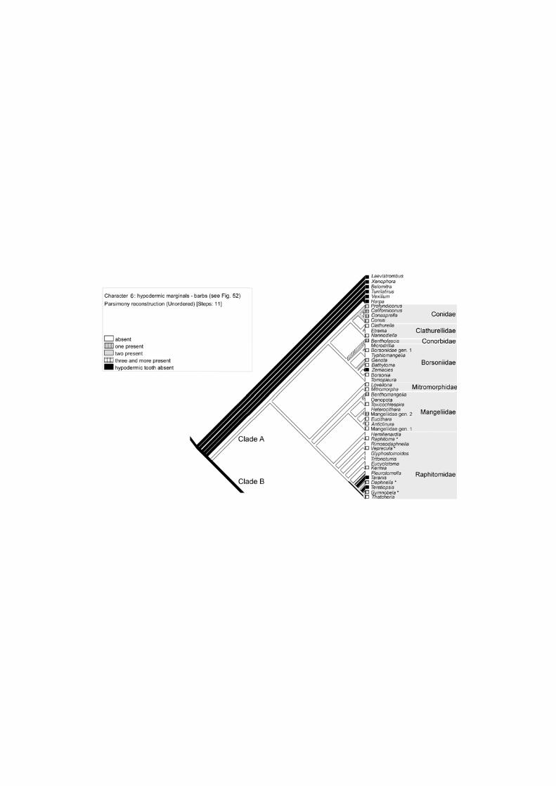
Character 6. Presence of a barb(s) (Fig. 54). A barb is a projection from the shaft of the tooth that has a cutting edge and joins
the shaft at an acute angle (Fig. 52). There can be from 0 to 5 barbs (Conus
californicus) (Kohn, Nishi & Pernet, 1999). The analysis suggests that the barbs
appeared independently in every family of clade A except Mitromorphidae, in which they
are absent.
Character 7. Presence of a blade (Fig. 55). The blade is a projection from the shaft of the tooth that has a cutting edge and
joins the shaft at an obtuse angle (Kohn et al.,, 1999) (Fig. 53). In some cases the
distinction between a barb and a blade is subtle. The analysis suggested that a blade
originated independently twice – in clade A and in Terebridae. Presence of a blade was
the most parsimonious ancestral state for clade A (Fig. 55).
Character 8. Presence of a ligament (Fig. 56). The ligament is an elongate, flexible stalk, attached to the base of the tooth and to
the membrane, when the latter is present (Fig. 51). In fresh radulae the ligament can be
subcircular in cross-section, but when air-dried the ligament is usually flat and
membrane-like. The presence of a ligament is often not recorded during radula
description.
A ligament has so far only been recorded in clade A, and its presence is the most
parsimonious ancestral state for the clade. It is present in Conidae, Borsoniidae,
Mitromorphinae and at least in one species of Raphitomidae (Thatcheria mirabilis – see
Taylor et al., 1993 -- fig. 23 c).
Character 9. Use of marginal teeth at the proboscis tip for stabbing prey (Fig. 57).
The analysis suggested that the use of marginal teeth at the proboscis tip is the
most parsimonious plesiomorphic state for the entire Conoidea.
12

Discussion
Origin of the conoidean feeding mechanism and general evolutionary trends
Use of separate marginal teeth one by one at the proboscis tip is one of the most
intriguing characters of conoidean evolution. Taylor et al. (1993) suggested that
conoidean feeding mechanisms gradually evolved within the group, but first appeared in
the early stages of conoidean evolution. We traced the use of separate marginal teeth
at the proboscis tip on a molecular tree using available published and unpublished
anatomical data. As was mentioned in the introduction, the base of the tooth is held by
special sphincter(s) and/or an epithelial pad of the buccal tube. Thus use of teeth at the
proboscis tip can be inferred from anatomical characters (presence of the sphincters of
the buccal tube). Although we do not have anatomical data for every species included in
our analysis, they are available for species of most of the genera and for every family,
allowing us to extrapolate to the remaining members of the clade. The analyses clearly
suggested that the origin of the peculiar feeding mechanism is an apomorphy of
Conoidea in general, and it appeared before the divergence of the two major clades (A
and B) (Fig. 57).
The initial divergence of Conoidea into clades A and B (that is the clades with
primarily hypodermic and primarily duplex marginal teeth, respectively) is an
unexpected inference from the conoidean molecular phylogeny. In previous cladistic
analyses based on morphological characters the representatives of clade A (referred to
as family Conidae by Taylor et al., 1993) appeared as a terminal clade, suggesting the
gradual transformation of radular morphology. The molecular-based results contradict
this hypothesis.
Although there is no fundamental difference in feeding mechanism between clades
A and B, there are nevertheless important differences in the anatomy of the radular
apparatus. In clade B, the radular apparatus consists of a more or less well-developed
odontophore with supporting musculature (it is absent only in a few species that lack a
radula, eg. Horaiclavus phaeocercus Sysoev, 2008, Horaiclavidae – Fedosov & Kantor,
2008), moderately strong continuous radular membrane and (not always) presence of
central and/or lateral teeth. An important character of the radula is that the marginal
teeth are attached to the membrane along a significant or even most of their length.
Exceptions are some Terebridae (discussed below).
13

In clade A an odontophore with muscles is absent and the subradular membrane is
very thin to vestigial. The teeth are attached to the membrane only by the very narrow
base of the tooth, sometimes through a flexible stalk – the ligament. The attachment of
the marginal tooth (of hypodermic type) to the membrane only by the base facilitates
rolling of the tooth, which may be formed by a few completely overlapping rolls in
Conidae (for more details see Kantor & Taylor, 2000), although usually the edges of the
tooth plate are only slightly overlapping (Fig. 51 – cross sections through the tooth).
Thus the molecular analysis suggests that appearance of the conoidean feeding
mechanism was the key apomorphy of the superfamily. We suggest that it may have
greatly improved prey capture and allowed rapid diversification and species radiation
that resulted in the modern hyperdiverse group that includes about 4600 Recent
described species and a larger number of still unnamed ones (Bouchet et al., 2009).
The splitting of Conoidea into two groups with different radular types and foregut
anatomies was the first major evolutionary event, taking place at the earliest stage of
evolution of the group after the initial appearance of the unique feeding mechanism.
Transformation of the non-hypodermic marginal teeth (clade B)
Non-hypodermic marginal teeth are found in clade B and are very variable,
although they can be reduced to 3 major morphological types – duplex (including semi-
enrolled), solid recurved and flat simple plate-like.
The solid recurved teeth were previously considered as the prototype marginal
teeth in Conoidea (eg. Kantor & Sysoev, 1990) and with the exception of three genera,
which were united in the Pseudomelatomidae (sensu Taylor et al., 1993), were also
found in some Terebridae (genera Duplicaria and Euterebra). The tooth morphology in
these two lineages is very similar. Plotting the character on a molecular tree clearly
indicated that this tooth type appeared independently twice and analysis suggested that
this type of marginal tooth developed secondarily from duplex teeth. Some important
differences can also be mentioned: in Pseudomelatomidae sensu Taylor et al., 1993 the
radula is long (about 100 rows of teeth in Tiariturris) and possesses large and broad
unicuspid central teeth; in Terebridae with these solid recurved teeth the radula is short
(about 20 rows of teeth) and lacks the central teeth.
The functioning of this type of radula remains largely unknown. The shape of the
rather strongly recurved teeth precludes their use separately at the proboscis tip. In
Duplicaria and Euterebra the venom gland as well as proboscis is absent, while the 14

odontophore is present (Rudman, 1969; Taylor, 1990). In Pseudomelatoma and
Hormospira the venom gland is present and well developed, the proboscis is long, but
the buccal tube lacks the sphincter that can hold the tooth (Kantor, 1988).
The three genera comprising Pseudomelatomidae sensu Taylor et al., 1993 (and
encompassing only six Recent species) have very limited distribution – all are found in
the Panamic province. It is possible that they comprise a local radiation connected to a
shift to some peculiar type of prey, although this needs further confirmation. The
presence of a large odontophore suggests that radulae with solid recurved marginal
teeth are primarily used as an entire organ (probably for tearing and rasping the prey).
In contrast, Terebridae with this type of radula (Taylor, 1990) have a broad distribution
in the Indo-Pacific – species are found from South Africa to Japan, including the
Solomon Islands and Oman, and some species have a broad Indo-Pacific distribution.
They lack a proboscis and venom gland and probably are more general feeders,
involving their long labial tube in prey capture.
Flat marginal teeth have also been considered as a prototype for the duplex teeth
(Taylor et al., 1993) and were found in some Drilliidae (among the genera used in our
analysis in Agladrillia, Splendrillia and Cerodrillia). The analysis demonstrates that this
condition is autapomorphic and this type of tooth most probably originated by
simplification of the duplex teeth, the presence of which is the plesiomorphic state for
the entire clade B and Drilliidae in particular.
Within the most common duplex type of marginal teeth many different
morphologies can be recognized (Figs. 16-21, 34-42), although they are very similar in
mode of formation. Kantor & Taylor (2000) studied maturing teeth along the radular
membrane and showed that they develop from a flat plate by thickening of the tooth
edges and elevation of the posterior edge (additional limb) from the membrane. This
thickening of the margins of duplex teeth, folding along the length and partial enrolling
(in the semi-enrolled teeth) can be explained as features that provide mechanical
strength. In mechanical terms, a simple flat plate is less stiff and more likely to buckle
when subject to a compressive force, than one with thickened edges or a hollow
cylinder (Wainwright et al., 1976). Teeth used at the proboscis tip need to be rigid to
pierce the prey’s integument.
Although marginal tooth shape appeared to be rather homoplastic, it is
characteristic for some of the well defined clades. For example, in Cochlespiridae the
accessory limb is large, nearly equal in size to the major limb. Therefore the tooth
appears to be folded lengthwise with a solid tip. Clavatulidae also possess rather 15

distinctive duplex teeth with a sharp-edged major limb and a deep socket where an
accessory limb is inserted, often with angulation distal to the socket. The well-supported
clade including genera Pilsbryspira, Zonulispira, and Pyrgospira is characterized by
semi-enrolled teeth with a similar shape and was previously considered a separate
subfamily Zonulispirinae. In some duplex teeth (eg. in Funa and Cheungbeia) the
secondary limb is minute, nearly obsolete. Kantor & Taylor (2000) suggested, based on
a morphological tree, that this is a derived state. The current analysis confirms this
hypothesis.
In the single genus Toxiclionella Powell, 1966 (not present in our analysis),
referred to Clavatulidae and that still possesses an odontophore, the teeth are hardly
distinguishable from true hypodermic teeth (Fig. 32) having two barbs at the tip and a
subterminally opening tooth canal. The marginal teeth are attached to the membrane
along most of their length, similarly to other species of clade B.
Transformations of the central radular segment (Clade B)
The peculiar feeding mechanism of Conoidea is associated with the anatomy of
the foregut. In all Conoidea the buccal mass with the radular diverticulum (and
odontophore with muscles in clade B) is situated at the proboscis base and often behind
proboscis in its contracted state. The odontophore cannot be protruded through the
mouth and therefore the radula has limited function as an integrated whole organ in
most conoideans. A few exceptions occur sporadically in different clades. In these
groups the buccal mass is secondarily shifted anteriorly or is able to evert through the
mouth along with the walls of the buccal tube (eg in Funa latisinuata – Taylor et al.,
1993, fig. 14).
The limited functioning of the radula as an integrated organ in adults is indirectly
confirmed our not observing traces of worn teeth (marginal, and central or laterals when
present) in radulae examined by us (except Drilliidae, see below). Thus the central and
lateral teeth are hardly functional in Conoidea (while the marginals detached from the
membrane are used individually at the proboscis tip) and therefore the adaptive value of
their morphological transformations may be reduced. This may explain the high
variability of the morphology of the central segment of the radula that includes the
central and lateral teeth.
In general the evolutionary transformation of the central (and lateral teeth) in clade
B is complicated. Analysis of the morphology of the central tooth with unordered 16

character states produced a shorter tree, but suggested initial reduction of the tooth in
the entire Conoidea and subsequent multiple (eight in our dataset) independent re-
appearances in different clades, that is multiple reversions (Fig. 5). This is not highly
parsimonious and it is especially difficult to explain these reversions from a functional
point of view due to the limited functions of the central and lateral teeth. On the contrary,
non-functionality of the central segment seems to be congruent with reduction and
complete loss of the central segment in different lineages as was suggested by the
stepmatrix analysis (Fig. 6).
Similarly we found the numerous losses of the lateral teeth (stepmatrix analysis –
Fig. 27) more probable (although less parsimonious) than initial reduction of the teeth in
the entire Conoidea and independent re-appearance in five clades (analysis with
unordered character states). Only in Drilliidae do the lateral teeth seem to be functional
(see below), while in other clades they are very weak plate-like structures, sometimes
appearing only as inconspicuous thickenings of the subradular membrane (eg in
Pusionella compacta – Fig. 29, vlt).
The central tooth is a highly variable structure in Conoidea, ranging from very
narrow unicuspid to broad with a large cusp and well developed lateral flaps. Kantor &
Sysoev (1991) and Taylor et al. (1993) suggested that the broad central tooth in some
groups may be the result of fusion of paired lateral teeth with the narrow unicuspid
central tooth. In some species, attributed here to Pseudomelatomidae (Crassiclava
turricula – Figs. 19-20, Antiplanes sanctiioannis (Smith, 1875) – Kantor & Sysoev, 1991,
figs. 27-28; Comitas onokeana vivens Dell, 1956 – Fig. 15), clearly separate plate-
shaped lateral teeth without cusps were found. In these species the central tooth (cusp)
is absent. In Comitas Finlay, 1926 and the related genus Knefastia Dall, 1919
intermediate stages can be found. In K. tuberculifera in addition to a very weak and
reduced central tooth (Fig. 18, ct) vestigial lateral plates (teeth) can be observed (vlt); in
Comitas pachycercus Sysoev et Bouchet, 2001, Comitas murrawolga (Garrard, 1961)
and Comitas sp. (Figs. 16-17) the central structure looks like a well defined central tooth
with a narrow cusp and broad lateral flaps. This transition row in closely related species
suggests a composite structure of the “central tooth” in this group, formed by fusion of
the central narrow tooth with cuspless laterals. Kantor (2006), after examination of the
radula of 64 species from 7 genera of Turrinae (= Turridae in the current classification),
confirmed the composite structure of the "central" tooth, which is formed by the fusion of
central and lateral teeth and suggested calling this structure the “central formation”. In
Turridae transitional conditions ranging from a clearly tripartite structure with a gap 17
between the cusp (=central tooth) and the lateral flaps (=lateral teeth) to a seemingly
solid central tooth occur, sometimes within a single genus. This range can be illustrated
by the different species of Turridrupa Hedley, 1922 (see Kantor, 2006, fig. 4) or
Gemmula (herein, Fig. 58-63). In Gemmula unilineata Powell, 1967 the central tooth is
well separated from the narrow laterals (Figs. 58-59); in Gemmula sp. 3 (sensu Kantor
2006) the lateral teeth are close to the central tooth, but separated by gaps (indicated
by arrows on Fig. 63), and finally in Gemmula rarimaculata the lateral and central teeth
are fused (Figs. 60-61). Finally in three clades – in Cochlespiridae, in that combining
Pseudomelatoma and Tiariturris (Pseudomelatomidae sensu Taylor et al., 1993), and in
Gemmuloborsonia there is no indication that the central tooth has a composite origin.
The posterior margin, bearing the cusp is equally developed along its width and
elevated over the radular membrane. Without further information we conclude that these
groups possess the broad unicuspid central tooth, while the lateral teeth are absent.
This conclusion could be rebutted by observations of embryonic development of the
radula, that may demonstrate a composite structure of the central teeth.
One of the unexpected results of our analysis is the possible secondary origin of
the multi-cuspidate separate and well formed lateral teeth in Drilliidae. Previously this
type of the tooth was considered prototypic for turrids (Powell, 1966; Kantor, Sysoev,
1991), and this hypothesis was the rationale for placing the Drilliidae as a separate
family from the other Turridae sensu Taylor et al. 1993. According to the analysis, the
multicuspid lateral teeth may be the ancestral state for clade B; they then disappeared
and re-appeared again in the Drilliidae plus Pseudomelatomidae clade, though
multicuspid teeth are present only in the former family. However, different runs of the
analysis with different coding of radular character states in the outgroups suggested
different most parsimonious plesiomorphic conditions in clade B, but in none of the runs
were multicuspid teeth the single plesiomorphic state. One of the reasons for these
inconclusive results may be the fact that the homology of different teeth in
Neogastropoda (including outgroups) is not yet finally established and the relationships
within the entire Neogastropoda are far from resolved.
From a general point of view it seems more probable that well pronounced
multicuspid lateral teeth in Drilliidae is the plesiomorphic condition in Conoidea, retained
due to peculiarities of their feeding mechanism. Unfortunately there is practically no
information on the diet and radula functioning of drilliids. The only published record is
that of Maes (1983) of the gut content of Drillia cydia (Bartsch, 1943). Finding intact
prey (sipunculid) in the posterior oesophagus suggested that the radula is used for 18

gripping and/or piercing, not rasping or tearing. Drilliidae studied by us possess large
and powerful odontophores, which may suggest active use of the radula as an
intgegrated whole organ. To confirm this we carefully examined the bending plane of
the radulae. In many species the laterals and even the central teeth were badly
damaged (Fig. 12 – broken parts of the teeth are marked by small arrows); damage on
the marginal teeth in the same specimens was not observed. These observations
suggest that lateral (and even small central) teeth are functional, but without additional
data on feeding in this group it is impossible to draw any final conclusions about the
mode of functioning.
One important question remains: why in clade B conoideans, which possess in
general the same feeding mechanism as clade A species, are the odontophore and its
musculature retained? The odontophore varies in size from large in Drilliidae and some
Pseudomelatomidae (Pseudomelatoma) (Taylor et al., 1993) to very small or nearly
obsolete (eg. in some Hindsiclava, Pseudomelatomidae – Kantor et al., 1997). There
are no data that explain this phenomenon. From what has been said above it is clear
that the functioning of the radula as an ingtegrated organ may be limited, possibly to
transferring the swallowed prey from the buccal cavity, situated at the proboscis base,
further to oesophagus. Only in very few conoideans is the buccal cavity plus radula
either shifted to the proboscis tip or able to evert through the mouth (for details see
Taylor et al., 1993). From the point of view of speciation, clade A conoideans are more
diverse, including 202 genera (not counting 82 genera recognized in the Conidae, the
phylogeny and taxonomy of which is not yet finally revised) versus 180 genera in clade
B. Thus absence of the odontophore does not seem to limit prey capture and feeding.
Moreover, clade A conoideans seem to have a broader prey range, including other
gastropods (numerous Conus species) and bivalves (Phymorhynchus, Raphitomidae –
Fujikura et al., 2004) and even fish (several species of Conus).
In some species of Conus there are ontogenetic changes of radular teeth probably
related to changes in diet (summarized by Nybakken, 1990) and therefore in prey
capture mechanism. We may imagine that similar changes can occur in clade B and
that the prey capture and feeding mechanisms may differ between young individuals
and adults and that at some ontogenetic stage the odontophore may be fully
operational. This supposition needs careful research to detect any ontogenetic radular
and foregut anatomy changes.
19

Transformation of the hypodermic marginal teeth
Hypodermic marginal teeth are found in clade A and some Terebridae. The
morphology of the hypodermic teeth is extremely variable and was traditionally used for
taxonomy. Some hypodermic teeth are very simple, semi-enrolled. Such teeth are found
among Mangeliidae (eg. Mangelia – Fig. 50). Unfortunately none of these species is
present in our molecular tree.
The hypodermic teeth of Conidae s.s. have been described in great detail and
correlation between tooth morphology and diet has been demonstrated (eg. Nybbaken,
1990). The representation of genera in our data matrix is relatively sparse and therefore
we could only trace a limited number of characters of the hypodermic teeth. It appeared
that the spurs and barbs of the teeth are homoplasic and evolved independently several
times. No clear trends were obvious, possibly due to the incompleteness of our dataset.
Terebridae radiation
One of the most remarkable findings of the molecular analysis (Puillandre et al.,
2008, 2011) is that the Terebridae do not represent a totally separate lineage, but are
included in clade B and are probably sister to the Turridae. Consequently, the
evolutionary history of radular transformation is rather different from that traditionally
accepted.
Common among all terebrids is complete loss of the central and lateral teeth. In
our analysis the first taxon to diverge among the Terebridae is Euterebra tristis
(Deshayes, 1859), characterized by the solid recurved teeth, superficially similar to that
in Pseudomelatoma and related genera (Pseudomelatomidae). Our analysis was not
able to resolve the most parsimonious ancestral state for Terebridae. Before the
molecular analysis of all Conoidea was performed (Puillandre et al., 2011), more
detailed analysis of Terebridae has been conducted (Holford et al., 2009). Among other
things it revealed that Pellifronia jungii (Lai, 2001) might be sister group to all the other
Terebridae (recent analyses confirm that P. jungi, E. tristis and all the other Terebridae
represent three distinct lineages, among which P. jungi is sister-group of a clade
including E. tristis and all the other Terebridae – Castelin unpublished results). The
radula of P. jungi appeared to have a new type of marginal teeth for the family, similar in
general arrangement to the duplex teeth of other families in clade B (Fig. 46). The
species possess a venom gland with a muscular bulb proboscis and small odontophore. 20

Thus within the Terebridae the entire transition can be found from species with
duplex teeth with a strong subradular membrane (and an odontophore) through solid
recurved teeth to species with typical hypodermic teeth, attached only by their bases to
a vestigial membrane (similar to the arrangement of the marginal teeth in clade A and
similarly lacking an odontophore). There are some species (not included in our analysis,
but whose position was inferred in the molecular phylogeny of Holford et al. 2009), eg.
Impages hectica (Linnaeus, 1758), that possess hypodermic teeth (penetrated by
numerous holes – Fig. 49) that are attached to a rather strong membrane along their
length, a condition similar to that in Toxiclionella (Clavatulidae).
Within this single clade the radula transformation repeats the evolution of the
radular apparatus within the entire Conoidea. This is a remarkable example of the
radular evolvability in Conoidea.
Acknowledgements
Material for this study was collected during a number of field trips and expeditions.
The PANGLAO 2004 Marine Biodiversity Project was funded by the Total Foundation
and the French Ministry of Foreign Affairs, the MNHN-IRD-PNI Santo 2006 expedition
was made possible by grants, among others, from the Total Foundation and the Stavros
Niarchos Foundation, and the AURORA 2007 cruise was made possible through a grant
from the Lounsbery Foundation. The Coral Sea and Solomon Islands cruises took place
on board R/V Alis deployed from Nouméa by the Institut de Recherche pour le
Développement (IRD), and Bertrand Richer de Forges was cruise leader for the
Solomons, Coral Sea and Vanuatu expeditions. Ellen Strong, Marie-Catherine
Boisselier and Sarah Samadi are thanked for their role in molecular sampling during
these expeditions. Cruises in the Gulf of Panama (2000) and the Lower Florida Keys
(2001) were on R/V Urraca and R/V Bellows, respectively, and we thank the captain
and crew of each and the respective expedition leaders H. Lessios (STRI, Panama) and
T. Collins (FIU, Miami).
Work was conducted in Paris by YK during visits under the MNHN visiting
curatorship programme. He expresses his thanks to Virginie Héros, Barbara Buge,
Philippe Maestrati and Pierre Lozouet for assistance during his time in Paris. This work
was supported by a grant from the Russian Foundation of Basic Research (RFBR) 11-
04-01284-а 'Evolution of digestive system of carnivorous gastropods: testing of
morphologically-based hypotheses by molecular data' (PI Y.Kantor). 21
The senior author conducted much of the study of the radulae of the Conoidea in
co-authorship with John Taylor and some of the images have been reproduced here
and used in discussion. We thank Dr. Alexander Fedosov for his comments on the
manuscript. Dr. Robert Cowie assisted in editing of the manuscript.
References
Bandel, K., 1984, The radulae of Carribean and other Mesogastropoda and
Neogastropoda. Zoologische Verhandelingen, 214: 1-188, 22 pls.
Bouchet, P., Y. Kantor, A. Sysoev & N. Puillandre, 2011, A new operational
classification of the Conoidea (Mollusca, Gastropoda). Journal of Molluscan
Studies, in press.
Bouchet, P., P. Lozouet & A. Sysoev, 2009, An inordinate fondness for turrids. Deep-
sea research. Part II-Topical studies in oceanography, 56, issue 19-20: 1724-1731.
Duda, T.F., A.J. Kohn & S.R. Palumbi, 2001, Origins of diverse feeding ecologies within
Conus, a genus of venomous marine gastropods. Biological Journal of the Linnean
Society, 73: 391–409.
Duda, T.F. & A.J. Kohn, 2005, Species-level phylogeography and evolutionary history of
the hyperdiverse marine gastropod genus Conus. Molecular Phylogenetics and
Evolution, 34: 257-272.
Espiritu, D.J.D., M.Watkins, V. Dia-Monje, G.E. Cartier, L.E. Cruz, & B.M. Olivera, 2001,
Venomous cone snails: molecular phylogeny and the generation of toxin diversity.
Toxicon, 39: 1899-1916.
Fedosov, A. & Yu. Kantor, 2008, Toxoglossan gastropods of the subfamily
Crassispirinae (Turridae) lacking a radula, and a discussion of the status of the
subfamily Zemaciinae. Journal of Molluscan Studies, 74: 27–35.
Fujikura, K., T. Sasaki, T. Yamanaka & T. Yoshida, 2009, Turrids whelk,
Phymorhynchus buccinoides feeds on Bathymodiolus mussels at a seep site in
Sagami Bay, Japan. Plankton & Benthos Research, 4: 23–30.
Heralde III, F. M., Yu. I. Kantor, M. A. Q. Astilla, A. O. Lluisma, R. Geronimo, P. M.
Aliño, M. Watkins, P. S. Corneli, B. M. Olivera, A. D. Santos & G. P. Concepcion,
2010, The Indo-Pacific Gemmula species in the subfamily Turrinae: Aspects of
field distribution, molecular phylogeny, radular anatomy and feeding ecology.
Philippine Science Letters, 3: 21-34. 22
Heralde, F.M., M.Watkins, J.-P. Ownby, P.K. Bandyopadhyay, A.D. Santos, G.P.
Concepcion & B.M. Olivera, 2007, Molecular phylogeny of some Indo-Pacific
genera in the subfamily Turrinae H. Adams and A. Adams, 1853 (1838)
(Gastropoda: Neogastropoda). Nautilus, 121: 131-138.
Hughes, R.N. & W.K. Emerson, 1987, Anatomical and taxonomic characteristics of
Harpa and Morum (Neogastropoda: Harpaidae). Veliger, 29: 349-358. Holford, M., N. Puillandre, Y. Terryn, C. Cruaud, B.M. Olivera & P. Bouchet, 2009,
Evolution of the Toxoglossa Venom Apparatus as Inferred by Molecular Phylogeny
of the Terebridae. Molecular Biology and Evolution, 26: 15-25.
Kantor, Yu.I., 1988, On the anatomy of Pseudomelatominae (Gastropoda, Toxoglossa,
Turridae) with notes of functional morphology and phylogeny of the subfamily.
Apex, 3: 1-19.
Kantor, Yu.I., 2006, On the morphology and homology of the “central tooth” in the
radulae of Turrinae (Conoidea: Turridae). Ruthenica, 16: 47-52.
Kantor, Yu.I., 2007, How much can Conus swallow? Observations on molluscivorous
species. Journal of Molluscan Studies, 73: 123-127.
Kantor, Y. I., A. Medinskaya & J. D. Taylor, 1997, Foregut anatomy and relationships of
the Crassispirinae (Gastropoda, Conoidea). Bulletin of the Natural History
Museum, London (Zoology), 63: 55-92.
Kantor, Yu.I. & A.V. Sysoev, 1989, On the morphology of toxoglossan gastropods
lacking a radula, with a description of new species and genus of Turridae. Journal
of Molluscan Studies, 55: 537-549.
Kantor, Yu.I. & A.V. Sysoev, 1990, Peculiarities of the morphology and evolution of the
anterior part of the digestive system of Toxoglossa. Pp. 91-134, in O.L.Rossolimo,
ed., Evolutionary morphology of molluscs (Regularities of morpho-functional
changes of radular apparatus). Moskva, Izdatel'stvo Moskovskogo Universiteta.
Kantor, Yu.I. & A.V. Sysoev, 1991,Molluscs of the genus Antiplanes (Gastropoda,
Turridae) of the northwestern Pacific Ocean. Nautilus, 105: 119-146.
Kantor, Y. I. & J. D. Taylor, 1991, Evolution of the toxoglossan feeding mechanism: new
information on the use of the radula. Journal of Molluscan Studies, 57: 129-134.
Kantor, Y. I. & J. D. Taylor, 1994, The foregut anatomy of Strictispira paxillus. Journal of
Molluscan Studies, 60: 343-346.
Kantor, Y. I. & J. D. Taylor, 2000, Formation of marginal radular teeth in Conoidea
(Neogastropoda) and the evolution of the hypodermic envenomation mechanism.
Journal of Zoology, London, 252: 251-262. 23

Kantor, Y. I. & J. D. Taylor, 2002, Foregut anatomy and relationships of raphitomine
gastropods (Gastropoda: Conoidea: Raphitominae). Bollettino Malacologico,
Supplement 5: 161-174.
Kilburn, R.N., 1985, Turridae (Mollusca: Gastropoda) of southern Africa and
Mozambique. Part 2. Subfamily Clavatulinae. Annals of the Natal Museum, 26:
417-470.
Kohn, A. J., 1956, Piscivorous gastropods of the genus Conus. Proceedings of the
National Academy of sciences of the United States of America, 42: 168-171.
Kohn, A. J., 1990. Tempo and mode of evolution in Conidae. Malacologia, 32: 55-67.
Kohn, A. J., M. Nishi & B. Pernet, 1999, Snail spears and scimitars: a character analysis
of Conus radular teeth. Journal of Molluscan Studies, 65: 461-481.
Maddison, W. P. & D.R. Maddison, 2007-2010, Mesquite: a modular system for
evolutionary analysis. Version 2.74 http://mesquiteproject.org
McLean, J.H., 1971, A revised classification of the family Turridae, with the proposal of
new subfamilies, genera, and subgenera from the Eastern Pacific. Nautilus, 14:
114-130.
Miller, J.A., 1989, The toxoglossan proboscis: structure and function. Journal of
Molluscan Studies, 55: 167-182.
Miller, J.A., 1990, The feeding and prey capture mechanism of Turricula nelliae spurius
(Hedley) (Gastropoda: Turridae). Pp. 979-992, in B. Morton, ed., Proceedings of
the Second International Marine Biological Workshop: The marine Flora and
Fauna of Hong Kong and Southern China, Hong Kong, 1986. Hong Kong
University Press, Hong Kong.
Nybakken, J, 1990, Ontogenetic change in the Conus radula, its form, distribution
among the radula types, and significance in systematics and ecology. Malacologia,
32: 35-54.
Olivera, B.M., 2006, Conus peptides: biodiversity-based discovery and exogenomics.
Journal of Biological Chemistry, 281: 31173-31177.
Olivera, B. M., J. Rivier, C. Clark, C. A. Ramilo, G. P. Corpuz, F. C. Abogadie, E. E.
Mena, S. R. Woodward, D. R. Hillyard & L. J. Cruz, 1990,. Diversity of Conus
neuropeptides. Science, 249: 257-263.
Powell, A.W.B., 1966, The molluscan families Speightiidae and Turridae. An evaluation
of the valid taxa, both recent and fossil, with lists of characteristics species. Bulletin
of the Auckland. Institute and Museum, 5: 5-184.
24

Puillandre, N. & M. Holford, 2010. The Terebridae and teretoxins: combining phylogeny
and anatomy for concerted discovery of bioactive compounds. BMC Chemical
Biology, 10, 7.
Puillandre N., S. Samadi, M.-C. Boisselier, A.V. Sysoev, Y.I. Kantor, C. Cruaud, A.
Couloux & P. Bouchet, 2008, Starting to unravel the toxoglossan knot: molecular
phylogeny of the ‘‘turrids” (Neogastropoda: Conoidea). Molecular Phylogenetics
and Evolution, 47: 1122–1134.
Puillandre N., Y. Kantor, A. Sysoev, A. Couloux, C. Meyer, T. Rawlings, J. Todd & P.
Bouchet, 2011, The dragon tamed? A molecular phylogeny of the Conoidea
(Mollusca, Gastropoda). Journal of Molluscan Studies, in press.
Rudman, W.B., 1969, Observations of Pervicacia trisits (Deshayes, 1859) and a
comparison with other toxoglossan gastropods. Veliger, 12: 53-64.
Shimek, R. L. & A. J. Kohn, 1981, Functional morphology and evolution of the
toxoglossan radula. Malacologia, 20: 423-438.
Sysoev, A.V. & Yu.I. Kantor, 1987, Deep-sea gastropods of the genus Aforia (Turridae)
of the Pacific: species composition, systematics, and functional morphology of the
digestive system. Veliger, 30: 105-126.
Sysoev, A.V. & Yu.I. Kantor, 1989, Anatomy of molluscs of the genus Splendrillia
(Gastropoda: Toxoglossa: Turridae) with description of two new bathyal species of
the genus from New Zealand. New Zealand Journal of Zoology, 16: 205-214.
Taylor, J. D., Y. I. Kantor & A. V. Sysoev, 1993, Foregut anatomy, feeding mechanisms,
relationships and classification of the Conoidea (= Toxoglossa) (Gastropoda).
Bulletin of the Natural History Museum, London (Zoology), 59: 125-170.
Taylor, J. D., 1990, The anatomy of the foregut and relationships in the Terebridae.
Malacologia 32: 19-34.
Tucker, J.K. & M.J. Tenorio, 2009, Systematic classification of recent and fossil
Conoidean gastropods. Conchbooks, Hackenheim, Germany. 296 pp.
Wainwright, S. A., W. D. Biggs, J. D. Currey & J. M. Gosline, 1976, Mechanical design
in organisms. London: Edward Arnold. 423 pp.
25
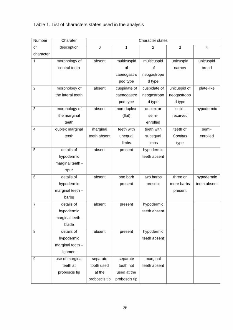
Table 1. List of characters states used in the analysis
Number
of
character
Charater
description
Character states
0 1 2 3 4
1 morphology of
central tooth
absent multicuspid
of
caenogastro
pod type
multicuspid
of
neogastropo
d type
unicuspid
narrow
unicuspid
broad
2 morphology of
the lateral teeth
absent cuspidate of
caenogastro
pod type
cuspidate of
neogastropo
d type
unicuspid of
neogastropo
d type
plate-like
3 morphology of
the marginal
teeth
absent non-duplex
(flat)
duplex or
semi-
enrolled
solid,
recurved
hypodermic
4 duplex marginal
teeth
marginal
teeth absent
teeth with
unequal
limbs
teeth with
subequal
limbs
teeth of
Comitas
type
semi-
enrolled
5 details of
hypodermic
marginal teeth -
spur
absent present hypodermic
teeth absent
6 details of
hypodermic
marginal teeth –
barbs
absent one barb
present
two barbs
present
three or
more barbs
present
hypodermic
teeth absent
7 details of
hypodermic
marginal teeth -
blade
absent present hypodermic
teeth absent
8 details of
hypodermic
marginal teeth –
ligament
absent present hypodermic
teeth absent
9 use of marginal
teeth at
proboscis tip
separate
tooth used
at the
proboscis tip
separate
tooth not
used at the
proboscis tip
marginal
teeth absent
26

Table 2. List of specimens (+ -- data available, - -- data unavailable)
Family Genus species Institutional registration number Expedition/locality, station and depth Radula preparation, or source of
information
Borsoniidae
Bathytoma neocaledonica Puillandre, et al., 2010 MNHN IM200717857 EBISCO, CP2551, 21°060'S, 158°350'E, 637-650m + Borsonia sp. MNHN IM200717932 Salomon 2, CP2197,8°24.40'S, 159°22.50'E, 897-1057 +
Borsoniidae gen. 1 sp. MNHN IM200717911 Panglao 2005, CP2333, 9°38.20'N, 123°43.50'E, 584-596m + Genota mitriformis (Wood, 1828) MNHN IM200742293 Angola, AF7, Pta. Das Lagostas +
Microdrillia cf. optima (Thiele, 1925) MNHN IM200717887 Panglao 2004, T36, 9°29.30'N, 123°51.50'E, 95-128m - Tomopleura reevii (C.B. Adams, 1850) MNHN IM200717875 Panglao 2004, T26,9°43.30'N, 123°48.80'E, 123-135m -
Typhlomangelia (cf.) sp. MNHN IM200717931 Salomon 2, CP2269, 7°45.10'S, 156°56.30'E, 768-890m - Zemacies excelsa Sysoev & Bouchet, 2001 MNHN IM200911056 Musorstom 4, DW226, 22°47'S, 167°22'E, 395m Radula-less species
Clathurellidae Clathurella nigrotincta (Montrouzier, 1872) MNHN IM200742607 Santo 2006, VM53, 15°31'S, 167°09'E, intertidal +
Etrema cf. tenera (Hedley, 1899) MNHN IM200717869 Panglao 2004, S21,9°41.70'N, 123°50.90'E, 4-12m - Nannodiella ravella (Hedley, 1922) MNHN IM200717904 Panglao 2004, T9, 9°33.5 'N, 123°49.50'E, 97-120m +
Clavatulidae
Clavatula xanteni Nolf & Verstraeten, 2006 MNHN IM200717829 Angola, AF1, 8°780'S, 13°230'E, 40-60m + Gemmuloborsonia colorata (Sysoev & Bouchet, 2001) MNHN IM200717849 EBISCO, DW2619, 20°060'S, 160°230'E, 490-550m +
Perrona subspirata (von Martens, 1902) MNHN IM200717833 Angola, AF10, 15°140'S, 12°290'E, 50m + Pusionella compacta Strebel, 1914 MNHN IM200717830 Angola, AF3, 10°510'S, 14°230'E, 5-10m +
Surcula nelliae spurius (Hedley, 1922) NHMUK MOEA 20100552 Off southern Hong Kong, South China Sea +
Cochlespiridae Cochlespira pulchella (Schepman, 1913) MNHN IM200717920 Panglao 2005, CP2340,9°29.40'N, 123°44.40'E, 271-318m + Sibogasyrinx sp. MNHN IM200717701 BOA1, CP2432, 14°59.70'S, 166°55.00'E, 630-705m +
Conidae
Californiconus californicus (Hinds, 1844) Monterey, California Tucker & Tenorio, 2009 Conasprella pagoda (Kiener, 1845) MNHN IM200717914 Panglao 2005, CP2380, 8°41.30'N, 123°17.80'E, 150-163m Tucker & Tenorio, 2009
Conus consors Sowerby I, 1833 MNHN IM200717939 Santo 2006, AT87,15°32.10'S, 167°16.10'E, 235-271m Tucker & Tenorio, 2009 Profundiconus teramachii (Kuroda, 1956) Philippines Tucker & Tenorio, 2009 Taranteconus chiangi Azuma, 1972 Philippines Tucker & Tenorio, 2009
Conorbidae Benthofascis lozoueti Sysoev & Bouchet, 2001 MNHN IM200742331 Norfolk 2, DW2147,22°50'S, 167°16'E, 496m +
Drilliidae
Agladrillia pudica (Hinds, 1843) NHMUK MOEA 20100543 Gulf of Panama, JTD-00-51, 08°36.41'N, 79°09.70'W, 73m McLean, 1971 Calliclava sp. NHMUK MOEA 20100546 Gulf of Panama, JTD-00-47, 08°31.83'N, 79°05.09'W, 21m McLean, 1971 Cerodrillia cybele(Pilsbry & Lowe, 1932) NHMUK MOEA 20100548 Gulf of Panama, JTD-00-18, 08°19.50'N, 78°47.71'W, 25-32m McLean, 1971
Clathrodrillia walteri (M. Smith, 1946) NHMUK MOEA 20100549 Gulf of Panama, JTD-00-46, 08°31.37'N, 79°05.79'W, 24-25m - Clavus canalicularis (Roeding, 1798) MNHN IM200717858 Panglao 2004, S12, 9°29.40'N, 123°56.00'E, 6-8m +
Conopleura striata Hinds, 1844 MNHN IM200717889 Panglao 2004, T41, 9°29.70'N, 123°50.20'E, 110-112m - Cruziturricula arcuata (Reeve, 1843) NHMUK MOEA 20100541 Gulf of Panama, JTD-00-34, 08°26.24'N, 79°09.14'W, 66-68m +
Drillia clavata (Sowerby III, 1870) NHMUK MOEA 20100550 Gulf of Panama, JTD-00-18, 08°19.50'N, 78°47.71'W, 25-32m McLean, 1971 Fusiturricula enae (Bartsch, 1934) INVEMAR MOL-1929 Colombia, E-73, 09°57.53'N, 79°07.71'W, 268-270m -
Imaclava unimaculata (Sowerby I, 1834) NHMUK MOEA 20100527 Gulf of Panama, JTD-00-46, 08°31.37'N, 79°05.79'W, 24-25m Shimek, Kohn, 1981 Iredalea pupoidea (H. Adams, 1872) MNHN IM200742556 Santo 2006, DB25, 15°37.7'S, 167°11.3'E, 10m +
Splendrillia sp. MNHN IM200717847 EBISCO, DW2617, 20°060'S, 160°220'E, 427-505m +
Horaiclavidae
Anacithara lita (Mellvill & Standen, 1896) MNHN IM200742614 Santo 2006, DS99, 15°32'S, 167°17'E, 100-105m + Anguloclavus sp. MNHN IM200717908 Panglao 2005, CP2332, 9°38.80'N, 123°45.90'E, 396-418m +
Carinapex minutissima (Garrett, 1873) MNHN IM200717868 Panglao 2004, B19, 9°29.40'N, 123°56.00'E, 17m + Ceritoturris pupiformis (Smith, 1884) MNHN IM200717888 Panglao 2004, T36, 9°29.30'N, 123°51.50'E, 95-128m +
Horaiclavidae gen. 1 sp. (juvenile) MNHN IM200742501 Salomon 2, CP2219, 7°58'S, 157°34'E, 650-836m + Horaiclavus splendidus (A.Adams, 1867) MNHN IM200717840 EBISCO, DW2631, 21°030'S, 160°440'E, 372-404m + Paradrillia sp. (juvenile) MNHN IM200742475 Panglao 2005, CP2396, 9°36'N, 123°42'E, 609-673m +
Mangeliidae Anticlinura sp. MNHN IM200742513 Salomon 2, CP2182, 8°47'S, 159°38'E, 762-1060m +
27

Benthomangelia cf. trophonoidea (Schepman, 1913) MNHN IM200717835 BOA1, CP2462, 16°37.50'S, 167°57.40'E, 618-641m + Eucithara cf. coronata (Hinds, 1843) MNHN IM200717900 Panglao 2004, B8, 9°37.10'N, 123°46.10'E, 3m +
Heterocithara sp. MNHN IM200717884 Panglao 2004, L46, 9°30.90'N, 123°41.20'E, 90-110m -
Mangeliidae gen. 1 sp. MNHN IM200717874 Panglao 2004, T26,9°43.30'N, 123°48.80'E, 123-135m Radula of Mangelia powisiana (Dautzenberg, 1887) used
Mangeliidae gen. 2 sp. MNHN IM200717872 Panglao 2004, S26, 9°41.50'N, 123°51.00'E, 21m - Oenopota sp. MNHN IM200742325 Hornsund, Svalbard, 1184-2001, -
Toxicochlespira pagoda Sysoev & Kantor, 1990 MNHN IM200717925 Salomon 2, CP2227, 6 37°20'S, 156°12.70'E, 508-522m +
Mitromorphidae Lovellona atramentosa (Reeve, 1849) MNHN IM200742552 Santo 2006, NR8, 15°35.7'S, 167°07.4'E, 11m + Mitromorpha metula (Hinds, 1843) MNHN IM200717898 Panglao 2004, B8, 9°37.10'N, 123°46.10'E, 3m +
Pseudomelatomidae
Carinodrillia dichroaPilsbry & Lowe, 1932 NHMUK MOEA 20100530 Gulf of Panama, JTD-00-18, 08°19.50'N, 78°47.71'W, 25-32m + Cheungbeia robusta (Hinds, 1839) NHMUK MOEA 20100556 Off southern Hong Kong, South China Sea, Sta. 70 +
Comitas sp. MNHN IM200717918 Panglao 2005, CP2388, 9°26.90'N, 123°34.50'E, 762-786m + Crassispira quadrilirata (E.A.Smith, 1882) MNHN IM200717755 Panglao 2004, L46, 9°30.90'N, 123°41.20'E, 90-110m +
Funa incerta (Smith, 1877) NHMUK MOEA 20100533 Off southern Hong Kong, South China Sea, Sta. 70 +
Hindsiclava alesidota (Dall, 1889) NHMUK MOEA 20100525 Lower Florida Keys, JTD-01-15, 24°33.47'N, 81°07.72'W, 117-148m +
Inquisitor sp. MNHN IM200717851 EBISCO, DW2625, 20°050'S, 160°190'E, 627-741m +
Knefastia tuberculifera (Broderip & Sowerby, 1829) NHMUK MOEA 20100533 Gulf of Panama, JTD-00-18, 08°19.50'N, 78°47.71'W, 25-32m +
Leucosyrinx sp. MNHN IM200717846 EBISCO, CP2600, 19°380'S, 158°460'E, 603-630m + Otitoma sp. MNHN IM200717905 Panglao 2005, CP2348, 9°29.60'N, 123°52.50'E, 196-216m +
Pilsbryspira jayana (C. B. Adams, 1850) USNM 857830 Carrie Bow Cay, Belize, intertidal + Pseudomelatoma moesta (Carpenter, 1865) California Kantor, 1988
Ptychobela suturalis (Gray, 1838) NHMUK MOEA 20100560 Luong SoN, 15km N. Nha Trang, Vietnam + Pyrgospira aenone (Dall, 1919) NHMUK MOEA 20100539 Gulf of Panama, JTD-00-18, 08°19.50'N, 78°47.71'W, 25-32m + Tiariturris spectabilis Berry, 1958 NHMUK MOEA 20100540 Gulf of Panama, JTD-00-34, 08°26.24'N, 79 09.14'W, 66-68m +
Zonulispira sp. NHMUK MOEA 20100536 Gulf of Panama, JTD-00-18, 08°19.50'N, 78°47.71'W, 25-32m +
Raphitomidae
Daphnella sp. MNHN IM200717927 Salomon 2, CP2260, 8°03.50'S, 156°54.50'E, 399-427m Radula of Daphnella mitrellaformis (Nomura, 1940) used
Eucyclotoma cymatodes (Hervier, 1899) MNHN IM200717903 Panglao 2004, S12, 9°29.40'N, 123°56.00'E, 6-8m - Glyphostomoides
(cf.) sp. MNHN IM200717892 Panglao 2004, T39, 9°30.10'N, 123°50.40'E, 100-138m -
Gymnobela sp. MNHN IM200717841 EBISCO, CP2648, 21°320'S, 162°300'E, 750-458m radula of Gymnobela yoshidai
(Kuroda et Habe in Habe, 1962) used
Hemilienardia calcicincta (Melvill & Standen, 1895) MNHN IM200717861 Panglao 2004, B14, 9°38.50'N, 123°49.20'E, 2-4m -
Kermia aureotincta (Hervier, 1897) MNHN IM200717878 Panglao 2004, B25, 9°29.40'N, 123°56.10'E, 16m radula of Kermia irretita (Hedley, 1899) used
Pleurotomella sp. MNHN IM200717848 EBISCO, DW2625, 20°050'S, 160°190'E, 627-741m - Raphitoma rubroapicata (E. A. Smith, 1885) MNHN IM200717890 Panglao 2004, L49, 9°36.50'N, 123°45.30'E, 90m radula of Raphitoma sp. used
Rimosodaphnella sp. MNHN IM200717836 BOA1, CP2462, 16°37.50'S, 167°57.40'E, 618-641m - Taranis sp. MNHN IM200742296 Aurora 07, CP2749, 15°57'N, 121°50'E, 743m Radula-less species
Teretiopsis cf. hyalina Sysoev & Bouchet, 2001 MNHN IM200717845 EBISCO, CP2651, 21°290'S, 162°360'E, 883-957m Radula-less species Thatcheria mirabilis (Angas, 1877) MNHN IM200717924 Salomon 2, CP2184, 8°16.90'S, 159°59.70'E, 464-523m Kantor, Taylor, 2002
Tritonoturris (cf.) sp. MNHN IM200717891 Panglao 2004, T39, 9°30.10'N, 123°50.40'E, 100-138m -
Veprecula sp. MNHN IM200717883 Panglao 2004, L46, 9°30.90'N, 123°41.20'E, 90-110m radula of Veprecula vepratica (Hedley, 1903) used
Terebridae Cinguloterebra cf. fenestrata (Hinds, 1844) MNHN IM200716735 Panglao 2005, CP2395, 9°36.2'N, 123°43.8'E, 382-434m +
28

Clathroterebra fortunei (Deshayes, 1857) MNHN IM200730401 Panglao 2005, CP2331, 9°39.2'N, 123°47.5'E, 255-268m Radula-less species Euterebra tristis (Deshayes, 1859) MNZ New-Zealand Rudman, 1969 Hastula strigilata (Linnaeus, 1758) MNHN IM200730420 Santo 2006, VM24, 15°35.2'S, 167°59.4'E, intertidal -
Hastulopsis amoena (Deshayes, 1859) MNHN IM200730480 Santo 2006, FR10, 15°36.9'S, 167 °10.5'E, 6-33m Radula-less species Myurella kilburni (Burch, 1965) MNHN IM200730459 Panglao 2004, S18, 9°35.7'N, 123°44.4'E, 0-2m Radula-less species Oxymeris maculatus (Linnaeus, 1758) MNHN IM200730371 Santo 2006, NR5, 15°28.7'S, 167°15.2'E, 19m Radula-less species
Strioterebrum plumbea (Quoy & Gaimard, 1833) MNHN IM200730558 Santo 2006, ED5, 15°31'S, 167°09'E, intertidal Radula-less species Terebra textilis Hinds, 1844 MNHN IM200717938 Santo 2006, LD28, 15°35.40'S, 166°58.70'E, 3-8m Radula-less species
Terenolla pygmaea (Hinds, 1844) MNHN IM200730449 Panglao 2004, S2, 9°37.4'N, 123°54.5'E, 4-5m Radula-less species
Turridae
Gemmula rarimaculata Kuroda & Oyama, 1971 MNHN IM200717838 EBISCO, DW2533, 22°180'S, 159°280'E, 360-370m + Iotyrris cingulifera (Lamarck, 1822) MNHN IM200717685 Santo 2006, FS84, 15°33.6'S, 167°16.6', 8-9m +
Lophiotoma acuta (Perry, 1811) MNHN IM200717860 Panglao 2004, R44, 9°33.30'N, 123°43.90'E, 2m + Lucerapex cf. casearia (Hedley & Petterd, 1906) MNHN IM200742448 Panglao 2005, CP2363, 9°06'N, 123°25'E, 437-439m +
Polystira albida (Perry, 1811) NHMUK MOEA 20110066
S. of Bahia Honda Key, Florida Keys, 24834.24’N; 81816.64’W, 30–34m Bandel, 1984
Ptychosyrinx carynae (Haas, 1949) USNM 832922 North Atlantic, 38°0.14'N, 70° 29.28'W, 3188-5300m - Turridrupa cf. armillata (Reeve, 1845) MNHN IM200717850 EBISCO, DW2607, 19°330'S, 158°400'E, 400-413m -
Turris babylonia Linnaeus, 1758 MNHN IM200717754 Panglao 2004, R42, 9°37.10'N, 123°52.60'E, 8-22m A.E. Fedosov, unpublished Xenuroturris legitima Iredale, 1929 MNHN IM200717684 Santo 2006, DR87, 15°38.5'S, 167°15.1'E, 13m +
Outgroups
Belomitra sp. MNHN IM200911057 Salomon 2, CP2184, 8°17'S, 160°00'E, 464-523m + Harpa kajiyamai Habe et Kosuge, 1970 MNHN IM200740569 Santo 2006, EP22,15°37.3'S, 167°05.8E, 78-91m Hughes & Emerson, 1987
Laevistrombus guidoi (Man in't Veld & De Turck, 1998) MNHN IM200911060 Santo 2006, LR3, 15°35.8'S, 167°06.1'E Bandel, 1984 Turrilatirus turritus (Gmelin , 1791) MNHN IM200911059 Santo 2006, FB52, 15°42.7'S, 167°15.1'E, 7m Radula of Latirus sp. used Vexillum costatum (Gmelin , 1791) MNHN IM200911058 Santo 2006, DR64, 15°27.6'S, 167°14.3'E, 6-35m +
Xenophora solarioides (Reeve, 1845) MNHN IM200911061 Santo 2006, AT55, 15°36.2'S, 167°02.5'E, 80-82m Bandel, 1984
29

Figure captions
Figs. 1-4. Diagrammatic sections through the anterior foregut of Conoidea. 1. Anterior foregut
of the Conoidea with non-hypodermic marginal radular teeth and odontophore
(generalized representative of the clade B). A duplex marginal tooth detached from the
subradular membrane is used at the proboscis tip for stabbing and envenomating the
prey. 2. Section of the tip of the proboscis with the duplex marginal tooth held by
sphincters of the buccal tube (actual specimen of Aforia kupriyanovi Sysoev & Kantor,
1988 – Cochlespiridae). 3. Anterior foregut of the Conoidea with hypodermic marginal
radular teeth and lacking odontophore (generalized representative of clade A). A
hypodermic marginal tooth detached from the subradular membrane is used at the
proboscis tip. 4. Section of the tip of the proboscis with the hypodermic marginal tooth
held by a sphincter of the buccal tube (actual specimen of Phymorhynchus wareni
Sysoev & Kantor, 1995 – Raphitomidae).
Abbreviations: bts – buccal tube sphincter; dmt – duplex marginal tooth at the proboscis tip;
hmt – hypodermic marginal tooth at the proboscis tip; mb – muscular bulb of the venom
gland; oe – oesophagus; pr – proboscis; rhs – rhynchostomal sphincter; rs – radular sac
without odontophore; rsod – radular sac with odontophore; vg – venom gland.
Fig. 5. Evolution of the central radular tooth morphology (character 1) mapped on the
conoidean molecular phylogeny (Puillandre et al., 2011). Character states are treated as
unordered. The different shading and tiling of the branches corresponds to the most
parsimonious ancestral state for the corresponding clade. The mixed shading and/or
tiling of the branch indicates that analysis was not able to resolve the single most
parsimonious state.
Fig. 6. Evolution of the central radular tooth morphology mapped on the conoidean molecular
phylogeny (character 1). A stepmatrix parsimony model was used, interdicting reversion
of the character states. Since central teeth are absent in the entire clade A, only clade B
is illustrated.
Figs. 7-13. Radulae of Drilliidae. If not otherwise mentioned, data for the specimens are given
in Table 2. 7-8. Splendrillia sp., MNHN IM200717847. 9. Clavus exasperatus (Reeve,
1843), MNHN, New-Caledonia, LIFOU 2000, st. 1420, 20°47.7'S, 167°09.35'E, 4-5m.
10. Imaclava pilsbryi (Bartsch, 1950), after Kantor and Taylor, 2000. 11. Cruziturricula
arcuata (Reeve, 1843), NHMUK MOEA 20100541. Semi-enrolled marginal teeth. 12-13.
Clavus sp. 3, MNHN uncataloged, BATHUS 2, DW714; 12 – bending plane of the
30

radula, arrows indicate strong wear of the teeth; 13 – central part of the same radula,
showing intact central and lateral teeth.
Abbreviations: ct – central tooth; lt – lateral tooth, mt – marginal tooth.
Figs. 14-26. Radulae of Pseudomelatomidae. If not otherwise mentioned, data for the
specimens are given in Table 2. 14. Tiariturris spectabilis Berry, 1958, NHMUK MOEA
20100540. Radula with solid recurved marginal teeth and broad central teeth. 15.
Comitas onokeana vivens Dell, 1956, MNHN, New-Caledonia, MONTROUZIER, st.
1269, after Kantor and Taylor, 2000. Radula with paired plate-like lateral teeth. 16-17.Comitas sp., MNHN IM200717918. 17 – enlarged central segment of the radula. 18.
Knefastia tuberculifera (Broderip & Sowerby, 1829), NHMUK MOEA 20100533.
Enlarged central segment of the radula. 19-20. Crassiclava turricula (Sowerby, 1834).
Costa Rica, Off Nacascola, West side of Bahia Culebra, after Kantor et al., 1997.
Radula with paired plate-like lateral teeth. 21. Zonulispira sp., NHMUK MOEA
20100536. Radula with semi-enrolled marginal teeth. 22. Ptychobela suturalis (Gray,
1838), NHMUK MOEA 20100560. Radula with semi-enrolled marginal teeth. 23-26.
Diagrammatic transverse sections of different duplex marginal teeth. Black horizontal
line represents the subradular membrane. 23 – Comitas-type; 24 – typical duplex tooth;
25 – semienrolled tooth of Zonulispira; 26 – semienrolled tooth of Ptychobela.
Abbreviations: al – accessory limb; ct – central tooth; lt – lateral tooth; ml – major limb; vlt –
vestigial lateral tooth.
Fig. 27. Evolution of the lateral radular tooth morphology (character 2) mapped on the
conoidean molecular phylogeny (Puillandre et al., 2011). A stepmatrix parsimony model
was used, interdicting reversion of the character states.
Figs. 28-33. Radulae of Clavatulidae. If not otherwise mentioned, data for the specimens are
given in Table 2. 28-29. Pusionella compacta Strebel, 1914, MNHN IM200717830. 29 –
enlarged central segment of the radula with vestigial lateral teeth. 30-31. Clavatula
xanteni Nolf & Verstraeten, 2006, MNHN IM200717829. 31 – enlarged central segment
of the radula. Arrow indicates the narrow central tooth. 32. Toxiclionella tumida
(Sowerby, 1870), South Africa, after Kantor, Taylor (2000). Semi-enrolled marginal
teeth. Left upper corner – diagrammatic section of the tooth. 33. Gemmuloborsonia
colorata (Sysoev & Bouchet, 2001), MNHN IM200717849.
Abbreviations: vlt – vestigial lateral teeth; ct – central tooth.
Figs. 34-42. Radulae of Turridae (34-35), Pseudomelatomidae (36-39), and Cochlespiridae
(40-42) with different duplex marginal teeth. If not otherwise mentioned, data for the
31

specimens are given in Table 2. 34. Xenuroturris legitima Iredale, 1929, MNHN
IM200717684. 35. Turridrupa acutigemmata (E. A. Smith, 1877), MNHN uncataloged,
New Caledonia. Radula with narrow central and plate-like lateral teeth. 36. Carinodrillia
dichroa Pilsbry & Lowe, 1932, NHMUK MOEA 20100530. 37. Funa incerta (Smith,
1877), NHMUK MOEA 20100554. 38. Cheungbeia robusta (Hinds, 1839), NHMUK
MOEA 20100557. 39. Inquisitor sp., MNHN IM200717851. 40-41. Cochlespira radiata
(Dall, 1889), MNHN, SE Brazil, after Kantor and Taylor, 2000. 42. Sibogasyrinx sp.,
MNHN IM200717701.
Abbreviations: al – accessory limb of the marginal duplex tooth; ct – central tooth; lt –lateral
tooth; ml – major limb of the marginal duplex tooth.
Fig. 43. Evolution of marginal tooth morphology (character 3) mapped on the conoidean
molecular phylogeny (Puillandre et al., 2011). Character states are treated as
unordered.
Fig. 44. Evolutionary transformations of duplex marginal tooth morphology (character 4)
mapped on the conoidean molecular phylogeny (Puillandre et al., 2011). Character
states are treated as unordered. Since duplex marginal teeth are absent in the entire
clade A, only clade B is illustrated.
Fig. 45. Presence or absence of a spur on marginal teeth (character 5) mapped on the
conoidean molecular phylogeny (Puillandre et al., 2011). Character states are treated as
unordered. The spur is absent in most of clade B, except some Terebridae. Therefore
for clarity only Terebridae from clade B are shown.
Figs. 46-53. Radula of Terebridae (46-49) and various hypodermic teeth from species of
clade A (50-53). If not otherwise mentioned, data for the specimens are given in Table
2. 46. Pellifronia jungi (Lai, 2001), MNHN 30591, Salomon 2, CP2195. 47.
Cinguloterebra cf. fenestrata Hinds, 1844, MNHN 30565, Panglao 05, st. CP2340. 48.
Duplicaria bernardi (Deshayes, 1857), Venus Bank, off NE end of Moreton Island,
Moreton Bay, Queensland, Australia, 27 º02'069" S, 153º19'00" E, 3.5-4.8 m, leg.
Glover, Taylor, 2008. 49. Impages hectica (Linnaeus, 1758), MNHN, uncataloged,
Philippines, Panglao Island, Alona Beach, intertidal, 2004. 50. Mangelia powisiana
(Dautzenberg, 1887). Plymouth, England, after Taylor et al. (1993). 51. Bathytoma
neocaledonica Puillandre et al., 2010, MNHN IM200717857. 52. Genota mitriformis
(Wood, 1828), MNHN IM200742293. 53. Benthomangelia trophonoidea (Schepman,
1913), MNHN IM200717835.
32

Fig. 54. Presence or absence of barbs on marginal teeth (character 6) mapped on the
conoidean molecular phylogeny (Puillandre et al., 2011). Character states are treated as
unordered. The barb(s) are present only in clade A and therefore clade B is omitted.
Fig. 55. Presence or absence of blade on marginal teeth (character 7) mapped on the
conoidean molecular phylogeny (Puillandre et al., 2011). Character states are treated as
unordered. The blade is absent in most of clade B, except some Terebridae. Therefore
for clarity only Terebridae from clade B are shown.
Fig. 56. Presence or absence of ligament of marginal teeth (character 8) mapped on the
conoidean molecular phylogeny (Puillandre et al., 2011). Character states are treated as
unordered. The ligament is present only in clade A and therefore clade B is omitted.
Fig. 57. Presence or absence of use of the separate marginal teeth on the proboscis tip
(character 9) mapped on the conoidean molecular phylogeny (Puillandre et al., 2011).
Character states are treated as unordered
Fig. 58-63. Radula of Gemmula Weinkauff, 1875, demonstrating different degree of fusion of
central and lateral teeth and the appearance of the “central formation”. 58-59. Gemmula
unilineata Powell, 1967, MNHN uncataloged, NORFOLK 2, New Caledonia, sta. DW
2097, bank Kaimon Maru, 24°44’S, 168°06’E, 580-583 m. 60-61. Gemmula rarimaculata
Kuroda et Oyama in Kuroda et al., 1971, MNHN uncataloged, MUSORSTOM 5, Coral
Sea, sta. DW263, 25°21’S, 159°46’E, 150-225 m. 62-63. Gemmula sp. 3, MNHN
uncataloged, MUSORSTOM 10, Fiji, sta. CP1354, 17°42.6’S, 178°55.0’E, 959-963 m.
33