EVIDENCE ON THE CARCINOGENICITY OF - OEHHA · relevant to the assessment of the evidence on the...
40
EVIDENCE ON THE CARCINOGENICITY OF C.I. DISPERSE YELLOW 3 August 2012 Reproductive and Cancer Hazard Assessment Branch Office of Environmental Health Hazard Assessment California Environmental Protection Agency
Transcript of EVIDENCE ON THE CARCINOGENICITY OF - OEHHA · relevant to the assessment of the evidence on the...

e
EVIDENCE ON THE CARCINOGENICITY OF
C.I. DISPERSE YELLOW 3
August 2012
Reproductive and Cancer Hazard Assessment Branch Office of Environmental Health Hazard Assessment California Environmental Protection Agency

C.I. Disperse Yellow 3 -i- August 2012 OEHHA
The Office of Environmental Health Hazard Assessment’s (OEHHA) Reproductive and Cancer Hazard Assessment Branch was responsible for the preparation of this document.
Primary Authors Kate Li, Ph.D., DABT Staff Toxicologist Cancer Toxicology and Epidemiology Section Jay Beaumont, Ph.D. Staff Toxicologist Cancer Toxicology and Epidemiology Section
Internal OEHHA Reviewers Martha S. Sandy, Ph.D. Chief, Cancer Toxicology and Epidemiology Section Reproductive and Cancer Hazard Assessment Branch Lauren Zeise, Ph.D. Deputy Director for Scientific Affairs

C.I. Disperse Yellow 3 -ii- August 2012 OEHHA
PREFACE Proposition 65 requires the publication of a list of chemicals “known to the state” to cause cancer or reproductive toxicity.1 It specifies that “a chemical is known to the state to cause cancer … if in the opinion of the state’s qualified experts the chemical has been clearly shown through scientifically valid testing according to generally accepted principles to cause cancer ...” The “state’s qualified experts” regarding findings of carcinogenicity are the members of the Carcinogen Identification Committee (CIC) of the Science Advisory Board.2 The lead agency for implementing Proposition 65 is the Office of Environmental Health Hazard Assessment (OEHHA) of the California Environmental Protection Agency. OEHHA selected C.I. Disperse Yellow 3 for preparation of hazard identification materials. Upon selection, the public was given the opportunity to submit information relevant to the assessment of the evidence on the carcinogenicity of C.I. Disperse Yellow 3. OEHHA reviewed and considered those submissions in preparing this document. OEHHA developed this document to provide the CIC with comprehensive information on C.I. Disperse Yellow 3’s carcinogenicity for use in its deliberations on whether or not the chemical should be listed under Proposition 65.
1 The Safe Drinking Water and Toxic Enforcement Act of 1986 (California Health and Safety Code 25249.5 et seq.) 2 Title 27 Cal. Code of Regs. §25302

C.I. Disperse Yellow 3 -iii- August 2012 OEHHA
TABLE OF CONTENTS 1 EXECUTIVE SUMMARY ................................................................................... 1
2 INTRODUCTION ............................................................................................... 2
2.1 Identity of C.I. Disperse Yellow 3 .................................................................... 2
2.2 Occurrence and Use ........................................................................................ 3
3 DATA ON CARCINOGENICITY 3.1 Carcinogenicity Studies in Humans ............................................................... 5
3.2 Carcinogenicity Studies in Animals ............................................................... 8
3.2.1 Long-term Dietary Studies in Rats ..................................................................... 8
3.2.2 Long-term Dietary Studies in Mice ................................................................... 10
3.3 Other Relevant Data ....................................................................................... 10 3.3.1 Pharmacokinetics and Metabolism .................................................................. 15
3.3.2 Genotoxicity ..................................................................................................... 15
3.3.3 In Vitro Transformation Studies ....................................................................... 19
3.3.4 Animal Tumor Pathology .................................................................................. 19
3.3.5 Structure-Activity Comparisons ........................................................................ 20
4 MECHANISMS ................................................................................................ 26
5 REVIEWS BY OTHER AGENCIES ................................................................. 26
6 SUMMARY AND CONCLUSIONS .................................................................. 27
6.1 Summary of Evidence ................................................................................... 27
6.2 Conclusions ................................................................................................... 29
7 REFERENCES ................................................................................................ 30

C.I. Disperse Yellow 3 -iv- August 2012 OEHHA
LIST OF TABLES
Table 1. Tumor Incidences in Male Rats Treated with C.I. Disperse Yellow 3 for 103 Weeks. .................................................................................................. 10
Table 2. Tumor Incidences in Male and Female Mice Treated with C.I. Disperse Yellow 3 ............................................................................................................ 12
Table 3. Genotoxicity Studies of C.I. Disperse Yellow 3 in Non-Mammalian Species. ............................................................................................................. 16
Table 4. In Vitro and in vivo Genotoxicity Studies of C.I. Disperse Yellow 3 in Mammalian Species ..................................................................................... 18
Table 5. Structure-Activity Comparisons for C.I. Disperse Yellow 3 and Its Expected Metabolites (Resulting from Reduction of the Azo Bond). .......... 22
LIST OF FIGURES
Figure 1. Chemical structure of C.I. Disperse Yellow 3 .............................................. 2
Figure 2. A Proposed Mechanism for Microsomal Azoreduction of Azo Dyes and the Resulting Amines ................................................................................ 14
Figure 3. C.I. Disperse Yellow 3 Metabolites ............................................................. 15

C.I. Disperse Yellow 3 -1- August 2012 OEHHA
1 EXECUTIVE SUMMARY C. I. Disperse Yellow 3 is a monoazo chemical used as a textile dye for coloring clothing, hosiery, carpeting products, polystyrene and other thermoplastics. It is also used as a direct dye in ink products, and in pulp and paper manufacture. Exposure may occur to the general public through uses of the dye in clothing, hosiery, carpets, inks, and other products, and to workers in the occupational setting. No epidemiology studies were identified that investigated the risk of cancer associated with documented exposure to CI Disperse Yellow 3. Four case-control studies were identified that examined bladder cancer risk associated with dye-related occupations in the textile industry; however, none provided information on exposures to specific dyes. Because C.I. Disperse Yellow 3 is only one of many disperse dyes used in the textile industry and because these studies did not collect information on exposures to specific dyes, they are inadequate to assess the relationship between exposures to C.I. Disperse Yellow 3 and cancer risk. Evidence for carcinogenicity of C.I. Disperse Yellow 3 comes primarily from 104-week diet studies conducted in male F344 rats and female and male B6C3F1 mice. Exposure to C.I. Disperse Yellow 3 in male rats resulted in statistically significant increases in benign, and combined benign and malignant liver tumors. Rare stomach tumors were also observed in treated male rats. In female mice, exposure to C.I. Disperse Yellow 3 resulted in statistically significant increases in malignant lymphoma and combined malignant lymphoma and leukemia as well as statistically significant increases in benign, and combined benign and malignant liver tumors. In male mice, exposure to C.I. Disperse Yellow 3 resulted in a statistically significant increase in benign lung tumors; the increase in combined benign and malignant lung tumors approached statistical significance. Positive findings in multiple genotoxicity test systems indicate that C.I. Disperse Yellow 3 may operate through a genotoxic mechanism. C.I. Disperse Yellow 3 induced mutations in multiple strains of Salmonella typhimurium and in mouse lymphoma cells, chromosomal aberrations in frog larvae, sister chromatid exchanges in Chinese hamster ovary cells, and unscheduled DNA synthesis in rat hepatocytes in vitro. 4-Aminoacetanilide and 2-amino-p-cresol, the expected metabolites arising from reduction of the azo bond in C.I. Disperse Yellow 3, are also genotoxic. C.I. Disperse Yellow 3 and five genotoxic carcinogens with shared structural similarity induce liver tumors in rodents. The structurally similar liver carcinogens include p-aminoazobenzene, o-aminoazotoluene, and 2,4-diaminotoluene, which like C.I.

C.I. Disperse Yellow 3 -2- August 2012 OEHHA
Disperse Yellow 3 induce liver tumors in more than one sex/species, and azobenzene and 2-aminotoluene, which induce liver tumors in one sex/species. 2 INTRODUCTION
2.1 Identity of C.I. Disperse Yellow 3 C.I. Disperse Yellow 3 is a monoazo dye that exists as a solid. It is soluble in acetone, ethanol and benzene and has low solubility in water. Its structure is given in Figure 1 and physical and chemical characteristics are given below (IARC, 1990).
NN
OH
CH3
NH
O
CH3
Figure 1. Chemical structure of C.I. Disperse Yellow 3 Molecular Formula: C15H15N3O2 Molecular Weight: 269.30 CAS Registry Number: 2832-40-8 IUPAC Systematic Name: 4' –[(6-Hydroxy-meta-tolyl)azo]acetanilide Synonyms: Dispersion Yellow 3; N-[4-(2-Hydroxy-5-methylphenylazo)-
phenyl]acetamide; 4-(2-Hydroxy-5-methylphenylazo)-acetanilide; 4-Acetamido-2' -hydroxy-5' -methylazobenzene; C.I. Solvent Yellow 77; C.I. Solvent Yellow 92; Solvent Yellow 99; 4'-[(2-hydroxy-5-methylphenyl) azo]acetanilide
Chemical Class: monoazo dye Chemical Appearance: powder Melting Point: 195°C Water Solubility: 1.18 mg/L (at 25°C) Henry's Law Constant: 1.5 x 10⁻¹¹ atm-m3/mole (at 25°C) Vapor Pressure: 5 x 10-11 mm Hg (at 25°C) Octanol-water coefficient: LogP = 3.98 The dye’s name is from the Color Index (CI) system of the Society of Dyers and Colourists (Hatch and Maibach, 2000). Each dye in the index has a name composed of
C.I. Disperse Yellow 3 -3- August 2012 OEHHA
an application class (such as “disperse”), the color emitted (such as “yellow”), and a number indicating the order of discovery (such as “3”) within an application class and color group. C.I. Disperse Yellow 3 is classified as a “disperse” dye because of its physical state in the dye-bath. Disperse dyes, due to extremely low water solubility, must be milled to small particle sizes (1-3 microns) and dispersed in water using a surfactant (dispersing agent) (Freeman and Mock, 2007). If the disperse dyeing is not done in accordance with best practice, for example over-drying, wrong textile substance, or incomplete removal of the carriers, there may be higher exposure to dyes when wearing garments dyed in this way (Santa Cruz Biotechnology, Inc., 2008). Disperse dyes are fat-soluble and dissolve in chemical fibers. Because of their small molecular size and high fat solubility, some disperse dyes may be easily absorbed through the skin (Santa Cruz Biotechnology, Inc., 2008). The color of C.I. Disperse Yellow 3 comes from its “azo” chromophore (the chemical group responsible for the color of a dye). C.I. Disperse Yellow 3’s chromophore is called “azo” because of its central azo structure (two nitrogen atoms joined by a double bond, with each nitrogen atom attached to another group). As a category, azo disperse dyes constitute about 60% of all disperse dyes (Freeman and Mock, 2007). C.I. Disperse Yellow 3 is considered to be a “low energy” disperse dye, where energy refers to heat and pressure required during the dyeing process. Low energy disperse dyes have been said to be easier to remove from the industrial atmosphere with exhaust ventilation (relative to higher energy disperse dyes), which could be a consideration in assessment of human occupational exposures (Freeman and Mock, 2007). 2.2 Occurrence and Use C.I. Disperse Yellow 3 is produced by coupling diazotized 4-acetamidoaniline with para-cresol. It is manufactured in the U.S., Europe, Japan, India and China. Production in the US was reported at more than 95 metric tons per years in 1983 (Bomberger and Boughton, 1983). More recent production data were not found. C.I. Disperse Yellow 3 is primarily used as a textile dye for coloring nylon, polyvinyl chloride and acrylic fibers, wools, furs, cellulose acetate, polystyrene, and other thermoplastics. Finished products containing C.I. Disperse Yellow 3 include clothing, hosiery, and carpeting (IARC, 1990). Other reported uses are as dyes in ink products,

C.I. Disperse Yellow 3 -4- August 2012 OEHHA
and in pulp and paper manufacture (Hewlett-Packard Company, 2011; Scorecard, 2011). Exposure may occur to workers in related industries, with the biggest consumer of disperse dyes being the textile industry, which uses disperse dyes to dye synthetic materials. Dyeing of synthetic materials may occur during any one of several stages, as follows (McIntyre, 2004):
• Yarn stage. Yarns are often dyed while they are still spooled in a process called “batch” dyeing. Workers may be exposed to dyes via skin contact with dyed yarn or possibly via inhalation of particles generated from dyed yarn. Work categories using dyed yarn may include warping and weaving (including knitting). Yarn spools are dyed in batches in machines made for the purpose.
• Fabric stage. This is the most common stage of dyeing in the textile industry. Most fabrics dyed a single solid color are dyed at this stage.
• Garment stage. Sometimes parts of garments or whole garments are dyed. Because the fastness of disperse dyes to synthetic fibers such as polyamide and acetate is limited (Stahlmann et al., 2006), exposure to C.I. Disperse Yellow 3 may occur to the general public through uses of the dye in clothing, hosiery, carpets and inks. Reports of allergic eczema associated with wearing nylon hosiery dyed with C.I. Disperse Yellow 3 provide evidence of exposure from the use of textiles dyed with C.I. Disperse Yellow 3 (NTP, 1982). Disperse dyes constitute 50% of all known textile dye contact allergens, and they can sensitize both before and after application to textiles (Gideon, 2010). C.I. Disperse Yellow 3 is listed among 39 disperse dyes known to cause occupational contact allergic dermatitis in the textile industry (Hatch and Maibach, 2000). C.I. Disperse Yellow 3 has been detected in wastewater from carpet dying plants in the U.S. In water treatment plant influents and effluents, levels of C.I. Disperse Yellow 3 have been reported to range from none detected to 0.436 ppm (IARC, 1990). According to the national occupational exposure survey from the Centers for Disease Control and Prevention (CDC), an estimated 27,130 people working in occupations related to the production of textile mill products were exposed to C.I. Disperse Yellow 3 between 1981 and 1983 (CDC, 1983). More recent data on occupational exposure in the US were not available. The U.S. EPA Toxics Release Inventory (TRI) reported that 1,538 pounds of C.I. Disperse Yellow 3 were released in 1995 and 500 pounds were released in 2001 (U.S. EPA, 2001).

C.I. Disperse Yellow 3 -5- August 2012 OEHHA
3 DATA ON CARCINOGENICITY 3.1 Carcinogenicity Studies in Humans There are no studies in humans that have investigated the risk of cancer associated with documented exposure to C.I. Disperse Yellow 3. Since it is used primarily to dye textiles, results of epidemiological studies of cancer risk associated with dye-related occupations in the textile industry are summarized here. Results for synthetic material-related occupations in the textile industry are also summarized (from the one study with data, Serra et al,. 2008) because C.I. Disperse Yellow 3 is used primarily on synthetic materials. Among the four studies identified, only one mentioned C.I. Disperse Yellow 3 in the article (Gonzales et al., 1988). It was notable that all four studies were case-control in design, that Spain was the location of three of the studies, and that the bladder was the only organ-site for which cancer risk data were reported. A strength of all four studies was that they adjusted for cigarette smoking, a known cause of bladder cancer. Serra et al. (2008) reported results for the textile industry and bladder cancer in a case-control study of 18 hospitals in Spain. The study was based on 1,182 incident cases (1,065 male and 117 female) identified from 1998 through 2001, and 1,221 controls from the same hospitals. Lifetime work histories were obtained via interviewer-administered questionnaires. A strength of the study was a questionnaire module designed specifically for the textile industry. The module was administered to subjects who reported working in the textile industry in the main questionnaire. The module completion rate was about 50 percent among subjects who, in the main questionnaire, had indicated that they worked in the textile industry. The module included 75 questions and gathered information for nine categories of occupation, 33 categories of location, and seven categories of type of material. Textile industry work would have occurred primarily in the geographic areas represented by the study’s participating hospitals in Asturias, Alicante, Barcelona, Tenerife, and Valles/Bages, Spain. The investigators did not mention disperse dyes, but disperse dyes would have been among the dyes used as they have been an important part of the worldwide textile industry for decades. Work with dyes in the Serra et al. study was identified by the occupation “finishers/dyers” and by the locations “dyeing, printing” and “dye room or house.” The occupation “finishers/dyers,” was not associated with bladder cancer risk (OR=0.94, 95% CI=0.46-1.95). The locations “dyeing, printing” (OR=1.03, 95% CI=0.43-2.49) and “dye room or house” (OR=1.49, 95% CI=0.48-4.65) also were not significantly

C.I. Disperse Yellow 3 -6- August 2012 OEHHA
increased. The investigators noted that “this lack of a strong finding is not surprising since preventive measures were extensively applied in past decades throughout Europe to prevent exposure to dyes as these were singled out as one of the main causes of bladder cancer in several industries.” Exposure to dyes may also occur during work with dyed textiles in locations where the following activities occur: winding, warping, and sizing. The strongest association with bladder cancer was found in the Serra et al. study for the combination of “winding, warping, and sizing” occupations and working with “synthetic” materials (OR=15.39, 95% CI=1.89-125.29, based on 11 exposed cases and one exposed control). This finding is interesting, in part, because disperse dyes are used primarily on synthetic materials (e.g., polyester is dyed only with disperse dyes). The overall odds ratio for work with “synthetic” materials (all occupations and locations) was significantly elevated when restricted to work duration of 10 or more years (OR=2.62, 95% CI 1.14-6.01, based on 21 exposed cases and nine exposed controls). The investigators noted “an overall tendency of an increased risk among workers exposed to synthetics.” “Winding” in textile manufacturing generally refers to the winding of freshly made, undyed yarn, but it could refer to the rewinding of dyed yarn. “Winding” overall (including all materials) was associated with increased but non-significant risk of bladder cancer (OR=2.70, 95% CI=0.92 to 7.98). “Warping” generally refers to creation of a lengthwise matrix of yarn (using “warp” yarn), to which yarn is added later in the crosswise direction (using “weft” yarn) during weaving or knitting. “Warping” overall (including all materials) was associated with statistically significant increased risk (seven exposed cases and zero exposed controls, odds ratio not calculable, p<0.05). Unfortunately, the study did not report results for the specific job of warping with synthetic yarns. “Sizing” generally is the adding of chemicals such as starch to improve the physical characteristics of the material, and sizing is generally removed prior to dyeing. “Sizing” overall (including all materials) was nonsignificantly associated with increased risk (OR=6.84, 95% CI=0.71-66.43). Unfortunately, the study did not report results for the specific job of sizing synthetic yarns. Thus, of the three occupations (“winding, warping, and sizing”) associated with the strongest epidemiology result, it appears that “warping” had the greatest potential for exposure to disperse dyes. However, according to Serra et al., winding, warping and sizing are usually located in the same physical area, so there may have been common exposures.

C.I. Disperse Yellow 3 -7- August 2012 OEHHA
In Serra et al. (2008) the combination of “weaving room” location and work with “synthetic” materials was nonsignificantly associated with bladder cancer (OR=2.55, 95% CI=0.81-8.02). Similarly, the combination of “weavers” occupational category and work with “synthetic” materials was nonsignificantly elevated (OR=2.59, 95% CI=0.82-8.17). Interestingly, when the analysis was restricted to 10 or more years of employment duration, the “weaving room” location overall (all materials) was significantly associated with bladder cancer (OR=2.94, 95% CI=1.24-7.01), and the “weavers” occupational group overall had borderline statistical significance (OR= 2.27, 95% CI=0.97-5.34). As with warping, weaving may have included contact with materials recently dyed with disperse dyes. In summary, the Serra et al. (2008) study found bladder cancer to be associated with warping and weaving occupations and with synthetic materials. Exposures to disperse dyes may have occurred during handling of synthetic yarns and fabrics already dyed, but C.I. Disperse Yellow 3 is only one of many disperse dyes. Thus, only an unknown portion of subjects who performed warping and weaving would possibly have been exposed to CI Disperse Yellow 3. With such a large amount of misclassification, a single dye would have to be a strong carcinogen to explain the elevated odds ratios found by Serra et al. In New Zealand, a nationwide case-control study of occupational risks for bladder cancer identified incident cases during 2003 and 2004 (Dryson et al., 2008). The study included 213 cases (165 male and 48 female) and 471 population controls. Lifetime work histories were obtained via interviewer-administered questionnaires, and the occupations and industries were categorized with a standard coding system. The occupation category “textile products machine operators - textile bleaching, dyeing, and cleaning,” was not significantly elevated (OR=0.81, 95% CI=0.19-3.54, based on three exposed cases and 10 exposed controls). C.I. Disperse Yellow 3 was not mentioned in the article. A case-control study of bladder cancer and occupations based at 12 hospitals in four regions of Spain identified 497 cases (438 male and 59 female) occurring from 1985 through 1986 (Gonzales et al., 1989). There were two control groups, one from the same 12 hospitals and one from the general population. Lifetime work histories were obtained via interviewer-administered questionnaires, and work histories were categorized with a standard occupation/industry coding system. The study found that among “textile dyers” the relative risk was negligible (based on 11 exposed cases and 17 exposed controls, OR=1.29, 95% CI=0.5-3.1). C.I. Disperse Yellow 3 was not mentioned in the article.
C.I. Disperse Yellow 3 -8- August 2012 OEHHA
A case-control study of bladder cancer among workers in the textile industry in Mataro County, Spain, included 57 cases identified at a single hospital or in a local death registry between 1978 and 1981, and 107 control subjects (Gonzales et al., 1988). Approximately two controls were selected for each case, matched on several factors, including date of death or hospitalization, age, and sex. One set of controls had cancers other than bladder or lung cancer; the other set had non-neoplastic diseases. Lifetime work histories were obtained via interviewer-administered questionnaires, but a major limitation of the study was that most subjects were deceased (75% of cases and 74% of controls) by the time of interview. Thus, most questionnaires were administered to spouses, family members, and friends who had less knowledge of the subjects’ work histories than the subjects themselves. The occupations and industries were categorized with a standard coding system. The study, after adjusting for tobacco smoking, found that that the risk of bladder cancer was elevated among individuals who worked in “textile dyeing or printing” jobs (OR=4.41, 95% CI=1.15-16.84, based on eight exposed cases and three exposed controls). The authors indicated that C.I. Disperse Yellow 3 was among 72 dyes that had been used in the Mataro textile factories. Other dyes noted by the authors were C.I. Direct Black 38 and C.I. Direct Blue 5, both of which are Proposition 65 carcinogens.
3.2 Carcinogenicity Studies in Animals A review of the scientific literature regarding the carcinogenicity of C.I. Disperse Yellow 3 in experimental animals identified long-term dietary studies conducted in male and female rats and mice (NTP, 1982). 3.2.1 Long-term Dietary Studies in Rats Male and female F344 rats (50/sex/group) were fed on diets containing C.I. Disperse Yellow 3 (87.6% purity3) at concentrations of 0, 5,000 (low-dose) or 10,000 (high-dose) ppm for 103 weeks (NTP, 1982). Gross and microscopic examinations were performed on all major organs from animals found dead and on all animals sacrificed at the end of these studies (104 weeks). Survival among male and female rats in treated groups was higher compared to controls in both the male study and the female study. In male rats, survival in the low dose group was significantly greater as compared to controls (p< 0.05). In females, the
3 NTP determined the test material was representative of commercially available C.I. Disperse Yellow 3, and thus suitable for use in these studies.

C.I. Disperse Yellow 3 -9- August 2012 OEHHA
high-dose group had significantly greater survival (p < 0.05) than did the controls. Mean body weights of treated rats were lower than those of controls in both the male and the female studies. Mean body weights in both sexes of rats were decreased in a dose-related manner. Among high-dose male rats, body weights were 23% lower than controls by the end of the study. Similarly, body weights of high-dose female rats were 24% lower than controls by the end of the study. Food intake by treated and control animals was comparable in both studies. Among male rats treated with C.I. Disperse Yellow 3 (Table 1), statistically significant increases in hepatocellular adenomas and combined hepatocellular adenomas and carcinomas were observed in both the low-dose (p< 0.01) and the high-dose (p < 0.05) groups as compared to controls. The incidences across dose groups showed statistically significant positive trends with dose (p < 0.05). Stomach tumors which are rare in rats were also observed in treated animals. Tumors of the glandular portion of the stomach were seen in two low-dose animals and one high-dose animal. Tumors of the non-glandular portion of the stomach were seen in two low-dose animals. No stomach tumors occurred in the control group.
After terminal sacrifice, histo-pathological findings of incidence of primary tumors were analyzed in female rats. No treatment-related tumors were observed in the female rat study of C.I. Disperse Yellow 3.

C.I. Disperse Yellow 3 -10- August 2012 OEHHA
Table 1. Tumor Incidencea in Male F344 Rats Fed C.I. Disperse Yellow 3 for 103 Weeks (NTP, 1982).
Organ Tumor Dose group (ppm) Trend
test p valueb 0 5000 10000
Liver
Hepatocellular adenomac 1/31 15/45** 10/39* <0.05
Combined hepatocelluar adenoma and carcinoma
2/31 15/45** 11/39* <0.05
Stomach
Glandular portion: Combined adenoma, mucinous adenocarcinoma, and sarcoma
0/30 2/45d 1/39e NS
Non-glandular portion: Combined squamous cell papilloma and fibrosarcoma
0/30 2/45f 0/39 NS
a Incidence is the number of tumor-bearing animals/ the number of animals alive at the time of the first occurrence of tumor at that site. The first occurrence of tumors in the liver was at 104 weeks, and in the stomach was at 104 weeks. b The p value from the Cochran-Armitage trend test; NS: not significant c At the time, NTP used the term “neoplastic nodule” to describe benign nodular hepato-proliferative lesions in rats. Since 1986, NTP refers to these lesions as “hepatocellular adenoma.” (IARC, 1990, p. 38) d One animal with an adenoma, another with a mucinous adenocarcinoma e One animal with an adenocarcinoma and sarcoma f One animal with a squamous cell papilloma, another with a fibrosarcoma. * p < 0.05, ** p < 0.01: pairwise comparison with controls by Fisher exact test 3.2.2 Long-term Dietary Studies in Mice Groups (50/sex/group) of male and female B6C3F1 mice received diets containing 0, 2,500 (low-dose), or 5,000 (high-dose) ppm of C.I. Disperse Yellow 3 (87.6% purity) for 103 weeks (NTP, 1982). Gross and microscopic examinations were performed on all major organs from animals found dead and on all animals sacrificed at the end of these studies (104 weeks). Survival among male and female mice in treated groups and controls was similar in both the male and the female studies. By the end of the male mouse study, the mean body weight of the low-dose group was 8% lower than that of controls, while the mean body weight of the high-dose group was 15% higher than that of the controls. At the end of

C.I. Disperse Yellow 3 -11- August 2012 OEHHA
the female mouse study, mean body weights were similar in the high-dose and control groups, while mean body weight in low-dose females was 13% lower than that of controls. Food intake was not affected by treatments in either study. Among male mice treated with C.I. Disperse Yellow 3, a statistically significant increase in alveolar /bronchiolar adenoma (p < 0.05) was observed in the high-dose group, as compared to controls (Table 2). An increase in combined alveolar/bronchiolar adenoma and carcinoma which approached statistical significance was also observed in the high-dose group (p = 0.055). The incidence of alveolar/bronchiolar adenoma and combined adenoma and carcinoma increased across doses with statistically significant positive trends (p < 0.05). In treated female mice, dose-dependent increases in tumors of the hematopoietic system and liver were observed (Table 2). Significant increases in malignant lymphoma and in combined malignant lymphoma and leukemia (p < 0.05) occurred in the high-dose group, as compared to controls. Increases in hepatocellular adenoma and combined hepatocellular adenoma and carcinoma were observed in both the low-dose (p < 0.05) and high-dose (p< 0.01) groups, as compared to controls.

C.I. Disperse Yellow 3 -12- August 2012 OEHHA
Table 2. Tumor Incidencea in Male and Female B6C3F1 Mice Treated with C.I. Disperse Yellow 3 (NTP, 1982).
Organ Tumor Dose group (ppm) Trend test
p valueb 0 2500 5000
Male mice
Lung
Alveolar/bronchiolar adenoma
2/47 6/42 9/46* < 0.05
Combined alveolar/bronchiolar
adenoma and carcinoma
3/47 7/42 9/46# < 0.05
Female mice
Hematopoietic System
Malignant lymphoma
10/50 16/50 19/50* < 0.05
Combined malignant
lymphoma and leukemia
10/50 17/50 20/50* <0.05
Liver
Hepatocellular adenoma
0/50 6/47* 12/46** <0.001
Hepatocelluar carcinoma
2/50 4/47 5/46 NS
Combined hepatocellular adenoma and
carcinoma
2/50 10/47* 17/46** <0.001
aIncidence is the number of tumor-bearing animals/ the number of animals alive at the time of the first occurrence of tumor at that site. In male mice, the first occurrence of tumor in the lung was at 91 weeks. In female mice, the first occurrence of tumor in the hematopoietic system was at 48 weeks, and in the liver was at 83 weeks. bThe p value from the Cochran-Armitage trend test; NS: not significant * p < 0.05; ** p < 0.01; # p=0.055: pairwise comparison with controls by Fisher exact test

C.I. Disperse Yellow 3 -13- August 2012 OEHHA
3.3 Other Relevant Data 3.3.1 Pharmacokinetics and Metabolism No specific information is available about the absorption, distribution, metabolism or excretion of C.I. Disperse Yellow 3 in mammals. Absorption of C.I. Disperse Yellow 3 through the skin is expected, however; based on the lipophilic properties of disperse dyes in general, and observed skin absorption for other disperse dyes (Stahlmann et al., 2006; Santa Cruz Biotechnology, Inc., 2008). In general, azo dyes, including disperse dyes, undergo reductive cleavage of the azo bond, resulting in the formation of aromatic amine metabolites (Levine, 1991). This generation of aromatic amines is thought to contribute to the carcinogenicity of many azo dyes (SCCNFP, 2002). Reduction of the azo bond can occur in mammalian cells (e.g., liver, skin), in bacteria present on the skin, and in bacteria present in the gastrointestinal tract (Levin, 1991; Chung et al., 1992; Platzek et al., 1999; SCCNFP, 2002; Stahlmann et al., 2006). A number of cytosolic and microsomal enzymes are capable of reducing the azo bond, including NADPH cytochrome P450 reductase, NAD(P)H:quinone oxidoreductase, and cytochrome P450s (Levine, 1991; Chung et al., 1992; Stahlmann et al., 2006). A proposed mechanism of microsomal azoreduction described by Levine (1991) is shown in Figure 2. In this scheme, two primary aromatic amines are formed following the transfer of two electrons and the addition of two protons to the parent azo compound. The aromatic amines that arise from reduction and cleavage of the azo bond can undergo further metabolism through N-oxidation by cytochrome P450s or flavin-containing monoxygenases to form N-hydroxylarylamines (Levin, 1991; Chung et al., 1992; SCCNFP, 2002). Under acidic pH, N-hydroxylarylamines can form reactive nitroxide radicals that can alkylate DNA bases, particularly the nucleophilic centers in guanine. N-Hydroxylarylamines may undergo glucuronidation (activation) or acetylation (inactivation) reactions (Levine, 1991).

C.I. Disperse Yellow 3 -14- August 2012 OEHHA
Figure 2. A Proposed Mechanism for Microsomal Azoreduction of Azo Dyes and the Resulting Amines (From Levine, 1991).
The two aromatic amine metabolites likely to be formed from C.I. Disperse Yellow 3 by the metabolic activity of skin bacteria and/or by metabolism in the skin are 4-aminoacetanilide (CAS No. 122-80-5) and 2-amino-p-cresol (2-Amino-4-methylphenol, CAS No. 95-84-1) (Figure 3) (Stahlmann et al., 2006). Indirect evidence of skin absorption and metabolism of C.I. Disperse Yellow 3 to these aromatic amines comes from studies of the skin sensitizing and allergenic properties of the dye and its azo cleavage products. Using a mouse model, Stahlmann et al. (2006) found that 2-amino-p-cresol was a strong allergen, and that C.I. Disperse Yellow 3 and 4-aminoacetamilide were weak sensitizers. More recently, Malinauskiene et al. (2012) assessed skin sensitization in seven individuals with a previous positive skin patch test

C.I. Disperse Yellow 3 -15- August 2012 OEHHA
result for C.I. Disperse Yellow 3. Six of those seven individuals had a positive C.I. Disperse Yellow 3 skin patch test in this study. All six also tested positive for 2-amino-p-cresol, and three of the six tested positive for 4-aminoacetanilide.
Figure 3. C.I. Disperse Yellow 3 Metabolites (From Stahlmann et al., 2006)
3.3.2 Genotoxicity Multiple studies have investigated the genotoxicity of C.I. Disperse Yellow 3 in non-mammalian assays and in mammalian cells in vitro. Two in vivo genotoxicity studies have been reported, one in rats, and another in mice. The findings are presented in Tables 3 and 4 below. C.I. Disperse Yellow 3 induced reverse mutations in Salmonella typhimurium strains by causing base substitution mutations in TA100 with S-9 metabolic activation, and frameshift mutations in TA97, TA98, TA1537 and TA1538 with or without metabolic activation (Table 3) (CCRIS, 2012; Cameron et al., 1987; Zeiger et al., 1988). The chemical did not induce mutations in the TA1535 strain (Cameron et al., 1987; Zeiger et al., 1988). A negative result was reported in the Drosophila sex-linked recessive lethal (SLRL) mutation assay (Foureman et al., 1994). C.I. Disperse Yellow 3 induced chromosomal aberrations in frog larvae (Table 3) (CIR, 1996; Gray et al., 1979).

C.I. Disperse Yellow 3 -16- August 2012 OEHHA
Table 3. Genotoxicity Studies of C.I. Disperse Yellow 3 in Non-Mammalian Species.
Endpoint Strain Concen-trations Tested
Results Activation System References
- S-9 + S-9
Salmonella typhimurium reverse mutation assay (basepair substitution)
TA100 1-333 µg/plate NT + Uninduced hamster
S-9 Cameron et al., 1987
TA100 333-10000 µg/plate
_ + Aroclor 1254 induced rat and hamster S-9
Cameron et al., 1987
TA1535 333-10000 µg/plate
_ _ Aroclor1254 induced rat and hamster S-9
Cameron et al., 1987
TA1535 10-1000 µg/plate _ _
Aroclor 1254 induced rat and hamster S-9
Zeiger et al., 1988; CCRIS, 2012
Salmonella typhimurium reverse mutation assay (frameshift mutation)
TA97 10-1000 µg/plate + +
Aroclor 1254 induced rat and hamster S-9
Zeiger et al., 1988; CCRIS, 2012
TA98 1-333 µg/plate NT + Aroclor 1254
induced hamster S-9 Cameron et al., 1987
TA98 333-10000 µg/plate
+ + Aroclor1254 induced rat and hamster S-9
Cameron et al., 1987
TA98 10-1000 µg/plate + +
Aroclor 1254 induced rat and hamster S-9
Zeiger et al., 1988; CCRIS, 2012
TA1537 333-10000 µg/plate
+ + Aroclor 1254 induced rat and hamster S-9
Cameron et al., 1987
TA1538 333-10000 µg/plate
+ + Aroclor 1254 induced rat and hamster S-9
Cameron et al., 1987
Sex-linked recessive lethal mutation assay
Drosophila 500-1000 ppm
_ NA Foureman et al.,
1994
Chromosomal aberrations
Frog (Rana clamitans) larvae
4 mg/ml + NA CIR, 1996; Gray
et al., 1979
+ = positive result; − = negative result; NT= not tested; NA=not applicable; S-9= supernatant fraction from liver homogenate

C.I. Disperse Yellow 3 -17- August 2012 OEHHA
In the mouse lymphoma forward mutation assay, C.I. Disperse Yellow 3 was positive in the presence of S-9 in two studies (Tennant et al., 1987b; McGregor et al., 1988), produced inconclusive results in one study (Seifried et al., 2006), and was negative in the absence of S-9 (Cameron et al., 1987; McGregor et al., 1988; Seifried et al., 2006) (Table 4). C.I. Disperse Yellow 3 induced sister chromatid exchange (SCE) in Chinese hamster ovary (CHO) cells in one study in the presence, but not the absence of S-9 (Ivett et al., 1989) and in another study in the absence of S-9 (Tennant et al., 1987b). C.I. Disperse Yellow 3 did not induce chromosomal aberrations in CHO cells in the presence or absence of S-9 (Ivett et al., 1989; Tennant et al., 1987b). C.I. Disperse Yellow 3 induced unscheduled DNA synthesis (UDS) in primary rat hepatocytes (Tennant et al., 1987a). In an in vivo mouse micronucleus assay, C.I. Disperse Yellow 3 did not increase the frequency of micronuclei in the bone marrow (Shelby et al., 1993). In an in vivo oral dosing study, C.I. Disperse Yellow 3 did not cause rat liver DNA damage, as determined in an alkaline elution assay for DNA single strand breaks, at doses up to 770 mg/kg (Kitchin and Brown, 1994). In summary, in non-mammalian systems, C.I. Disperse Yellow 3 induced frameshift mutations with and without S-9 activation across multiple Salmonella strains, basepair substitution mutations in Salmonella TA100 strain with S-9, and chromosomal aberrations in frog larvae. In mammalian in vitro assays, C.I. Disperse Yellow induced mutations in the mouse lymphoma assay in two out of four reports, SCE in CHO cells, and UDS in rat hepatocytes in vitro. In vivo, C.I. Disperse Yellow 3 did not induce micronuclei in mouse bone marrow, or DNA single strand breaks in rat liver. In addition, the expected metabolites of C.I. Disperse Yellow 3 arising from azoreduction, namely 4-aminoacetanilide and 2-amino-p-cresol, have also been tested for genotoxicity. 4-Aminoacetanilide was mutagenic in Salmonella typhimurium strain TA98 in the presence of S-9 (Zeiger et al., 1988) and induced chromosome aberrations in mouse bone marrow cells in vivo (Ben Mansour et al., 2010). 4-Aminoacetanilide did not induce unscheduled DNA synthesis in primary rat hepatocytes (Yoshimi et al., 1988). 2-Amino-p-cresol induced reverse mutations in Salmonella typhimurium strains TA97 and TA100 without metabolic activation (Zeiger et al., 1988) and caused forward mutations in L5178Y mouse lymphoma cells with or without metabolic activation (CCRIS, 2012).

C.I. Disperse Yellow 3 -18- August 2012 OEHHA
Table 4. In Vitro and In Vivo Genotoxicity Studies of C.I. Disperse Yellow 3 in Mammalian Species.
Endpoint Assay System Concentra-tions Tested
Results Activation System References
- S-9 + S-9
In vitro studies
Forward mutations
L5178Y mouse lymphoma cells (forward mutation at TK locus)
7-487 µg/ml − −/+ Aroclor 1254 induced rat S-9
Seifried et al., 2006
7-357 µg/ml _ NT NA Cameron et al., 1987
2.5-40 µg/ml _ + Aroclor 1254 induced rat S-9
McGregor et al., 1988
10 µg/ml NT + Aroclor 1254 induced rat S-9
Tennant et al., 1987b
Chromosomal aberrations
Chinese hamster ovary cells
150 – 1000 µg/ml for no S-9; 996-1500 µg/ml for with S-9
− − Aroclor 1254 induced rat S-9
Ivett et al., 1989
1500 µg/ml _ NT NA Tennant et al., 1987b
Sister chromatid exchanges (SCE)
Chinese hamster ovary cells
1.5 –15 µg/ml for no S-9; 150-1500 µg/ml for with S-9
_ + Aroclor 1254 induced rat S-9
Ivett et al., 1989
5 µg/ml + NT NA Tennant et al., 1987b
Unscheduled DNA synthesis
Rat hepatocytes not specified + NT NA Tennant et
al., 1987a
In vivo studies
Micronucleus induction
Mouse, bone marrow cells
250-1000 mg/kg − NA Shelby et al.,
1993
DNA single strand breaks Rat, liver 770 mg/kg _ NA Kitchin and
Brown, 1994
+ = positive result; − = negative result; +/- = inconclusive result; NT= not tested; NA=not applicable; S-9= supernatant fraction from liver homogenate

C.I. Disperse Yellow 3 -19- August 2012 OEHHA
3.3.3 In Vitro Transformation Studies C.I. Disperse Yellow 3 was tested at four doses (0.045 – 1.577 mM) in two sets of in vitro cell transformation assays using BALB/c-3T3 cells without metabolic activation (Matthews et al., 1993). The assay is designed to detect a change in growth pattern of cells that is indicative of loss of contact inhibition, a phenotype that is characteristic of cancer cells. In two experiments of the transformation assay, C.I. Disperse Yellow 3 did not induce cell transformation. 3.3.4 Animal Tumor Pathology C.I. Disperse Yellow 3 significantly increased the incidence of benign and combined benign and malignant liver tumors in male F344 rats. Benign and malignant tumors of the glandular and non-glandular portions of the stomach were also seen in male rats treated with C.I. Disperse Yellow 3. The liver tumors observed in treated male rats were identified by NTP as neoplastic nodules and hepatocellular carcinomas (NTP, 1982). At the time of this study, NTP used the term “neoplastic nodule” to describe benign nodular hepato-proliferative lesions. Since 1986, NTP refers to these lesions as “hepatocellular adenoma” (IARC, 1990). The hepatocellular adenomas observed in dosed males were frequently multiple, of moderate to small size, and composed of basophilic hepatocytes. A few adenomas were composed of eosinophilic cells. Hepatocellular carcinomas were the trabecular type. Hepatocellular adenomas and carcinomas arise from the same cell type, and adenomas can progress to carcinomas. For this reason, these two tumor phenotypes are aggregated when evaluating study results (IARC, 2006; McConnell et al., 1986). Various types of stomach tumors were observed in treated male rats in the glandular and non-glandular portions of the stomach. Tumors identified in the glandular stomach were an adenoma, a mucinous adenocarcinoma, an adenocarcinoma, and a sarcoma. Tumors identified in the non-glandular stomach (i.e., forestomach) were a squamous cell papilloma, and a fibrosarcoma. Forestomach tumors are rare in F344 rats (Boorman et al., 1990) and there was one squamous cell papilloma out of 1004 male control F344 rats in 20 NTP studies conducted by the oral route. Similarly, glandular stomach tumors are rarely observed in F344 rats. The combined incidence of tumors of the glandular stomach observed in control male F344 rats in these 20 NTP studies was 0/1004 (NTP, 1999).

C.I. Disperse Yellow 3 -20- August 2012 OEHHA
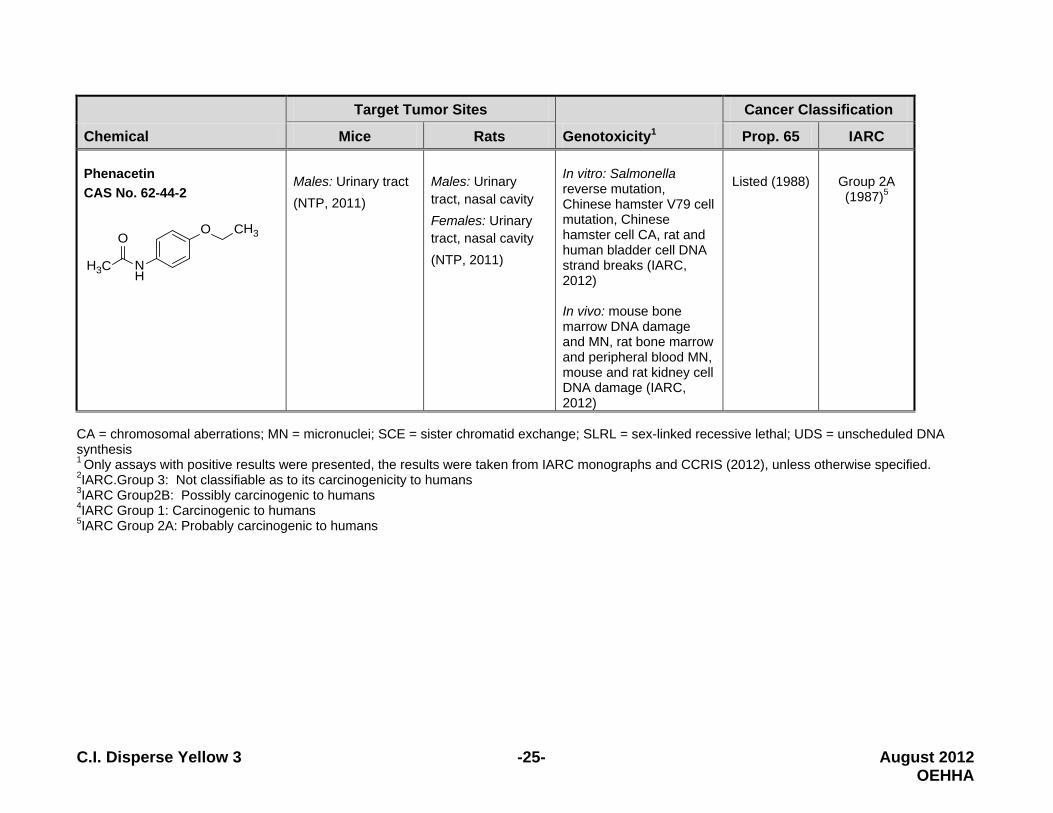
In male B6C3F1 mice, C.I. Disperse Yellow 3 significantly increased the incidence of benign lung tumors, accompanied by a nearly statistically significant increase in combined benign and malignant lung tumors. In female mice, significant increases in malignant lymphoma, combined malignant lymphoma and leukemia, benign liver tumors, and combined benign and malignant liver tumors occurred. The lung tumors observed in treated male mice were alveolar/bronchiolar adenomas and carcinomas. These tumors arise from the same cell of origin, thus they are combined when evaluating study results (IARC, 2006; McConnell et al., 1986). The malignant lymphomas observed in treated female mice occurred in multiple organs, and were of either the lymphocytic or histiocytic type. Lymphocytic leukemias were also observed in treated female mice. When evaluating treatment-related effects on hematopoietic tumors, malignant lymphomas and lymphocytic leukemias were combined (McConnell et al., 1986). The liver tumors observed in treated female mice included hepatocellular adenomas and carcinomas. The adenomas were composed of well-circumscribed solid sheets of cells which had basophilic or eosinophilic cytoplasm. Hepatocellular carcinomas had prominent trabecular areas. Hepatocellular adenomas and carcinomas arise from the same cell type, and adenomas can progress to carcinomas. For this reason, these two tumor phenotypes are combined when evaluating study results (IARC, 2006; McConnell et al., 1986). 3.3.5 Structure-Activity Comparisons C.I. Disperse Yellow 3 is a monoazo dye that shares structural similarity with several other compounds. The expected metabolites formed as a result of reduction of the azo bond, 4-aminoacetanilide and 2-amino-p-cresol, also present concerns for potential carcinogenicity, based on positive findings in genotoxicity assays. C.I. Disperse Yellow 3 and its expected metabolites were compared with seven structurally similar carcinogens with regard to target tumor sites, genotoxic activity, and cancer classification status as reported under Proposition 65 and the International Agency for Research on Cancer (IARC). As summarized in Table 5, C.I. Disperse Yellow 3 was compared to four structurally similar monoazo compounds, 4-aminoacetanilide was compared to one structurally similar compound, and 2-amino-p-cresol was compared to two structurally similar compounds, all of which are listed under Proposition 65 as causing cancer. 2-Aminotoluene, with structural similarity to 2-amino

C.I. Disperse Yellow 3 -21- August 2012 OEHHA
-p-cresol, is classified by IARC as carcinogenic to humans (Group 1). Phenacetin, with structural similarity to 4-aminoacetanilide, is classified by IARC as probably carcinogenic to humans (Group 2A). Four of the compounds in Table 5 are classified by IARC as possibly carcinogenic to humans (Group 2B): p-aminoazobenzene, o-aminoazotoluene, and Oil Orange SS, which are each monoazo compounds, and 2,4-diaminotoluene, which has structural similarity to 2-amino-p-cresol. Many of the carcinogens included in Table 5 induced tumors in experimental animals at multiple sites, and all induce tumors in more than one sex/species. In particular, liver tumors were induced in more than one sex/species by C.I. Disperse Yellow 3 and several structurally similar compounds, including p-aminoazobenzene, o-aminoazotoluene, and 2,4-diaminotoluene. Two additional chemicals induced liver tumors in one sex/species: azobenzene (male mice) and 2-aminotoluene (female mice). Each of the structurally similar carcinogens included in Table 5 were positive in one or more in vitro genotoxicity assays and three (2,4-diaminotoluene, 2-aminotoluene and phenacetin) were positive in one or more in vivo genotoxicity assays.

C.I. Disperse Yellow 3 -22- August 2012 OEHHA
Table 5. Structure-Activity Comparisons for C.I. Disperse Yellow 3 and Its Expected Metabolites (Resulting from Reduction of the Azo Bond).
Chemical
Target Tumor Sites
Genotoxicity1
Cancer Classification
Mice Rats Prop. 65 IARC
C. I. Disperse Yellow 3 CAS No. 2832-40-8
NN
OH
CH3
NH
O
CH3
Males: Lung Females: Liver, hematopoietic system (NTP, 1982)
Males: Liver, stomach (NTP, 1982)
In vitro: Salmonella reverse mutation, mouse L5178Y lymphoma cells forward mutation, Chinese hamster ovary cells SCE, primary rat hepatocyte UDS In vivo: frog larvae CA
Under evaluation
Group 32
(1990)
4-Aminoacetanilide expected metabolite CAS No. 122-80-5
NH2
NH
O
CH3
Not tested Not tested In vitro: Salmonella reverse mutation In vivo: Mouse bone marrow CA
Not evaluated
Not evaluated
2-Amino-p-cresol expected metabolite CAS No. 95-84-1
H3C NH2
OH
Not tested Not tested In vitro: Salmonella reverse mutation, mouse L5178Y lymphoma cells forward mutation
Not evaluated
Not evaluated

C.I. Disperse Yellow 3 -23- August 2012 OEHHA
Chemical
Target Tumor Sites
Genotoxicity1
Cancer Classification
Mice Rats Prop. 65 IARC
Azobenzene CAS No. 103-33-3
NN
Males: Liver (IARC, 1975a)
Males: Spleen and other abdominal organs Females: Spleen and other abdominal organs (NTP,1979)
In vitro: Salmonella reverse mutation, rat hepatocyte DNA damage, human lymphocyte UDS (US EPA, 1988) In vivo: Rat bone marrow MN (George, 1990)
Listed (1990) Group 3 (1987)
p-Aminoazobenzene (Solvent Yellow 1) CAS No. 60-09-3
NN
H2N
Males: Liver (IARC,1975b)
Males: Liver, skin (IARC,1975b)
In vitro: Salmonella reverse mutation, rodent hepatocyte UDS
Listed (1990) Group 2B3 (1987)
o-Aminoazotoluene (Solvent Yellow 3) CAS No. 97-56-3
NN
NH2
CH3
CH3
Males and females: Liver, lung Males: Liver Females: Soft tissues (fibrosarcoma) (NTP, 2011)
Males: Liver, lung; Females: Liver, lung (NTP, 2011)
In vitro: Salmonella reverse mutation, mouse L5178Y lymphoma cells forward mutation
Listed (1987) Group 2B (1987)

C.I. Disperse Yellow 3 -24- August 2012 OEHHA
Chemical
Target Tumor Sites
Genotoxicity1
Cancer Classification
Mice Rats Prop. 65 IARC
Oil Orange SS (C.I. Solvent Orange 2) CAS No. 2646-17-5
NN
OH
CH3
Males and females: Bladder Males: Intestinal (IARC,1975c)
None (IARC 1975c)
In vitro: Salmonella reverse mutation
Listed (1988)
Group 2B (1987)
2,4-Diaminotoluene CAS No. 95-80-7
CH3
H2N NH2
Females: Liver (NTP, 2011)
Males: Liver, kidney; skin Females: Liver, mammary gland, lymphoma (NTP, 2011)
In vitro: Salmonella reverse mutation, rat hepatocyte and human HepG2 cell UDS, human HepG2 cell MN, Chinese hamster lung cell and ovary cell CA (IARC, 1978) In vivo: rat hepatocyte MN (Narumi et al, 2012), Drosophila SLRL (IARC, 1978)
Listed (1988)
Group 2B (1987)
2-Aminotoluene CAS No. 95-53-4
CH3
NH2
Males: Blood-vessels (hemangioma and hemangiosarcoma); Females: Liver and blood vessels (NTP, 2011)
Males: Abdominal, scrotum, skin, spleen Females: Mammary, bladder, spleen; (NTP, 2011)
In vitro: Salmonella reverse mutation, Chinese hamster V79 cell mutation, rat hepatocyte UDS, rodent cell CA, human cell SCE and UDS In vivo: rat peripheral blood reticulocyte MN, Drosophila somatic mutation
Listed (1989) Group 1 (2010)4
C.I. Disperse Yellow 3 -25- August 2012 OEHHA
Chemical
Target Tumor Sites
Genotoxicity1
Cancer Classification
Mice Rats Prop. 65 IARC Phenacetin CAS No. 62-44-2
CH3O
NH
H3C
O
Males: Urinary tract (NTP, 2011)
Males: Urinary tract, nasal cavity Females: Urinary tract, nasal cavity (NTP, 2011)
In vitro: Salmonella reverse mutation, Chinese hamster V79 cell mutation, Chinese hamster cell CA, rat and human bladder cell DNA strand breaks (IARC, 2012) In vivo: mouse bone marrow DNA damage and MN, rat bone marrow and peripheral blood MN, mouse and rat kidney cell DNA damage (IARC, 2012)
Listed (1988)
Group 2A (1987)5
CA = chromosomal aberrations; MN = micronuclei; SCE = sister chromatid exchange; SLRL = sex-linked recessive lethal; UDS = unscheduled DNA synthesis 1 Only assays with positive results were presented, the results were taken from IARC monographs and CCRIS (2012), unless otherwise specified. 2IARC.Group 3: Not classifiable as to its carcinogenicity to humans 3IARC Group2B: Possibly carcinogenic to humans 4IARC Group 1: Carcinogenic to humans 5IARC Group 2A: Probably carcinogenic to humans

C.I. Disperse Yellow 3 -26- August 2012 OEHHA
4 MECHANISMS C.I. Disperse Yellow 3 increased the incidence of liver tumors in treated male F344 rats and female B6C3F1 mice, hematopoietic cancers in female B6C3F1 mice, and lung tumors in male B6C3F1 mice. In addition, rare stomach tumors were observed in treated male F344 rats. The mechanism by which C.I. Disperse Yellow 3 induces tumors in these various tissues is unknown. However, a body of evidence suggests that C.I. Disperse Yellow 3 is likely to operate by a genotoxic mechanism or mechanisms. C.I. Disperse Yellow 3 tested positive in a variety of genotoxicity assays (described in Section 3.3.2 Genotoxicity). Evidence for genotoxicity includes positive tests for mutagenicity in multiple strains of Salmonella and in mouse lymphoma cells, chromosomal aberrations in frog larvae, SCE in Chinese hamster ovary cells, and UDS in rat hepatocytes exposed in vitro. Furthermore, the expected aromatic amine metabolites arising from azoreduction of C.I. Disperse Yellow 3, namely 4-aminoacetanilide and 2-amino-p-cresol, have also demonstrated genotoxic activity. In summary, while the mechanism(s) of tumorigenic action of C.I. Disperse Yellow 3 remain unknown, the available evidence suggests that genotoxicity is involved. This includes evidence from tests for mutagenicity, clastogenicity and DNA synthesis, expected metabolism to genotoxic aromatic amines, structural similarity of C.I. Disperse Yellow 3 with other genotoxic and carcinogenic monoazo compounds, and structural similarity of the azoreduction products to other genotoxic and carcinogenic compounds. Other mechanisms yet to be elucidated may also be operative. 5 REVIEWS BY OTHER AGENCIES C.I. Disperse Yellow 3 has not been classified as to its potential carcinogenicity by the U.S. EPA, IARC, the U.S. Food and Drug Administration, the National Toxicology Program, or the National Institute for Occupational Safety and Health.

C.I. Disperse Yellow 3 -27- August 2012 OEHHA
6 SUMMARY AND CONCLUSIONS 6.1 Summary of Evidence No epidemiology studies were identified that investigated the risk of cancer associated with documented exposure to C.I. Disperse Yellow 3. Four case-control studies were identified that examined bladder cancer risk associated with dye-related occupations in the textile industry. Studies evaluating other tumor sites were not found. None of the studies provided information on exposures to specific dyes, and only one study mentioned C.I. Disperse Yellow 3. That study, by Gonzales et al. (1988), reported an elevated risk of bladder cancer among individuals who worked in “textile dyeing or printing jobs,” after adjusting for tobacco smoking (OR=4.41, 95% CI=1.15-16.84). Because C.I. Disperse Yellow 3 is only one of many disperse dyes used in the textile industry and because the available studies did not collect information on exposures to specific dyes, these studies are inadequate to assess the relationship between exposure to C.I. Disperse Yellow 3 and cancer risk. In 104-week animal studies, dietary administration of C.I. Disperse Yellow 3 significantly increased the incidence of liver tumors in treated male F344 rats and female B6C3F1 mice, hematopoietic cancers in female B6C3F1 mice, and lung tumors in male B6C3F1 mice. Rare stomach tumors were also observed in treated male F344 rats (NTP, 1982). Specifically, the following increases in tumors were observed in these studies: Liver tumors
• In male rats, C.I. Disperse Yellow 3 significantly increased the incidence of hepatocellular adenoma, and combined hepatocellular adenoma and carcinoma in the low- and high-dose groups as compared with the control group. Significant positive dose-related trends in tumor incidence were observed.
• In female mice, C.I. Disperse Yellow 3 significantly increased the incidence of hepatocellular adenoma and combined hepatocellular adenoma and carcinoma in the low- and high-dose groups as compared with the control group. Significant positive dose-related trends in tumor incidence were observed.
Tumors of the hematopoietic system
• In female mice, C.I. Disperse Yellow 3 significantly increased the incidence of malignant lymphoma and combined malignant lymphoma and leukemia in the high-dose group as compared with the control group. Significant positive dose-related trends in tumor incidence were observed.

C.I. Disperse Yellow 3 -28- August 2012 OEHHA
Lung tumors • In male mice, C.I. Disperse Yellow 3 significantly increased the incidence of
alveolar/bronchiolar adenoma in the high-dose group as compared with the control group. The increase in combined alveolar/bronchiolar adenoma and carcinoma approached statistical significance in the high-dose group as compared with the control group. Significant positive dose-related trends in tumor incidence were observed.
Stomach tumors
• In male rats, rare stomach tumors (glandular and non-glandular) were observed in treated animals. No stomach tumors were seen in the control group. Tumors of the glandular stomach were seen in two low-dose animals (an adenoma and a mucinous adenocarcinoma) and in one high-dose animal (an adenocarcinoma and a sarcoma). Tumors of the non-glandular stomach were seen in two low-dose animals (a squamous papilloma and a fibrosarcoma).
Evidence of genotoxicity comes from several test systems. C.I. Disperse Yellow 3 induced:
• Basepair substitution reverse mutations in Salmonella typhimurium in the presence of exogenous metabolic activation;
• Frameshift reverse mutations in Salmonella typhimurium in the absence or presence of exogenous metabolic activation;
• Forward mutations in mouse lymphoma cells in the presence of exogenous metabolic activation;
• Chromosomal aberrations in frog larvae; • SCEs in CHO cells in the absence or presence of metabolic activation; • Unscheduled DNA synthesis in rat hepatocytes.
The expected aromatic amine metabolites of C.I. Disperse Yellow 3 and structurally similar compounds present concern regarding the carcinogenicity of C.I. Disperse Yellow 3:
• The expected aromatic amine metabolites (4-aminoacetanilide and 2-amino-p-cresol) of C.I. Disperse Yellow 3 are genotoxic.
• Like C.I. Disperse Yellow 3 and its expected aromatic amine metabolites, each of the seven structurally similar carcinogens are genotoxic.
• C.I. Disperse Yellow 3 and five of the seven structurally similar carcinogens induce liver tumors in rodents.
o C.I. Disperse Yellow 3, p-aminoazobenzene, o-aminoazotoluene, and 2,4-diaminotoluene induced liver tumors in more than one sex/species.
o Azobenzene and 2-aminotoluene induce liver tumors in one sex/species.

C.I. Disperse Yellow 3 -29- August 2012 OEHHA
6.2 Conclusions Evidence for carcinogenicity of C.I. Disperse Yellow 3 comes primarily from 104-week diet studies conducted in male F344 rats and female and male B6C3F1 mice. Exposure to C.I. Disperse Yellow 3 in male rats resulted in statistically significant increases in benign, and combined benign and malignant liver tumors. Rare stomach tumors were also observed in treated male rats. In female mice, exposure to C.I. Disperse Yellow 3 resulted in statistically significant increases in malignant lymphoma and combined malignant lymphoma and leukemia as well as statistically significant increases in benign, and combined benign and malignant liver tumors. In male mice exposure to C.I. Disperse Yellow 3 resulted in a statistically significant increase in benign lung tumors; the increase in combined benign and malignant lung tumors approached statistical significance. Positive findings in multiple genotoxicity test systems indicate that C.I. Disperse Yellow 3 may operate through a genotoxic mechanism. C.I. Disperse Yellow 3 induced mutations in multiple strains of Salmonella typhimurium and in mouse lymphoma cells in vitro, SCEs in CHO cells in vitro, and UDS in rat hepatocytes in vitro, and chromosomal aberrations in frog larvae in vivo,. 4-Aminoacetanilide and 2-amino-p-cresol, the expected aromatic amine metabolites arising from azoreduction of C.I. Disperse Yellow 3, are also genotoxic. C.I. Disperse Yellow 3 and five genotoxic carcinogens with shared structural similarity induce liver tumors in rodents. The structurally similar liver carcinogens include p-aminoazobenzene, o-aminoazotoluene, and 2,4-diaminotoluene, which like C.I. Disperse Yellow 3 induce liver tumors in more than one sex/species, and azobenzene and 2-aminotoluene, which induce liver tumors in one sex/species.

C.I. Disperse Yellow 3 -30- August 2012 OEHHA
7 REFERENCES Bomberger DC, Boughton RL (1983). Wastes from manufacture of azo dyes and pigments (excluding benzidine and its congeners). Industrial environmental research laboratory, Office of research and development, US EPA. Contract No. 68-03-2944, reported by SRI International. Boorman GA, Chapin RE, Mitsumori K (1990). Oral cavity, esophagus, and stomach. In: Pathology of the Fischer Rat. G Boorman, S Eustis, M Elwel, CA Montgomery, Jr. and WF MacKenzie (Eds.). San Diego: Academic Press, Inc., pp.9-29. Cameron TE, Hughes TJ, Kirby EE, Fung VA, Dunkel VC (1987). Mutagenic activity of 27 dyes and related ehemicals in the Salmonella microsome and mouse lymphoma TK+ 1-assays. Mutat Res 189:223-261. CDC (Center For Disease Control And Prevention, 1983). National Occupational Exposure Survey 1981-1983. http://www.cdc.gov/noes/noes5/m2104ctr.html (Accessed in November, 2011). Chemical Carcinogenesis Research Information System (CCRIS, 2012). Available at URL: http://toxnet.nlm.nih.gov (accessed on January 27, 2012). Chung KT, Stevens SE Jr, Cerniglia CE (1992). The reduction of azo dyes by the intestinal microflora. Crit Rev Microbiol 18(3):175-90. CIR (Cosmetic Ingredient Review, 1996). Final report on the safety assessment of disperse yellow 3. J Am Coll Tox 15:311-319. Dryson E, Mannetje A, Walls C, McLean D, McKenzie F, Maule M, Cheng S, Cunningham C, Kromhout H, Boffetta P, Blair A, Pearce N (2008). Case-control study of high risk occupations for bladder cancer in New Zealand. Int J Cancer 122:1340-6. Foureman P, Mason JM, Valencia R, Zimmering S (1994). Chemical mutagenesis testing in Drosophila. X. Results of 70 coded compounds tested for the National Toxicology Program. Environ Mol Mutagen 23:208-227. Freeman HS, and Mock GN (2007). Dye Application, manufacture of dye intermediates and dyes. In: Kent and Riegel’s Handbook of Industrial Chemistry and Biotechnology (editor Kent JA), published by Springer Science, New York, NY.

C.I. Disperse Yellow 3 -31- August 2012 OEHHA
George E, Andrews M, Westmoreland C (1990). Effects of azobenzene and aniline in the rodent bone marrow micronucleus test. Carcinogenesis 11(9):1551-6. Gideon (2010). Information Preview for Textile Dyes. In GIDEON online. Retrieved from http://www.gideononline.com on May 9, 2012. Gonzales CA, Riboli E, Lopez-Abente G (1988). Bladder cancer among workers in the textile industry: results of a Spanish case-control study. Am J Ind Med 14:673-80. González CA, López-Abente G, Errezola M, Escolar A, Riboli E, Izarzugaza I, Nebot M (1989). Occupation and bladder cancer in Spain: A multi-centre case-control study. Int J Epidemiol 18:569-77. Gray PS, Hunter R Jr, Patterson RM (1979). Chromosomal aberrations induced by Dispersion Yellow 3 in Rana clamitans larvae during tail regeneration. Cytobios 25:175-82. Hatch KI, and Maibach HI (2000). Textiles. In: Handbook of Occupational Dermatology (editors Kanerva L, Elsner P, Wahlberg JE and Maibach HI). Springer-Verlag, Berlin Heidelberg, Germany. Hewlett-Packard Company (2011). Material Safety Data Sheet: CD406 series-inkjet printing. Avaiable at URL: http://www.hp.com/hpinfo/globalcitizenship/environment/productdata/Countries/sg/lf_cd406series_sg_eng_v1.pdf IARC (International Agency for Research on Cancer, 1975a). IARC Monographs On The Evaluation Of Carcinogenic Risks To Humans. Some aromatic azo compounds: Azobenzene. Volume 8. IARC, Lyon, p.75-81. IARC (International Agency for Research on Cancer, 1975b). IARC Monographs On The Evaluation Of Carcinogenic Risks To Humans. Some aromatic azo compounds: Para-dimethylamino-azobenzene. Volume 8. IARC, Lyon, France. p.125-146. IARC (International Agency for Research on Cancer, 1975c). IARC Monographs On The Evaluation Of Carcinogenic Risks To Humans. Some aromatic azo compounds: Oil Orange SS. Volume 8. IARC, Lyon, p.165-171. IARC (International Agency for Research on Cancer, 1978). IARC Monographs On The Evaluation Of Carcinogenic Risks To Humans. Some aromatic amines and related nitro

C.I. Disperse Yellow 3 -32- August 2012 OEHHA
compounds (hair dyes, colouring agents and miscellaneous industrial chemicals): 2,4-Diaminotoluene. Volume 16. IARC, Lyon, p.83-95. IARC (International Agency for Research on Cancer,1987). Overall evaluations of carcinogenicity: An updating of IARC Monographs. Volumes 1-42, Supplement 7. IARC, Lyon, France. IARC (International Agency for Research on Cancer, 1990). IARC Monographs on the evaluation of carcinogenic risks to humans. Some flame retardants and textile chemicals, and exposures in the textile manufacturing industry. Volume 48. IARC, Lyon, France. p.33-40; p.149-159. IARC (International Agency for Research on Cancer, 2006). IARC Monographs on the Evaluation of Carcinogenic Risks to Humans. Preamble. IARC, Lyon, France. Available at URL: http://monographs.iarc.fr/ENG/Preamble/CurrentPreamble.pdf. IARC (International Agency for Research on Cancer, 2010). IARC Monographs On The Evaluation Of Carcinogenic Risks To Humans. Some aromatic amines, organic dyes, and related exposures: 2-Aminotoluene. Volume 99. IARC, Lyon, France. p.407-470. IARC (International Agency for Research on Cancer, 2011). Agents Classified by the IARC Monographs, Volumes 1–105. World Health Organization. Lyon, France. Available at URL: http://monographs.iarc.fr/ENG/Classification/index.php IARC (International Agency for Research on Cancer, 2012). IARC Monographs On The Evaluation Of Carcinogenic Risks To Humans. A review of human carcinogens: pharmaceuticals: Phenacetin. Volume 100A. IARC, Lyon, France. p.379-400. Ivett JL, Brown BM, Rodgers C, Anderson BE, Resnick MA, Zeiger E (1989). Chromosomal aberrations and sister chromatid exchange tests in Chinese hamster ovary cells in vitro. IV. Results with 15 chemicals. Environ Mol Mutagen 14:165-87. Kitchin KT, Brown JL (1994).Dose-response relationship for rat liver DNA damage caused by 49 rodent carcinogens.Toxicology 88:31-49. Levine WG (1991). Metabolism of azo dyes: implication for detoxication and activation. Drug Metab Rev 23:253-309. Malinauskiene L, Zimerson E, Bruze M, Ryberg K, Isaksson M (2012). Patch testing with the textile dyes Disperse Orange 1 and Disperse Yellow 3 and some of their

C.I. Disperse Yellow 3 -33- August 2012 OEHHA
potential metabolites, and simultaneous reactions to para-amino compounds. Contact Dermatitis. doi: 10.1111/j.1600-0536.2012.02080.x. Mansour H, Ayed-Ajmi Y, Mosrati R, Corroler D, Ghedira K, Barillier D, Chekir-GhediraL (2010). Acid violet 7 and its biodegradation products induce chromosome aberrations, lipid peroxidation, and cholinesterase inhibition in mouse bone marrow. Environ Sci Pollut Res Int 17(7):1371-8. Matthews EJ, Spalding JW, Tennant RW (1993). Transformation of BALB/c-3T3 cells: V. Transformation responses of 168 chemicals compared with mutagenicity in Salmonella and carcinogenicity in rodent bioassays. Environ Health Perspect 101:347-482. McConnell EE, Solleveld HA, Swenberg JA, Boorman GA (1986). Guidelines for combining neoplasms for evaluation of rodent carcinogenesis studies. J Natl Cancer Inst 76:283-9. McGregor DB, Brown A, Cattanach P, Edwards I, McBride D, Riach C, Caspary WJ (1988). Responses of the L5178Y tk+ /tk- mouse lymphoma cell forward mutation assay. III. 72 coded chemicals. Environ Mol Mutagenesis 12:85-154. McIntyre JE (2004). Synthetic fibers: nylon, polyester, acrylic, polyolefin. The Textile Institute. Woodhead Publishing, Cambridge, and CRC Press, New York. p80. Narumi K, Ashizawa K, Takashima R, Takasawa H, Katayama S, Tsuzuki Y, Tatemoto H, Morita T, Hayashi M, Hamada S (2012). Development of a repeated-dose liver micronucleus assay using adult rats: An investigation of diethylnitrosamine and 2,4-diaminotoluene. Mutat Res 747(2):234-9. NTP (National Toxicology Program, 1979). National Cancer Institute carcinogenesis technical report series: Bioassay of azobenzene for possible carcinogenicity. TR-154, NIH Publication No. 79-1710. NTP (National Toxicology Program, 1982). NTP technical report on the carcinogenesis bioassay of C.I. Disperse Yellow 3 (CAS No. 2832-40-8) in F344/N rats and B6C3F1 mice (Feed Study). TR-222, NTP-81-80, NIH Publication No. 82-1778. NTP (National Toxicology Program, 1999). NTP historical controls report oral feed F344 rats. Department of Health and Human Services. December, 1999. Available at URL: http://ntp.niehs.nih.gov/?objectid=92E6AAA5-F1F6-975E-71C88528A3E7B315#9

C.I. Disperse Yellow 3 -34- August 2012 OEHHA
NTP (National Toxicology Program, 2011). Report on carcinogens, 12th edition. U.S. Department of Health and Human Services, June 2011. Platzek T, Lang C, Grohmann G, Gi US, Baltes W (1999). Formation of a carcinogenic aromatic amine from an azo dye by human skin bacteria in vitro. Hum Exp Toxicol 18:552-9. Santa Cruz Biotechnology, Inc. (2008). Chronic Health Effects. In: Material Safety Data Sheet: Disperse Yellow 3, sc-214923. Available at URL: http://datasheets.scbt.com/sc-214923.pdf SCCPNFP (Scientific Committee on Cosmetic Products and Non-food products, 2002). Opinion of the scientific committee on cosmetic products and non-food products intended for consumers: Concerning the safety review of the use of certain azo-dyes in cosmetic products. The 19th Plenary Meeting. European Commission, February 2002. Scorecard (2011). C.I. Disperse Yellow 3: Chemical profiles-industrial uses. Available at URL: http://scorecard.goodguide.com/chemical-profiles/uses.tcl?edf_substance_id=2832-40-8 Seifried HE, Seifried RM, Clarke JJ, Junghans TB, San RH (2006). A compilation of two decades of mutagenicity test results with the Ames Salmonella typhimurium and L5178Y mouse lymphoma cell mutation assays. Chem Res Toxicol 19:627-44. Serra C, Kogevinas M, Silverman DT, Turuguet D, Tardon A, Garcia-Closas R, Carrato A, Castaño-Vinyals G, Fernandez F, Stewart P, Benavides FG, Gonzalez S, Serra A, Rothman N, Malats N, Dosemeci M (2008). Work in the textile industry in Spain and bladder cancer. Occup Environ Med 65:552-9. Shelby MD, Erexson GL, Hook GJ, Tice RR (1993). Evaluation of a three-exposure mouse bone marrow micronucleus protocol: Results with 49 chemicals. Environ Molec Mutag 21:160-179. Stahlmann R, Wegner M, Riecke K, Kruse M, Platzek T (2006). Sensitizing potential of four textile dyes and some of their metabolites in a modified local lymph node assay. Toxicology 219(1-3):113-23. Tennant RW, Spalding JW, Stasiewicz S, Caspary WD, Mason JM, Resnick MA (1987a). Comparative evaluation of genetic toxicity patterns of carcinogens and non-
C.I. Disperse Yellow 3 -35- August 2012 OEHHA
ccinogens: strategies for predictive use of short-term assay. Environ Health Perspect 75:87-95. Tennant RW, Margolin BH, Shelby MD, Zeiger E, Haseman lK, Spalding J, Caspary W, Resnick M, Stasiewicz S, Anderson B, Minor R (1987b). Prediction of chemical carcinogenicity in rodents from in vitro genetic toxicity assays. Science 236:933-941. US EPA (1988) Integrated Risk Information System (IRIS): Azobenzene (CASRN 103-33-3). Available at URL: http://www.epa.gov/iris/subst/0351.htm US EPA Toxics Release Inventory (TRI, 2001). Chemical Reports on C.I. Disperse Yellow for 1995, 1998, 1999, 2000 and 2001. Available at URL: http://iaspub.epa.gov/triexplorer/tri_getcounties.getcounties?report=tri_release.chemical01&scriptname=chemical&state=USA&c_year=2001&c_industry=ALL&c_chemical=PICKCHEM&c_indlist=&c_chemlist=&c_coreyear=&c_usrState=&c_fips=00000&c_zip=&c_tabrpt=1&c_chk0=true&c_chk1=true&c_chk2=true&c_chk3=true&c_chk4=true&c_chk5=true&c_chk6=true&c_chk7=true&c_chk8=true&c_chk9=true&c_chk10=true Yoshimi N, Sugie S, Iwata H, Niwa K, Mori H, Hashida C, Shimizu H (1988). The genotoxicity of a variety of aniline derivatives in a DNA repair test with primary cultured rat hepatocytes. Mutat Res 206(2):183-91. Zeiger E, Anderson B, Haworth S, Lawlor T, Mortelmans K (1988). Salmonella mutagenicity tests: IV. Results from the testing of 300 chemicals. Environ Mol Mutagen 11:1-157.