Evidence for quasispecies distributions in the human hepatitis A virus genome
9
Evidence for quasispecies distributions in the human hepatitis A virus genome Glo `ria Sa ´nchez, a Albert Bosch, a, * Gema Go ´mez-Mariano, b Esteban Domingo, b and Rosa M. Pinto ´ a a Grup Virus Ente `rics, Department of Microbiology, University of Barcelona, 08028 Barcelona, Spain b Centro de Biologı ´a Molecular “Severo Ochoa,” CSIC-UAM, Cantoblanco, 28049 Madrid, Spain Received 7 April 2003; returned to author for revision 20 May 2003; accepted 12 June 2003 Abstract Nucleotide sequence analysis of multiple molecular clones of the hepatitis A virus (HAV), generated by reverse transcription-PCR of two capsid-coding regions, revealed a degree of heterogeneity compatible with a quasispecies structure in three clinical samples. Passage of plaque-purified reference strain HAV pHM175 43c in FRhK-4 cells documented the generation of a mutant distribution of HAV genomes. The mutant spectra showed mutation frequencies in the range of 1 10 3 to 1 10 4 substitutions per nucleotide, with a dominance of transition over transversion mutations. While in the VP3-coding region, nonsynonymous mutations were predominant; in the VP1-coding region they were uncommon. Around 50% of the amino acid replacements involved residues located at or near antigenic sites. Most of the detected mutations occurred at or in the vicinity of rare codons, suggesting a dynamics of mutation-selection, predominantly at and around rare codons. The results indicate that despite antigenic conservation, HAV replicates as a complex distribution of mutants, a feature of viral quasispecies. © 2003 Elsevier Inc. All rights reserved. Keywords: HAV; Quasispecies; Antigenic sites; Codon-usage; Rare codon; Normal codon Introduction Hepatitis A virus (HAV), classified as the type species of the genus Hepatovirus within the Picornaviridae family (van Regenmortel et al., 2000), is a hepatotropic virus which represents a significant problem for human health (Battegay and Feinstone, 1997; Hollinger and Emerson, 2001). The virion capsid is composed of the structural proteins VP1, VP2, VP3, and possibly VP4, encoded in the P1 region of the genome (Hollinger and Emerson, 2001; Racaniello, 2001). Some degree of nucleotide sequence heterogeneity of the P1 genomic region has been observed among independent HAV isolates from different regions of the world (Lemon et al., 1987; Robertson et al., 1992; Taylor, 1997; Arauz-Ruiz et al., 2001; Costa-Mattioli et al., 2001). However, this variability at the nucleotide level is not reflected in an equivalent degree of variation at the amino acid level (Lemon and Robertson, 1993; Hollinger and Emerson, 2001; Sa ´nchez et al., 2003). The high degree of conservation of the amino acid sequences of the capsid proteins of HAV entails a low antigenic diversity, and therefore, only a single serotype of human HAV has been recognized (Hollinger and Emerson, 2001). This suggests the operation of severe structural constraints in the HAV capsid (Sa ´nchez et al., 2003). HAV shows a high codon usage bias, with the repeated occurrence of 22 rare codons for 14 amino acids (Sa ´nchez et al., 2003). Most of the carboxy-terminal regions of -barrels and -helices, that are predicted in the capsid proteins (Luo et al., 1988), include residues encoded by conserved rare codons, suggesting a potential function of such codons in a decrease of the rate of translation to facilitate the proper folding of the capsid proteins (Sa ´nchez et al., 2003). The molecular basis of the genetic variability of RNA * Corresponding author. Department of Microbiology, School of Biol- ogy, University of Barcelona, Av. Diagonal 645, 08028 Barcelona, Spain. Fax: 34-934034629. E-mail address: [email protected] (A. Bosch). R Available online at www.sciencedirect.com Virology 315 (2003) 34 – 42 www.elsevier.com/locate/yviro 0042-6822/03/$ – see front matter © 2003 Elsevier Inc. All rights reserved. doi:10.1016/S0042-6822(03)00483-5
-
Upload
gloria-sanchez -
Category
Documents
-
view
212 -
download
0
Transcript of Evidence for quasispecies distributions in the human hepatitis A virus genome

Evidence for quasispecies distributions in the humanhepatitis A virus genome
Gloria Sanchez,a Albert Bosch,a,* Gema Gomez-Mariano,b Esteban Domingo,b
and Rosa M. Pinto´a
a Grup Virus Enterics, Department of Microbiology, University of Barcelona, 08028 Barcelona, Spainb Centro de Biologıa Molecular “Severo Ochoa,” CSIC-UAM, Cantoblanco, 28049 Madrid, Spain
Received 7 April 2003; returned to author for revision 20 May 2003; accepted 12 June 2003
Abstract
Nucleotide sequence analysis of multiple molecular clones of the hepatitis A virus (HAV), generated by reverse transcription-PCR oftwo capsid-coding regions, revealed a degree of heterogeneity compatible with a quasispecies structure in three clinical samples. Passageof plaque-purified reference strain HAV pHM175 43c in FRhK-4 cells documented the generation of a mutant distribution of HAV genomes.The mutant spectra showed mutation frequencies in the range of 1� 10�3 to 1 � 10�4 substitutions per nucleotide, with a dominance oftransition over transversion mutations. While in the VP3-coding region, nonsynonymous mutations were predominant; in the VP1-codingregion they were uncommon. Around 50% of the amino acid replacements involved residues located at or near antigenic sites. Most of thedetected mutations occurred at or in the vicinity of rare codons, suggesting a dynamics of mutation-selection, predominantly at and aroundrare codons. The results indicate that despite antigenic conservation, HAV replicates as a complex distribution of mutants, a feature of viralquasispecies.© 2003 Elsevier Inc. All rights reserved.
Keywords: HAV; Quasispecies; Antigenic sites; Codon-usage; Rare codon; Normal codon
Introduction
Hepatitis A virus (HAV), classified as the type species ofthe genusHepatovirus within the Picornaviridae family(van Regenmortel et al., 2000), is a hepatotropic virus whichrepresents a significant problem for human health (Battegayand Feinstone, 1997; Hollinger and Emerson, 2001). Thevirion capsid is composed of the structural proteins VP1,VP2, VP3, and possibly VP4, encoded in the P1 region ofthe genome (Hollinger and Emerson, 2001; Racaniello,2001).
Some degree of nucleotide sequence heterogeneity of theP1 genomic region has been observed among independentHAV isolates from different regions of the world (Lemon et
al., 1987; Robertson et al., 1992; Taylor, 1997; Arauz-Ruiz etal., 2001; Costa-Mattioli et al., 2001). However, this variabilityat the nucleotide level is not reflected in an equivalent degreeof variation at the amino acid level (Lemon and Robertson,1993; Hollinger and Emerson, 2001; Sa´nchez et al., 2003). Thehigh degree of conservation of the amino acid sequences of thecapsid proteins of HAV entails a low antigenic diversity, andtherefore, only a single serotype of human HAV has beenrecognized (Hollinger and Emerson, 2001). This suggests theoperation of severe structural constraints in the HAV capsid(Sanchez et al., 2003). HAV shows a high codon usage bias,with the repeated occurrence of 22 rare codons for 14 aminoacids (Sa´nchez et al., 2003). Most of the carboxy-terminalregions of�-barrels and�-helices, that are predicted in thecapsid proteins (Luo et al., 1988), include residues encoded byconserved rare codons, suggesting a potential function of suchcodons in a decrease of the rate of translation to facilitate theproper folding of the capsid proteins (Sa´nchez et al., 2003).
The molecular basis of the genetic variability of RNA
* Corresponding author. Department of Microbiology, School of Biol-ogy, University of Barcelona, Av. Diagonal 645, 08028 Barcelona, Spain.Fax: �34-934034629.
E-mail address: [email protected] (A. Bosch).
R
Available online at www.sciencedirect.com
Virology 315 (2003) 34–42 www.elsevier.com/locate/yviro
0042-6822/03/$ – see front matter © 2003 Elsevier Inc. All rights reserved.doi:10.1016/S0042-6822(03)00483-5

viruses has been associated with the absence of a 3� 3 5�exonuclease proofreading activity in viral RNA-dependentRNA polymerases and reverse transcriptases, together withlack of postreplicative repair mechanisms that can act onDNA but not on RNA (Holland et al., 1992; Steinhauer etal., 1992; Williams and Loeb, 1992; Domingo and Holland,1997). Mutation rates for a variety of RNA viruses rangebetween 10�4 and 10�5 substitutions per nucleotide copied(Batschelet et al., 1976; Drake, 1993; Drake and Holland,1999). As a consequence, RNA viruses replicate as complexdynamic mutant distributions, termed viral quasispecies (Ei-gen and Biebricher, 1988; Holland et al., 1992; Domingoand Holland, 1997; Domingo et al., 2001). The open readingframe of the putative HAV 3D (the RNA-dependent RNApolymerase) does not provide any evidence for the presenceof a proofreading or error-correcting activity known to beassociated with several DNA- dependent DNA polymerases(Kunkel, 1988; Zimmern, 1988; Friedberg et al., 1995).Therefore, mutation rates and frequencies for HAV are notexpected to differ significantly from those of other picorna-viruses and RNA viruses in general (Drake and Holland,1999). In this view, the 90% or higher amino acid sequenceidentities among independent strains and isolates of HAV(Hollinger and Emerson, 2001) would be the result of neg-ative selection on many newly arising mutants and conver-gence of consensus or average sequences (Eigen andBiebricher, 1988; Holland et al., 1992; Domingo et al.,2001). Yet if mutational pressure originated a number ofmutants hidden in a mutant spectrum, such mutants wouldprovide evidence of quasispecies dynamics, implying thepresence of a variant reservoir for HAV adaptation. Despitethe biological significance of this population structure, nosuch analyses of HAV mutant spectra have been reported.
In the present work, results suggesting the occurrence ofdistributions of related genomes in HAV are presented, bothwith clinical isolates of HAV and in populations evolved incell culture from a biological clone of HAV. The nature ofthe mutations detected in the mutant spectra supports theoperation of structural constraints in the capsid of HAV.Furthermore, the results of HAV evolution in cell culturerevealed the presence of antigenic variants that were gen-erated in the absence of immune selection-as-previouslyobserved with other viruses (Domingo et al., 1993).
Results
A mutant spectrum in clinical isolates of HAV
To test whether the limited amino acid variation amongconsensus sequences of independent HAV isolates was par-alleled by a homogeneous mutant spectrum within a HAVpopulation, a clonal analysis of three clinical samples ofHAV was performed. The samples were Val9 (stool), Val10(stool), and Val12 (serum) (Sanchez et al., 2002), whoseorigin is described under Materials and methods. A mean of
25 molecular clones from a single reverse transcription(RT)-PCR amplification of a fragment of the VP3 (fromnucleotides 1470 to 1839, encoding amino acids 1 to 123)and a fragment of the VP1 (from nucleotides 2459 to 2943,encoding amino acids 85 to 245), which span sequencesencoding the main antigenic sites of HAV (Nainan et al.,1992; Ping and Lemon, 1992; Bosch et al., 1998), wereanalyzed for each sample. Minimum mutation frequencies(counting repeated mutations only once) ranged between1.2 � 10�3 and 1.4 � 10�4 substitutions per nucleotide(Table 1). In some analyses, the maximum mutation fre-quency (counting all mutations relative to the consensus)increased over the minimal mutation frequency. The valuesof Shannon entropy (a measure of the proportion of differ-ent sequences in the set analyzed) indicated heterogeneity ofthe mutant spectra. Control experiments (described in detailunder Materials and methods) indicated that the mutationfrequencies observed cannot be the result of misincorpora-tions during RT-PCR amplification of HAV RNA. Statisti-cal analysis (nonparametric Mann–Whitney U test andANOVA) revealed significant differences between the mu-tation frequencies of the patient samples and the error rateof the RT-PCR system, except in the Val10 sample. How-ever, despite this lack of statistical significance, the local-ization of the mutations (see below) make it extremelyunlikely that they could be just due to the occurrence ofrandom misincorporations. The mutation-frequencies andShannon entropies of the mutant spectra are in the rangeobserved in other viral quasiespecies (see Discussion).
A mutant spectrum in serially passaged HAVpHM175 43c
The sequence comparisons indicated the occurrence inthe HAV isolates of dominant genomic sequences togetherwith several variants. These results suggest generation ofmutants in the course of replication of each individual HAV.However, a definitive proof of mutant generation requires ademonstration that multiple variants are produced de novoupon replication of a single HAV genome (Domingo et al.,1978; Domingo, 1996). To this effect, the reference HAVstrain pHM175 43c, adapted to FRhK-4 cells, was subjectedto three successive plaque isolations to produce pHM17543c P0, and then this clonal population was subjected to 26serial passages in the same cells (about 106 PFU infecting106 cells per passage), as detailed under Materials andmethods. The resulting population is termed pHM175 43cP26. No mutations were detected in the consensus se-quences of the VP1- and VP3-coding regions analyzed, atpassages 1, 5, 11, 16, and 26. However, a clonal analysisrevealed 23 mutations in 100 molecular clones from passage26 (Table 2). The minimum mutation frequencies were7.0�10�4 and 3.3�10�4 substitutions per nucleotide forthe VP3- and VP1-coding regions, respectively. Only forthe VP3-coding region the maximum mutation frequencywas slightly higher than the minimum mutation frequency.
35G. Sanchez et al. / Virology 315 (2003) 34–42

Statistical analysis (nonparametric Mann–Whitney U testand ANOVA) revealed significant differences betweenthese mutation frequencies and the experimental error rate.The values of Shannon entropy indicated heterogeneity ofthe mutant spectra. The consensus and the dominant se-quences were identical. Thus, the results document con-stancy of the average or consensus sequences, and the gen-eration of a mutant spectrum upon replication of abiological clone of HAV. These are features of quasispeciesdynamics (Eigen and Biebricher, 1988). Experiments are inprogress to elucidate whether the virus mutant distributionvaries with cell passage.
Types and location of mutations: mutation clustering
A dominance of transitions over transversions was ob-served in all HAV mutant spectra analyzed (Tables 1 and 2).In contrast, the ratio of synonymous-to-nonsynonymous
mutations was 33 for the VP1-coding region and 0.5 for theVP3-coding region (average for all mutations detected inthe four mutant spectra analyzed, Tables 1 and 2). Thissuggests higher constraints for variation at the amino acidlevel for VP1 than for VP3. As expected, no bias wasobserved in the few mutations recorded in the control am-plification of an RNA transcript (basal RT-PCR error, de-scribed under Materials and methods).
The positions of the mutations and amino acid substitu-tions indicate the occurrence of several clusters of mutationsin the mutant spectra of both clinical samples and in theclonal population pHM175 43c P26 (Fig. 1). Clusters ofsubstitutions at amino acid positions 37, 40, 41 and at 82,83, 84 of VP3 in the mutant spectra of pHM175 43c P26,and at 179, 180, and 181 of VP1 in the mutant spectra of theclinical samples were remarkable. Some substitutions wereobserved in independent mutant spectra (i.e., at positions93, 104, 116, 131, 140, 181, and 217 of VP1), suggesting
Table 1Characterization of the mutant spectrum of HAV in clinical samples
Genomicregion
Mutationsa
nucleotidessequenced
Nucleotide mutation frequencyc Maximum aminoacid mutationfrequencyc
SNd
Tsb Tv
b Nsynb Synb Indel or stopb Minimum Maximum
VP3 6/8773 3 3 4 2 0 6.8 � 10�4 6.8 � 10�4 1.3 � 10�3 0.31Val9VP3 1/6960 1 0 0 1 0 1.4 � 10�4 1.4 � 10�4 0 0.05Val10VP3 5/9913 4 1 4 1 0 4.0 � 10�4 5.0 � 10�4 1.2 � 10�3 0.22Val12VP1 40/13,035 38 2 0 40 0 1.2 � 10�3 3.1 � 10�3 0 0.50Val9VP1 2/9962 2 0 0 2 0 2.0 � 10�4 2.0 � 10�4 0 0.12Val10VP1 19/12,248 19 0 0 19 0 6.5 � 10�4 1.5 � 10�3 0 0.26Val12
a Mutant residues are those that vary relative to the corresponding consensus sequencesb Detected mutations were classified into transitions (Ts) versus transversions (Tv) nonsynonymous (inducing an amino acid substitution) versus
synonymous or silent (not inducing an amino acid substitution) and insertion/deletion mutations (inducing an erroneous reading frame)c The minimum nucleotide mutation frequency is the number of different mutations found divided by the total number of nucleotides sequenced. The
maximum nucleotide mutation frequency is the total number of mutations found divided by the total number of nucleotides sequenced. The maximum aminoacid mutation frequency is the total number of nonsynonymous mutations divided by the number of amino acids encoded in the sequence analyzed. Mutationfrequencies are expressed as substitutions per nucleotide or amino acid substitutions per amino acid.
d The normalized Shannon entropy was calculated as Sn � [�i (pi � 1npi)]/1nN, where pi is the frequency of each sequence and N is the total number ofsequences in the spectrum of mutants.
Table 2Characterization of the mutant spectrum of the pHM175 43c P26
Genomicregion
Mutationsa
nucleotidessequenced
Tsb Tv
b Nsynb Synb Indel or stopb Nucleotide mutation frequencyc Maximum amino
acid mutationfrequencyc
SNd
Minimum Maximum
VP3 15/18,500 12 3 10 5 0 7.0 � 10�4 8.1 � 10�4 1.6 � 10�3 0.31VP1 8/24,150 7 1 2 5 1 Stop 3.3 � 10�4 3.3 � 10�4 2.5 � 10�4 0.20
a Mutant residues are those that vary in comparison with the consensus sequence at P26.b Detected mutations were classified as indicated in footnote b of Table 1.c Mutation frequencies are as defined in footnote c of Table 1.d The normalized Shannon entropy was calculated as indicated in footnote d of Table 1.
36 G. Sanchez et al. / Virology 315 (2003) 34–42

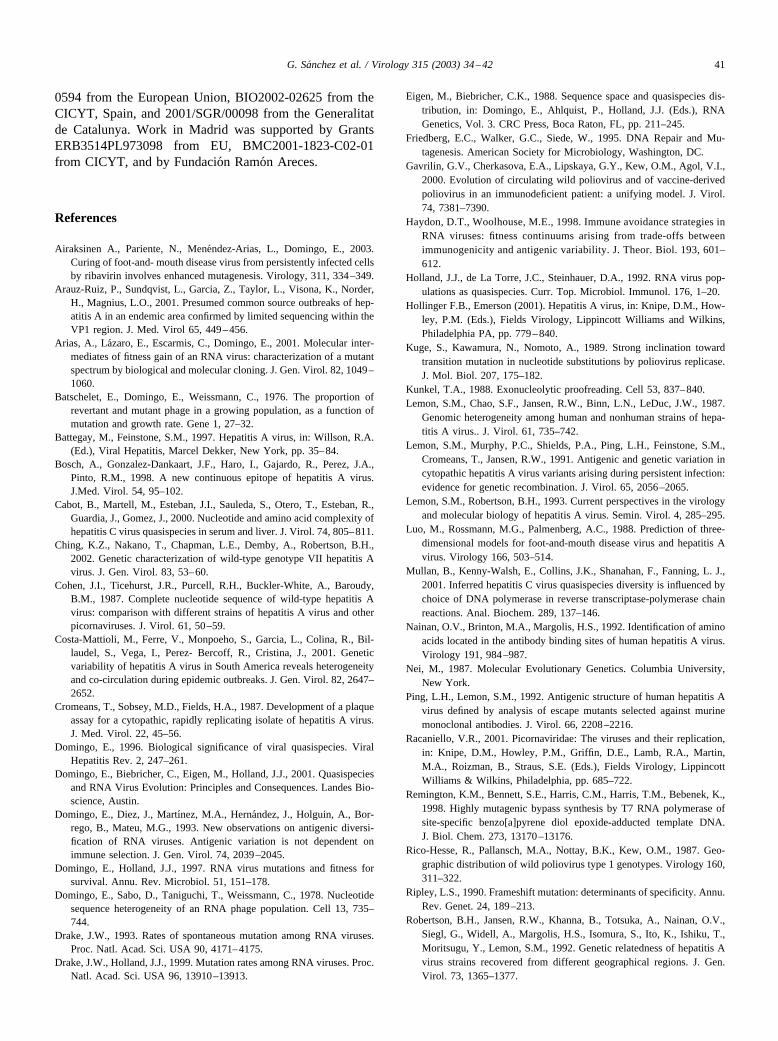
Fig. 1. Amino acid sequence of the VP3 and VP1 fragments analyzed in the present study. (A) VP3 sequence of the pHM175 43c strain (B) VP3 sequenceof the clinical samples. (C) VP1 sequence of the pHM175 43c strain. (D) VP1 sequence of the clinical samples. In bold are depicted those amino acidsencoded by rare codons. Arrows indicate those positions in which a mutation occurred. Regarding the codon usage (Sanchez et al., 2003), the letter ndesignates a normal (frequent) new codon;1n, a significantly more normal (frequent) new codon than the old one;2n, a significantly less normal (frequent)new codon than the old one; and r, a rare new codon. Numbers in parentheses indicate the number of clones with a given substitution. At those positionsin which a nonsynonymous substitution occurred, the new amino acid is depicted over the sequence row. At position 72 in (B) either a valine or an isoleucinemay be present depending on the clinical sample.
37G. Sanchez et al. / Virology 315 (2003) 34–42

again the operation of constraints that limit the number ofpositions that tolerate replacements, giving the appearanceof mutational hot spots (see Discussion). Seven of 15 aminoacid substitutions were either at residues previously identi-fied as belonging to antigenic sites (i.e., 65, 72, and 115 ofVP3) or at residues in the neighborhood of antigenic sites(i.e., 78, 83, 84, and 91 of VP3) (Ping and Lemon, 1992;Bosch et al., 1998; Hollinger and Emerson, 2001).
A large proportion of mutations in the mutant spectraanalyzed involved either rare codons, defined as thosecodons used at low frequencies (Gavrilin et al., 2000) ornormal codons located in the neighborhood of rare codons(at a maximum distance of three triplets from a rare codon)(Fig. 1). The majority of the mutations involving rarecodons (90%) resulted in the change to a normal codon.However, most of the resulting normal codons (78%) werecontiguous to a rare codon (Fig. 1). In contrast, most mu-tations involving normal codons (74%) induced changeswhich significantly affected the normality of the codon, anda high percentage of them (72%) were again closely locatedto rare codons (Fig. 1). Therefore, both the types of muta-tions and the types of codons affected suggest variationconstraints in HAV.
Discussion
Despite the invariance of consensus sequences, at theVP3- and VP1-coding regions examined, upon serial pas-sage of a biological clone of HAV, each of the populationsanalyzed consisted of a mutant spectrum in agreement witha quasispecies population structure for this important humanpathogen (Tables 1 and 2). From the minimum mutationfrequencies and since the HAV genome is about 7500 nu-cleotides in size [specifically, the genome of the originalHM175 wild-type HAV is 7493 nucleotides long (Cohen etal., 1987) and the genome of the cell culture-adapted strainpHM175 43c is 7503 nucleotides long (Lemon et al.,1991)], the average number of mutations per genome (rel-ative to the consensus sequence) for Val9, Val10, Val12,and the clonal population pHM175 43c P26 is 7.0, 1.2, 3.9,and 3.8, respectively. This calculation assumes that themutation frequencies for the analyzed genomic regions re-flect those of other genomic regions, a point that wouldrequire sequencing of other genomic regions from compo-nents of mutant spectra. However, comparison of nucleotidesequences from different HAV isolates suggests a consid-erable similarity in the degree of conservation of differentgenomic regions (Hollinger and Emerson, 2001; Ching etal., 2002; Sanchez et al., 2003). In these comparisons, themost variable genomic regions are those encoding 2B, 2C,and 3B (representing 25% of the genome), with an averageof 1.1-fold the genetic distance calculated for the analyzedVP3- and VP1-coding regions, the most conserved genomicregions are the highly conserved noncoding regions andthose encoding 3C and 3D (representing 39% of the ge-
nome), with an average of 0.8-fold the genetic distancecalculated for the VP3- and VP1-coding regions. Since inother viruses conservation among independent isolates oftenparallels conservation within mutant spectra (Arias et al.,2001; Domingo et al., 2001), the heterogeneity quantitatedon the basis of the VP3- and VP1-coding regions analyzedis unlikely to differ substantially from the heterogeneity forthe entire HAV genome. With this assumption, the propor-tion of genomes with no mutations (the dominant se-quence), calculated from the Poisson, distribution, would be0.09, 30, 2.0, and 2.2% for Val9, Val10, Val12, and pHM175 43c P26 respectively. These values are comparable tothose estimated for clonal populations of other RNA virusessuch as bacteriophage Q� (Domingo et al., 1978) or FMDV(Sobrino et al., 1983; Arias et al., 2001; review in Domingoet al., 2001). Thus, in these HAV populations the nonmu-tated class of genomes were a minority of the total.
Of the analyzed mutant spectra, only Val10 had a level ofheterogeneity that could be influenced by misincorporationsduring RT-PCR amplification. Several arguments supportthe fact that the great majority of the mutations found, evenin Val10, was present in the RNA populations under studyand were not the result of misincorporations during theRT-PCR procedure employed. First, a basal error rate forthe RT-PCR procedures was experimentally determined forthe same VP3- and VP1-coding regions under study. Thiscontrol experiment (detailed under Materials and methods)used recombinant clones to prepare VP3 and VP1 tran-scripts with T7 DNA-dependent RNA polymerase. Thetranscripts were then subjected to the same RT-PCR ampli-fication used with HAV RNA and 50 cDNA clones weresequenced. The final error rate attributable to the system is7.9 � 10�5 by 8.1 � 10�5 substitutions per nucleotide forthe VP3- and VP1-coding regions, respectively. Moreover,the mutations that occurred during the RNA synthesis andthe RT-PCR amplification (three transitions and two trans-versions, three nonsynonymous and two synonymous mu-tations) did not show any of the mutational type bias ormutation clustering observed in the HAV populations. Forobvious statistical reasons, the probability of mutation clus-tering as an RT-PCR artifact is negligible. The mutations inthe mutant spectrum of Val10 were located in the vicinity ofthe sites where mutations for the other samples have beenmapped. Therefore, the vast majority of mutations scored inthe mutant spectra must have been present in the HAV RNApopulations examined. Although it cannot be excluded thatsome mutation could have been generated during the invitro amplification procedure, its exclusion would not sig-nificantly modify the quantifications and conclusions onmutant spectrum complexity of HAV. The narrower mutantspectrum in Val10 could not be the result of a limitation inthe number of RNA template molecules in the sample, sincein all cases a dilution of at least 1:100 of the preparation ofthe RNAs used as template produced a positive amplifica-tion, which excludes a bias in the sequence repertoire(Airaksinen et al., 2003). The narrower mutant spectrum of
38 G. Sanchez et al. / Virology 315 (2003) 34–42

this isolate could result from a shorter time from a clonalorigin of the infection, a limited HAV replication, or highersequence constraints than for the other isolates, among otherpossibilities (Domingo et al., 2001).
A complex mutant spectrum for HAV isolates poses aproblem regarding the concept of strain for this virus. Forhepatitis C virus, a genomic distance higher than 5%, in agenomic fragment encompassing the E2-NS2 junction, isrequired for two HCVs to be considered a different strain(Cabot et al., 2000), and different HAV and poliovirusgenotypes are those with higher than 15% nucleotide se-quence divergence in the VP1-2A-coding region (Rico-Hesse et al., 1987; Hollinger and Emerson, 2001). As moreconsensus sequences and data on mutant spectra of HAVbecome available, it may be necessary to redefine limits forstrain and genotype classifications.
The mean ratio of transition to transversion mutations inthe RNA of the analyzed populations was 8.6, probablyreflecting the misincorporations tendencies of RNA depen-dent RNA polymerases (Domingo et al., 1978; Kuge et al.,1989; Schnerder and Roossinck, 2000). Also, the proportionof transitions tends to decrease with the divergence of thegenes compared (Villanueva et al., 1983; Nei, 1987), sup-porting a recent occurrence of the mutations observed inthese HAV samples. Examination of the sequence context inwhich mutations are found indicates that 25% of the muta-tions in clusters are located at the ends of short oligo (A) oroligo (U) stretches, suggesting a possible contribution ofpolymerase slippage in the generation of some of thesemutations (Ripley, 1990; Arias et al., 2001). However notall homopolymeric tracts were associated with mutationsonly 2 of 14 (2/14) A3 tracts; 3/33 U3; 1/7 A4; 1/10 U4; 2/3A5; 4/8 U5.
Several amino acid substitutions in components of themutant spectra are located at or near recognized antigenicsites of HAV (Fig. 1). In particular in population pHM17543c P26 passaged in cell culture in the absence of immuneselection, substitutions at positions 65 (Pro 3 Leu), 83(Pro3 Ser), and 84 (Tyr3 Asn) of VP3 are related to theimmunodominant site of HAV (Nainan et al., 1992; Luo etal., 1988), and 115 (Leu3 Phe) and 91 (Thr3 Lys) affecta continuous epitope of VP3 (Bosch et al., 1998; M. Luo,personal communication). Antigenic variation in the ab-sence of immune selection has been observed in severalother viruses (reviewed in Domingo et al., 1993) and hasbeen attributed to higher tolerance to replacements of sur-face residues which are relatively free of structural con-straints (Domingo et al., 1993; Haydon and Woolhouse,1998). Replacements at or in the neighborhood of antigenicsites were also found in the mutant spectra of Va19 [sub-stitution Va13 Ile at position 72 of VP3, which is involvedin the epitope defined by the monoclonal antibody K34C8(Sanchez et al., 2002)] or of Val12 [substitutions Ile3 Valat position 72 of VP3, and Val3Leu at position 78 of VP3].These replacements are in the vicinity of the immunodom-inant site (Luo et al., 1988).
One of the unusual features of HAV is the abundance ofrare codons at some capsid sites, compatible with a modu-lating effect on HAV translation (Sanchez et al., 2003). Thefact that the mutations observed tended to maintain a min-imal frequency of rare codons suggests a constraint in thedynamics of mutation-selection to preserve the previouslypostulated balance between rate of translation and capsidprotein folding (Sanchez et al., 2003). In HAV, this may bean important evolutionary constraint, additional to thosegenerally accepted to limit evolutionary rates of RNA vi-ruses despite high mutation rates (Simmonds and Smith,1999).
A quasispecies dynamics for HAV may be of conse-quence for the natural history of this pathogen and itscontrol in the human population. Pathogenic manifestationsof HAV are quite variable (reviewed in Hollinger and Em-erson, 2001). Patients may be asymptomatic despite activeviral replication, but relapsing and fulminant forms of hep-atitis, as well as extrahepatic manifestations such as enceph-alopathy, have been described. Although a strong host com-ponent undoubtedly must influence disease outcome, apossible participation of virus variants to respond to hostdefense mechanisms or to favor replication in the face ofphysiological alterations cannot be excluded. In this con-text, it is remarkable that the pattern of mutations found inthe analyzed VP3 and VP1 regions differed between thepHM175 and the clinical isolates, likely due to differentselective pressures. The existence of variant reservoirs inHAV populations should also be taken into consideration inthe design of preventive and therapeutic treatments, despiteantigenic conservation of the virus.
Material and methods
Cells, viruses, and infections
The cytopathogenic pHM175 43c strain of HAV (cour-tesy of T. Cromeans, Centers for Disease Control, Atlanta,GA) was three times plaque-purified in FRhK-4 cells, aspreviously described (Cromeans et al., 1987), and a biolog-ical clone (pHM175 43c P0) was serially passaged 26 timesin the same cell line, as previously described (Bosch et al.,1998) to yield population pHM175 43c P26.
Clinical samples
Three HAV strains were isolated from three differentpatients of an outbreak of acute hepatitis A associated withthe consumption of coquina clams in Valencia, Spain, dur-ing autumn–winter 1999 (Sanchez et al., 2002). One virusstrain (Va19) was isolated from 60 �l of serum, and twoadditional strains (Va110 and Va112) were isolated from 60�l of extracted feces as previously described (Sanchez et al.,2002). All strains belonged to genotype IB and presented anoverall nucleotide homology of the consensus sequences of
39G. Sanchez et al. / Virology 315 (2003) 34–42

99.7% in a fragment of the 5�NCR and 99.3% in the regionencoding the VP1-2A junction (Sanchez et al., 2002), whichindicates an epidemic relationship among them.
Molecular cloning and sequencing
RNA extracted from pHM175 43c P26 or from the clin-ical samples was retrotranscribed to a cDNA with the M-MLV reverse transcriptase (Promega), and the cDNA wascopied and amplified by the thermostable Pwo pol fromPyrococcus woesei, which has proofreading activity (errorrate of 3.2�10�6 substitutions/nucleotide) (Mullan et al.,2001). Two genomic regions coding for capsid proteinswere amplified. A fragment of the VP3-coding region (nu-cleotides 1470 to 1839, corresponding to amino acids 1 to123) and a fragment of the VP1-coding region (nucleotides2459 to 2943, corresponding to amino acids 85 to 245),which include most of the epitopes so far described in HAV(Nainan et al., 1992; Ping and Lemon, 1992; Bosch et al.,1998). The primers used to copy and amplify the VP3-coding region are NH2-VP3 (5� TCTACCTGAAT-GATATTTGG 3�) for the cDNA synthesis, and NH2-VP3and VP3-1431B (5� CTTGGATCCCACTCAATGTTT-TAGCTAGA 3�) for, PCR amplification. The primers usedto copy and amplify the VP1-coding regions are VP1-2965(5� TCTGTGACAGACAGACAAATAACAAC 3�) for thecDNA synthesis, and VP1–2965 and VP1-2428 (5� GAGG-GATCCGACATACATCAGATCATATGTC 3�) for PCRamplification. The synthesis of cDNAs was performed in afinal volume of 25 �l containing 8 units of M-MLV RT, 0.2mM of each nucleotide, 0.5 �M primer, 50 mM Tris–HC1,75 mM KCl, 3 mM MgCl2, and 10 mM DTT. Ten microli-tus of HAV RNA was denatured at 99°C for 5-min andincubated at 45°C for 1 h. DNA amplification was per-formed following the manufacturer’s specifications in afinal volume of 50 �l containing 0.5 U of the Pwo pol, 0.2mM of each nucleotide 0.5 �M of each primer, 10 mMTris–HCl, 25 mM KC1, 5 mM (NH4)2SO4, 2 mM MgSO4,and 10 �l of the RT product, and using an annealing tem-perature of 50°C in both reactions. Since the DNA frag-ments produced by Pwo pol are blunt-ended, and primersVP3–1431B and VP1–2428 were designed to include aBamHI restriction site at their 5� end, the amplificationproducts were cloned into pGEM-3Zf(�). PCR productswere digested with BamHI and the plasmid vector with bothBamHI and HincII. Digested DNAs were purified with theHigh Pure PCR Product Purification Kit (Roche) followingthe directions of the manufacturer. DNA ligations wereperformed overnight at 16°C using T4 DNA ligase. Ligationproducts were transformed in Escherichia coli DH5�, andtransformant clones were screened first by the standardwhite/blue �-galactosidase colorimetric reaction and thenconfirmed by colony hybridization with specific digoxige-nin-labeled probes. Plasmid DNA from each clone waspurified by using the Wizard Plus SV Minipreps Kit (Pro-mega). Nucleotide sequencing was carried out in an ABI
PRISM 377 automated DNA sequencer, with the ABIPRISM BigDye Terminator Cycle Sequencing Ready Re-action Kit (Applied Biosystems) and using vector-derivedprimers, as described elsewhere (Arias et al., 2001). Allmutations were confirmed by sequencing both strands ofDNA.
Error rate of the RT-PCR system
To ensure that the observed heterogeneity was due to theHAV polymerase and not to artifactual misincorporationsintroduced by the RT or Pwo polymerases during the am-plification procedure, a control experiment to determine theerror rate of the system was carried out. One recombinantclone of the VP3 fragment and one recombinant clone of theVP1 fragment, obtained by copying RNA from pHM17543c, were in vitro transcribed with the T7 polymerase. TheRNA transcripts, diluted 1/1000, were subjected to the orig-inal RT-PCR procedure and the amplified products weresubcloned. The sequence of 50 clones from each VP3- andVP1-coding region yielded an error rate for the amplifica-tion system of 1.1 � 10�4 and 1.7 � 10�4 mutations pernucleotide, respectively. Since the error rate of the T7 RNApolymerase is about 2.9 �10�5 (Remington et al., 1998),the final error rate of the system was calculated to be8.1�10�5 and 1.4�10�4 for the VP3- and VP1-codingregions, respectively. The ANOVA and the nonparametricMann–Whitney U tests were employed to compare viralmutation frequencies and the system error rates, revealingstatistical significance in all but one case (Va110). Thesedata, together with the types and location of the mutationsscored (described under Results and Discussion) indicatethat the vast majority of mutations detected must have beenpresent in the mutant spectra of the HAV samples analyzed.
Sequence analysis
The quasispecies complexity was analyzed by calculat-ing the mutation frequencies and the Shannon entropy.Minimum and maximum mutation frequencies were deter-mined as previously described (Arias et al., 2001). Normal-ized Shannon entropies were calculated following the for-mula Sn � �[�i (pi � lnpi)]/lnN, where pi is the frequencyof each sequence and N is the total number of sequences inthe spectrum of mutants (Airaksinen et al., 2003). Sn rangesfrom 0 (no diversity) to 1 (maximum diversity). The codonusage table of HAV defined previously (Sanchez et al.,2003) was used in the analyses of codon abundances.
Acknowledgments
We acknowledge the technical expertise of the ServeisCientıfic-Tecnics of the University of Barcelona. Workin Barcelona was supported in part by GrantsERB3514PL973098, QLRT- 1999-0634, and QLRT-1999-
40 G. Sanchez et al. / Virology 315 (2003) 34–42
0594 from the European Union, BIO2002-02625 from theCICYT, Spain, and 2001/SGR/00098 from the Generalitatde Catalunya. Work in Madrid was supported by GrantsERB3514PL973098 from EU, BMC2001-1823-C02-01from CICYT, and by Fundacion Ramon Areces.
References
Airaksinen A., Pariente, N., Menendez-Arias, L., Domingo, E., 2003.Curing of foot-and- mouth disease virus from persistently infected cellsby ribavirin involves enhanced mutagenesis. Virology, 311, 334–349.
Arauz-Ruiz, P., Sundqvist, L., Garcia, Z., Taylor, L., Visona, K., Norder,H., Magnius, L.O., 2001. Presumed common source outbreaks of hep-atitis A in an endemic area confirmed by limited sequencing within theVP1 region. J. Med. Virol 65, 449–456.
Arias, A., Lazaro, E., Escarmis, C., Domingo, E., 2001. Molecular inter-mediates of fitness gain of an RNA virus: characterization of a mutantspectrum by biological and molecular cloning. J. Gen. Virol. 82, 1049–1060.
Batschelet, E., Domingo, E., Weissmann, C., 1976. The proportion ofrevertant and mutant phage in a growing population, as a function ofmutation and growth rate. Gene 1, 27–32.
Battegay, M., Feinstone, S.M., 1997. Hepatitis A virus, in: Willson, R.A.(Ed.), Viral Hepatitis, Marcel Dekker, New York, pp. 35–84.
Bosch, A., Gonzalez-Dankaart, J.F., Haro, I., Gajardo, R., Perez, J.A.,Pinto, R.M., 1998. A new continuous epitope of hepatitis A virus.J.Med. Virol. 54, 95–102.
Cabot, B., Martell, M., Esteban, J.I., Sauleda, S., Otero, T., Esteban, R.,Guardia, J., Gomez, J., 2000. Nucleotide and amino acid complexity ofhepatitis C virus quasispecies in serum and liver. J. Virol. 74, 805–811.
Ching, K.Z., Nakano, T., Chapman, L.E., Demby, A., Robertson, B.H.,2002. Genetic characterization of wild-type genotype VII hepatitis Avirus. J. Gen. Virol. 83, 53–60.
Cohen, J.I., Ticehurst, J.R., Purcell, R.H., Buckler-White, A., Baroudy,B.M., 1987. Complete nucleotide sequence of wild-type hepatitis Avirus: comparison with different strains of hepatitis A virus and otherpicornaviruses. J. Virol. 61, 50–59.
Costa-Mattioli, M., Ferre, V., Monpoeho, S., Garcia, L., Colina, R., Bil-laudel, S., Vega, I., Perez- Bercoff, R., Cristina, J., 2001. Geneticvariability of hepatitis A virus in South America reveals heterogeneityand co-circulation during epidemic outbreaks. J. Gen. Virol. 82, 2647–2652.
Cromeans, T., Sobsey, M.D., Fields, H.A., 1987. Development of a plaqueassay for a cytopathic, rapidly replicating isolate of hepatitis A virus.J. Med. Virol. 22, 45–56.
Domingo, E., 1996. Biological significance of viral quasispecies. ViralHepatitis Rev. 2, 247–261.
Domingo, E., Biebricher, C., Eigen, M., Holland, J.J., 2001. Quasispeciesand RNA Virus Evolution: Principles and Consequences. Landes Bio-science, Austin.
Domingo, E., Diez, J., Martınez, M.A., Hernandez, J., Holguin, A., Bor-rego, B., Mateu, M.G., 1993. New observations on antigenic diversi-fication of RNA viruses. Antigenic variation is not dependent onimmune selection. J. Gen. Virol. 74, 2039–2045.
Domingo, E., Holland, J.J., 1997. RNA virus mutations and fitness forsurvival. Annu. Rev. Microbiol. 51, 151–178.
Domingo, E., Sabo, D., Taniguchi, T., Weissmann, C., 1978. Nucleotidesequence heterogeneity of an RNA phage population. Cell 13, 735–744.
Drake, J.W., 1993. Rates of spontaneous mutation among RNA viruses.Proc. Natl. Acad. Sci. USA 90, 4171–4175.
Drake, J.W., Holland, J.J., 1999. Mutation rates among RNA viruses. Proc.Natl. Acad. Sci. USA 96, 13910–13913.
Eigen, M., Biebricher, C.K., 1988. Sequence space and quasispecies dis-tribution, in: Domingo, E., Ahlquist, P., Holland, J.J. (Eds.), RNAGenetics, Vol. 3. CRC Press, Boca Raton, FL, pp. 211–245.
Friedberg, E.C., Walker, G.C., Siede, W., 1995. DNA Repair and Mu-tagenesis. American Society for Microbiology, Washington, DC.
Gavrilin, G.V., Cherkasova, E.A., Lipskaya, G.Y., Kew, O.M., Agol, V.I.,2000. Evolution of circulating wild poliovirus and of vaccine-derivedpoliovirus in an immunodeficient patient: a unifying model. J. Virol.74, 7381–7390.
Haydon, D.T., Woolhouse, M.E., 1998. Immune avoidance strategies inRNA viruses: fitness continuums arising from trade-offs betweenimmunogenicity and antigenic variability. J. Theor. Biol. 193, 601–612.
Holland, J.J., de La Torre, J.C., Steinhauer, D.A., 1992. RNA virus pop-ulations as quasispecies. Curr. Top. Microbiol. Immunol. 176, 1–20.
Hollinger F.B., Emerson (2001). Hepatitis A virus, in: Knipe, D.M., How-ley, P.M. (Eds.), Fields Virology, Lippincott Williams and Wilkins,Philadelphia PA, pp. 779–840.
Kuge, S., Kawamura, N., Nomoto, A., 1989. Strong inclination towardtransition mutation in nucleotide substitutions by poliovirus replicase.J. Mol. Biol. 207, 175–182.
Kunkel, T.A., 1988. Exonucleolytic proofreading. Cell 53, 837–840.Lemon, S.M., Chao, S.F., Jansen, R.W., Binn, L.N., LeDuc, J.W., 1987.
Genomic heterogeneity among human and nonhuman strains of hepa-titis A virus.. J. Virol. 61, 735–742.
Lemon, S.M., Murphy, P.C., Shields, P.A., Ping, L.H., Feinstone, S.M.,Cromeans, T., Jansen, R.W., 1991. Antigenic and genetic variation incytopathic hepatitis A virus variants arising during persistent infection:evidence for genetic recombination. J. Virol. 65, 2056–2065.
Lemon, S.M., Robertson, B.H., 1993. Current perspectives in the virologyand molecular biology of hepatitis A virus. Semin. Virol. 4, 285–295.
Luo, M., Rossmann, M.G., Palmenberg, A.C., 1988. Prediction of three-dimensional models for foot-and-mouth disease virus and hepatitis Avirus. Virology 166, 503–514.
Mullan, B., Kenny-Walsh, E., Collins, J.K., Shanahan, F., Fanning, L. J.,2001. Inferred hepatitis C virus quasispecies diversity is influenced bychoice of DNA polymerase in reverse transcriptase-polymerase chainreactions. Anal. Biochem. 289, 137–146.
Nainan, O.V., Brinton, M.A., Margolis, H.S., 1992. Identification of aminoacids located in the antibody binding sites of human hepatitis A virus.Virology 191, 984–987.
Nei, M., 1987. Molecular Evolutionary Genetics. Columbia University,New York.
Ping, L.H., Lemon, S.M., 1992. Antigenic structure of human hepatitis Avirus defined by analysis of escape mutants selected against murinemonoclonal antibodies. J. Virol. 66, 2208–2216.
Racaniello, V.R., 2001. Picornaviridae: The viruses and their replication,in: Knipe, D.M., Howley, P.M., Griffin, D.E., Lamb, R.A., Martin,M.A., Roizman, B., Straus, S.E. (Eds.), Fields Virology, LippincottWilliams & Wilkins, Philadelphia, pp. 685–722.
Remington, K.M., Bennett, S.E., Harris, C.M., Harris, T.M., Bebenek, K.,1998. Highly mutagenic bypass synthesis by T7 RNA polymerase ofsite-specific benzo[a]pyrene diol epoxide-adducted template DNA.J. Biol. Chem. 273, 13170–13176.
Rico-Hesse, R., Pallansch, M.A., Nottay, B.K., Kew, O.M., 1987. Geo-graphic distribution of wild poliovirus type 1 genotypes. Virology 160,311–322.
Ripley, L.S., 1990. Frameshift mutation: determinants of specificity. Annu.Rev. Genet. 24, 189–213.
Robertson, B.H., Jansen, R.W., Khanna, B., Totsuka, A., Nainan, O.V.,Siegl, G., Widell, A., Margolis, H.S., Isomura, S., Ito, K., Ishiku, T.,Moritsugu, Y., Lemon, S.M., 1992. Genetic relatedness of hepatitis Avirus strains recovered from different geographical regions. J. Gen.Virol. 73, 1365–1377.
41G. Sanchez et al. / Virology 315 (2003) 34–42

Sanchez, G., Bosch, A., Pinto, R.M., 2003. Genome variability and capsidstructural constraints of hepatitis a virus. J. Virol. 77, 452–459.
Sanchez, G., Pinto, R.M., Vanaclocha, H., Bosch, A., 2002. Molecularcharacterization of hepatitis a virus isolates from a transcontinentalshellfish-borne outbreak. J. Clin. Microbiol. 40, 4148–4155.
Schneider, W.L., Roossinck, M.J., 2000. Evolutionarily related Sindbis-like plant viruses maintain different levels of population diversity in acommon host. J. Virol. 74, 3130–3134.
Simmonds, P., Smith, D.B., 1999. Structural constraints on RNA virusevolution. J. Virol. 73, 5787–5794.
Sobrino, F., Davila, M., Ortin, J., Domingo, E., 1983. Multiple geneticvariants arise in the course of replication of foot-and-mouth diseasevirus in cell culture. Virology 128, 310–318.
Steinhauer, D.A., Domingo, E., Holland, J.J., 1992. Lack of evidence forproofreading mechanisms associated with an RNA virus polymerase.Gene 122, 281–288.
Taylor, M.B., 1997. Molecular epidemiology of South African strains ofhepatitis A virus: 1982–1996. J. Med. Virol. 51, 273–279.
van Regenmortel, M.H.V., Fauquet, C.M., Bishop, D.H.L., Carstens, E.B.,Estes, M.K., Lemon, S.M., Maniloff, J., Mayo, M.A., Mc Geoch, D.J.,Pringle, C.R., Wickner, R.B., 2000. Virus Taxonomy. Seventh Reportof the International Committee on Taxonomy of Viruses. AcademicPress, San Diego.
Villanueva, N., Davila, M., Ortin, J., Domingo, E., 1983. Molecular clon-ing of cDNA from foot-and-mouth disease virus C1-Santa Pau (C-S8).Sequence of protein-VP1-coding segment. Gene 23, 185–194.
Williams, K.J., Loeb, L.A., 1992. Retroviral reverse transcriptases: errorfrequencies and mutagenesis. Curr. Top. Microbiol. Immunol. 176,165–180.
Zimmern, D., 1988. Evolution of RNA viruses, in: Domingo, E., Holland,J.J., Ahlquist, P. (Eds.), RNA Genetics, Vol. 2, CRC Press, FL, pp.211–240.
42 G. Sanchez et al. / Virology 315 (2003) 34–42