Relevance, relevance, relevance! A call to arms (hands, fingers and thumbs) for mobile research
Evidence for Immune Relevance of Prevotella copri, a Gut Microbe, in
Patients with Rheumatoid Arthritis
Annalisa Pianta1, Sheila Arvikar
1, Klemen Strle
1, Elise E. Drouin
1†, Qi Wang
2,
Catherine E. Costello2, and Allen C. Steere
1.
This work was supported by grants from the American College of Rheumatology
Innovative Grant Program, “Within our Reach, Finding a Cure for RA”; the Ounsworth-
Fitzgerald Foundation; Mathers Foundation; English, Bonter, Mitchell Foundation; Littauer
Foundation; Lillian B. Davey Foundation, and the Eshe Fund (to A.C.S.), and by NIH grants P41
GM104603, S10 RR020946 and S10 OD010724 (to C.E.C.). S.A. received support from a
Scientist Development Award from the Rheumatology Research Foundation.
1 Annalisa Pianta, PhD, Sheila Arvikar, MD, Klemen Strle, PhD, Elise E. Drouin, PhD,
Allen C. Steere, MD: Center for Immunology and Inflammatory Diseases, Division of
Rheumatology, Allergy and Immunology, Massachusetts General Hospital, Harvard Medical
School, Boston, Massachusetts, USA; 2
Qi Wang, PhD, Catherine E. Costello, PhD: Center for
Biomedical Mass Spectrometry, Boston University School of Medicine, Boston, Massachusetts,
USA; †Current address: Agenus Inc, 02421 Lexington (MA), United States.
Address correspondence to Annalisa Pianta, PhD, Center for Immunology and
Inflammatory Diseases, Massachusetts General Hospital, CNY 149/8301, 55 Fruit Street,
Boston, MA 02114, United States. Tel.: +16177261530; fax: +16177261544.
E-mail address: [email protected]
Full Length Arthritis & RheumatologyDOI 10.1002/art.40003
This article has been accepted for publication and undergone full peer review but has not beenthrough the copyediting, typesetting, pagination and proofreading process which may lead todifferences between this version and the Version of Record. Please cite this article as an‘Accepted Article’, doi: 10.1002/art.40003© 2016 American College of RheumatologyReceived: Aug 25, 2016; Revised: Oct 20, 2016; Accepted: Nov 15, 2016
This article is protected by copyright. All rights reserved.

2
Objective. Prevotella copri, an intestinal microbe, may over-expand in stool samples of
patients with new-onset rheumatoid arthritis (NORA), but it is not yet clear whether the
organism has immune relevance in RA pathogenesis.
Methods. HLA-DR-presented peptides (T cell epitopes) from P. copri were sought directly
from patients’ synovial tissue or peripheral blood mononuclear cells (PBMC) using tandem mass
spectrometry, followed by testing the antigenicity of peptides or their source proteins using
samples from RA patients or comparison groups. T cell reactivity was determined by ELISpot
assays; antibody responses were measured by ELISA, and cytokine/chemokine determinations
were made by Luminex. 16S rDNA of P. copri was sought in serum and synovial fluid samples
using nested PCR.
Results. In PBMC, we identified an HLA-DR-presented peptide from a 27-kD protein of P.
copri (Pc-p27), which stimulated Th1 responses in 42% of NORA patients. In both NORA and
chronic RA patients, one subgroup had IgA antibody responses to Pc-p27 or the whole organism,
which correlated with Th17 cytokine responses and frequent anti-citrullinated protein antibodies
(ACPA). The other subgroup had IgG P. copri antibodies, which were associated with Prevotella
DNA in synovial fluid, P. copri-specific Th1 responses, and less frequent ACPA. In contrast, P.
copri antibody responses were rarely found in patients with other rheumatic diseases or in
healthy controls.
Conclusion. Subgroups of RA patients have differential IgG or IgA immune reactivity with
P. copri, which appears to be specific for this disease. These observations provide evidence that
P. copri is immune-relevant in RA pathogenesis.
Page 2 of 41
John Wiley & Sons
Arthritis & Rheumatology
This article is protected by copyright. All rights reserved.

3
Rheumatoid arthritis (RA) results from a complex interplay between genetic and
environmental factors (1,2). Great progress has been made in the identification of genetic factors
and inflammatory pathways that influence the disease (1,3), but environmental factors are only
now being determined (4). A key hypothesis is that specific organisms in the mouth or gut
microbiota, the composition of which is strongly influenced by environmental cues, may shape
mucosal and systemic immune responses that affect joints in RA patients (4-7).
Using high-through-put sequencing, Scher et al. showed that Prevotella copri in the gut
microbiota was over-expanded in stool samples from patients with new-onset RA (NORA)
compared with patients with chronic RA (CRA), psoriatic arthritis, or healthy people (8). In
NORA patients, Prevotella abundance in the gut was at the expense of Bacteroides fragilis, an
organism that is important for Treg function (9,10). A second metagenome-wide analysis of
fecal samples in RA patients showed dysbiosis in the gut, but also in the mouth and salivary
glands (11). Moreover, a recent study in mice showed that dysbiosis contributes to arthritis
development via activation of autoreactive T cells in the intestine (12). However, it is unclear
whether over-expansion of P. copri in the gut in human patients has the potential to affect
immune cell functions at both mucosal and systemic sites, thereby contributing to RA disease
pathogenesis.
Whereas the previous studies used unbiased, discovery-based approaches to assess
dysbiosis of microorganisms in the oral or gut microbiome, we developed an unbiased,
discovery-based approach to identify novel, immunogenic T cell epitopes in patients with
chronic inflammatory arthritis. With this approach, in vivo HLA-DR presented peptides are
identified directly from patients’ synovial tissue, synovial fluid mononuclear cells (SFMC), or
peripheral blood mononuclear cells (PBMC) by tandem mass spectrometry (LC-MS/MS)
Page 3 of 41
John Wiley & Sons
Arthritis & Rheumatology
This article is protected by copyright. All rights reserved.
4
(13,14), followed by testing the antigenicity of identified peptides and their source proteins using
patients’ samples (15-19).
Recently, we used this approach to search for T cell epitopes of proteins derived from
microbes implicated in RA. We report here the identification of an HLA-DR-presented peptide
(T cell epitope) derived from a P. copri 27-kD protein (Pc-p27), which stimulated Th1 responses
in 42% of RA patients. We then found that P. copri induced differential antibody responses to
this protein or the whole organism in a substantial portion of RA patients. These observations
provide evidence for immune relevance of P. copri in RA pathogenesis.
Patients and Methods are described in Supplementary text.
RESULTS
Identification of naturally presented, microbial HLA-DR-presented peptides. Using
tandem mass spectrometry (LC-MS/MS), we identified HLA-DR-presented peptides in synovial
tissue (N=4), SFMC (N=3) or PBMC (N=2) from 5 patients with NORA or chronic RA (CRA).
From the 17 HLA-DR-presented peptides identified from the PBMC of one CRA patient (called
here RA1), one P. copri sequence was found (Figure 1A). In contrast, no sequences from P.
gingivalis or from B. burgdorferi, the Lyme disease agent, were identified in any sample.
At disease onset, patient RA1, who had 2 copies of the RA “shared epitope” (HLA-
DRB1*0401 and 0101), had severe symmetrical polyarthritis affecting large and small joints.
During the course of the disease, she developed a positive test for ACPA, but not RF. Despite
treatment with disease modifying anti-rheumatic drugs (DMARDs), she had recurrent episodes
of knee swelling with evidence of destructive changes in cartilage and bone. The HLA-DR-
Page 4 of 41
John Wiley & Sons
Arthritis & Rheumatology
This article is protected by copyright. All rights reserved.

5
presented peptide derived from P. copri was identified from PBMC obtained during one such
episode 7 years after disease onset.
The peptide sequence of 19 amino acids had 100% sequence homology with part of the
signal sequence of a 27-kD protein of P. copri (Pc-p27, WP_022121928.1) (Figure 1A). The
peptide had minimal sequence homology with any human peptide, suggesting that it was not a
human protein erroneously assigned with a microbial database. Using signalP 4.0 software (20),
this HLA-DR-presented P. copri peptide was predicted to be part of the Sec secretion signal
peptide sequence (D score = 0.869), strongly suggesting that the peptide would be cleaved from
the source protein. This signal peptide was not predicted to be lipidated (LipoP 1.0 Server). In
addition, the algorithm TEPITOPE predicted that the peptide was highly promiscuous, as is
typical of signal peptides, and would bind all 25 HLA-DR molecules modeled in the program
(21), including the patient’s DRB1*0101 and 0401 molecules. When her PBMC were stimulated
with this P. copri peptide in an IFN-γ ELISpot assay, her T cells secreted levels of IFN-γ that
were >3 times background (Figure 1A insert), attesting to the peptide’s immunogenicity.
T cell reactivity to P. copri peptides in NORA patients. To determine the
immunogenicity of HLA-DR-presented peptides of Pc-p27 more broadly, we used PBMC
obtained from our cohort of patients with new-onset RA (NORA) seen prior to DMARD therapy,
the time when immune responses would be expected to be most robust. All patients met the
ACR/EULAR criteria for that disease (22).
When PBMC from 39 NORA patients and from the case patient RA1 (a CRA patient)
were stimulated with Pc-p27 peptide 1, 17 of the 40 patients (42%) secreted levels of IFN-γ that
were >3 SD above the mean value of HC (P=0.0002), using an IFN-γ/IL-17 Double-Color
ELISpot assay (Figure 1B). In comparison, patients with Lyme arthritis (LA) lacked reactivity
Page 5 of 41
John Wiley & Sons
Arthritis & Rheumatology
This article is protected by copyright. All rights reserved.

6
with this peptide (P<0.0001). Stimulation of cells with PHA, used as a positive control, verified
the viability of cells in all patients. The predominant response to stimulation with Pc-p27 in the
RA patients was a Th1-type response, whereas PBMC from only one RA patient secreted small
amounts of IL-17 (data not shown).
To determine whether patients had reactivity with other epitopes of the Pc-p27 protein,
TEPITOPE was used to predict 2 additional promiscuous peptides derived from the same protein
(Pc-p27 peptides 2 and 3). The 2 peptides together were predicted to be presented by all 25
HLA-DR molecules in the program, and therefore, these peptides were pooled for testing. PBMC
from 14 of the 40 patients (35%) secreted levels of IFN-γ to peptides 2 and 3 that were >3 SD
above the mean value of HC (P=0.0007) or patients with LA (P=0.006) (Figure 1C). Altogether,
PBMC from 24 of the 40 patients (60%) had reactivity with 1 or more of the 3 P. copri peptide
sequences, showing that Th1 immune responses to this protein were common in NORA patients.
Because of the importance of anti-citrullinated protein antibodies (ACPA) in the
diagnosis and pathogenesis of RA (1,23), peptide 1 was re-synthesized with a citrulline in place
of the only arginine in the peptide, which was predicted to be in the – P1 flanking position of the
HLA-DR binding pocket. However, the results suggested that the Pc-p27 signal peptide
sequence was probably not citrullinated in vivo (supplementary Figure 1). This does not preclude
citrullination of other parts of the protein, including B cell epitopes.
IgG and IgA antibody responses to Pc-p27 and whole P. copri. We next determined
antibody responses to Pc-p27 in serum samples from 303 individuals. These included samples
from 127 patients with new-onset or chronic RA (CRA), 28 patients with connective tissue
diseases (14 with systemic lupus, 4 with mixed connective tissue disease, 4 with scleroderma,
and 6 with Sjogren’s syndrome), 28 patients with spondyloarthropathy (SpA) (15 with psoriatic
Page 6 of 41
John Wiley & Sons
Arthritis & Rheumatology
This article is protected by copyright. All rights reserved.

7
arthritis, 10 with ankylosing spondylitis, and 3 with reactive arthritis), 70 patients with Lyme
arthritis, and 50 healthy subjects.
Of the 78 NORA patients, 10 (13%) had IgG antibody responses to Pc-p27 that were >3
SD above those in HC (P<0.0001) (Figure 2A). Moreover, 10 of 49 patients (20%) with CRA
had IgG antibody responses to the protein (P<0.0001), including patient RA1 in whom 4 serial
samples obtained 4 to 9 years after disease onset yielded positive results. In contrast, only 1
patient with SpA and 1 healthy subject had borderline positive IgG antibody responses to the
protein.
Because the first interactions between P. copri and immune cells would presumably
occur in the gut mucosa, we also determined IgA antibody responses to the organism. About
10% of the patients in both the NORA and CRA groups had IgA antibody responses to Pc-p27
(P<0.0002 and P<0.02, respectively), and the responses tended to be more robust in NORA
patients (Figure 2B). In contrast, only 1 patient with LA and 1 healthy subject had IgA antibody
reactivity with the protein. Except for 2 RA patients who had both IgG and IgA responses to Pc-
p27, the other Pc-p27-positive patients had either an IgG or IgA response, but not both.
Altogether, 24% of the 127 RA patients had IgG or IgA antibody responses to Pc-p27.
When both T and B cell responses were considered together, 3 of the 24 patients who had
T cell reactivity with Pc-p27 peptides also had IgG Pc-p27 antibody responses, but none had IgA
responses to the protein. In comparison, among 16 patients lacking T cell reactivity to Pc-p27
peptides, only 1 had an IgG antibody response to the protein, but 5 had IgA responses (P=0.05).
The frequencies of “shared epitope” alleles in patients with P. copri T or B cell responses was
not significantly different than those in patients who lacked these responses (data not shown).
Page 7 of 41
John Wiley & Sons
Arthritis & Rheumatology
This article is protected by copyright. All rights reserved.

8
In an effort to confirm these findings, we determined IgG and IgA antibody responses to
whole P. copri using the same set of 303 serum samples. Using PCR, we confirmed that this
strain expressed Pc-p27 (data not shown). Six of the 78 NORA patients (8%) and 5 of the 49
CRA patients (10%) had IgG antibody responses to P. copri (Figure 2C). Similarly, 6 of 78
NORA patients (8%) had IgA antibody responses to P. copri (P<0.004), and 7 of 49 patients
(14%) with CRA had elevated IgA antibody levels to the organism (P<0.002) (Figure 2D).
Among the 19 patients who had positive IgG or IgA responses to P. copri, only 5 (26%) had both
responses. No patient with CTD, SpA, or LA had IgG or IgA antibodies to the organism.
Altogether, 15% of 127 RA patients had P. copri IgG or IgA antibody responses.
When the antibody responses to whole P. copri or recombinant Pc-p27 were combined,
41 (32%) of the 127 RA patients had IgG or IgA antibody reactivity with the organism. Thus,
antibody responses to P. copri were common in RA patients, both early and late in the disease,
yet they were rarely found in patients with other types of arthritis, implying specificity in RA.
Antibody responses to other oral or commensal bowel flora. To examine the
specificity of antibody responses to P. copri in RA patients, the same serum samples were also
tested for reactivity with whole P. gingivalis, an oral periodontal pathogen implicated in RA
(24), and with 2 common gut commensal organisms, Bacteroides fragilis and Escherichia coli.
Similar to previous reports (25,26), IgG antibody responses to P. gingivalis were found in
about 25% of our NORA and CRA patients, and these responses tended to be higher in RA
patients than in the other comparison groups (Figure 3A). However, in contrast with P. copri
antibody responses, IgG antibodies to P. gingivalis were also present in small percentages of
patients with other rheumatic diseases or healthy control subjects, and IgA antibody reactivity
with P. gingivalis was not increased in RA patients compared with the other groups (Figure 3B).
Page 8 of 41
John Wiley & Sons
Arthritis & Rheumatology
This article is protected by copyright. All rights reserved.
9
Moreover, in contrast with the dichotomy between IgG and IgA antibody responses to P. copri,
all RA patients with IgA antibodies to P. gingivalis also had IgG responses to this microbe.
Importantly, 66 of the 127 RA patients (52%) had antibody responses to either P. copri or P.
gingivalis, but only 8 of the 66 patients (12%) had antibody responses to both microbes. Thus,
minimal overlap was observed in the antibody responses to these 2 organisms, indicating that
these responses are largely independent, and only the response to P. copri was specific for RA.
Very few RA patients or those with other rheumatic diseases had IgG or IgA antibody
responses to B. fragilis or E. coli that were >3 SD above the mean values in healthy control
subjects (Figures 3C-F). However, IgG absorbance values for B. fragilis were significantly lower
in NORA patients than in patients in the other groups (P<0.03) (Figure 3C), consistent with the
decrease in B. fragilis abundance noted previously in NORA patients (8). Conversely, IgG and
IgA absorbance values to B. fragilis in the CTD group were significantly higher than those in the
other groups. Thus, in contrast with P. copri antibody responses, antibody levels to B. fragilis
and E. coli were similar or less in RA patients than those in patients with other types of arthritis
or in healthy subjects.
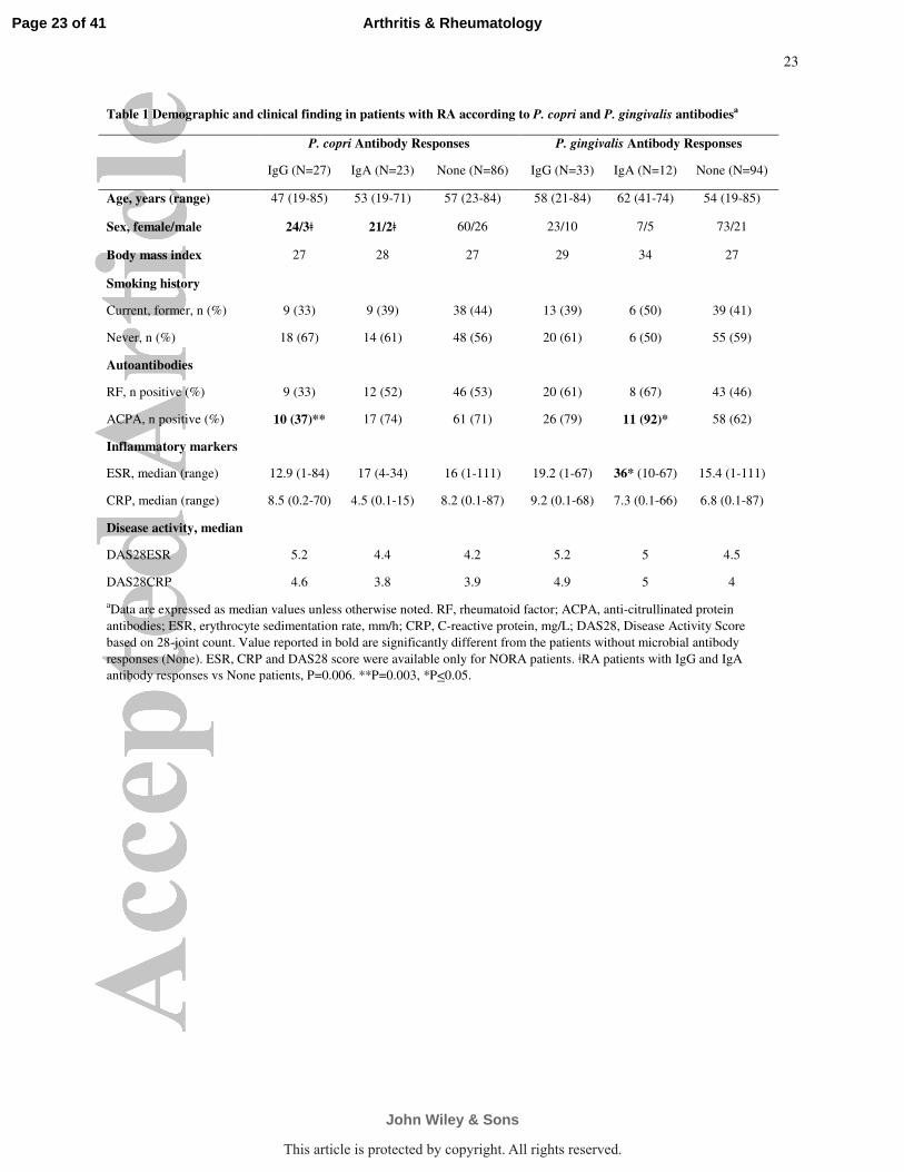
Clinical parameters according to P. copri or P. gingivalis antibody responses.
Because P. copri and P. gingivalis have both been implicated in RA, clinical parameters in our
cohorts of NORA and CRA patients were compared in patients who did or did not have IgG or
IgA antibody responses to these microbes. Because the findings were similar in NORA and CRA
patients, these groups were combined for presentation here.
Several significant differences were found between these groups (Table 1). First, 45 of
the 50 patients (90%) with P. copri antibody responses were female compared with 60 of the 86
patients (70%) who lacked such responses (P=0.006). In contrast, the percentages of female and
Page 9 of 41
John Wiley & Sons
Arthritis & Rheumatology
This article is protected by copyright. All rights reserved.

10
male patients were not significantly different in those with P. gingivalis antibody responses.
Second, only 37% of the RA patients with IgG P. copri antibody responses had ACPA compared
with 74% of those who had IgA P. copri antibodies (P=0.01) and 72% who lacked P. copri
antibodies (P=0.003). There was a similar trend with RF (P=0.08). In contrast, patients with P.
gingivalis IgG and IgA antibody responses had higher frequencies of ACPA and RF than patients
who lacked these responses. Finally, at study entry, there was a trend toward higher disease
activity scores (DAS28) in NORA patients with either P. copri or P. gingivalis IgG antibody
responses. There were no significant differences among the groups in age, body mass index
(BMI), or smoking history. Thus, P. copri antibodies were found primarily in women and ACPA
were less common in patients with IgG P. copri antibodies, whereas neither of these factors
correlated with P. gingivalis antibody responses, again indicating that these microbes induce
distinct responses.
Correlation of P. copri antibodies with serum cytokine and chemokine levels. In an
effort to link P. copri with inflammatory responses and autoantibody production, IgG and IgA P.
copri antibody values were correlated with serum cytokine levels in 120 of the 127 RA patients
in whom sufficient serum samples were still available. The 14 cytokines and chemokines
measured were representative of innate, Th1 and Th17 immune responses. The assays were
performed with Heteroblock to limit the possible interference by rheumatoid factor.
Because the results were similar in NORA and CRA patients, they were combined for
presentation here.
When the magnitude of P. copri IgG or IgA antibodies in the 37 antibody-positive
patients were correlated with cytokine levels, strong positive correlations were found between
IgA antibody values and the levels of 3 innate cytokines (IFN-α, MIP-1α, and MIP-1β), 2 Th1-
Page 10 of 41
John Wiley & Sons
Arthritis & Rheumatology
This article is protected by copyright. All rights reserved.

11
associated cytokines (IFN-γ and IL-12), and 3 Th17-associated cytokines (IL-17F, IL-22, and IL-
1β) (Figure 4A-C). In contrast, IgG P. copri antibody values correlated only with levels of the
Th1 chemoattractant CXCL10 (Figure 4B).
When P. copri IgA absorbance values in all 120 RA patients, including those with
positive and negative values, were correlated with cytokine levels, even stronger associations
were found with innate (MIP-1α and MIP-1β), Th1 (IFN-γ and IL-12), and Th17 cytokines (IL-
23, IL-22, IL-17A, IL-17E, and IL-17F) (Supplementary Figure 2A). In contrast, IgG absorbance
values did not correlate with any cytokine or chemokine levels. Similarly, P. gingivalis IgG and
IgA antibody did not correlate with any cytokine or chemokine level (Supplementary Figure 2B),
further indicating that these microbes induce distinct responses at different mucosal sites.
Detection of P. copri 16S rDNA in synovial fluid. Because P. copri IgG antibody
responses were suggestive of a systemic immune response to the organism, we investigated
whether P. copri itself may spread to joints. For this purpose, we designed nested PCR primers
to detect DNA for the 16S ribosomal RNA gene of P. copri (16S rDNA) in patients’ samples. Of
18 patients in whom paired serum and SF samples were available, 10 were obtained from NORA
patients, and 8 were collected from CRA patients who were seen from 3-to-50 years after disease
onset. Five of the 18 patients had IgG antibody responses to P. copri; 2 had IgA antibody
reactivity with the organism, and 11 did not have P. copri antibody responses. Although the
numbers were small, the clinical correlations in each of these groups, including disease duration,
DAS28 scores, ACPA and RF frequencies, were similar to those in the larger groups of RA
patients (Supplementary Table 1).
Of the 5 patients (RA1 to RA5) with IgG P. copri antibodies, 3 had P. copri 16S rDNA
detected in SF. In 2 of the 3 patients, 16S rDNA was found in samples obtained prior to
Page 11 of 41
John Wiley & Sons
Arthritis & Rheumatology
This article is protected by copyright. All rights reserved.

12
DMARD therapy. In the third patient (CRA patient RA1), in whom the original HLA-DR-
presented P. copri peptide was identified in PBMC collected 7 years after disease onset (Figure
1A), P. copri 16S rDNA was detected in SF obtained 9 years after disease onset (Figure 5A). In
contrast, P. copri DNA was not detected in SF samples from the remaining 13 patients, 2 with
IgA P. copri antibodies and 11 without P. copri antibodies. Serum samples from all 18 patients
had negative PCR results. Thus, 3 of the 5 patients with IgG P. copri antibodies had positive
PCR results for Prevotella DNA in SF compared with none of 13 patients in the other 2 groups
(P=0.01).
For comparison, nested PCR primers were designed to detect 16S rDNA from B. fragilis,
another gut commensal. Of the 18 patients, one (RA15) had B. fragilis 16S rDNA in SF; the
sequence of this amplicon had 98% homology to B. fragilis 16S rDNA (Supplementary Figure
3). This patient did not have positive tests for P. copri DNA or P. copri antibody responses, and
none of the 18 patients had IgG or IgA responses to B. fragilis.
All positive results were confirmed by sequencing. Amplicons from patient RA2 had
100% identity with the sequence for P. copri 16S rDNA in the NCBI database, which was
obtained from a Japanese isolate. Interestingly, patient RA2 grew up in Korea. Amplicons from
patient RA1 and from patient RA5 annealed with P. copri sequences (DSM18205), but they had
86% and 89% sequence homology, respectively (Figure 5B), suggesting that the sequence could
be from another Prevotella species (Figure 5B). Alternately, even though cultures of P. copri
from stool were not available for analysis, the differences in P. copri 16S rDNA sequences may
be explained by strain variation since the 3 patients grew up in widely different geographic
locations (Korea, the United States, or the Caribbean Islands). This is consistent with a study in
Page 12 of 41
John Wiley & Sons
Arthritis & Rheumatology
This article is protected by copyright. All rights reserved.
13
which P. copri was ranked as the second-most variable member of the human gut microbiota
between continents (27).
DISCUSSION
In this study, using a discovery-based search for HLA-DR-presented peptides derived
from P. copri, one spectrum-to-peptide match was identified from the PBMC of 1 RA patient.
This peptide sequence was found in the signal domain of a 27-kD protein of P. copri, which was
predicted to be a secreted protein. Signal sequences, which are cleaved prior to secretion, can
accumulate in transmembrane locations; they are often highly antigenic, and they typically bind
many different HLA-DR molecules (28). In addition, the secreted portion of the protein may
become an immunogenic soluble antigen (6,29). We then found that the signal sequence peptide
and two other peptides from the Pc-p27 protein induced Th1 responses in 60% of patients with
RA. Although RA SE alleles correlated inversely with P. copri overexpansion in the gut (8), our
study showed no significant correlations between these T cell epitopes and SE alleles, consistent
with the fact that these epitopes are promiscuous HLA-DR binders.
Whereas PBMC were only available in NORA patients, we were able to test for antibody
responses to P. copri in both NORA and CRA patients. Although over-expansion of P. copri was
previously detected only in the stool of NORA patients (8), we found similar frequencies of P.
copri antibody responses and similar clinical associations in NORA and CRA patients,
suggesting that once initiated, these antibody responses may persist for years. Importantly, in
both patient groups, P. copri antibody responses were specific for RA. First, P. copri antibodies
were rarely found in patients with other rheumatic diseases or in healthy controls. Second,
antibody levels to 2 other gut commensals, B. fragilis and E. coli, were similar or less in RA
Page 13 of 41
John Wiley & Sons
Arthritis & Rheumatology
This article is protected by copyright. All rights reserved.

14
patients than those in patients with other arthritides or in healthy subjects. Third, there was little
overlap between patients with P. gingivalis antibodies and those with P. copri antibodies.
In both NORA and CRA patients, P. copri antibody responses were found primarily in
women. When the 2 cohorts of patients were combined, P. copri-negative patients had a sex ratio
of 2.3 to 1 in favor of women, which is close to the ratio of 3 to 1 generally reported in RA
cohorts. In contrast, the patients with P. copri antibody responses had a sex ratio of 9 to 1,
implying that this organism may be a substantial contributor to the female predominance in RA.
In mice, gut symbiotic gram-negative bacteria may disseminate systemically to other sites
and induce IgG antibody responses (30). These homeostatic IgG responses may help later in
protection against invasion by pathogenic gram-negative bacteria. Our studies showed a high
background in E. coli and B. fragilis antibody assays, perhaps reflecting positive responses to
these bacteria in some individuals. However, antibody responses to these bacteria were not
greater in RA patients than in other comparison groups, including healthy controls, whereas P.
copri antibody responses were significantly higher in RA patients and correlated with
inflammatory cytokine levels. Thus, our findings could not be explained simply by homeostatic
P. copri antibody responses.
In our study, the magnitude of IgA P. copri antibody responses in RA patients correlated
with serum levels of a range of cytokines and chemokines associated with innate, Th1 and Th17
immune responses. Although we did not find Pc-p27-specific Th17 cells in PBMC of these
patients, it is likely that such cells were present in their intestinal mucosa. Th17 responses are
presumably important initially in containing the organism in the bowel, but with chronic
antigenic stimulation they can promote systemic inflammation and autoimmunity (31). This idea
is supported here by strong correlations among IgA antibody values, serum levels of Th17-
Page 14 of 41
John Wiley & Sons
Arthritis & Rheumatology
This article is protected by copyright. All rights reserved.
15
associated cytokines, and high frequencies of ACPA, which could react with citrullinated
proteins in joints (32).
Conversely, other RA patients had IgG P. copri antibody responses associated with
Prevotella DNA in joints, inflammatory Th1 responses, and infrequent ACPA. In these patients,
we postulate that P. copri, a strict anaerobe, may spread systemically within phagocytic cells,
including to joints. In 2 of 3 patients, Prevotella DNA was detected in SF obtained early in the
disease. However, in the remaining patient (RA1), in whom the Pc-p27 HLA-DR-presented
peptide was identified in PBMC obtained 7 years after disease onset, P. copri DNA was detected
in SF collected 9 years after disease onset. This suggests that P. copri may spread from the
bowel in repeated waves over a period of years, perhaps explaining the persistence of antibody
responses to P. copri in CRA patients. The finding of 16S rDNA of B. fragilis in the SF of one
additional patient suggests that escape of other commensal bacteria from the bowel may occur,
but this patient lacked an antibody response to the organism.
There are parallels between the potential role of P. copri in RA pathogenesis and that of
the periodontal pathogen, Porphyromonas gingivalis (24,33). In patients with periodontitis, the
composition of the oral flora shifts in favor of organisms, particularly anaerobes such as P.
gingivalis, which thrive in an inflammatory environment (34). IgG antibody responses to P.
gingivalis in RA patients correlate strongly with systemic inflammation and co-existent
periodontal disease (24,26,35). Furthermore, P. gingivalis may disseminate from the gingiva,
presumably via dendritic cells, also leading to infection and inflammation at distant sites (6,36),
and DNA from periodontal bacteria has been detected by PCR in SF of a few RA patients with
periodontal disease (37).
Page 15 of 41
John Wiley & Sons
Arthritis & Rheumatology
This article is protected by copyright. All rights reserved.
16
In conclusion, our study provides evidence for immune relevance of P. copri in RA and
suggests that bowel pathology may be a feature of the disease in this subgroup of patients. These
new insights are likely to have implications for both the diagnosis and treatment of RA. For
example, P. copri IgG antibody responses could provide support for the diagnosis in RA patients
who lack ACPA or RF. Moreover, dietary interventions or specifically tailored antibiotic
regimens targeting P. copri could have an adjunctive role to DMARDs in the treatment of this
disease. Previously, patients with early RA who received tetracycline or its derivatives had
significantly better outcomes than placebo-treated patients (38,39). These results are often
attributed to the anti-inflammatory effects of tetracyclines, but recent insights about the
microbiota suggest an additional explanation. Importantly, insights about specific immune-
relevant commensal organisms in the microbiota promise a new era in the understanding and
treatment of RA.
ACKNOWLEDGMENTS
We thank John Branda, Judith M. Holden, Gail McHugh for help with microbial cultures,
Dan Littman and Hannah Fehlner-Peach for providing P. copri DNA to clone the Pc-p27 protein,
Chunxiang Yao for help with mass spectrometry analyses, Deborah Collier and Marcy Bolster
for help with patient care, Dennis Burke and John Aversa for help in obtaining synovial tissue,
Mandakolathur Murali for RF and ACPA analyses, the American Red Cross HLA laboratory for
HLA-DR typing of patients, Katherine Sulka for help in the preparation of samples for HLA-DR
typing, and the CCIB DNA CORE facility at MGH for sequencing of P. copri 16S rDNA
amplicons. This work was presented in part at the annual meeting of the American College of
Rheumatology in San Francisco 2015 (40).
Page 16 of 41
John Wiley & Sons
Arthritis & Rheumatology
This article is protected by copyright. All rights reserved.
17
AUTHOR CONTRIBUTIONS
All authors were involved in drafting the article or revising it critically for important
intellectual content. Dr. Steere had full access to the data in the study and takes responsibility for
the integrity of the data and the accuracy of data analysis.
Study conception and design. Pianta, Drouin, Costello, Steere
Acquisition of data. Pianta, Arvikar, Strle, Wang, Steere
Analysis and interpretation of data. Pianta, Arvikar, Strle, Drouin, Wang, Costello, Steere
REFERENCES
1. McInnes IB, Schett G. The pathogenesis of rheumatoid arthritis. N Engl J Med 2011;
365:2205-19.
2. Firestein GS. Evolving concepts of rheumatoid arthritis. Nature 2003; 423:356-61.
3. Plenge RM. Rheumatoid arthritis genetics: 2009 update. Curr Rheumatol Rep 2009;
11:351-6.
4. Catrina AI, Deane KD, Scher JU. Gene, environment, microbiome and mucosal immune
tolerance in rheumatoid arthritis. Rheumatology (Oxford) 2014; 55:391-402.
5. Longman RS, Littman DR. The functional impact of the intestinal microbiome on
mucosal immunity and systemic autoimmunity. Curr Opin Rheumatol 2015; 27:381-7.
6. Han YW, Wang X. Mobile microbiome: oral bacteria in extra-oral infections and
inflammation. J Dent Res 2013; 92:485-91.
7. Scher JU, Abramson SB. The microbiome and rheumatoid arthritis. Nat Rev Rheumatol
2011; 7:569-78.
Page 17 of 41
John Wiley & Sons
Arthritis & Rheumatology
This article is protected by copyright. All rights reserved.
18
8. Scher JU, Sczesnak A, Longman RS, Segata N, Ubeda C, Bielski C et al. Expansion of
intestinal Prevotella copri correlates with enhanced susceptibility to arthritis. Elife 2013;
2:e01202.
9. Atarashi K, Tanoue T, Shima T, Imaoka A, Kuwahara T, Momose Y et al. Induction of
colonic regulatory T cells by indigenous Clostridium species. Science 2011; 331:337-41.
10. Round JL, Lee SM, Li J, Tran G, Jabri B, Chatila TA et al. The Toll-like receptor 2
pathway establishes colonization by a commensal of the human microbiota. Science
2011; 332:974-7.
11. Zhang X, Zhang D, Jia H, Feng Q, Wang D, Liang D et al. The oral and gut microbiomes
are perturbed in rheumatoid arthritis and partly normalized after treatment. Nat Med
2015; 21:895-905.
12. Maeda Y, Kurakawa T, Umemoto E, Motooka D, Ito Y, Gotoh K et al. Dysbiosis
contributes to arthritis development via activation of autoreactive T cells in the intestine.
Arthritis Rheumatol 2016; doi:10.1002/art.39783.
13. Seward RJ, Drouin EE, Steere AC, Costello CE. Peptides presented by HLA-DR
molecules in synovia of patients with rheumatoid arthritis or antibiotic-refractory Lyme
arthritis. Mol Cell Proteomics 2011; 10:M110 002477.
14. Wang Q, Drouin EE, Yao C, Zhang J, Huang Y, Leon DR et al. Immunogenic HLA-DR-
presented self-peptides identified directly from clinical samples of synovial tissue,
synovial fluid, or peripheral blood in patients with rheumatoid arthritis or Lyme arthritis.
J Proteome Res 2016 (Epub ahead of print).
15. Crowley JT, Strle K, Drouin EE, Pianta A, Arvikar SL, Wang Q et al. Matrix
metalloproteinase-10 is a target of T and B cell responses that correlate with synovial
Page 18 of 41
John Wiley & Sons
Arthritis & Rheumatology
This article is protected by copyright. All rights reserved.

19
pathology in patients with antibiotic-refractory Lyme arthritis. J Autoimmun 2016;
69:24-37.
16. Crowley JT, Drouin EE, Pianta A, Strle K, Wang Q, Costello CE et al. A highly
expressed human protein, apolipoprotein B-100, serves as an autoantigen in a subgroup
of patients with Lyme disease. J Infect Dis 2015; 212:1841-50.
17. Drouin EE, Seward RJ, Strle K, McHugh G, Katchar K, Londono D et al. A novel human
autoantigen, endothelial cell growth factor, is a target of T and B cell responses in
patients with Lyme disease. Arthritis Rheum 2013; 65:186-96.
18. Pianta A, Drouin EE, Crowley JT, Arvikar S, Strle K, Costello CE et al. Annexin A2 is a
target of autoimmune T and B cell responses associated with synovial fibroblast
proliferation in patients with antibiotic-refractory Lyme arthritis. Clin Immunol 2015;
160:336-41.
19. Pianta A, Drouin EE, Wang Q, Arvikar S, Costello CE, Steere AC. Identificatiom of N-
acetylglucosamine-6-sulfatase and filamin A as novel targets of autoimmune T and B cell
responses in rheumatoid arthritis [abstract]. Ann Rheum Dis 2015; 74(Suppl2): 112.
20. Petersen TN, Brunak S, von Heijne G, Nielsen H. SignalP 4.0: discriminating signal
peptides from transmembrane regions. Nat Methods 2011; 8:785-6.
21. Sturniolo T, Bono E, Ding J, Raddrizzani L, Tuereci O, Sahin U et al. Generation of
tissue-specific and promiscuous HLA ligand databases using DNA microarrays and
virtual HLA class II matrices. Nat Biotechnol 1999; 17:555-61.
22. Aletaha D, Neogi T, Silman AJ, Funovits J, Felson DT, Bingham CO, 3rd et al. 2010
Rheumatoid arthritis classification criteria: an American College of
Page 19 of 41
John Wiley & Sons
Arthritis & Rheumatology
This article is protected by copyright. All rights reserved.

20
Rheumatology/European League Against Rheumatism collaborative initiative. Arthritis
Rheum 2010; 62:2569-81.
23. Brink M, Hansson M, Mathsson L, Jakobsson PJ, Holmdahl R, Hallmans G et al.
Multiplex analyses of antibodies against citrullinated peptides in individuals prior to
development of rheumatoid arthritis. Arthritis Rheum 2013; 65:899-910.
24. Mikuls TR, Payne JB, Yu F, Thiele GM, Reynolds RJ, Cannon GW et al. Periodontitis
and Porphyromonas gingivalis in patients with rheumatoid arthritis. Arthritis Rheumatol
2014; 66:1090-100.
25. de Smit M, Westra J, Vissink A, Doornbos-van der Meer B, Brouwer E, van Winkelhoff
AJ. Periodontitis in established rheumatoid arthritis patients: a cross-sectional clinical,
microbiological and serological study. Arthritis Res Ther 2012; 14:R222.
26. Arvikar SL, Collier DS, Fisher MC, Unizony S, Cohen GL, McHugh G et al. Clinical
correlations with Porphyromonas gingivalis antibody responses in patients with early
rheumatoid arthritis. Arthritis Res Ther 2013; 15:R109.
27. Schloissnig S, Arumugam M, Sunagawa S, Mitreva M, Tap J, Zhu A et al. Genomic
variation landscape of the human gut microbiome. Nature 2013; 493:45-50.
28. Kovjazin R, Volovitz I, Daon Y, Vider-Shalit T, Azran R, Tsaban L et al. Signal peptides
and trans-membrane regions are broadly immunogenic and have high CD8+ T cell
epitope densities: Implications for vaccine development. Mol Immunol 2011; 48:1009-
18.
29. Yang Y, Torchinsky MB, Gobert M, Xiong H, Xu M, Linehan JL et al. Focused
specificity of intestinal TH17 cells towards commensal bacterial antigens. Nature 2014;
510:152-6.
Page 20 of 41
John Wiley & Sons
Arthritis & Rheumatology
This article is protected by copyright. All rights reserved.

21
30. Zeng MY, Cisalpino D, Varadarajan S, Hellman J, Warren HS, Cascalho M et al. Gut
microbiota-induced immunoglobulin G controls systemic infection by symbiotic bacteria
and pathogens. Immunity 2016; 44:1-12.
31. Ruff WE, Kriegel MA. Autoimmune host-microbiota interactions at barrier sites and
beyond. Trends Mol Med 2015; 21:233-44.
32. Romero V, Fert-Bober J, Nigrovic PA, Darrah E, Haque UJ, Lee DM et al. Immune-
mediated pore-forming pathways induce cellular hypercitrullination and generate
citrullinated autoantigens in rheumatoid arthritis. Sci Transl Med 2013; 5:209ra150.
33. Pischon N, Pischon T, Kroger J, Gulmez E, Kleber BM, Bernimoulin JP et al.
Association among rheumatoid arthritis, oral hygiene, and periodontitis. J Periodontol
2008; 79:979-86.
34. Hajishengallis G. Periodontitis: from microbial immune subversion to systemic
inflammation. Nat Rev Immunol 2015; 15:30-44.
35. Scher JU, Abramson SB. Periodontal disease, Porphyromonas gingivalis, and rheumatoid
arthritis: what triggers autoimmunity and clinical disease? Arthritis Res Ther 2013;
15:122.
36. Carrion J, Scisci E, Miles B, Sabino GJ, Zeituni AE, Gu Y et al. Microbial carriage state
of peripheral blood dendritic cells (DCs) in chronic periodontitis influences DC
differentiation, atherogenic potential. J Immunol 2012; 189:3178-87.
37. Martinez-Martinez RE, Abud-Mendoza C, Patino-Marin N, Rizo-Rodriguez JC, Little
JW, Loyola-Rodriguez JP. Detection of periodontal bacterial DNA in serum and synovial
fluid in refractory rheumatoid arthritis patients. J Clin Periodontol 2009; 36:1004-10.
Page 21 of 41
John Wiley & Sons
Arthritis & Rheumatology
This article is protected by copyright. All rights reserved.
22
38. O'Dell JR, Haire CE, Palmer W, Drymalski W, Wees S, Blakely K et al. Treatment of
early rheumatoid arthritis with minocycline or placebo: results of a randomized, double-
blind, placebo-controlled trial. Arthritis Rheum 1997; 40:842-8.
39. Stone M, Fortin PR, Pacheco-Tena C, Inman RD. Should tetracycline treatment be used
more extensively for rheumatoid arthritis? Metaanalysis demonstrates clinical benefit
with reduction in disease activity. J Rheumatol 2003; 30:2112-22.
40. Pianta A, Drouin EE, Arvikar S, Strle K, Crowley JT, Wang Q, Costello CE, Steere AC.
Identification of a broadly immunogenic Prevotella copri T cell epitope in patients with
rheumatoid arthritis [abstract]. Arthritis Rheumatol 2015; 67 (suppl 10).
Page 22 of 41
John Wiley & Sons
Arthritis & Rheumatology
This article is protected by copyright. All rights reserved.
23
Table 1 Demographic and clinical finding in patients with RA according to P. copri and P. gingivalis antibodiesa
P. copri Antibody Responses P. gingivalis Antibody Responses
IgG (N=27) IgA (N=23) None (N=86) IgG (N=33) IgA (N=12) None (N=94)
Age, years (range) 47 (19-85) 53 (19-71) 57 (23-84) 58 (21-84) 62 (41-74) 54 (19-85)
Sex, female/male 24/3ǂ 21/2ǂ 60/26 23/10 7/5 73/21
Body mass index 27 28 27 29 34 27
Smoking history
Current, former, n (%) 9 (33) 9 (39) 38 (44) 13 (39) 6 (50) 39 (41)
Never, n (%) 18 (67) 14 (61) 48 (56) 20 (61) 6 (50) 55 (59)
Autoantibodies
RF, n positive (%) 9 (33) 12 (52) 46 (53) 20 (61) 8 (67) 43 (46)
ACPA, n positive (%) 10 (37)** 17 (74) 61 (71) 26 (79) 11 (92)* 58 (62)
Inflammatory markers
ESR, median (range) 12.9 (1-84) 17 (4-34) 16 (1-111) 19.2 (1-67) 36* (10-67) 15.4 (1-111)
CRP, median (range) 8.5 (0.2-70) 4.5 (0.1-15) 8.2 (0.1-87) 9.2 (0.1-68) 7.3 (0.1-66) 6.8 (0.1-87)
Disease activity, median
DAS28ESR 5.2 4.4 4.2 5.2 5 4.5
DAS28CRP 4.6 3.8 3.9 4.9 5 4
aData are expressed as median values unless otherwise noted. RF, rheumatoid factor; ACPA, anti-citrullinated protein
antibodies; ESR, erythrocyte sedimentation rate, mm/h; CRP, C-reactive protein, mg/L; DAS28, Disease Activity Score
based on 28-joint count. Value reported in bold are significantly different from the patients without microbial antibody
responses (None). ESR, CRP and DAS28 score were available only for NORA patients. ǂRA patients with IgG and IgA
antibody responses vs None patients, P=0.006. **P=0.003, *P<0.05.
Page 23 of 41
John Wiley & Sons
Arthritis & Rheumatology
This article is protected by copyright. All rights reserved.

24
FIGURE LEGEND
Figure 1. Identification of a Broadly Immunogenic P. copri T Cell Epitope. (A) LC/MS/MS
spectrum of the Pc-p272-20 peptide. Consensus peptide identification was achieved as
KRIILILTVLLAMLGQ(deamidated)VAY by OMSSA and X!Tandem. The insert panel shows
the IFN-γ ELISpot assay using matching patient’s PBMC stimulated with the peptide (1, 2 and 4
µM). Reactivity of >3 times background (no antigen) was considered positive. (B) IFN-
γ ELISpot assay using PBMC from patients with rheumatoid arthritis (RA), Lyme arthritis (LA),
and healthy controls (HC) incubated with the HLA-DR-presented peptide identified from the
PBMC of patient RA1 (Peptide1, 1 µM). (C) IFN-γ secretion of PBMC from patients and
controls incubated with 2 predicted promiscuous HLA-DR binding peptides from Pc-p27 (1 µM
each). A positive response was defined as >3 SD above the mean value of the HC (area above the
shaded region). The value for patient RA1 is indicated with a star. Horizontal lines represent the
mean values of each group.
Figure 2. IgG and IgA Responses to P. copri in RA Patients and Control Subjects. Serum
samples from 303 individuals with NORA, CRA, other rheumatic diseases or from healthy
controls were tested for P. copri antibodies. IgG (A) and IgA (B) ELISAs against the bacterial
protein Pc-p27; IgG (C) and IgA (D) ELISA against 1% formalin-inactivated P. copri. Positivity
was defined as >3 SD above the mean value of healthy controls (area above the shaded region).
Symbols represent values in individual patients and horizontal lines show mean values. The
value for patient RA1 is indicated with a star. Only significant P values relative to HC are shown.
Page 24 of 41
John Wiley & Sons
Arthritis & Rheumatology
This article is protected by copyright. All rights reserved.
25
HC, healthy control; CTD, connective tissue diseases; SpA, spondyloarthropathies; LA, Lyme
arthritis; NORA, new onset rheumatoid arthritis; CRA, chronic rheumatoid arthritis.
Figure 3. IgG and IgA Responses to Other Organisms in RA Patients and Controls. Serum
samples from the same 303 individuals, shown in figure 2, were tested for antibody responses to
other bacteria. IgG (A) and IgA (B) ELISAs against 1% formalin-inactivated P. gingivalis.
Positivity was defined as >2SD above the mean value of normal control negative for
periodontitis as determined in our previous publication (26). IgG (C) and IgA (D) ELISAs
against 1% formalin-inactivated B. fragilis; IgG (E) and IgA (F) ELISAs against 1% formalin-
inactivated E. coli. For these analyses, positivity was defined as >3 SD above the mean value of
healthy controls (area above the shaded region). Symbols represent values in individual patients
and horizontal lines show mean values. Only significant P values are shown. HC, healthy
control; CTD, connective tissue diseases; SpA, spondyloarthropathies; LA, Lyme arthritis;
NORA, new onset rheumatoid arthritis; CRA, chronic rheumatoid arthritis.
Figure 4. Correlation between Cytokine Levels and P. copri Antibody Responses in
Antibody-Positive Patients. Correlation between P. copri specific IgG or IgA responses and
cytokines associated with innate immunity (A), Th1 immunity (B), or Th17 immunity (C).
Correlations were performed using the Pearson's correlation test.
Figure 5. PCR Testing for Prevotella copri 16S rDNA in Concomitant Serum and SF
Samples of RA Patients. (A) Nested PCR of P. copri 16S rDNA amplicons (254 bp) analyzed
on 1.5% agarose gels stained with ethidium bromide. Results from serum and SF samples from
Page 25 of 41
John Wiley & Sons
Arthritis & Rheumatology
This article is protected by copyright. All rights reserved.
26
the 3 positive patients are shown. Patient RA1 had 4 paired serum and SF samples; patient RA2
had 1 serum and 2 SF samples, and patient RA5 had 1 serum and 1 SF samples. In patients RA2
and RA5, enough material was available for testing in duplicate. M, 100 bp DNA ladder; +,
positive control (P. copri DSM 18205); H, water control. (B) Sequence alignment of the 16S
gene amplicons obtained from patient RA1, RA2 and RA5 using CLC Genomic Workbench
software. The sequence of P. copri (DSM 18205) 16S gene is shown as the reference and the
conservation of all sequence positions is shown below the alignment.
Supplementary Figure 1. T cell reactivity to peptide 1 with or without citrullination. (A)
IFN-γ secretion of PBMC from patients and controls stimulated with peptide 1 with and without
citrullination. (B) Comparison of T cell reactivity between citrullinated and uncitrullinated
peptide 1 in RA patients, with values in the same patient connected by a line. SFU = spot
forming units.
Supplementary Figure 2. Correlation between Cytokine Levels and P. copri or P. gingivalis
IgA Values in All RA Patients. Correlation between innate, Th1, or Th17 cytokines levels and P.
copri (A) or P. gingivalis (B) specific IgA responses in all 120 RA patients, including those with
positive or negative absorbance values. Correlations were performed using the Pearson's
correlation test.
Supplementary Figure 3. PCR Testing for B. fragilis 16S rDNA in SF Samples of RA
Patients. (A) Nested PCR of B. fragilis 16S rDNA amplicons (266 bp) analyzed on 1.5%
Page 26 of 41
John Wiley & Sons
Arthritis & Rheumatology
This article is protected by copyright. All rights reserved.

27
agarose gels. All patients had only one SF sample available, except patient RA2 who had 2 SF
samples. M, 100 bp DNA ladder; +, positive control (B. fragilis DNA); H, water control. (B)
Sequence alignment of the 16S gene amplicon obtained from patient RA15 using CLC Genomic
Workbench software. The sequence of B. fragilis 16S gene is shown as the reference.
Page 27 of 41
John Wiley & Sons
Arthritis & Rheumatology
This article is protected by copyright. All rights reserved.

254x338mm (300 x 300 DPI)
Page 28 of 41
John Wiley & Sons
Arthritis & Rheumatology
This article is protected by copyright. All rights reserved.
254x338mm (300 x 300 DPI)
Page 29 of 41
John Wiley & Sons
Arthritis & Rheumatology
This article is protected by copyright. All rights reserved.

254x338mm (300 x 300 DPI)
Page 30 of 41
John Wiley & Sons
Arthritis & Rheumatology
This article is protected by copyright. All rights reserved.

254x338mm (300 x 300 DPI)
Page 31 of 41
John Wiley & Sons
Arthritis & Rheumatology
This article is protected by copyright. All rights reserved.
254x338mm (300 x 300 DPI)
Page 32 of 41
John Wiley & Sons
Arthritis & Rheumatology
This article is protected by copyright. All rights reserved.