Evidence for direct activation of an anthocyanin promoter by the ...
15
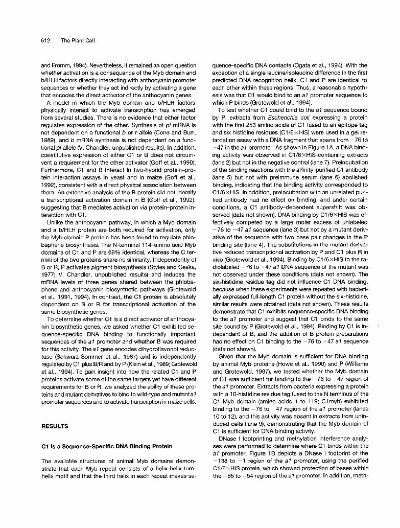
The Plant Cell, Vol. 9, 61 1-625, April 1997 O 1997 American Society of Plant Physiologists Evidence for Direct Activation of an Anthocyanin Promoter by the Maize Cl Protein and Comparison of DNA Binding by Related Myb Domain Proteins Manuel B. Sainz,aErich Grotewold,b and Vicki L. Chandleragl a lnstitute of Molecular Biology, University of Oregon, Eugene, Oregon 97403 Cold Spring Harbor Laboratory, Cold Spring Harbor, New York 11724 The enzyme-encoding genes of two classes of maize flavonoid pigments, anthocyanins and phlobaphenes, are differ- entially regulated by distinct transcription factors. Anthocyanin biosynthetic gene activation requires the Myb domain C1 protein and the basic helix-loop-helix B or R proteins. In the phlobaphene pathway, a subset of C1-regulated genes, including a7, are activated by the Myb domain P protein independently of BIR. We show sequence-specific binding to the a1 promoter by C1 in the absence of B. Activation is decreased by mutations in the C1 DNA binding domain or in a1 sequences bound by C1, providing direct evidence for activation of the anthocyanin biosynthetic genes by C1. The two C1 binding sites in the a7 promoter are also bound by P. One site is bound with higher affinity by P relative to C1, whereas the other site is bound with similar lower affinity by both proteins. Interestingly, either site is sufficient for C1 plus B/R or P activation in vivo, demonstrating that differences in DNA binding affinities between P and C1 are insuffi- cient to explain the differential requirement for B. Results of DNA binding site-selection experiments suggest that C1 has a broader DNA binding specificity than does P, which may help C1 to activate a more diverse set of promoters. INTRODUCTION Regulation of flavonoid pigment biosynthesis has been an exceilent system for the study of combinatorial regulation of an entire set of biosynthetic genes (reviewed in Dooner et al., 1991; van der Meer et al., 1993). Two classes of maize fla- vonoid pigments are the red and purple anthocyanins and the red phlobaphenes. The anthocyanin and phlobaphene bio- synthetic pathways share at least three enzymatic steps and then diverge. Maize genes encoding most of the flavonoid pigment biosynthetic enzymes as well as severa1 regulatory transcription factors have been identified and cloned. Numer- ous experiments have demonstrated that flavonoid biosyn- thetic gene activation requires developmental and tissue- specific expression of the transcription factors regulating the anthocyanin and phiobaphene biosynthetic pathways. Two classes of transcription factors are required for the regulation of anthocyanin biosynthesis in maize. A functional allele of c7 or pl together with a functional allele of b or r are required to coordinately induce mRNA levels of the anthocya- nin biosynthetic genes (Cone et ai., 1986, 1993; Chandler et al., 1989; Ludwig et al., 1989). The c7 (Paz-Ares et al., 1987) and pl (Cone et al., 1993) loci encode functionally equivalent proteins that have 80% identity but are expressed in differ- To whom correspondence should be addressed. E-mail chandler @molbio.uoregon.edu; fax 541 -346-501 1. Address after May 1 : De- partment of Plant Sciences, 303 Forbes Hall, University of Arizona, Tucson, AZ 85721. E-mail [email protected]; fax 520-621 - 7186. ent tissues. These proteins share an N-terminal Myb motif, originally identified as the DNA binding domain of the v-Myb oncoprotein (Biedenkapp et ai., 1988) and consisting of two (or three) amino acid repeats. Each Myb repeat forms a he- lix-helix-turn-helix structure (Ogata et al., 1994). The C ter- minus of C1 functions as an independent transcriptional activation domain in maize and in yeast when fused to the DNA binding domain of the yeast GAL4 protein (Goff et al., 1991; Sainz et al., 1997). The b and r genes encode function- ally duplicate proteins that share 78% identity and are ex- pressed in diverse tissues (Dellaporta et al., 1988; Chandler et ai., 1989; Ludwig et al., 1989; Radicella et al., 1991). B and R have a basic helix-loop-helix (WHLH) motif characteristic of a large family of transcriptional activators (reviewed in Ludwig and Wessler, 1990). In various proteins of this class, the ba- sic region functions in DNA binding, whereas the HLH do- main mediates homodimerization or heterodimerization (Weintraub et al., 1991). Previous studies have demonstrated that the Myb domain and b/HLH factors induce transcription of the anthocyanin biosynthetic genes. In the presence of functionalpl and b alle- les, both steady state mRNA levels and transcription rates of the anthocyanin genes increase (Cone et al., 1993; Patterson et al., 1993). cis-Acting sequences that are necessary and sufficient for transcriptional activation by C1 plus B/R map to upstream promoter regions of the biosynthetic genes, which are 5’ of the start of transcription (Roth et ai., 1991; Bodeau and Walbot, 1992; Grotewold et al., 1994; Tuerck
Transcript of Evidence for direct activation of an anthocyanin promoter by the ...

The Plant Cell, Vol. 9, 61 1-625, April 1997 O 1997 American Society of Plant Physiologists
Evidence for Direct Activation of an Anthocyanin Promoter by the Maize C l Protein and Comparison of DNA Binding by Related Myb Domain Proteins
Manuel B. Sainz,a Erich Grotewold,b and Vicki L. Chandleragl a lnstitute of Molecular Biology, University of Oregon, Eugene, Oregon 97403
Cold Spring Harbor Laboratory, Cold Spring Harbor, New York 1 1724
The enzyme-encoding genes of two classes of maize flavonoid pigments, anthocyanins and phlobaphenes, are differ- entially regulated by distinct transcription factors. Anthocyanin biosynthetic gene activation requires the Myb domain C1 protein and the basic helix-loop-helix B or R proteins. In the phlobaphene pathway, a subset of C1-regulated genes, including a7, are activated by the Myb domain P protein independently of BIR. We show sequence-specific binding to the a1 promoter by C1 in the absence of B. Activation is decreased by mutations in the C1 DNA binding domain or in a1 sequences bound by C1, providing direct evidence for activation of the anthocyanin biosynthetic genes by C1. The two C1 binding sites in the a7 promoter are also bound by P. One site is bound with higher affinity by P relative to C1, whereas the other site is bound with similar lower affinity by both proteins. Interestingly, either site is sufficient for C1 plus B/R or P activation in vivo, demonstrating that differences in DNA binding affinities between P and C1 are insuffi- cient to explain the differential requirement for B. Results of DNA binding site-selection experiments suggest that C1 has a broader DNA binding specificity than does P, which may help C1 to activate a more diverse set of promoters.
INTRODUCTION
Regulation of flavonoid pigment biosynthesis has been an exceilent system for the study of combinatorial regulation of an entire set of biosynthetic genes (reviewed in Dooner et al., 1991; van der Meer et al., 1993). Two classes of maize fla- vonoid pigments are the red and purple anthocyanins and the red phlobaphenes. The anthocyanin and phlobaphene bio- synthetic pathways share at least three enzymatic steps and then diverge. Maize genes encoding most of the flavonoid pigment biosynthetic enzymes as well as severa1 regulatory transcription factors have been identified and cloned. Numer- ous experiments have demonstrated that flavonoid biosyn- thetic gene activation requires developmental and tissue- specific expression of the transcription factors regulating the anthocyanin and phiobaphene biosynthetic pathways.
Two classes of transcription factors are required for the regulation of anthocyanin biosynthesis in maize. A functional allele of c7 or pl together with a functional allele of b or r are required to coordinately induce mRNA levels of the anthocya- nin biosynthetic genes (Cone et ai., 1986, 1993; Chandler et al., 1989; Ludwig et al., 1989). The c7 (Paz-Ares et al., 1987) and pl (Cone et al., 1993) loci encode functionally equivalent proteins that have 80% identity but are expressed in differ-
To whom correspondence should be addressed. E-mail chandler @molbio.uoregon.edu; fax 541 -346-501 1. Address after May 1 : De- partment of Plant Sciences, 303 Forbes Hall, University of Arizona, Tucson, AZ 85721. E-mail [email protected]; fax 520-621 - 71 86.
ent tissues. These proteins share an N-terminal Myb motif, originally identified as the DNA binding domain of the v-Myb oncoprotein (Biedenkapp et ai., 1988) and consisting of two (or three) amino acid repeats. Each Myb repeat forms a he- lix-helix-turn-helix structure (Ogata et al., 1994). The C ter- minus of C1 functions as an independent transcriptional activation domain in maize and in yeast when fused to the DNA binding domain of the yeast GAL4 protein (Goff et al., 1991; Sainz et al., 1997). The b and r genes encode function- ally duplicate proteins that share 78% identity and are ex- pressed in diverse tissues (Dellaporta et al., 1988; Chandler et ai., 1989; Ludwig et al., 1989; Radicella et al., 1991). B and R have a basic helix-loop-helix (WHLH) motif characteristic of a large family of transcriptional activators (reviewed in Ludwig and Wessler, 1990). In various proteins of this class, the ba- sic region functions in DNA binding, whereas the HLH do- main mediates homodimerization or heterodimerization (Weintraub et al., 1991).
Previous studies have demonstrated that the Myb domain and b/HLH factors induce transcription of the anthocyanin biosynthetic genes. In the presence of functionalpl and b alle- les, both steady state mRNA levels and transcription rates of the anthocyanin genes increase (Cone et al., 1993; Patterson et al., 1993). cis-Acting sequences that are necessary and sufficient for transcriptional activation by C1 plus B/R map to upstream promoter regions of the biosynthetic genes, which are 5’ of the start of transcription (Roth et ai., 1991; Bodeau and Walbot, 1992; Grotewold et al., 1994; Tuerck
612 The Plant Cell
and Fromm, 1994). Nevertheless, it remained an open question whether activation is a consequence of the Myb domain and b/HLH factors directly interacting with anthocyanin promoter sequences or whether they act indirectly by activating a gene that encodes the direct activator of the anthocyanin genes.
A model in which the Myb domain and b/HLH factors physically interact to activate transcription has emerged from severa1 studies. There is no evidence that either factor regulates expression of the other. Synthesis of pl mRNA is not dependent on a functional b or r allele (Cone and Burr, 1989), and b mRNA synthesis is not dependent on a func- tional pl allele (V. Chandler, unpublished results). In addition, constitutive expression of either C1 or B does not circum- vent a requirement for the other activator (Goff et al., 1990). Furthermore, C1 and B interact in two-hybrid protein-pro- tein interaction assays in yeast and in maize (Goff et al., 1992), consistent with a direct physical association between them. An extensive analysis of the B protein did not identify a transcriptional activation domain in B (Goff et al., 1992), suggesting that B mediates activation via protein-protein in- teraction with C1.
Unlike the anthocyanin pathway, in which a Myb domain and a b/HLH protein are both required for activation, only the Myb domain P protein has been found to regulate phlo- baphene biosynthesis. The N-terminal 11 4-amino acid Myb domains of C1 and P are 69% identical, whereas the C ter- mini of the two proteins share no similarity. lndependently of B or R, P activates pigment biosynthesis (Styles and Ceska, 1977; V. Chandler, unpublished results) and induces the mRNA levels of three genes shared between the phloba- phene and anthocyanin biosynthetic pathways (Grotewold et al., 1991, 1994). In contrast, the C1 protein is absolutely dependent on B or R for transcriptional activation of the same biosynthetic genes.
To determine whether C1 is a direct activator of anthocya- nin biosynthetic genes, we asked whether C1 exhibited se- quence-specific DNA binding to functionally important sequences of the a7 promoter and whether B was required for this activity. The a7 gene encodes dihydroflavonol reduc- tase (Schwarz-Sommer et al., 1987) and is independently regulated by C1 plus B/R and by P (Klein et al., 1989; Grotewold et al., 1994). To gain insight into how the related C1 and P proteins activate some of the same targets yet have different requirements for B or R, we analyzed the ability of these pro- teins and mutant derivatives to bind to wild-type and mutantal promoter sequences and to activate transcription in maize cells.
RESULTS
C1 1s a Sequence-Specific DNA Binding Protein
The available structures of animal Myb domains demon- strate that each Myb repeat consists of a helix-helix-turn- helix motif and that the third helix in each repeat makes se-
quence-specific DNA contacts (Ogata et al., 1994). With the exception of a single leucine/isoleucine difference in the first predicted DNA recognition helix, C1 and P are identical to each other within these regions. Thus, a reasonable hypoth- esis was that C1 would bind to an a7 promoter sequence to which P binds (Grotewold et al., 1994).
To test whether C1 could bind to the a7 sequence bound by P, extracts from fscherichia coli expressing a protein with the first 253 amino acids of C1 fused to an epitope tag and six histidine residues (Cl/GxHIS) were used in a gel re- tardation assay with a DNA fragment that spans from -76 to -47 in the a7 promoter. As shown in Figure lA, a DNA bind- ing activity was observed in Cl/GxHIS-containing extracts (lane 2) but not in the negative control (lane 7). Preincubation of the binding reactions with the affinity-purified C1 antibody (lane 5) but not with preimmune serum (lane 6) abolished binding, indicating that the binding activity corresponded to C1/6xHIS. In addition, preincubation with an unrelated puri- fied antibody had no effect on binding, and under certain conditions, a C1 antibody-dependent supershift was ob- served (data not shown). DNA binding by Cl/GxHIS was ef- fectively competed by a large molar excess of unlabeled -76 to -47 a7 sequence (lane 3) but not by a mutant deriv- ative of the sequence with two base pair changes in the P binding site (lane 4). The substitutions in the mutant deriva- tive reduced transcriptional activation by P and C1 plus R in vivo (Grotewold et al., 1994). Binding by C1/6XHIS to the ra- diolabeled -76 to -47 a7 DNA sequence of the mutant was not observed under these conditions (data not shown). The six-histidine residue tag did not influence C1 DNA binding, because when these experiments were repeated with bacteri- ally expressed full-length C1 protein without the six-histidine, similar results were obtained (data not shown). These results demonstrate that C1 exhibits sequence-specific DNA binding to the a7 promoter and suggest that C1 binds to the same site bound by P (Grotewold et al., 1994). Binding by C1 is in- dependent of B, and the addition of B protein preparations had no effect on C1 binding to the -76 to -47 a7 sequence (data not shown).
Given that the Myb domain is sufficient for DNA binding by animal Myb proteins (Howe et al., 1990) and P (williams and Grotewold, 1997), we tested whether the Myb domain of C1 was sufficient for binding to the -76 to -47 region of the a7 promoter. Extracts from bacteria expressing a protein with a 10-histidine residue tag fused to the N terminus of the C1 Myb domain (amino acids 1 to 119; Clmyb) exhibited binding to the -76 to -47 region of the a7 promoter (lanes 1 O to 12), and this activity was absent in extracts from unin- duced cells (lane 9), demonstrating that the Myb domain of C1 is sufficient for DNA binding activity.
DNase I footprinting and methylation interference analy- ses were performed to determine where C1 binds within the a7 promoter. Figure 16 depicts a DNase I footprint of the -138 to -1 region of the a7 promoter, using the purified C1/6xHIS protein, which showed protection of bases within the -65 to -54 region of t hea l promoter. In addition, meth-

C1 DNA Binding to a? and Comparison to P 613
I.ANI- 1 2 1 4 s 6 7 8 9 10 11 12protein -- CI/6xHIS » TsrA -- un Cluiyb »coiifietilor -- -- wt muianlilxxly -- -- -- -- ("1 pi
free DNA
wild-lype -76 CGGGTCAGTGTPfCTACTAArCTITAAArAr -47mutant T T
I.ANH 1 2 1 4(5 free hound free
-MC
-65 A
Figure 1. Sequence-Specific DNA Binding by C1/6xHIS to a Site inthe -76 to -47 a 1 Promoter.
(A) Sequence-specific binding by C1/6XHIS to the -76 to -47 re-gion of the a? promoter. A gel retardation assay was performed withextracts from bacteria expressing C1/6xHIS (lanes 2 to 6) and a ra-diolabeled -76 to -47 region of the at sequence (shown as wildtype below). Dashes indicate the absence of a given component.Lane 1 contains the free DNA; lane 7 shows the DNA binding activityin extracts from bacteria expressing TsrA (Ames and Parkinson,1994), which serves as a control for bacterial DNA binding proteinsinduced by protein overexpression. Competitions were performedusing the wild-type sequence (wt; lane 3) or a mutant derivative(mut; lane 4), with two base pair changes (indicated below the wild-type sequence) in the boxed P binding site (Grotewold et al., 1994).Reactions were preincubated with the C1 antibody (C1; lane 5) orpreimmune serum (pi; lane 6). In a different gel (lanes 8 to 12), ex-tracts from bacteria expressing the C1 Myb domain (C1 myb) for 1 hr(lane 10), 2 hr (lane 11), and 3 hr (lane 12) were used in gel retarda-tion assays with the radiolabeled -76 to -47 a 7 promoter se-quence. Lane 8, free DNA; lane 9, extract from uninduced (un) cells.
ylation interference assays indicated that C1/6XHIS wasprevented from binding by methylation of the six guaninebases in this region (cytosines on the strand shown in Figure1B; data not shown). Thus, like P (Grotewold et al., 1994),C1 binds to the -65 to -54 site in the a1 promoter.
C1 and P Bind to Two Functionally Important Sitesin the at Promoter
Sequence-specific DNA binding by C1 and P to the -76 to-47 region of the a1 promoter provides an explanation forthe ability of this region of a? to confer C1 plus R and P in-ducibility to a heterologous minimal promoter (Grotewold etal., 1994). However, a region of the a 1 promoter from -140to -79 can also confer inducibility by C1 plus B and by P(Tuerck and Fromm, 1994). Activation of the -140 to -79region of the a1 promoter could be mediated by C1 and Pdirectly binding to this sequence or through the binding ofother proteins to this region that recruit C1 or P to the DNA.
To explore further their DNA binding characteristics, C1and P proteins were purified from crude bacterial extracts.Unfortunately, full-length P is not expressed as well as C1 in£. co// and is highly susceptible to proteolysis (data notshown). We overcame this problem by using a fusion proteinwith the Myb DNA binding domain of P fused to the C termi-nus of C1/6XHIS (Pmyb/C1/6xHIS), which was expressedto the same extent as C1/6xHIS in bacteria (data notshown). Several lines of evidence suggest that Pmyb/C1functions similarly to P. Like P, Pmyb/C1 activates a1 inde-pendently of R when expressed in maize cells (Grotewold etal., 1994). Furthermore, the P Myb domain is sufficient forDNA binding (Williams and Grotewold, 1997). We were ableto purify C1/6XHIS and Pmyb/C1/6xHIS from bacterial ex-tracts to the same extent (^80% pure; data not shown), fa-cilitating comparative analyses of the DNA binding activitiesmediated by the C1 and P Myb domains in the context ofthe C1/6XHIS C terminus.
To test the hypothesis that C1 and P directly activate the-140 to -79 region of the a1 promoter, we examinedwhether C1/6XHIS could bind to a distal (-133 to -83) alsequence in gel retardation assays. As shown in Figure 2A,C1/6XHIS bound to the distal a1 sequence (lane 2), and C1/6xHIS binding was diminished by the addition of the C1 an-tibody (lane 4) but not by preimmune serum (lane 3). Specificbinding to the distal a 7 sequence was competed by an excessof unlabeled distal DNA (lane 6) and even more effectively by
(B) DNase I footprint of C1/6XHIS bound to a site at -65 to -54 inthe a 1 promoter. DNase I footprinting was performed using purifiedC1/6XHIS and a radiolabeled -138 to -1 region of the a7 pro-moter. Lane 1 contains the Maxam and Gilbert G sequencing reac-tion (G); lanes 2 and 4 contain free DNA (free); lane 3 contains C1/6xHIS bound DNA. The sequence shown is to the strand oppositethe one labeled. The lines at left denote the two overlapping P bind-ing sites previously reported by Grotewold et al. (1994).

614 The Plant Cell
LANE 1 2 3 4 5 6 7 8 9 10protein - Cl/6xHIS ————————————competitor -- — — — pro dis A B C Dantibody - -
-83.GGATGCGCTCA'CCTACGCGAGT
-98 CCCTCGAGGG -89-108 CCCTCGAGGG -99
-118 CCCTCGAGGG-109D-128 CCCTCGAGGG -119
BI.ANF. 1 2 3 4
G free bound freeLANE 1 2 3 4
G free bound free
Pmyb/Cl/6xHIS Cl/6xHIS
Figure 2. C1/6XHIS and Pmyb/C1/6xHIS DNA Binding to a Site inthe -133 to -83 Region of the a? Promoter.
(A) Sequence-specific binding by C1/6XHIS to the -133 to -83 re-gion of the a1 promoter. Gel retardation assays were done withequal amounts of the purified C1/6XHIS protein (lanes 2 to 10) andthe radiolabeled wild-type -133 to -83 a1 sequence. Dashes indi-cate the absence of a given component. Lane 1 contains the freeDNA. Reactions were preincubated with preimmune serum (pi; lane3), C1 antibody (C1; lane 4), or unlabeled competitor DNA (lanes 5 to10) before the addition of radiolabeled DNA. The competitor DNAused in lanes 5 and 6 are the proximal (pro; -76 to -47) and distal(dis; -133 to -83) a1 sequences, respectively. The sequenceschanged in the mutant a1 promoter derivatives used as competitorDNA (A to D; lanes 7 to 10) and corresponding to the top strand ofthe wild-type sequence are shown, along with their coordinates. Theboxed region is bound by C1/6XHIS and Pmyb/C1/6xHIS in DNaseI footprinting assays (B).(B) DNase I footprints of Pmyb/C1/6xHIS and C1/6XHIS bound to asite at -116 to -124 in the distal a 7 promoter. DNase I footprintingwas performed using purified Pmyb/C1/6xHIS or C1/6xHIS and aradiolabeled -219 to +7 sequence of the a1 promoter with a mu-tated -65 to -54 binding site. Lanes 1 contain the Maxam and Gilbert
equivalent amounts of the unlabeled proximal (-76 to -47)a1 DNA (lane 5).
To identify where C1/6XHIS was binding within the distala1 promoter, DNA sequences with specific mutations wereused to compete with radiolabeled wild-type distal DNA forbinding by C1/6XHIS (Figure 2A). These mutations hadbeen shown previously to decrease activation in vivo(Tuerck and Fromm, 1994). DNA fragments with mutations Aand B were as effective competitors as the wild-type -133to -83 region of a? DNA (Figure 2A, lanes 7 and 8), suggest-ing that C1/6xHIS bound to fragments with these mutatedsequences with the same affinity with which it bound to thewild type. In contrast, DNA fragments with mutations C or Dwere less effective as competitors compared with the wild-type distal fragment (Figure 2A, lanes 9 and 10), suggestingthat C1/6XHIS does not bind well to the mutated sequencesin the C and D variants.
Similar results were obtained in experiments involving di-rect binding of C1/6XHIS to radiolabeled wild-type and mu-tant DNA fragments. C1/6xHIS bound -30% of the wild-type fragment and —30% of the fragments with the A and Bmutations; in contrast, only 5% of DNA sequences bearingthe C and D mutations were bound by an equivalent amountof C1/6XHIS (data not shown). These data are consistentwith the competition results, and together they suggest thatC1/6XHIS binds to the a1 promoter in the -128 to -109 re-gion spanned by the C and D mutations. The binding andcompetition experiments were repeated with Pmyb/C1/6xHIS, with results similar to those obtained with C1/6XHIS(data not shown), indicating that the P Myb domain also me-diates binding to the distal a7 sequence.
DNase I footprinting of the wild-type a1 promoter failed toreveal C1/6xHIS (data not shown) or P (Grotewold et al.,1994) binding tp distal a1 sequences. This result, combinedwith the more effective competition exhibited by the proxi-mal a7 fragment relative to the distal sequence (Figure 2A,lanes 5 and 6), suggests that C1 and P bind with higher af-finity to the overlapping ACCT/AACC sequence at -65 to-54 in the a7 promoter. To identify more precisely the C1and P binding sites within the distal a7 sequence, a -219 to+ 7 a7 sequence with a mutated -65 to -54 proximal bind-ing site was used in DNase I footprinting experiments. Asseen in Figure 2B, when the proximal site is mutated, C1/6xHIS and Pmyb/C1/6xHIS exhibit binding to a site at-116 to -124 (AACTACCGG), in the opposite orientationrelative to the -65 to -54 binding site. This sequence iswithin the region covered by the C and D mutations in thedistal a7 sequence, as predicted from the results of the gelretardation experiments.
G sequencing reaction (G); lanes 2 and 4 contain free DNA (free);lanes 3 contain Pmyb/C1/6xHIS or C1/6XHIS bound DNA (bound)as indicated. The sequence shown is to the strand opposite the onelabeled.

C1 DNA Binding to a7 and Comparison to P 61 5
Our identification of C1 and P binding sites in each of the regions sufficient for activation by C1 plus B and by P (Grotewold et al., 1994; Tuerck and Fromm, 1994) suggests that C1 and P bind to both sites in a7 independently and that a single site suffices to assemble a functional transcrip- tion complex. Previous studies have shown that mutations within either binding site in the context of the full-length a7 promoter had detectable but minor effects (Grotewold et al., 1994; Tuerck and Fromm, 1994). We now hypothesize that this was because C1 or P could still bind to the other site and activate transcription. A prediction of this hypothesis is that mutating both binding sites should have a more severe effect on activation by C1 plus B and by P.
To test this hypothesis, we assayed C1 plus B and P acti- vation of the wild-type a7 promoter, promoter derivatives with mutations in each of the C1 and P binding sites, or a derivative with both sites mutated. Activation of wild-type and mutant a7 promoters was tested in cultured maize cells transiently transformed by microprojectile bombardment; the constructs used are depicted in Figure 3A. As seen in Figure 3B, changes in either of the C1 and P binding sites moderately reduced C1 plus B and P activation, in agree- ment with previous studies (Grotewold et al., 1994; Tuerck and Fromm, 1994). Mutation of both binding sites had a more severe effect on activation by C1 plus B and by P (Fig- ure 38). Mutation of the proximal or distal binding sites de- creased activation by P similarly and was 18 and 16% of the levels observed with the wild-type a7 promoter, respec- tively. When both binding sites were mutated, activation by P was only threefold over background levels, suggesting that P binding sites mediate the major portion of a7 activa- tion by P in vivo. The addition of B did not alter activation by P (data not shown). For activation by C1 plus B, mutations of the proximal and distal C1 binding sites had similarly mild effects and were 35 and 53% of wild-type a1 promoter ac- tivity, respectively. C1 plus B activation of the a7 promoter double mutant was further decreased to 17% of wild-type a7 levels, yet it remained 12-fold over background. For acti- vation by both C1 plus B and by P, the phenotype of the double-site mutant was additive relative to the single-site mutations, suggesting that C1 and P bind independently to the two sites. A summary of our results, combined with pre- viously published data, is presented in Figure 3C.
Although the identified C1 binding sites are important for activation, other sequences within the a7 promoter may contribute as well. When mutated, the -98 to -89 region caused a major decrease in C1 plus B activation of a7 (Fig- ure 3C, mutant A; Tuerck and Fromm, 1994). Nevertheless, C1 binding to the -133 to -83 a7 promoter fragment in the absence of the distal binding site was extremely weak, and mutations of the -98 to -89 region did not affect C1 bind- ing to the distal a7 site (Figure 2A). These results suggest that other proteins important for activation by C1 plus B may be binding in the -98 to -89 region. If true, this may ac- count for the residual level of C1 plus B activation we ob- served when both C1 binding sites in a7 were mutated. A
logical candidate for a protein binding to the -98 to -89 re- gion is B or R. However, when using an in vitro-translated B protein or B protein preparations from E. coli or Sf9 insect cells, we did not observe DNA binding to any functionally important anthocyanin promoter sequence (data not shown). Potentially, B may require an HLH dimerization partner to bind to DNA.
C1 DNA Binding Activity 1s lmportant for Activation of the a7 Promoter
If C1 DNA binding activity is important for transcriptional ac- tivation of the anthocyanin promoters, then a C1 mutant that fails to bind to DNA in vitro should be unable to activate in vivo. The C1 Myb domain is sufficient for DNA binding (Fig- ure IA) and for interacting with B (Goff et al., 1992), providing two independent functional assays. This enables the identifi- cation of C1 mutants specifically defective in DNA binding or the interaction with B. Mutations that result in misfolded pro- teins would be predicted to affect both activities.
A candidate C1 DNA binding mutant was noted in previ- ous studies. A C1 mutant in which the aspartate (D) at posi- tion 101 was changed to glutamate (E) was isolated from a naturally occurring dominant inhibitor allele of c7 called C7-I (Paz-Ares et al., 1990; Goff et al., 1991). D-101 is conserved in mouse Myb. Based on the available structure of the mouse Myb repeats bound to DNA (Ogata et al., 1994), D-101 is at the beginning of the putative DNA recognition helix in the second Myb repeat of C1. Previous experiments had shown that the C1:DlOl E mutant was unable to activate the bronze7 anthocyanin promoter in maize cells (Goff et al., 1991). Nevertheless, C1:DlOlE was able to interact with B as well as wild-type C1 in maize two-hybrid assays, leading to the prediction that C1 :D1 O1 E was a DNA binding mutant (Goff et al., 1992).
To test this prediction, wild-type and mutant C1 proteins were assayed for in vitro DNA binding to the proximal a7 se- quence. As seen in Figure 4A, DNA binding by C1 :D1 O1 E/ 6XHIS (lane 5) was extremely weak, with a faint band appar- ent only in very long exposures (data not shown). In contrast, a 1:10 dilution of wild-type C1/6XHIS (lane 4) generated a visible band in this experiment. Thus, C1:DlOl E/GxHIS is defective in DNA binding. A DNA binding defect is also caused by the analogous D117E mutation in MYB.Ph3, a plant Myb domain protein from petunia (Solano et al., 1995).
Even though C1 and C1 :D1 O1 E interacted similarly with B in maize two-hybrid assays (Goff et al., 1992), we are unable to quantify expression of the two proteins in this system. Because C1 and C1 :D1 O1 E are expressed similarly in yeast (data not shown), we used yeast two-hybrid assays to con- firm that wild-type and mutant C1 proteins interact similarly with B. The wild-type Cl/GAL4 activation domain and the B/GAL4 DNA binding domain fusion proteins generated a robust level of p-galactosidase activity when coexpressed in yeast cells bearing a GAL7 promoter-lacZ reporter gene. As

61 6 The Plant Cell
- P : pAlDBL
bkgd : pAlDBL
C1+ B : pAlWT -
A Expression vectors
t
pZmP
p35SB
B + Reporter constructs
C1+ B : pAlPRO
C1+ B : pAlDIS
C1+ B : pAlDBL
bkgd : pAlDBL
C b b % wild type
-128 -19 -16 -47 a1 activation TATG CGGTA GT CAGCGT GTGGTGTTGA ATGGAGGATG CGCTCAATCG CGCGGGTCAG TG CCTACC AACC TAAAC AC
P A T A c c & T C G C A CACCACAACT TACCTCCTAC WGAGTTAGC EGCCCAGTC A C $ Z Z L $ A T T T G TG - c 1 + B - D 60% 15% - distal* 53% 16%
C 40% 12% B 26% 32%
A 8% 30% Z 111% 112%
proximal* 35% 18% double * 17% 1%
Figure 3. Activation of Wild-Type and Mutant a7 Promoters by P and by C1 plus B.
(A) Constructs used in maize transient transformation assays. Plasmids designed to express the activator proteins (pZmP, pZmC1, and p35SB) are shown. Reporter constructs with the wild-type (wt) -219 to f7 region of the a7 promoter (PAI WT) or mutant derivatives fused to a firefly Iu- ciferase (LUC) gene are shown. The proximal mutant (PAI PRO) has base pair changes in the C1 and P binding site at -65 to -54 (pro). The dis- tal mutant (pAlDIS) changes the C1 and Pmyb/CI binding site at -116 to -124 (dis), and the double (dbl) mutant a7 promoter reporter (PAI DBL) has both the proximal and dista1 binding site mutations. (B) Activation by P and by C1 plus B of wild-type and mutant a7 promoters. Cultured maize cells were transiently transformed with expression vectors, reporter constructs, and a constitutively expressed p-glucuronidase reporter gene, which is included as a transformation and extraction control. The ratio of luciferase to p-glucuronidase activity in extracts from the transiently transformed cells was determined and then expressed as the fold induction observed in the presence of the activators relative to the expression observed in their absence. Fold induction for the mu- tant promoter constructs was normalized to the fold induction by P or by C1 plus B of pA1 WT, which was set at 100%. The observed induction of pAlWT is robust: an average of 272-fold for P and 90-fold for C1 plus B. The amount of background (bkgd) activation was similarly low for all three promoter constructs. Each histogram represents the average of 10 to 12 samples from two separate experiments (except C1 alone, n = 6 in one experiment; C1 plus B, n = 16 in three experiments). Error bars represent the standard error. (C) Summary of a7 promoter studies. The large arrows denote the two regions of the a7 promoter that can independently confer inducibility by C1 plus B/R and inducibility by P when fused to a heterologous minimal promoter (Grotewold et al., 1994; Tuerck and Fromm, 1994). C1 and P binding sites are boxed. Linker scanning mutations are depicted by lines underneath the altered sequences; those denoted by an asterisk repre- sent data from (B), whereas others had been published previously (Tuerck and Fromm, 1994). The proximal linker scanner had been previously tested for activation by C1 plus R and by P, with results similar to ours (Grotewold et al., 1994).

C1 DMA Binding to a 1 and Comparison to P 617
LANE 1 2 3 4 5 6protein - Cl/6xHIS-» DIOlETsrAdilution -- 1:1 1:2 1:10 1:1 1:1
B
free DNA
-76 CGGGTCAGTGTACCTACCAACCTTAAACAC -47
Reporter constructURA3-.:GALl-lacZ
GAL1lacZ
PGK
GPD
GPD
Expression vectors> pYBGAL
B |GAL4DB}
i pYClGAL InteractionCl |GAL4AD|—— 100% ±12
OR, pYDEGAL
|GAL4AD|—— 132%+11
i wild-type Cl + B a I activation25 50 75 100 125
Figure 4. Functional Assays of the C1:D101E Mutant.
(A) C1 :D101 E mutant DNA binding activity in gel retardation assays.Gel retardation assays were done with crude extracts from bacteriaexpressing wild-type C1/6XHIS or the mutant C1:D101E/6xHISproteins and the radiolabeled -76 to -47 region of the a 7 promoter.Dashes indicate the absence of a given component. Wild-type C1/6xHIS (lane 2) and dilutions (lanes 3 and 4) were used for compari-sons to DNA binding by C1:D101E/6xHIS (D101E; lane 5). Lane 1contains the free DNA; lane 6 contains the TsrA negative controlfrom the same gel.(B) C1:D101E mutant activity in yeast two-hybrid protein-protein in-teraction assays with B. The cDNAs encoding the C1 (pYCIGAL) orC1:D101E (pYDEGAL) proteins fused to sequences coding for the
seen in Figure 4B, C1:D101E interacted with B at least aswell as with wild-type C1 in yeast two-hybrid assays. Hence,the D101E mutation only affects the DNA binding activity ofC1 and not its ability to interact with B, strongly suggestingthat the DNA binding defect is not due to a general misfold-ing of the mutant protein.
To assess the importance of C1 DNA binding activity fora?activation, the ability of C1:D101E to activate the a? promoterwas assayed using transient transformation of cultured maizecells. As seen in Figure 4C, C1 :D101 E activated the wild-typea1 promoter at only 11% of wild-type C1 levels in the pres-ence of B, similar to the reduction in activation observed whenboth C1 binding sites in a 1 were mutated (Figure 3B). De-creased activation by C1:D101E, a mutant specifically af-fected in DNA binding, strongly argues that C1 DNA bindingactivity is important in activating the a7 promoter.
Quantitative Comparison of C1 and P DNA BindingActivities
We have shown that C1 and P bind to the same al se-quences in vitro. If both proteins bound to their sites with
GAL4 activation domain in a yeast expression vector were cotrans-formed into yeast along with a plasmid designed to express a B/GAL4DNA binding domain fusion protein (pYBGAL; Goff et al., 1992). PGKand GPD indicate the yeast 3-phosphoglycerate kinase and glyc-eraldehyde-3-phosphate dehydrogenase promoters, respectively;GAL4 DB and GAL4 AD indicate the yeast GAL4 protein DNA bind-ing and activation domains, respectively. The interaction of C1 orC1:D101E with B was monitored by activation of a GAL1-lacZ re-porter present in the yeast strain, as measured by fi-galactosidaseactivity in the transformed cells. The background (i-galactosidaseactivity generated by the empty vector plus pYBGAL averaged <1Miller unit. pYCIGAL plus pYBGAL generated an average of 225Miller units of (3-galactosidase activity in these experiments. Datafrom pYDEGAL plus pYBGAL were normalized to the p-galactosi-dase activity generated by pYCIGAL plus pYBGAL and representthe average of 10 to 15 independent transformants from two sepa-rate experiments, ±SE.(C) Activation of the a1 promoter by the C1:D101E mutant. Activa-tion by C1 plus B and C1 :D101 E plus B was compared in transientlytransformed cultured maize cells. Transformations included the full-length (1.4-kb) a1 promoter fused to a firefly luciferase reporter gene(pAILuc; Klein et al., 1989) and a constitutively expressed fS-glucu-ronidase reporter gene, which serves as a transformation and ex-traction control. Data were calculated as the ratio of luciferase-to-p-glucuronidase activity in extracts from the transiently transformedcells and normalized to wild-type C1 plus B activity, which was setat 100%. Activation by C1 plus B averaged 42-fold over the back-ground observed in the absence of C1 and B. Histograms representthe average of 18 samples from three separate experiments (exceptC1 alone and B alone, n = 12 from two experiments; C1:D101Ealone, n = 6 from one experiment). Error bars represent the stan-dard error.

61 8 The Plant Cell
12- 1 ° -
3 8 -
3 4 .
8 6 .
2 -
0 -
the same affinity in vivo, then one might expect both of them to activate transcription, because both proteins have activa- tion domains (Goff et al., 1991; E. Grotewold, M.B. Sainz, and V.L. Chandler, unpublished results). However, C1 is completely dependent on B for activation, despite the ab- sence of a transcriptional activation domain in B (Goff et al., 1992; Sainz et al., 1997). Thus, one simple model is that C1 requires B to bind to DNA with high affinity. A prediction of this model is that C1 alone would have a significantly lower DNA binding affinity than would P in vitro.
To test this prediction, we cornpared the relative DNA binding affinities of the C1 and P Myb domains for the high- affinity site in the proximal a7 promoter. Proteins with a 10- histidine residue tag fused to the Myb domains of C1 (Clmyb) and of P (Pmyb) were expressed in and purified from E. coli and used in gel retardation assays with the -76 to -47 a7 sequence. Free and bound bands were quanti- fied, and the data were subjected to Scatchard analysis (Scatchard, 1949); representative graphs are depicted in Figures 5A and 56. When we used the average of six differ-
Kd = 48 nM
e \ - - 0.65 O l l 0.15 0.5 0.35
C
42 e
3 5 1 ~
Kd = 860 nM RMS = 0.84
ent experiments that yielded curves with root mean square (RMS) values 20.8, Pmyb exhibited an equilibrium dissocia- tion constant (Kd) of 52 nM ? 4 SE for the high-affinity a7 site, which is comparable to the 28 nM ? 3 value recently reported by Williams and Grotewold (1997). In contrast, Clmyb had a Kd of 330 nM ? 50 SE, an approximately six- fold lower affinity for the proximal a7 sequence relative to Pmyb. These Kd values are within the range previously re- ported for mouse Myb domain binding to its high-affinity binding site under experimental conditions similar to ours (Rarnsay et al., 1992). Although binding constants have not been determined for nearly full-length proteins, similar affin- ity differences have also been observed for these proteins, because Pmyb/Cl/GxHIS binds to the high-affinity a7 site with approximately three- to fourfold greater affinity than does Cl/GxHIS (data not shown).
To address whether C1 and P exhibited differences in af- finity for other functionally important sequences, we deter- mined the DNA binding affinity of the C1 and P Myb domains for the low-affinity site in the dista1 a7 promoter.
B
251 Kd = 400 nM RMS = 0.86
D
Kd = 120 nM RMS = 0.90 24
30 - Kd = 120 nM RMS = 0.90
6 -
i 0.04 0.05 0.06 0.07 0.08
boundfree
Figure 5. Scatchard Analysis of Clmyb and Pmyb Binding to the High- and Low-Affinity Binding Sites on the a7 Promoter.
Representative curves are shown, with RMS and equilibrium Kd values close to the mean values from the six experiments done for each protein and each binding site, as indicated. (A) Pmyb binding to the high-affinity a7 binding site. (B) Clmyb binding to the high-affinityal binding site. (C) Pmyb binding to the low-affinity a7 binding site. (D) C1 myb binding to the low-affinity a7 binding site.

C1 DNA Binding to a7 and Comparison to P 619
Representative Scatchard plots are shown in Figures 5C and 5D. When we used the average of six different experiments that yielded curves with RMS values 20.8, C1 myb and Pmyb exhibited equivalent dissociation constants for the distal a7 se- quence of 780 nM ? 70 and 860 nM 2 150 SE, respectively.
To determine what DNA sequence C1 most prefers to bind, we performed polymerase chain reaction (PCR)-based site selection, which involved cycles of C1 binding to a ran- dom pool of annealed oligonucleotides, immunoprecipita- tion of the protein-DNA complexes, and PCR amplification of the selected sequences (Pollock and Treisman, 1990). In contrast to P, which selected primarily sequences with an ACCT/AACC motif (Grotewold et al., 1994), C1 selected 26 diverse sequences from the same random pool of oligonu- cleotides used for the P experiment. Only some of these fragments had sites similar to the C1 binding sites in the a7 promoter. The binding of C1 to the various site-selected se- quences was quantified relative to the proximal (-76 to -47) a7 sequence, using the same amounts of radiolabeled fragments and C1 protein in gel retardation assays. No de- tectable C1 binding to nine of the selected sequences was observed; the remaining 17 sequences are ranked in order of affinity for C1 in Figure 6. None of the selected sequences bound C1 with greater affinity than did the proximal a7 se- quence, suggesting that C1, like P, binds with highest affin- ity to the proximal a7 binding site.
Although we have not directly identified where C1 is bind- ing within the selected sequences, many of them have sites similar to an Ac/ACT/AAc/AC motif present in the a7 promoter C1 binding sites. This motif, originally identified in plant fla- vonoid biosynthetic gene promoters (Lois et al., 1989; Loake et al., 1992), is the consensus binding site for plant Myb do- main proteins from diverse species (Grotewold et al., 1994; Sablowski et al., 1994; Solano et al., 1995). None of the se- quences selected by C1 had animal Myb consensus binding sites (T/,AACT/,G; Biedenkapp et al., 1988), and the three selected sequences with the highest affinity for C1/6xHIS possess sites with no mismatches relative to the Ac/ACT/AAC/AC motif, suggesting that C1/6xHIS prefers to bind to se- quences similar to the plant Myb domain protein consensus binding site. C1/6XHIS binds to the distal a7 binding site with 42% of the affinity with which it binds to the high-affin- ity binding site in the -74 to -47 region of the a7 promoter (Figure 5). Thus, although binding constants have not been determined, the four site-selected sequences with the high- est affinity for C1/6XHIS (Figure 6) are bound by the protein with an affinity comparable (within twofold) to that of the functionally important -133 to -83 distal a7 promoter sequence.
DlSCUSSlON
Sequence-specific DNA binding by C1/6XHIS to two func- tionally important regions of the a1 promoter provides direct
evidence that C1 directly activates the transcription of an- thocyanin biosynthetic genes. The importance of C1 DNA binding activity in the activation of a7 is demonstrated by several lines of evidence. First, C1 binding sites have been identified in each of the a7 regions that independently confer C1 plus B inducibility to a heterologous promoter. Second, the significance of C1 DNA binding activity to C1 plus B-medi- ated activation is indicated by the large reduction in activ- ation observed when both C1 binding sites in a1 were mutated and also by the reduced activation exhibited by the C1:DlOlE mutant, which is specifically affected in DNA binding activity. Given that C1 binds to the a1 promoter and interacts with the absolutely corequired B protein, B is also likely to be directly involved in activation of the a7 gene.
The diversity of sequences identified by site selection suggests that C1 has a broader DNA binding specificity than does P. Although the selected sequences with the highest relative affinities for C1 show similarity to the plant Myb do- main protein consensus binding site (Ac/ACT/AAc/AC), several sequences selected by C1 have two mismatches relative to this consensus motif. An example is clone 4, to which C1/ 6XHIS binds with 25% of the affinity of the high-affinity a7 binding site (Figure 6). The recognition of diverse sequences by C1 makes it difficult to identify potential C1 binding sites in anthocyanin promoters by sequence scanning. In a previ- ous study, mutation of an animal Myb consensus binding site in the bronze7 promoter decreased C1 plus B activation to 10% of wild-type levels and led to the hypothesis that C1 binds to this site (Roth et al., 1991). C1 exhibits sequence- specific DNA binding to the bronze7 anthocyanin promoter (M. Sainz and V. Chandler, unpublished results), and the hy- pothesis that C1 binds to the animal Myb consensus site is currently being tested. Nevertheless, it appears that C1 binds with high affinity to another site in the bronze7 pro- moter (M. Sainz and V. Chandler, unpublished results).
Severa1 lines of evidence suggest that C1 does not require B or R to bind to anthocyanin promoter sequences in vivo. First, C1 exhibits sequence-specific DNA binding activity to functionally important sites in the a7 promoter in vitro in the absence of B. Second and most importantly, the C1 and P Myb domains have similar affinities for the distal a7 pro- moter in vitro, but only C1 requires B to activate this pro- moter fragment in vivo (Tuerck and Fromm, 1994). Hence, differences in DNA binding affinity do not correlate with dif- ferential requirements for B by C1 and P in activating the distal a7 promoter sequence. Third, although C1 myb exhib- its a sixfold lower affinity than does Pmyb for the high-affin- ity a7 binding site, this difference is not sufficient to explain why there is no activation by C1 alone. If B or R were re- quired solely to boost C1 DNA binding affinity, residual acti- vation caused by weak DNA binding and activation by C1 in the absence of B or R would be expected. No such residual activation by C1 alone has been observed with any of the anthocyanin promoters studied to date (Klein et al., 1989; Goff et al., 1990; Roth et al., 1991; Bodeau and Walbot, 1992; Grotewold et al., 1994; Tuerck and Fromm, 1994; M. Lesnick

620 The Plant Cell
# SITES WITH % -76 TO -47 MISMATCHES =
CLONE SEOUENCE 1 2 a1 BINDING O
- 7 6 to -47 a1 CGGGTCAGTGTACCTACCACTTAFLACAC
1 ccccgggtATGGATGAAAAAGTTTC-TTtggatcc
2 ccccgggtACAGCCTAGGGTAACATACCTACCCAtggatcc
3 ggatccaATCAACTACCTACCAGCTTCCCTCGTacccgggg
4 CcccgggtGATGATCATGCTTCCTAAACACGTGGtggatcc
5 ggatccaTTATTTTATGCTCATGTATCCTGACTacccgggg
6 ggatccaACCGACCTGATGTTTCTGCTCCAACCacccgggg
7 ggatccaGCGACACGTAATGTCTTTCTTC-cgggg
8 ggatccaAGACTGATCCAACGACCAGAAATGAGacccgggg
9 ccccgggtTGTGTTAAGTATATCTACCACCCGAAtggatcc
10 ggatccaTTATTTAGCACTACGGGAGTGCTTCAacccgggg
11 ggatccaAAAATGTCTGCCTGCCAATCCATCCGacccgggg
12 ggatccaTTGTCCACGAACCGATACACCTTTTCacccgggg
13 ggatccaTCTAGAACTCTAACCACAAAGGTGTacccgggg
14 ggatccaCAGAGAGCTATTGTGGTATACT-cgggg
15 ggatccaTTTCGCTTACCATCCGCTCGACTGAAacccgggg
16 ggatcccaTGATACCGCCTTCCCCAACCGGTCAacccgggg
17 ccccgggtTCCCTTAGCCTATTATCTGTAAA?+GAtggatcc
Figure 6. PCR Site Selection by C1.
100%
79%
35%
32%
25%
13%
12%
12%
8%
5%
5%
5%
4%
4%
4%
4%
3%
3%
2 1
1
1 2
2 5
1
5
2
1
3
1
1
1
1
1
4
2
5
4
3
3
4
5
1
5
2
1
3
Selected sequences are ranked in order of C1/6xHIS binding affinity. The fraction of bound radiolabeled DNA was determined and normalized to the bound fraction of the -76 to -47 a7 sequence (set = 100%) in the same gel. The high-affinity C1 binding site in the -76 to -47 a7 se- quence is in boldface; lowercase letters represent linker (nonrandom) sequences. Sites within these sequences with the fewest mismatches (zero or one) relative to an ACIACTIAACIAC motif are underlined. One site in clone 9 is on the opposite strand. The number of sites with zero, one, or two mismatches relative to the Ac/ACT/AAc/AC motif are indicated. Clone 17 only had sites with three or more mismatches.
and V. Chandler, unpublished results). The arguments de- tailed above suggest that lower DNA binding affinity is insuf- ficient to explain why C1 requires 8. Nevertheless, B may act to increase C1 DNA binding specificity, and possibly af- finity, for functional sites in anthocyanin promoters. It is also possible that P requires additional uncharacterized proteins to increase its DNA binding affinity in vivo.
One hypothesis that explains why C1 requires B/R to acti- vate is that the interaction between the C1 Myb domain and B/R alters the activation potential of C1. We speculate that the C1 Myb domain/B interaction may function at least in part to relieve an inhibitory masking of the C1 activation do- main by the C1 Myb domain. Studies to date suggest that
the C1 C-terminal activation domain is the only one present in the CI/B complex. An extensive analysis of B did not re- veal a transcriptional activation domain in B (Goff et al., 1992). Furthermore, mutations in the C1 activation domain cause equivalent decreases in activation when assayed as fusions of the C1 activation domain to a heterologous DNA binding domain in the absence of B and when assayed in the context of otherwise native C1 activating in concert with B (Sainz et al., 1997). A C1 Myb domain/GAL4 activation do- main fusion protein remains dependent on B for activating the bronze7 and a7 promoters (Goff et al., 1991; M. Sainz and V. Chandler, unpublished results), suggesting that if the model of C1 Myb domain inhibition of activation domain

C1 DNA Binding to a7 and Comparison to P 621
function is correct, the C1 Myb domain interacts with and masks the GAL4 activation domain. The C1 activation do- main is not masked by all Myb domains, because it can function independently of R (Grotewold et al., 1994) or B (M. Sainz and V. Chandler, unpublished results) when fused to the P Myb domain. Fusions of the C1 and P Myb domains to other transcriptional activation domains may further address this issue. Alternatively, B or R may be required for efficient nuclear localization of C1 but not of P. Nuclear localization sequences have been identified in R (Shieh et al., 1993), and similar analyses of C1 and P are in progress.
Regulation of the a1 promoter by C1 plus B/R and by P illus- trates fundamental principles of eukaryotic gene expression. The modular structure of the a1 promoter is demonstrated by the presence of two binding sites of roughly equal functional importance for activation by C1 plus B and by P in vivo. The importance of combinatorial interactions of transcription fac- tors in the activation of a7 is demonstrated by the selective requirement for B or R by C1 but not by P. Future experi- ments with chimeric proteins and other anthocyanin pro- moters should further elucidate the differential regulation of maize flavonoid biosynthetic genes by distinct sets of tran- scription factors.
METHODS
Bacterial Expression and Purification of Proteins
For expression of C1/6xHIS and Pmyb/Cl/GxHIS, an Ndel site was engineered by site-directed mutagenesis (Ausubel et al., 1987) at the first ATG codons of the cDNAs encoding C1 (Cone et al., 1986) and P (Grotewold et al., 1994). The sequences encoding the Pmyb/Cl protein (P amino acids 1 to 11 8 plus C1 amino acids 126 to 253) were fused as previously described (Grotewold et al., 1994). C1 or C1 :D101 E (amino acids 1 to 253) and Pmyb/Cl were cloned as Ndel- Xhol fragments into pET-25b (Novagen, Madison, WI), generating C-terminal translational fusions to the herpes simplex virus epitope and six histidine residues encoded by the vector. Full-length C1 (amino acids 1 to 273) was cloned as an Ndel-BamHI fragment into pBH500 (Hoopes et al., 1992), enabling expression to be driven by the T7 promoter. Sequences encoding the C1 and P Myb domains (amino acids 1 to 119) were amplified using polymerase chain reac- tion (PCR) primers with appropriate restriction sites and then cloned as Xhol-BamHI fragments into pET-I 9b (Novagen), generating N-terminal translational fusions to the 1 O histidine residues encoded by the vector.
Escherichia coli BL21 (DE3) was cotransformed with the constructs described above and a plasmid expressing the E. coli dnaY gene, which encodes a rare arginine tRNA (Brinkmann et al., 1989) and is required for overexpression of C1 proteins. Cultures with an ODsoo of 0.8 to 1 .O were induced by adding isopropyl P-D-thiogalactopyrano- side to 0.4 mM and then harvested after 1 hr (CI, CI/GxHIS, and Pmyb/CI/GxHIS) or 3 hr (C1 myb or Pmyb) at 30°C. Denatured crude protein extracts of C1, CVGxHIS, and Pmyb/CI/GxHIS were pre- pared essentially as described for Myb (Garcia et al., 1991), except that cells were lysed by sonication on ice. For protein purification, crude extracts were in lysis buffer minus EDTA (10 mM Tris-HCI, pH 7.8, 50 mM NaCl, 6 M urea, 1 mM phenylmethylsulfonyl fluoride
[PMSF], 1 mM leupeptin, 0.3 pM aprotinin, 1 pM leupeptin, and 1 pM pepstatin). For Clmyb and Pmyb, cell pellets were resuspended in HP buffer (50 mM Na,HP04/NaH,P0, pH 8.0, 300 pM NaCI, 6 M urea, 10 mM EDTA, 1 pM PMSF, 0.3 mM aprotinin, 1 pM leupeptin, and 1 pM pepstatin) before sonication.
All proteins were purified from denatured crude extracts by Ni+ af- finity chromatography (Abate et al., 1990) by using Ni+-nitriloacetic acid agarose (Qiagen, Chatsworth, CA) or HIS-bind resin (Novagen) and the conditions for purification recommended by Novagen. Pro- teins were eluted in elution buffer (0.5 M NaCI, 400 mM imidazole, 6 M urea, 20 mM Tris-HCI, pH 7.9). CI/GxHIS and Pmyb/CI/GxHIS proteins were concentrated using a Centricon-30 (Amicon, Beverly, MA) per the manufacturer's instructions and stored at -20°C. Dena- tured crude extracts or purified protein preparations of C1, C1/ GxHIS, or Pmyb/CI/GxHIS used for gel retardation assays were renatured by dilution, essentially as described by Garcia et al. (1991). The C1/6xHIS or Pmyb/Cl/GxHIS preparations used for DNase I footprinting were renatured by dialysis at 4°C against renaturing buffer (50 mM NaCI, 10 mM Tris-HCI, pH 7.8,1 mM EDTA, 20% glyc- erol, 1 mM PMSF, 0.3 pM aprotinin, 1 pM leupeptin, and 1 pM pep- statin). For Clmyb and Pmyb, eluted proteins were renatured by dialysis at 4°C against WB buffer (50 mM NaZHPO,/NaHzPO4, pH 8.0, 300 mM NaCI, 1 % Tween 20, 10% glycerol, 5 mM 2-mercaptoetha- nol, 10 mM EDTA, 1 mM PMSF, 0.3 pM aprotinin, 1 pM leupeptin, and 1 pM pepstatin). All renatured proteins were stored in small ali- quots at -70°C. Protein concentrations were determined by Lowry assays (Lowry et al., 1951). Expression and the extent of purification for all proteins were assayed on Coomassie Brilliant Blue R 250- stained SDS-polyacrylamide gels by using standard techniques (Ausubel et al., 1987). Wild-type Cl/GxHIS and CI:DlOlE/6xHIS mutant proteins were expressed to the same extent (data not shown).
Gel Retardation Assays
Synthetic oligonucleotides of the appropriate a7 promoter region were end-labeled using T4 polynucleotide kinase (New England Bio- labs, Beverly, MA) and a molar excess of Y-~~P-ATP (>7000Ci/mM; ICN Biomedicals, Costa Mesa, CA) for 1 hr at 37°C. An equimolar amount of the complementary oligonucleotide and NaCl to 50 mM NaCl were added to the kinase reaction, and the oligonucleotides were annealed by cooling from 90°C to room temperature over ~2 hr and purified on native polyacrylamide gels.
Gel retardation assays were as described by Garcia et al. (1991), except that binding reactions included 1 O0 pg/mL poly(d1-dC) (Phar- macia Biotechnology), 4 pg/mL sheared salmon sperm DNA, -20,000 to 60,000 cpm (-1 O to 120 fmol) of labeled DNA, and -5 to 50 pg of C1, Cl/GxHIS, or Pmyb/CI/GxHIS protein. For quantitative binding curves, 25-pL reactions were used, the salmon sperm DNA was omitted, and reactions included 0.05 to 2 pg of Clmyb or Pmyb protein in WB buffer. Appropriate reactions were preincubated with antisera, purified antibody, or unlabeled DNA for 20 to 30 min at 4"C, followed by the addition of labeled DNA. The amount of unlabeled DNA varied depending on the experiment (225 pmol per reaction for crude bacterial protein extracts, 450 pmol per reaction using purified C1/6xHIS and Pmyb/CI/GxHIS, and 0.25 to 128 pmol per reaction for quantitative binding analysis). Competition with unrelated com- petitor DNA had little or no effect on C1/6xHIS DNA binding under our conditions (data not shown).
Binding was for 20 to 30 min at 4"C, and binding reactions were loaded on 1.5-mm-thick 5% polyacrylamide gels (80:l acrylamide-

622 The Plant Cell
bisacrylamide) in 0.25 x TBE buffer (1 x TBE is 89 mM Tris base, 89 mM H,BO,, and 2 mM EDTA) and run at 20 V/cm for 70 min (Cl, C1/ GXHIS, and Pmyb/Cl/GxHIS) or 90 min (Clmyb and Pmyb) at 4°C. Gels were prerun for at least 2 hr. Free and bound DNA complexes were visualized by autoradiography of dried gels and were quantified using an AMBIS (San Diego, CA) or Phosphorlmager (Molecular Dy- namics, Sunnyvale, CA) radioanalytic scanner, according to the man- ufacturer's instructions.
DNase I Footprinting
DNA was radioactively labeled using the Klenow fragment of DNA polymerase I (Boehringer Mannheim) to fill in a restriction site with only G and C residues (BssHII) or A and T residues (EcoRI) by using a twofold molar excess of each of the two appropriate radioactive deoxynucleotide triphosphates (Du Pont-New England Nuclear) rela- tive to sites at which they could potentially be incorporated. DNase I protection assays were performed essentially as described by Andrews et al. (1987). Binding reactions were performed as they were for the gel retardation assays, except that ~8 to 80 pg of par- tially purified Cl/GxHIS or Pmyb/Cl/GxHIS protein was used in a 45-pL volume, and binding reactions included 2.5 mM MgCI2, 0.5 mM CaCI,, and 400,000 cpm of radiolabeled DNA. lmmediately be- fore loading, 5 pL of a fresh dilution of RNase-free DNase I (Boeh- ringer Mannheim) in DNase I buffer (50% glycerol, 50 mM NaCl, 50 mM Tris-HCI, pH 7.2, 10 mM MgS04, 100 pg/mL BSA, and 1 mM DTT) was added, and reactions were incubated for 30 sec at 4°C and then stopped by the addition of 5 pL of 0.5 M EDTA, pH 8.0. Gels were run as described for the gel retardation assays, wrapped, and exposed to film, and the free and bound fragments were cut out, electroeluted, and ethanol precipitated. Free and bound DNA was re- suspended at equivalent cpm/pL, run on a 6% sequencing gel, dried, and exposed to film.
PCR Site Selection
The pool of DNA used has a central 26-bp random sequence flanked by 25-bp linkers and was constructed by annealing a primer to the constant linker sequence of single-stranded oligonucleotides, ex- tending with the Klenow fragment and purifying the double-stranded products on native polyacrylamide gels, as previously described (Grotewold et al., 1994). Five rounds of selection were performed. Double-stranded oligonucleotides (0.4 ng in rounds 1 and 2 or 0.2 ng in rounds 3 to 5) were used in 50-pL binding reactions, as was de- scribed for the gel retardation assays.
In rounds 1 and 2, purified C1/6xHIS protein was used. Protein- DNA complexes were immunoprecipitated by the addition of 1 pg of herpes simplex virus monoclonal antibody (Novagen) and 5 pL of a 50% slurry of protein A-Sepharose (Pharmacia). PCR was performed using primers to the constant linker sequences as follows: 5 min at 94"C, then 1 min at 94"C, 1 min at 50"C, and 30 sec at 72°C for 15 to 20 cycles, followed by a final 20-min extension at 72°C. PCR prod- ucts to be used in subsequent rounds were gel purified on native polyacrylamide gels.
For rounds 3 through 5, crude extracts from bacteria expressing full-length C1 protein were used in the binding reactions. Gel retar- dation assays were performed, and the shifted complexes were eluted, ethanol precipitated, and amplified by PCR as before. After the final round, the PCR products were digested with EcoRl and BamHl and cloned into pBluescript KS+ (Stratagene, La Jolla, CA),
and 26 clones were sequenced (Ausubel et al., 1987). A constant amount of purified C1/6xHIS protein was used in gel retardation as- says with radiolabeled selected DNA sequences, as described above. Binding was quantified as the percentage of DNA bound, us- ing a Phosphorlmager per the manufacturer's instructions. Se- quences with similarity to Ac/ACT/AAc/AC or T/CAACT/GG motifs were found using the FlND program in the Genetics Computer Group (Madison, WI) sequence analysis package (Devereux et al., 1984).
Yeast Two-Hybrid Assays and Protein Gel Blot Analyses
The pYBGAL and pYC1 GAL vectors, designed to express the B/GAL4 DNA binding domain (PGK promoter and terminator, CENIARS, LEU2) and the Cl/GAL4 activation domain (GPD promoter, 2-pm, PGK terminator, HIS3) fusion proteins, respectively, have been de- scribed previously (Goff et al., 1992). The wild-type sequence encod- ing C1 amino acids 1 to 144 in pYClGAL was replaced with a sequence bearing the DlOlE mutation on a BamHI-Aatll fragment, generating pYDEGAL. The presence of the DlOl E mutation in pYDE- GAL was confirmed by sequencing (Ausubel et al., 1987). Yeast strain GGY::171 @aMA gal80A bis3 leu2 URA3::Gall-lacZ) (Gil1 and Ptashne, 1987) was transformed using a modified Li+ transformation protocol (Schiestl and Gietz, 1989), with pYBGAL and pYC1GAL or pYDEGAL. Cells were plated and grown on synthetic defined me- dium lacking leucine and histidine at 30°C. lndependent transfor- mants were transferred to liquid culture and grown to saturation under selection. p-Galactosidase assays and units of activity were calculated as described by Miller (1972). Cultures used for p-galac- tosidase assays were pooled and used to inoculate fresh selective medium at a 1:lO dilution, grown to saturation, and used to make protein extracts. Yeast protein extracts and protein gel blot analysis were as described previously (Sainz et al., 1997), except that sec- ondary antibodies coupled to horseradish peroxidase (Bio-Rad) were detected using a chemiluminescence kit (Durrant, 1990).
Maize Transient Transformation Assays
A mutation in the dista1 C1 and P binding site at -1 16 to -124 in the a7 promoter, changing the sequence from AACTACCGG to TCTA- GAGGG, was generated by PCR site-directed mutagenesis (Ausubel et al., 1987) in plasmids with either the wild-type -219 to +7 a7 promoter or a derivative with a mutated (ACCTACCAACC to ACC- CGATCGTC) proximal C1 and P binding site (Grotewold et al., 1994). The pABR4 plant reporter vector was made by cloning the sequence containing the first intron of the maize Adhl-S alcohol dehydroge- nase gene, the firefly luciferase coding region, and the Agrobacte- rium tumefaciens nopaline synthase terminator (nos 3' end) from pJD312 (Luehrsen et al., 1992) downstream of the polylinker of the pUC6s vector (Vieira and Messing, 1991). Plasmids with the wild- type sequence (pAlWT), either of the single binding site mutations (pAl PRO and pAl DIS) or both mutations (pAl DBL), were generated by cloning the appropriate a7 promoter sequences upstream of the maize Adbl intron in pABR4.
Vectors designed to express full-length C1, Cl:DlOlE, or P in maize cells were constructed and named pZmCl, pZmDE, and pZmP, respectively. Coding regions of the respective proteins with the 45-nucleotide shortened c7 5' untranslated leader sequence (Goff et al., 1992) were cloned downstream of the cauliflower mosaic virus 35s promoter and Adbl intron and upstream of the nos 3' end in pMF6 (Callis et al., 1987). The pZmP vector includes 3' p cDNA un-

C1 DNA Binding to a7 and Comparison to P 623
translated trailer sequences and the bacteriophage T7 gene 1 O ter- minator upstream of the nos 3’ end in pMF6. The B-Peru cDNA driven by the 35s cauliflower mosaic virus promoter (p35SB; Goff et al., 1990) and the pAlLuc reporter plasmid featuring the 1.4-kb a7 promoter fused to the firefly luciferase gene (Klein et al., 1989) have been described previously. The constitutively expressed p-glucu- ronidase gene plasmid p35SIG (Bodeau and Walbot, 1992) was in- cluded as a transformation and extraction control. All plasmid DNA was purified by CsCI, equilibrium gradient ultracentrifugation and re- suspended at 4 mg/mL. For the -219 to +7 a7 promoter reporter constructs and for pZmC1, pZmDE, and pZmP, two different plasmid DNA preparations were purified and tested in separate experiments.
Maize transient transformation by microprojectile bombardment was performed as previously described (Sainz et al., 1997). Precipi- tations onto 1 -pm gold microprojectiles included 1 O pg each of re- porter plasmid and p35SIG control plasmid together with a total of 2 pg of expression vector: 1 pg of p35SB plus 1 pg of pZmCl or pZmDE, 1 pg each of pZmP and pMF6, or simply 2 pg of pMF6 (empty vector background). Tissue samples consisting of 0.4 mL of packed L6 maize suspension cells (Ciba-Geigy Agricultura1 Biotech- nology Unit, Research Triangle Park, NC) spread on N6 media (Lowe et al., 1985) were bombarded with gold microprojectiles accelerated by a helium gas shock wave. Plates were incubated for -36 hr in the dark at 28°C. Luciferase was assayed as previously described (Goff et al., 1992). Luciferase activity was expressed as light units detected by using a luminometer (model 3010; Analytical Scientific Instru- ments, Richmond, CA; or model TD-20e; Turner Designs, Sunnyvale, CA) at room temperature for 10 or 15 sec, respectively. p-Glucu- ronidase assays (Jefferson, 1987) were performed as previously de- scribed (Sainz et al., 1997). Activation was calculated for each sample as the ratio of luciferase to p-glucuronidase activity, minus the respective backgrounds.
ACKNOWLEDGMENTS
We appreciate the helpful comments of Alice Barkan and Marc Lesnick. We thank Devon Brown for construction of the pABR4 plas- mid and Steve Goff for the C1:DlOlE mutant. M.B.S. was a Howard Hughes Medical lnstitute predoctoral fellow, and the work was sup- ported by National Science Foundation Grants No. MCB-9406233 to E.G. and No. MCB-9304687 to V.L.C.
Received November 14,1996; accepted February 14, 1997.
REFERENCES
Abate, C., Luk, D., Gentz, R., Rauscher 111, F.J., and Curran, T. (1990). Expression and purification of the leucine zipper and DNA- binding domains of Fos and Jun: Both Fos and Jun contact DNA directly. Proc. Natl. Acad. Sci. USA 87, 1032-1036.
Ames, P., and Parkinson, J.S. (1 994). Constitutively signaling frag- ments of Tsr, the Escherichia coli serine chemoreceptor. J. Bacte- riol. 176, 6340-6348.
Andrews, B.J., Beatty, L.G., and Sadowski, P.D. (1987). lsolation of intermediates in the binding of FLP recombinase of the yeast
plasmid 2-micron circle to its target sequence. J. MOI. Biol. 193,
Ausubel, F.M., Brent, R., Kingston, R.E., Moore, D.D., Seidman, J.G., Smith, J.A., and Struhl, K., eds (1987). Current Protocols in Molecular Biology. (New York: Wiley-lnterscience).
Biedenkapp, H., Borgmeyer, U., Sippel, A.E., and Klempnauer, K.-H. (1 988). Vira1 myb oncogene encodes a sequence-specific DNA-binding activity. Nature 335, 835-837.
Bodeau, J.P., and Walbot, V. (1 992). Regulated transcription of the maize Bronze-2 promoter in electroporated protoplasts requires the C7 and R gene products. MOI. Gen. Genet. 233, 379-387.
Brinkmann, U., Mattes, R.E., and Buckel, P. (1989). High-leve1 expression of recombinant genes in Escherichia coli is depen- dent on the availability of the dnaY gene product. Gene 85,
Callis, J., Fromm, M., and Walbot, V. (1987). lntrons increase gene expression in cultured maize cells. Genes Dev. 1, 1183-1200.
Chandler, V.L., Radicella, J.P., Robbins, T.P., Chen, J., and Turks, D. (1989). Two regulatory genes of the maize anthocyanin pathway are homologous: lsolation of B utilizing R genomic sequences. Plant Cell 1, 11 75-1 183.
Cone, K.C., and Burr, B. (1989). Molecular and genetic analysis of the light requirement for anthocyanin synthesis in maize. In The Genetics of Flavonoids, D.E. Styles, G.A. Gavazzi, and M.L. Racchi, eds (Milan: Edizioni Unicopli), pp. 143-145.
Cone, K.C., Burr, F.A., and Burr, B. (1986). Molecular analysis of the maize anthocyanin regulatory locus C7. Proc. Natl. Acad. Sci.
Cone, K.C., Cocciolone, S.M., Burr, F.A., and Burr, B. (1993). Maize anthocyanin regulatory gene pl is a duplicate of c7 that functions in the plant. Plant Cell 5, 1795-1805.
Dellaporta, S.L., Greenblatt, I., Kermicle, J.L., Hicks, J.B., and Wessler, S.R. (1 988). Molecular cloning of the maize R-nj allele by transposon tagging with Ac. In Chromosome Structure and Func- tion: lmpact of New Concepts, 18th Stadler Genetics Symposium, J.P. Gustafson and R. Appels, eds (New York: Plenum Press), pp.
Devereux, J., Haeberli, P., and Smithies, O. (1984). A comprehen- sive set of sequence analysis programs for the VAX. Nucleic Acids Res. 12,387395,
Dooner, H.K., Robbins, T.P., and Jorgensen, R.A. (1991). Genetic and developmental control of anthocyanin biosynthesis. Annu. Rev. Genet. 25, 173-1 99.
Durrant, 1. (1 990). Light-based detection of biomolecules. Nature
Garcia, A., LaMontagne, K., Reavis, D., Stober-Grasser, U., and Lipsick, J.S. (1991). Determinants of sequence-specific DNA- binding by p48v-mYb. Oncogene 6, 265-273.
Gill, G., and Ptashne, M. (1987). Mutants of GAL4 protein altered in an activation function. Cell51, 121-126.
Goff, S.A., Klein, T.M., Roth, B.A., Fromm, M.E., Cone, K.C., Radicella, J.P., and Chandler, V.L. (1 990). Transactivation of anthocyanin biosynthetic genes following transfer of B regulatory genes into maize tissues. EMBO J. 9, 2517-2522.
Goff, S.A., Cone, K.C., and Fromm, M.E. (1991). ldentification of functional domains in the maize transcriptional activator C1:
345-358.
109-1 14.
USA 83,9631-9635.
263-282.
346,297-298.

624 The Plant Cell
Comparison of wild-type and dominant inhibitor proteins. Genes Dev. 5,298309,
Goff, S.A., Cone, K.C., and Chandler, V.L. (1992). Functional analy- sis of the transcriptional activator encoded by the maize B gene: Evidence for a direct functional interaction between two classes of regulatory proteins. Genes Dev. 6, 864-875.
Grotewold, E., Athma, P., and Peterson, T. (1991). Alternatively spliced products of the maize P gene encode proteins with homology to the DNA-binding domain of myb-like transcription factors. Proc. Natl. Acad. Sci. USA 88, 4587-4591.
Grotewold, E., Drummond, B.J., Bowen, B., and Peterson, T. (1 994). The myb-homologous P gene controls phlobaphene pig- mentation in maize floral organs by directly activating a flavonoid biosynthetic gene subset. Cell76,543-553.
Hoopes, B.C., LeBlanc, J.F., and Hawley, D.K. (1992). Kinetic analysis of yeast TFIID-TATA box complex formation suggests a multi-step pathway. J. Biol. Chem. 267, 11539-11547.
Howe, K.M., Reakes, C.F.L., and Watson, R.J. (1990). Character- ization of the sequence-specific interaction of mouse c-myb pro- tein with DNA. EMBO J. 9, 161-169.
Jefferson, R.A. (1987). Assaying chimeric genes in plants: The GUS gene fusion system. Plant MOI. Biol. Rep. 5, 387-405.
Klein, T.M., Roth, B.A., and Fromm, M.E. (1989). Regulation of anthocyanin biosynthetic genes introduced into intact maize tissues by microprojectiles. Proc. Natl. Acad. Sci. USA86,6681-6685.
Loake, G.J., Faktor, O., Lamb, C.J., and Dixon, R.A. (1992). Com- bination of H-box [CCTACC(N),CT] and G-box (CACGTG) cis ele- ments is necessary for feed-forward stimulation of a chalcone synthase promoter by the phenylpropanoid-pathway intermediate p-coumaric acid. Proc. Natl. Acad. Sci. USA 89,9230-9234.
Lois, R., Dietrich, A., Hahlbrock, K., and Schulz, W. (1989). A phe- nylalanine ammonia-lyase gene from parsley: Structure, regula- tion, and identification of elicitor and light-responsive cis-acting elements. EMBO J. 8, 1641-1648.
Lowe, K.S., Taylor, D.B., Ryan, P.L., and Paterson, K.P. (1985). Plant regeneration via organogenesis and embryogenesis in the maize inbred line 873. Plant Sci. 41, 125-132.
Lowry, O.H., Rosebrough, N.J., Farr, A.L., and Randall, R.J. (1951). Protein measurement with the Folin phenol reagent. J. Biol. Chem. 193,265-275.
Ludwig, S.R., and Wessler, S.R. (1990). Maize R gene family: Tis- sue-specific helix-loop-helix proteins. Cell 62, 849-851.
Ludwig, S.R., Habera, L.F., Dellaporta, S.L., and Wessler, S.R. (1989). Lc, a member of the maize R gene family responsible for tissue-specific anthocyanin production, encodes a protein similar to transcriptional activators and contains the myc-homology region. Proc. Natl. Acad. Sci. USA 86, 7092-7096.
Luehrsen, K.R., de Wet, J.R., and Walbot, V. (1992). Transient expression analysis in plants using the firefly luciferase reporter gene. Methods Enzymol. 216,397-414.
Miller, J.H. (1972). Assay of P-galactosidase. In Experiments in Molecular Genetics (Cold Spring Harbor, NY: Cold Spring Harbor Laboratory), pp. 352-355.
Ogata, K., Morikawa, S., Nakamura, H., Sekikawa, A., Inoue, T., Kanai, H., Sarai, A., Ishii, S., and Nishimura, Y. (1994). Solution structure of a specific DNA complex of the Myb DNA-binding domain with cooperative recognition helices. Cell79,639-648.
Patterson, G.I., Thorpe, C.J., and Chandler, V.L. (1993). Paramu- tation, an allelic interaction, is associated with a stable and herita- ble reduction of transcription of the maize b regulatory gene. Genetics 135, 881-894.
Paz-Ares, J., Ghosal, D., Wienand, U., Peterson, P.A., and Saedler, H. (1987). The regulatory c7 locus of Zea mays encodes a protein with homology to myb proto-oncogene products and with structural similarities to transcriptional activators. EMBO J. 6,
Paz-Ares, J., Ghosal, D., and Saedler, H. (1990). Molecular analy- sis of the C1-l allele from Zea mays: A dominant mutant of the regulatory C1 locus. EMBO J. 9,315-321.
Pollock, R., and Treisman, R. (1990). A sensitive method for the determination of protein-DNA binding specificities. Nucleic Acids Res. 18,6197-6204.
Radicella, J.P., Turks, D., and Chandler, V.L. (1991). Cloning and nucleotide sequence of a cDNA encoding €?-Peru, a regulatory protein of the anthocyanin pathway in maize. Plant MOI. Biol. 17,
Ramsay, R.G., Ishii, S., and Gonda, T.J. (1992). lnteraction of the Myb protein with specific DNA binding sites. J. Biol. Chem. 267,
Roth, B.A., Goff, S.A., Klein, T.M., and Fromm, M.E. (1991). C7- and R-dependent expression of the maize 627 gene requires sequences with homology to mammalian myb and myc binding sites. Plant Cell 3, 31 7-325.
Sablowski, R.W.M., Moyano, E., Culianez-Macia, F.A., Schuch, W., Martin, C., and Bevan, M. (1994). A flower-specific Myb pro- tein activates transcription of phenylpropanoid biosynthetic genes. EMBO J. 13,128-137.
Sainz, M.B., Goff, S.A., and Chandler, V.L. (1997). Extensive mutagenesis of a transcriptional activation domain identifies sin- gle hydrophobic and acidic amino acids important for activation in vivo. MOI. Cell. Biol. 17, 115-122.
Scatchard, G. (1949). The attractions of proteins for small mole- cules and ions. Ann. N.Y. Acad. Sci. 51,660-672.
Schiestl, R.H., and Gietz, R.D. (1 989). High efficiency transforma- tion of intact yeast cells using single stranded nucleic acids as carrier. Curr. Genet. 16,339346,
Schwarz-Sommer, Z., Shepherd, N., Tacke, E., Gierl, A., Rohde, W., Leclerq, L., Mattes, M., Berndtgen, R., Petersen, P.A., and Saedler, H. (1987). lnfluence of transposable elements on the struc- ture and function of theA7 gene of Zea mays. EMBO J. 6,287-294.
Shieh, M.W., Wessler, S.R., and Raikhel, N.V. (1 993). Nuclear tar- geting of the maize R protein requires two nuclear localization sequences. Plant Physiol. 101, 353-361.
Solano, R., Nieto, C., Avila, J., Canas, L., Diaz, I., and Paz-Ares, J. (1995). Dual DNA binding specificity of a peta1 epidermis-spe- cific MYB transcription factor (MYB.Ph3) from Petunia hybrida.
Styles, E.D., and Ceska, O. (1977). The genetic control of flavonoid synthesis in maize. Can. J. Genet. Cytol. 19, 289-302.
Tuerck, J.A., and Fromm, M.E. (1994). Elements of the maize A7 promoter required for transactivation by the anthocyanin 6IC7 or phlobaphene P regulatory genes. Plant Cell 6, 1655-1 663.
Van der Meer, I.M., Stuitje, AR., and MOI, J.N.M. (1993). Regula- tion of general phenylpropanoid and flavonoid gene expression. In
3553-3558.
127-1 30.
5656-5662.
EMBO J. 14,1773-1784.

C1 DNA Binding to a7 and Comparison to P 625
Control of Plant Gene Expression, D.P.S. Verma, ed (Boca Raton, FL: CRC Press), pp. 125-1 55.
Vieira, J., and Messing, J. (1991). New pUC-derived cloning vec- tors with different selectable markers and DNA replication origins. Gene 100,189-1 94.
Weintraub, H., Davis, R., Tapscott, S., Thayer, M., Krause, M., Benezra, R., Blackwell, T.K., Turner, D., Rupp, R., Hollenberg,
S., Zhuang, Y., and Lassar, A. (1991). The myoD gene family: Nodal point during specification of the muscle cell lineage. Sci- ence 251,761-766.
williams, c.E., and ~,.,,t~~,,ld, E. (1 997). ~ i f f ~ ~ ~ ~ ~ ~ ~ between plant and animal Myb domains are fundamental for DNA-binding activity and chimeric Myb domains have nove1 DNA-binding spec- ificities. J. Biol. Chem. 272, 563-571.


![Genetic Dissection of a Major Anthocyanin QTL Contributing ... · anthocyanin (pink) pigment was estimated as [(R + B)/2] 2 G. QTL affecting anthocyanin concentration in the backcross](https://static.fdocuments.net/doc/165x107/5e6421962a91715ff42dfa60/genetic-dissection-of-a-major-anthocyanin-qtl-contributing-anthocyanin-pink.jpg)