Evidence for a role of orcokinin-related peptides in the ...
10
2794 Introduction Orcokinins are a family of arthropod neuropeptides that have been identified in decapod crustaceans and, more recently, in two species of insects (Stangier et al., 1992; Bungart et al., 1995; Huybrechts et al., 2003; Pascual et al., 2004; Hofer et al., 2005). All orcokinins are highly conserved between species and, in the investigated crustaceans, occur in multiple isoforms within a given species (Bungart et al., 1995; Yasuda-Kamatani and Yasuda, 2000; Skiebe et al., 2002; Huybrechts et al., 2003; Fu et al., 2005). The insect orcokinins identified to date, the tetradecapeptide Scg-OK1 from the locust Schistocerca gregaria and the dodecapeptide Blatella-OKL from the cockroach Blatella germanica, are closely related to crustacean orcokinins by identical N-terminal NFDEIDRSGF-sequence with crustacean Asn 13 -, Val 13 - and Ala 13 -orcokinins. Immunocytochemistry with an antiserum against Asn 13 - orcokinin revealed widespread distribution of orcokinins in the brain of polyneopteran insects, such as the cockroach Leucophaea maderae and the locust Schistocerca gregaria, but complete lack of immunostaining in the brain of endopterygote insects (Hofer et al., 2005). Labeling of brain interneurons and processes in the neurohemal retrocerebral complex in the cockroach and locust suggests that orcokinin-related peptides function both as hormones and as neuromodulators in these insects, as described for crustaceans. In the present study, we have analyzed the role of orcokinin- related peptides in the circadian system of the cockroach Leucophaea maderae. As in the fruitfly Drosophila melanogaster, neurons with arborizations in the accessory medulla (AMe), a small neuropil at the anterior base of the medulla, constitute the master circadian clock in the brain of the cockroach (Reischig and Stengl, 2003a) (reviewed by Homberg et al., 2003; Helfrich-Förster, 2005). The cockroach AMe consists of a core of dense nodular neuropil, embedded in and surrounded by coarse neuropil (Petri et al., 1995; Reischig and Stengl, 1996; Reischig and Stengl, 2003b). Six groups of somata near the AMe send neurites into the AMe neuropil (Reischig and Stengl, 2003b). Neurons of the distal tract connect the medulla to the AMe (Reischig and Stengl, 1996; Reischig and Stengl, 2003b; Petri et al., 2002) and are candidates for mediating light entrainment of the clock through photoreceptors of the compound eye (Roberts, 1965; The accessory medulla (AMe), a small neuropil in the optic lobe, houses the master circadian clock in the brain of the cockroach Leucophaea maderae and controls circadian rhythms in locomotor activity. Recently, members of the orcokinin family of crustacean neuropeptides were identified in a cockroach and a locust and were shown by immunocytochemistry to be prominently present in the AMe. In the cockroach L. maderae, about 30 neurons in five of six established cell groups of the AMe showed orcokinin immunostaining. By means of tracer injections into one AMe and immunostaining with anti-orcokinin antiserum, we show here that one orcokinin-immunoreactive ventral neuron and three ventromedian neurons directly connect both AMae. To determine a possible circadian function of orcokinin in the cockroach, we injected 150·fmol Asn 13 - orcokinin into the vicinity of the AMe at different circadian times. These experiments resulted in stable phase-dependent phase shifts of circadian locomotor activity of the cockroach. The shape of the resulting phase-response curve closely matched the phase-shifting effects of light pulses, and its amplitude was dependent on the amount of the injected peptide. Together with the anatomical data, the results suggest that orcokinin-related peptides play an important role in light entrainment pathways to the circadian clock via the contralateral compound eye. This study, furthermore, provides the first evidence for a physiological role of an orcokinin-related peptide in insects. Key words: orcokinin, insect brain, circadian rhythms, accessory medulla, phase-response curve, light entrainment, cockroach, Leucophaea maderae. Summary The Journal of Experimental Biology 209, 2794-2803 Published by The Company of Biologists 2006 doi:10.1242/jeb.02307 Evidence for a role of orcokinin-related peptides in the circadian clock controlling locomotor activity of the cockroach Leucophaea maderae Sabine Hofer and Uwe Homberg* Fachbereich Biologie, Tierphysiologie, Philipps Universität Marburg, D-35032 Marburg, Germany *Author for correspondence (e-mail: [email protected]) Accepted 3 May 2006 THE JOURNAL OF EXPERIMENTAL BIOLOGY
Transcript of Evidence for a role of orcokinin-related peptides in the ...
2794
IntroductionOrcokinins are a family of arthropod neuropeptides that have
been identified in decapod crustaceans and, more recently, intwo species of insects (Stangier et al., 1992; Bungart et al.,1995; Huybrechts et al., 2003; Pascual et al., 2004; Hofer etal., 2005). All orcokinins are highly conserved between speciesand, in the investigated crustaceans, occur in multiple isoformswithin a given species (Bungart et al., 1995; Yasuda-Kamataniand Yasuda, 2000; Skiebe et al., 2002; Huybrechts et al., 2003;Fu et al., 2005). The insect orcokinins identified to date, thetetradecapeptide Scg-OK1 from the locust Schistocercagregaria and the dodecapeptide Blatella-OKL from thecockroach Blatella germanica, are closely related to crustaceanorcokinins by identical N-terminal NFDEIDRSGF-sequencewith crustacean Asn13-, Val13- and Ala13-orcokinins.Immunocytochemistry with an antiserum against Asn13-orcokinin revealed widespread distribution of orcokinins inthe brain of polyneopteran insects, such as the cockroachLeucophaea maderae and the locust Schistocerca gregaria, butcomplete lack of immunostaining in the brain of endopterygoteinsects (Hofer et al., 2005). Labeling of brain interneurons and
processes in the neurohemal retrocerebral complex in thecockroach and locust suggests that orcokinin-related peptidesfunction both as hormones and as neuromodulators in theseinsects, as described for crustaceans.
In the present study, we have analyzed the role of orcokinin-related peptides in the circadian system of the cockroachLeucophaea maderae. As in the fruitfly Drosophilamelanogaster, neurons with arborizations in the accessorymedulla (AMe), a small neuropil at the anterior base of themedulla, constitute the master circadian clock in the brain ofthe cockroach (Reischig and Stengl, 2003a) (reviewed byHomberg et al., 2003; Helfrich-Förster, 2005). The cockroachAMe consists of a core of dense nodular neuropil, embeddedin and surrounded by coarse neuropil (Petri et al., 1995;Reischig and Stengl, 1996; Reischig and Stengl, 2003b). Sixgroups of somata near the AMe send neurites into the AMeneuropil (Reischig and Stengl, 2003b). Neurons of the distaltract connect the medulla to the AMe (Reischig and Stengl,1996; Reischig and Stengl, 2003b; Petri et al., 2002) and arecandidates for mediating light entrainment of the clock throughphotoreceptors of the compound eye (Roberts, 1965;
The accessory medulla (AMe), a small neuropil in theoptic lobe, houses the master circadian clock in the brainof the cockroach Leucophaea maderae and controlscircadian rhythms in locomotor activity. Recently,members of the orcokinin family of crustaceanneuropeptides were identified in a cockroach and a locustand were shown by immunocytochemistry to beprominently present in the AMe. In the cockroach L.maderae, about 30 neurons in five of six established cellgroups of the AMe showed orcokinin immunostaining.By means of tracer injections into one AMe andimmunostaining with anti-orcokinin antiserum, we showhere that one orcokinin-immunoreactive ventral neuronand three ventromedian neurons directly connect bothAMae. To determine a possible circadian function oforcokinin in the cockroach, we injected 150·fmol Asn13-orcokinin into the vicinity of the AMe at different
circadian times. These experiments resulted in stablephase-dependent phase shifts of circadian locomotoractivity of the cockroach. The shape of the resultingphase-response curve closely matched the phase-shiftingeffects of light pulses, and its amplitude was dependent onthe amount of the injected peptide. Together with theanatomical data, the results suggest that orcokinin-relatedpeptides play an important role in light entrainmentpathways to the circadian clock via the contralateralcompound eye. This study, furthermore, provides the firstevidence for a physiological role of an orcokinin-relatedpeptide in insects.
Key words: orcokinin, insect brain, circadian rhythms, accessorymedulla, phase-response curve, light entrainment, cockroach,Leucophaea maderae.
Summary
The Journal of Experimental Biology 209, 2794-2803Published by The Company of Biologists 2006doi:10.1242/jeb.02307
Evidence for a role of orcokinin-related peptides in the circadian clockcontrolling locomotor activity of the cockroach Leucophaea maderae
Sabine Hofer and Uwe Homberg*Fachbereich Biologie, Tierphysiologie, Philipps Universität Marburg, D-35032 Marburg, Germany
*Author for correspondence (e-mail: [email protected])
Accepted 3 May 2006
THE JOURNAL OF EXPERIMENTAL BIOLOGY
2795Orcokinin in the circadian clock
Nishiitsutsuji-Uwo and Pittendrigh, 1968). Neural connectionsbetween both brain hemispheres of L. maderae serve forbilateral coupling of the clocks and, in addition, for lightentrainment of the clock through the contralateral compoundeye (Page, 1978; Page, 1981; Page, 1983a). Commissuralneurons between both AMae, which might mediate thesefunctions, have been identified (Loesel and Homberg, 2001;Reischig and Stengl, 2002; Reischig et al., 2004).Immunocytochemical studies and injections of neuroactivesubstances suggest that several neuropeptides and �-aminobutyric acid (GABA) play roles as neuromediators in thecircadian system of the cockroach (Petri et al., 1995; Petri etal., 2002; Petri and Stengl, 1997; Schneider and Stengl, 2005).Neurons immunoreactive for �-pigment-dispersing factor(PDF) may serve as pacemakers of the clock; some of theseneurons with projections to the lamina and selected parts of themidbrain might also serve as outputs of the clock (Reischig etal., 2004), while others with fibers in the anterior and posterioroptic commissure transmit coupling information into thecontralateral clock (Petri and Stengl, 1997; Reischig et al.,2004). GABA-immunoreactive neurons of the distal tract andallatotropin-related peptides in local interneurons of the AMeare part of the ipsilateral light entrainment pathway, assuggested by injection experiments (Petri et al., 2002). Finally,both GABA and PDF contribute to synchronize electricalactivity among clusters of neurons in the circadian clock of thecockroach (Schneider and Stengl, 2005).
Orcokinin-related peptides were detectedimmunocytochemically in the AMe of the cockroach (Hofer etal., 2005; Hofer and Homberg, 2006). Detailed mappingshowed that staining was present in about 30 neurons of theAMe (Hofer and Homberg, 2006). Staining in AMe neuronswith axonal fibers in the posterior optic commissure wasparticularly prominent, suggesting that orcokinin-ir neuronsparticipate in coupling of the bilateral clocks. The presentstudy shows that four pairs of orcokinin-ir neurons connectboth AMae. Microinjections of Asn13-orcokinin into thevicinity of the AMe result in phase-dependent phase shifts incircadian wheel-running activity resembling the phase-shiftingeffects of light. These experiments are the first to demonstratea physiological role of orcokinins in insects and suggest thatthese peptide(s) plays a role in light entrainment of the clockvia the contralateral compound eye.
Materials and methodsAnimals
Male adult cockroaches Leucophaea maderae Fabricius1792 were taken from crowded colonies at the University ofMarburg, Germany. Animals were reared under 12·h:12·hlight:dark (LD) photoperiod, at about 60% relative humidity,and a temperature of 28°C.
Dextran injections
Animals (N=22) were anesthetized with CO2 and fixed in amounting device. A small window was cut into the head
capsule above the left optic lobe to expose the brain. Anamount of 1–3·nl Texas Red–dextran solution (TRed-D,dextran conjugated with Texas Red, 3000·kDa, lysine fixable,Molecular Probes Inc., USA; 0.1·mg·ml–1 in water) waspressure-injected with a microinjector (Microinjector 5242,Eppendorf, Germany) under stereomicroscopic control intoone AMe with a glass capillary (Clark, Pangbourne Reading,England). The capillary was pulled to a pipette as used forpatch clamp experiments, with a tip diameter of 1–3·�m. Afterthe injection, the head capsule was closed with wax to allowintracellular transport of the dye in those insects that survivedovernight.
Immunocytochemistry
The next day the injected brains of the cockroaches wereremoved and fixed for 4·h or overnight in 4%paraformaldehyde/7.5% saturated picric acid in sodiumphosphate buffer (0.1·mol·l–1, pH·7.4) at room temperature.The brains were embedded in gelatine/albumin (4.8% gelatineand 12% ovalbumin in demineralized water) and postfixed in8% formalin in sodium phosphate buffer (0.1·mol·l–1, pH·7.4).The brains were sectioned in the frontal plane at 40·�mthickness using a vibrating blade microtome (Leica, Nussloch,Germany). The brain sections were washed in Tris-bufferedsaline (TBS; 0.1·mol·l–1 Tris-HCl/0.3·mol·l–1 NaCl, pH·7.4)containing 0.1% Triton X-100 (TrX). They were preincubatedin TBS with 0.5% TrX and 5% normal goat serum (NGS;Dako, Hamburg, Germany). Primary antiserum, anti-Asn13-orcokinin (provided by Dr H. Dircksen, Department ofZoology, Stockholm), was diluted at 1:4000 in TBS containing0.5% TrX and 1% NGS and was applied to the sections for18–20·h at room temperature. The sections were washed inTBS containing 0.1% TrX and incubated with Cy2-conjugatedgoat anti-rabbit antiserum (GAR; Dianova Hamburg,Germany, diluted 1:300) in TBS containing 0.5% TrX and 1%NGS for 1·h. Afterwards, the sections were thoroughly washed,and mounted on chromalum/gelatine coated microscope slides.
Specificity controls
The anti-Asn13-orcokinin antiserum has been characterized(Bungart et al., 1994) by testing HPLC-fractions of differentastacidean crustaceans with an enzyme-linked immunosorbentassay (ELISA). On cockroach brain sections, specificity of theantiserum was determined by liquid-phase preadsorption of thediluted primary antiserum with various concentrations(10–4·mol·l–1 up to 10–11·mol·l–1) of Asn13-orcokinin[NFDEIDRSGFGFN-OH (Stangier et al., 1992); Bachem,Heidelberg, Germany], before adding the combined solution tothe preparation. Immunostaining was abolished afterpreadsorption with 1·nmol·l–1 Asn13-orcokinin for 18–20·h atroom temperature.
Evaluation and visualization
Microscopic images were captured with a Zeiss microscopeequipped with a 2-megapixel digital camera (Polaroid,Cambridge, MA, USA). A Leica TCS SP2 confocal laser scan
THE JOURNAL OF EXPERIMENTAL BIOLOGY
2796
microscope equipped with a spectrophotometric emission lightdetection system was used to evaluate the double-stainingexperiments. All scans were performed using a Leica HPX PLapochromate 40�/1.25 oil immersion objective. To excludecrosstalk artifacts, the specimens were scanned sequentially,and the detection ranges were separated as far as possible. Cy2fluorescence was excited with the 488·nm line of an argon laserand detected between 505 and 525·nm. Texas Red fluorescencewas excited with the 543·nm line of a helium/neon laser anddetected between 585 and 625·nm.
Operation and orcokinin injection in behavioral experiments
All manipulations were performed in dim red light using amicroinjector (see above). The experimental animals wereremoved at different circadian times from their running wheelsand mounted in metal tubes. The animals were anesthetizedwith CO2. A small window was cut in the head capsule, andone optic lobe was injected with 2·nl of Asn13-orcokininsolution or saline in the vicinity of the AMe, following theprocedure described above for dextran injections. After theinjection, the excised piece of cuticle was waxed back and theanimal was returned to the running wheel. The time ofinjections did not take more than 15·min. The injection volume(150·fmol in 2·nl saline with blue food dye [FD+C blue No. 1;McCormick, Baltimore, MD, USA]) was controlled before andafter injection with test injections in mineral oil. Theconcentration of 10–4·mol·l–1 was chosen because similar doseshad been effective in previous peptide injection experiments(Petri and Stengl, 1997; Petri et al., 2002). Concentrationsof 10–8·mol·l–1 and 10–12·mol·l–1 orcokinin were tested atcircadian time (CT) 13–15 of the circadian cycle,corresponding to the peak of the phase-response curve, toinvestigate dose-dependency of the response. Controlinjections consisted of 10% blue food dye in saline withoutorcokinin.
Behavioral assays and data analysis
Circadian behavior was analyzed from cockroaches kept inconstant darkness (DD) and constant temperature (28°C) andhumidity (60%). Locomotor activity was recorded withrunning wheels (Wiedenmann, 1977) equipped with amagnetic reed switch. One revolution of the running wheelresulted in one impulse. Impulses were continuously countedby a computer over 1·min intervals and condensed andprocessed by a custom-designed PC-compatible software(developed by H. Fink, University of Konstanz). The data wereplotted in double plot activity histograms. The free-runningperiod � and the induced phase shifts were estimated byconverting the raw data into ASCII format. They were thenmerged into 30·min intervals and analyzed with Chrono IIsoftware (provided by Till Roenneberg) (Roenneberg andMorse, 1993) on a Macintosh computer. The �2-periodogramswere calculated with Tempus 1.6 (Reischig, 2003), an add-infor Microsoft Excel, on an IBM-compatible PC. Data wereevaluated from 110 of the 157 animals used. The remaining 47cockroaches died after the operation. The free-running periods
S. Hofer and U. Homberg
before and after injection were calculated by linear regressionthrough daily activity onset. Changes in � (��=�after–�before)were calculated, with periods estimated by regression throughactivity onsets and by �2-periodogram analysis (Enright, 1965;Sokolove and Bushell, 1978). Phase shifts were determined astime differences between the regression lines before and afterinjection extrapolated to the day after treatment. Phase delayswere plotted as negative values and phase advances as positivevalues. Time on the x-axis of the resulting phase-responsecurve is shown as CT, with CT 12:00=activity onset=beginning of the subjective night. Daily activity onsetswere determined by using Chrono II (Roenneberg and Morse,1993).
The behavioral data were merged into 2·h time intervals andthe means and standard deviations (s.d.) were calculated foreach bin. Changes of phases and periods in a given timeinterval were considered to be significantly different from zero,if the calculated 95% confidence interval of phase shifts andperiods in a respective time interval did not contain the valuezero. The differences of phase and period changes were testedfor each pair of orcokinin and control injections with a two-tailed Student’s t-test. In addition, phase shifts caused by eitherorcokinin or control injections were tested separately aginst allother respective values applying ANOVA with Tukey’s post-hoc test. Significant differences in all cases were assumed atP<0.05. The statistical analyses were performed with SPSS11.0 (Superior Performing Software Systems; SPSS Inc.) andExcel XP (Microsoft). Smoothed phase-response curves wereproduced with Excel.
ResultsTo determine whether subsets of orcokinin-ir accessory
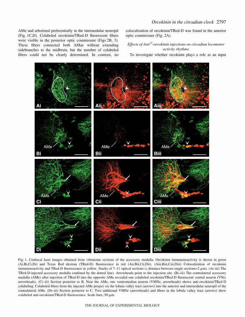
medulla (AMe) neurons qualify for direct, monosynapticcoupling of both AMae, we injected Texas Red-conjugateddextran (TRed-D) as neuronal tracer into one AMe. Brainswere immunostained with anti-orcokinin antiserum (N=21),and the AMe contralateral to the injection site andcommissures in the midbrain were examined for doublefluorescence (Figs·1, 2). These experiments showed that onebilateral pair of orcokinin-ir ventral neurons (VNe) and threepairs of ventro-median neurons (VMNe) provide directconnections between both AMae. To examine whetherorcokinin influences circadian locomotor activity of thecockroach, we injected Asn13-orcokinin into the vicinity of oneAMe of the cockroach. These experiments resulted in phase-dependent phase shifts in circadian locomotor activityresembling the phase-shifting effects of light.
Injection of TRed-D into the AMe
After injection of approximately 2 nl TRed-D into one AMe(Fig.·1A), we found up to four colabeled orcokinin/TRed-Dsomata in the contralateral AMe (Fig.·1B–D, Fig.·3). Thesesomata could be identified as one orcokinin/TRed-D colabeledVNe and three colabeled VMNe. Prominent orcokinin/TRed-D colabeled fibers projected via the lobula valley tract into the
THE JOURNAL OF EXPERIMENTAL BIOLOGY
2797Orcokinin in the circadian clock
AMe and arborized preferentially in the internodular neuropil(Fig.·1C,D). Colabeled orcokinin/TRed-D fluorescent fiberswere visible in the posterior optic commissure (Figs·2B, 3).These fibers connected both AMae without extendingsidebranches to the midbrain, but the number of colabeledfibers could not be clearly determined. In contrast, no
colocalization of orcokinin/TRed-D was found in the anterioroptic commissure (Fig.·2A).
Effects of Asn13-orcokinin injections on circadian locomotoractivity rhythms
To investigate whether orcokinin plays a role as an input
Fig.·1. Confocal laser images obtained from vibratome sections of the accessory medulla. Orcokinin immunoreactivity is shown in green(Ai,Bi,Ci,Di) and Texas Red dextran (TRed-D) fluorescence in red (Aii,Bii,Cii,Dii). (Aiii,Biii,Ciii,Diii) Colocalization of orcokininimmunoreactivity and TRed-D fluorescence in yellow. Stacks of 7–11 optical sections (z-distance between single sections=2·�m). (Ai–iii) TheTRed-D-injected accessory medulla (outlined by the dotted line). Arrowheads point to the injection site. (Bi–iii) The contralateral accessorymedulla (AMe) after injection of TRed-D into the opposite AMe revealed one colabeled orcokinin/TRed-D fluorescent ventral neuron (VNe;arrowheads). (Ci–iii) Section posterior to B. Near the AMe, one ventromedian neuron (VMNe, arrowheads) shows anti-orcokinin/TRed-Dcolabeling. Colabeled fibers from the injected AMe project via the lobula valley tract (arrows) into the anterior and internodular neuropil of thecontralateral AMe. (Di–iii) Section posterior to C. Two additional VMNe (arrowheads) and fibers in the lobula valley tract (arrows) showcolabeled anti-orcokinin/TRed-D fluorescence. Scale bars, 50·�m.
THE JOURNAL OF EXPERIMENTAL BIOLOGY
2798
signal to the circadian clock, we examined whether the peptideinfluences circadian locomotor activity of the cockroach. Asn13-orcokinin was injected into the vicinity of one AMe at differentcircadian times, and locomotor activities of the free-running
S. Hofer and U. Homberg
cockroaches were recorded before and after the injections(Fig.·4). Control injections with carrier solution alone (bluefood dye in saline) did not cause significant phase shifts incircadian locomotor rhythm, except for a small but significant
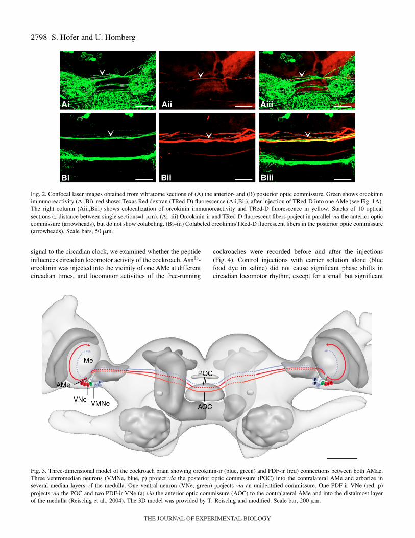
Fig.·2. Confocal laser images obtained from vibratome sections of (A) the anterior- and (B) posterior optic commissure. Green shows orcokininimmunoreactivity (Ai,Bi), red shows Texas Red dextran (TRed-D) fluorescence (Aii,Bii), after injection of TRed-D into one AMe (see Fig.·1A).The right column (Aiii,Biii) shows colocalization of orcokinin immunoreactivity and TRed-D fluorescence in yellow. Stacks of 10 opticalsections (z-distance between single sections=1·�m). (Ai–iii) Orcokinin-ir and TRed-D fluorescent fibers project in parallel via the anterior opticcommissure (arrowheads), but do not show colabeling. (Bi–iii) Colabeled orcokinin/TRed-D fluorescent fibers in the posterior optic commissure(arrowheads). Scale bars, 50·�m.
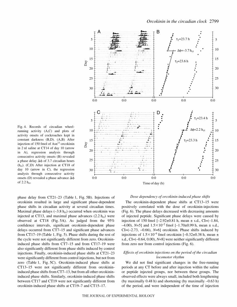
Fig.·3. Three-dimensional model of the cockroach brain showing orcokinin-ir (blue, green) and PDF-ir (red) connections between both AMae.Three ventromedian neurons (VMNe, blue, p) project via the posterior optic commissure (POC) into the contralateral AMe and arborize inseveral median layers of the medulla. One ventral neuron (VNe, green) projects via an unidentified commissure. One PDF-ir VNe (red, p)projects via the POC and two PDF-ir VNe (a) via the anterior optic commissure (AOC) to the contralateral AMe and into the distalmost layerof the medulla (Reischig et al., 2004). The 3D model was provided by T. Reischig and modified. Scale bar, 200·�m.
THE JOURNAL OF EXPERIMENTAL BIOLOGY
2799Orcokinin in the circadian clock
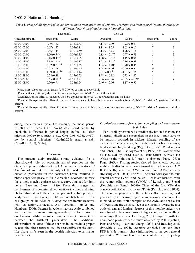
phase delay from CT21–23 (Table·1, Fig.·5B). Injections oforcokinin resulted in large and significant phase-dependentphase shifts in circadian activity at several circadian times.Maximal phase delays (–3.8·hct) occurred when orcokinin wasinjected at CT13, and maximal phase advances (2.2·hct) wereobserved at CT18 (Fig.·5A). As judged from the 95%confidence intervals, significant orcokinin-dependent phasedelays occurred from CT7–15 and significant phase advancesfrom CT17–19 (Table·1, Fig.·5). Phase shifts during the rest ofthe cycle were not significantly different from zero. Orcokinin-induced phase shifts from CT7–15 and from CT17–19 werealso significantly different from phase shifts induced by controlinjections. Finally, orcokinin-induced phase shifts at CT21–23were significantly different from control injections, but not fromzero (Table·1, Fig.·5C). Orcokinin-induced phase shifts atCT13–15 were not significantly different from orcokinin-induced phase shifts from CT7–13, but from all other orcokinin-induced phase shifts. Similarly, orcokinin-induced phase shiftsbetween CT17 and CT19 were not significantly different fromorcokinin-induced phase shifts at CT19–7 and CT15–17.
Dose dependency of orcokinin-induced phase shifts
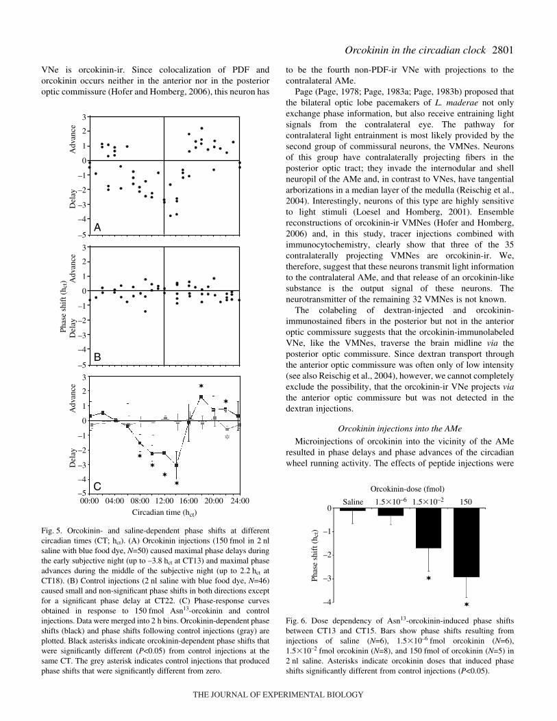
The orcokinin-dependent phase shifts at CT13–15 werepositively correlated with the dose of orcokinin-injections(Fig.·6). The phase delays decreased with decreasing amountsof injected peptide. Significant phase delays were caused byinjection of 150·fmol [–2.92±0.81·h, mean ± s.d., CI=(–1.84,–4.00), N=5] and 1.5�10–2·fmol [–1.70±0.99·h, mean ± s.d.,CI=(–2.73, –0.66), N=6] orcokinin. Phase shifts induced byinjections of 1.5�10–6·fmol orcokinin [–0.32±0.38·h, mean ±s.d., CI=(–0.64, 0.00), N=8] were neither significantly differentfrom zero nor from control injections (Fig.·6).
Effects of orcokinin injections on the period of the circadianlocomotor rhythm
We did not find significant changes in the free-runningperiods at any CT before and after injection within the salineor peptide injected groups, nor between these groups. Theobserved effects were always small, included both lengthening(by maximally 0.48·h) and shortening (by maximally –0.63·h)of the period, and were independent of the time of injection
τ1=23.7 h
Δφ=–3.7 hct
τ2=23.6 h
τ1=23.4 h
Δφ=2.2 hct
τ2=23.3 h
Day
BA
30
25
20
15
10
5
30
25
20
15
10
5
0:00:00:00:0 0:0
Time of day (h)
Day
30
25
20
15
10
5
30
25
20
15
10
5
0:00:00:00:0 0:0
C D
Fig.·4. Records of circadian wheel-running activity (A,C) and plots ofactivity onsets of cockroaches kept inconstant darkness (B,D). (A,B) Afterinjection of 150·fmol of Asn13-orcokininin 2·nl saline at CT14 of day 10 (arrowin A), regression analysis throughconsecutive activity onsets (B) revealeda phase delay � of 3.7 circadian hours(hct). (C,D) After injection at CT18 ofday 10 (arrow in C), the regressionanalysis through consecutive activityonsets (D) revealed a phase advance �of 2.2·hct.
THE JOURNAL OF EXPERIMENTAL BIOLOGY
2800 S. Hofer and U. Homberg
during the circadian cycle. On average, the mean period(23.58±0.23·h, mean ± s.d., N=96) was altered neither byorcokinin [difference in period lengths before and afterinjection 0.00±0.19·h, mean ± s.d., CI=(–0.05, 0.06), N=50]nor by control injections [–0.04±0.22·h, mean ± s.d.,CI=(–0.11, 0.02), N=46].
DiscussionThe present study provides strong evidence for a
physiological role of orcokinin-related peptides in thecircadian system of the cockroach L. maderae. Injections ofAsn13-orcokinin into the vicinity of the AMe, a mastercircadian pacemaker in the cockroach brain, resulted inphase-dependent phase shifts in circadian locomotor activitythat closely match the phase-response curve obtained for lightpulses (Page and Barrett, 1989). These data suggest aninvolvement of orcokinin-related peptides in circuits relayingphotic information to the circadian pacemaker. In a previousstudy, we showed that up to 30 neurons distributed in fivecell groups of the AMe of L. maderae are immunoreactivewith an antiserum against Asn13-orcokinin (Hofer andHomberg, 2006). Dextran injections into one AMe combinedwith orcokinin immunostaining revealed that four pairs oforcokinin-ir AMe neurons provide direct connectionsbetween the bilateral pacemakers. The additionalarborizations of some of these neurons in the medulla propersuggest that these neurons may be responsible for the light-like phase shifts seen in the peptide injection experiments(see below).
Orcokinin-ir neurons form a direct coupling pathway betweenboth AMae
For a well-synchronized circadian rhythm in behavior, thebilaterally distributed pacemakers in the insect brain have tobe mutually coupled. In crickets, bilateral coupling of theclocks is relatively weak, but in the cockroach L. maderae,bilateral coupling is strong (Page et al., 1977; Wiedenmannand Loher, 1984; Ushirogawa et al., 1997), and is assumed tobe mediated by direct neuronal connections between theAMae in the right and left brain hemisphere (Page, 1983a;Page, 1983b). Tracing studies showed that anterior neuronswith cell bodies in two clusters termed MC I (4 cells) and MCII (35 cells) near the AMe connect both AMae directly(Reischig et al., 2004). The MC I neurons correspond to fourventral neurons (VNe), and the MC II cells are identical withthe ventromedian neurons (VMNe) of Reischig and Stengl(Reischig and Stengl, 2003b). Three of the four VNe thatconnect both AMae directly are PDF-ir (Reischig et al., 2004).The neurons project via the anterior (two neurons) andposterior (one neuron) optic commissures, innervate theinternodular and shell neuropils of the AMe, and send a fanof fibers along the distal surface of the medulla toward the firstoptic chiasm and lamina. Neurons of this morphological typewere found to be unresponsive to light stimuli in intracellularrecordings (Loesel and Homberg, 2001). Together with thenon-photic phase-response curve obtained by PDF injection,Petri and Stengl (Petri and Stengl, 2002) and Reischig et al.(Reischig et al., 2004), therefore concluded that the threePDF-ir VNe transmit phase information to the contralateralpacemaker. We show here that one contralaterally projecting
Table·1. Phase shifts (in circadian hours) resulting from injections of 150·fmol orcokinin and from control (saline) injections atdifferent times of the circadian cycle (circadian time)
Phase shift 95% CI N
Circadian time (h) Orcokinin Saline Orcokinin Saline Orcokinin Saline
01:00–03:00 0.39±1.12d –0.12±0.33 3.17 to –2.39 –0.92 to 0.69 3 303:00–05:00 –0.07±1.27d –0.02±0.11 1.11 to –1.25 –1.03 to 0.10 7 305:00–07:00 –0.45±1.44d –0.30±0.59 3.13 to –4.03 –1.76 to 1.16 3 207:00–09:00 –1.50±0.54a,f –0.09±0.35 –0.83 to –2.17b –0.97 to 0.79 5 309:00–11:00 –2.16±0.49a,f –0.10±0.85 –1.38 to –2.94b –1,15 to 0.96 4 311:00–13:00 –2.13±1.11a,f 0.11±0.17 –1.08 to –3.18b –0.16 to 0.38 3 513:00–15:00 –2.92±0.87a,c,f –0.12±0.55 –1.84 to –4.00b –0.70 to 0.45 5 415:00–17:00 –0.21±1.19d 0.12±0.45 1.04 to –1.46 –0.50 to 0.84 6 617:00–19:00 1.39±0.50a,d,e –0.15±0.44 2.01 to 0.77b –1.01 to 1.06 5 419:00–21:00 0.58±0.88d 0.15±0.53 1.98 to –0.82 –0.72 to 1.27 4 421:00–23:00 0.65±0.88a,d –0.58±0.21 2.54 to –0.24 –0.85 to –0.19b 2 423:00–01:00 0.20±0.91d –0.28±0.24 2.46 to –2.06 –1.17 to 0.30 3 4
Phase shift values are means ± s.d.; 95% CI = lower limit to upper limit.aPhase shifts significantly different from control injections (P<0.05, two-tailed t-test).bSignificant phase shifts as judged by the 95% confidence interval (CI; see Materials and methods).cPhase shifts significantly different from orcokinin-dependent phase shifts at other circadian times (d) (P<0.05, ANOVA, post-hoc test after
Tukey).ePhase shifts significantly different from orcokinin-dependent phase shifts at other circadian times (f) (P<0.05, ANOVA, post-hoc test after
Tukey).
THE JOURNAL OF EXPERIMENTAL BIOLOGY
2801Orcokinin in the circadian clock
VNe is orcokinin-ir. Since colocalization of PDF andorcokinin occurs neither in the anterior nor in the posterioroptic commissure (Hofer and Homberg, 2006), this neuron has
to be the fourth non-PDF-ir VNe with projections to thecontralateral AMe.
Page (Page, 1978; Page, 1983a; Page, 1983b) proposed thatthe bilateral optic lobe pacemakers of L. maderae not onlyexchange phase information, but also receive entraining lightsignals from the contralateral eye. The pathway forcontralateral light entrainment is most likely provided by thesecond group of commissural neurons, the VMNes. Neuronsof this group have contralaterally projecting fibers in theposterior optic tract; they invade the internodular and shellneuropil of the AMe and, in contrast to VNes, have tangentialarborizations in a median layer of the medulla (Reischig et al.,2004). Interestingly, neurons of this type are highly sensitiveto light stimuli (Loesel and Homberg, 2001). Ensemblereconstructions of orcokinin-ir VMNes (Hofer and Homberg,2006) and, in this study, tracer injections combined withimmunocytochemistry, clearly show that three of the 35contralaterally projecting VMNes are orcokinin-ir. We,therefore, suggest that these neurons transmit light informationto the contralateral AMe, and that release of an orcokinin-likesubstance is the output signal of these neurons. Theneurotransmitter of the remaining 32 VMNes is not known.
The colabeling of dextran-injected and orcokinin-immunostained fibers in the posterior but not in the anterioroptic commissure suggests that the orcokinin-immunolabeledVNe, like the VMNes, traverse the brain midline via theposterior optic commissure. Since dextran transport throughthe anterior optic commissure was often only of low intensity(see also Reischig et al., 2004), however, we cannot completelyexclude the possibility, that the orcokinin-ir VNe projects viathe anterior optic commissure but was not detected in thedextran injections.
Orcokinin injections into the AMe
Microinjections of orcokinin into the vicinity of the AMeresulted in phase delays and phase advances of the circadianwheel running activity. The effects of peptide injections were
Fig.·5. Orcokinin- and saline-dependent phase shifts at differentcircadian times (CT; hct). (A) Orcokinin injections (150·fmol in 2·nlsaline with blue food dye, N=50) caused maximal phase delays duringthe early subjective night (up to –3.8·hct at CT13) and maximal phaseadvances during the middle of the subjective night (up to 2.2·hct atCT18). (B) Control injections (2·nl saline with blue food dye, N=46)caused small and non-significant phase shifts in both directions exceptfor a significant phase delay at CT22. (C) Phase-response curvesobtained in response to 150·fmol Asn13-orcokinin and controlinjections. Data were merged into 2·h bins. Orcokinin-dependent phaseshifts (black) and phase shifts following control injections (gray) areplotted. Black asterisks indicate orcokinin-dependent phase shifts thatwere significantly different (P<0.05) from control injections at thesame CT. The grey asterisk indicates control injections that producedphase shifts that were significantly different from zero.
A
B
0
1
–1
–2
–3
–4
2
3
00:00 04:00 08:00 12:00 16:00 20:00 24:00–5
0
1
–1
–2
–3
–4
2
3
–5
Circadian time (hct)
Phas
e sh
ift (
h ct)
C
0
1
–1
–2
–3
–4
2
3
–5
Adv
ance
Del
ayA
dvan
ceD
elay
Adv
ance
Del
ay
Orcokinin-dose (fmol)
Saline 1501.5�10–21.5�10–6
0
–1
–2
–3
–4
Phas
e sh
ift (
h ct)
Fig.·6. Dose dependency of Asn13-orcokinin-induced phase shiftsbetween CT13 and CT15. Bars show phase shifts resulting frominjections of saline (N=6), 1.5�10–6·fmol orcokinin (N=6),1.5�10–2·fmol orcokinin (N=8), and 150·fmol of orcokinin (N=5) in2·nl saline. Asterisks indicate orcokinin doses that induced phaseshifts significantly different from control injections (P<0.05).
THE JOURNAL OF EXPERIMENTAL BIOLOGY
2802
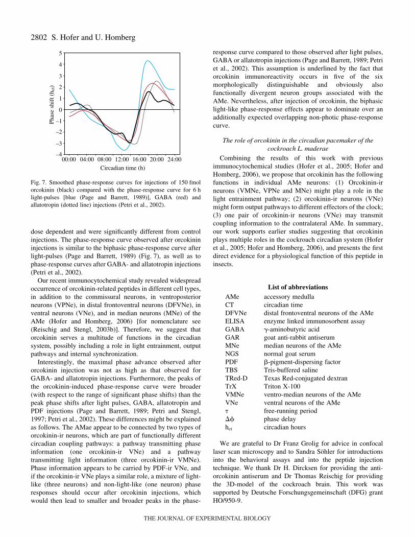
dose dependent and were significantly different from controlinjections. The phase-response curve observed after orcokinininjections is similar to the biphasic phase-response curve afterlight-pulses (Page and Barrett, 1989) (Fig.·7), as well as tophase-response curves after GABA- and allatotropin injections(Petri et al., 2002).
Our recent immunocytochemical study revealed widespreadoccurrence of orcokinin-related peptides in different cell types,in addition to the commissural neurons, in ventroposteriorneurons (VPNe), in distal frontoventral neurons (DFVNe), inventral neurons (VNe), and in median neurons (MNe) of theAMe (Hofer and Homberg, 2006) [for nomenclature see(Reischig and Stengl, 2003b)]. Therefore, we suggest thatorcokinin serves a multitude of functions in the circadiansystem, possibly including a role in light entrainment, outputpathways and internal synchronization.
Interestingly, the maximal phase advance observed afterorcokinin injection was not as high as that observed forGABA- and allatotropin injections. Furthermore, the peaks ofthe orcokinin-induced phase-response curve were broader(with respect to the range of significant phase shifts) than thepeak phase shifts after light pulses, GABA, allatotropin andPDF injections (Page and Barrett, 1989; Petri and Stengl,1997; Petri et al., 2002). These differences might be explainedas follows. The AMae appear to be connected by two types oforcokinin-ir neurons, which are part of functionally differentcircadian coupling pathways: a pathway transmitting phaseinformation (one orcokinin-ir VNe) and a pathwaytransmitting light information (three orcokinin-ir VMNe).Phase information appears to be carried by PDF-ir VNe, andif the orcokinin-ir VNe plays a similar role, a mixture of light-like (three neurons) and non-light-like (one neuron) phaseresponses should occur after orcokinin injections, whichwould then lead to smaller and broader peaks in the phase-
S. Hofer and U. Homberg
response curve compared to those observed after light pulses,GABA or allatotropin injections (Page and Barrett, 1989; Petriet al., 2002). This assumption is underlined by the fact thatorcokinin immunoreactivity occurs in five of the sixmorphologically distinguishable and obviously alsofunctionally divergent neuron groups associated with theAMe. Nevertheless, after injection of orcokinin, the biphasiclight-like phase-response effects appear to dominate over anadditionally expected overlapping non-photic phase-responsecurve.
The role of orcokinin in the circadian pacemaker of thecockroach L. maderae
Combining the results of this work with previousimmunocytochemical studies (Hofer et al., 2005; Hofer andHomberg, 2006), we propose that orcokinin has the followingfunctions in individual AMe neurons: (1) Orcokinin-irneurons (VMNe, VPNe and MNe) might play a role in thelight entrainment pathway; (2) orcokinin-ir neurons (VNe)might form output pathways to different effectors of the clock;(3) one pair of orcokinin-ir neurons (VNe) may transmitcoupling information to the contralateral AMe. In summary,our work supports earlier studies suggesting that orcokininplays multiple roles in the cockroach circadian system (Hoferet al., 2005; Hofer and Homberg, 2006), and presents the firstdirect evidence for a physiological function of this peptide ininsects.
List of abbreviationsAMe accessory medullaCT circadian timeDFVNe distal frontoventral neurons of the AMeELISA enzyme linked immunosorbent assayGABA �-aminobutyric acidGAR goat anti-rabbit antiserumMNe median neurons of the AMeNGS normal goat serumPDF �-pigment-dispersing factorTBS Tris-buffered salineTRed-D Texas Red-conjugated dextranTrX Triton X-100VMNe ventro-median neurons of the AMe VNe ventral neurons of the AMe� free-running period� phase delay hct circadian hours
We are grateful to Dr Franz Grolig for advice in confocallaser scan microscopy and to Sandra Söhler for introductionsinto the behavioral assays and into the peptide injectiontechnique. We thank Dr H. Dircksen for providing the anti-orcokinin antiserum and Dr Thomas Reischig for providingthe 3D-model of the cockroach brain. This work wassupported by Deutsche Forschungsgemeinschaft (DFG) grantHO/950-9.
00:00 04:00 08:00 12:00 16:00 20:00 24:00
0
1
–1
–2
–3
–4
2
3
4
5
Circadian time (h)
Phas
e sh
ift (
h ct)
Fig.·7. Smoothed phase-response curves for injections of 150·fmolorcokinin (black) compared with the phase-response curve for 6·hlight-pulses [blue (Page and Barrett, 1989)], GABA (red) andallatotropin (dotted line) injections (Petri et al., 2002).
THE JOURNAL OF EXPERIMENTAL BIOLOGY
2803Orcokinin in the circadian clock
ReferencesBungart, D., Dircksen, H. and Keller, R. (1994). Quantitative determination
and distribution of the myotropic neuropeptide orcokinin in the nervoussystem of astacidean crustaceans. Peptides 15, 393-400.
Bungart, D., Hilbich, C., Dircksen, H. and Keller, R. (1995). Occurrenceof analogues of the myotropic neuropeptide orcokinin in the shore crab,Carcinus maenas: evidence for a novel neuropeptide family. Peptides 16,67-72.
Enright, J. T. (1965). The search for rhythmicity in biological time series. J.Theor. Biol. 8, 426-468.
Fu, Q., Kutz, K. K., Schmidt, J. J., Hsu, Y.-W. A., Messinger, D. I., Cain,S. D., De La Iglesia, H. O., Christie, A. E. and Li, L. (2005). Hormonecomplement of the Cancer productus sinus gland and pericardial organ: ananatomical and mass spectrometric investigation. J. Comp. Neurol. 493,607-626.
Helfrich-Förster, C. (2005). Neurobiology of the fruit fly’s cicadian clock.Genes Brain Behav. 4, 65-76.
Hofer, S. and Homberg, U. (2006). Orcokinin immunoreactivity in theaccessory medulla of the cockroach Leucophaea maderae. Cell Tissue Res.DOI: 10.1007/s00441-00609155-y.
Hofer, S., Dircksen, H., Tollbäck, P. and Homberg, U. (2005). Novel insectorcokinins: characterization and neuronal distribution in the brains ofselected dicondylian insects. J. Comp. Neurol. 490, 57-71.
Homberg, U., Reischig, T. and Stengl, M. (2003). Neural organization of thecircadian system of the cockroach Leucophaea maderae. Chronobiol. Int.20, 577-591.
Huybrechts, J., Nusbaum, M. P., Bosch, L. V., Baggerman, G., De Loof,A. and Schoofs, L. (2003). Neuropeptidomic analysis of the brain andthoracic ganglion from the Jonah crab, Cancer borealis. Biochem. Biophys.Res. Commun. 308, 535-544.
Loesel, R. and Homberg, U. (2001). Anatomy and physiology of neuronswith processes in the accessory medulla of the cockroach Leucophaeamaderae. J. Comp. Neurol. 439, 193-207.
Nishiitsutsuji-Uwo, J. and Pittendrigh, C. S. (1968). Central nervous systemcontrol of circadian rhythmicity in the cockroach. III. The pathway of lightsignals that entrain the rhythm. Z. Vergl. Physiol. 58, 1-13.
Page, T. L. (1978). Interactions between bilaterally paired components of thecockroach circadian system. J. Comp. Physiol. A 124, 225-236.
Page, T. L. (1981). Effects of localized low-temperature pulses on thecockroach circadian pacemaker. Am. J. Physiol. 240, 144-150.
Page, T. L. (1983a). Regeneration of the optic tracts and circadian pacemakeractivity in the cockroach Leucophaea maderae. J. Comp. Physiol. A 152,231-240.
Page, T. L. (1983b). Effects of optic-tract regeneration on internal couplingin the circadian system of the cockroach. J. Comp. Physiol. A 153, 353-363.
Page, T. L. and Barrett, R. K. (1989). Effects of lights on circadianpacemaker development. II. Responses to light. J. Comp. Physiol. A 165,41-49.
Page, T. L., Caldarola, P. C. and Pittendrigh, C. S. (1977). Mutualentrainment of bilaterally distributed circadian pacemakers. Proc. Natl.Acad. Sci. USA 74, 1277-1281.
Pascual, N., Castresana, J., Valero, M.-L., Andreu, D. and Bellés, X.(2004). Orcokinins in insects and other invertebrates. Insect Biochem. Mol.Biol. 34, 1141-1146.
Petri, B. and Stengl, M. (1997). Pigment-dispersing hormone shifts the phaseof the circadian pacemaker of the cockroach Leucophaea maderae. J.Neurosci. 17, 4087-4093.
Petri, B., Stengl, M., Würden, S. and Homberg, U. (1995).Immunocytochemical characterization of the accessory medulla in thecockroach Leucophaea maderae. Cell Tissue Res. 282, 3-19.
Petri, B., Homberg, U., Loesel, R. and Stengl, M. (2002). Evidence for arole of GABA and Mas-allatotropin in photic entrainment of the circadianclock of the cockroach Leucophaea maderae. J. Exp. Biol. 205, 1459-1469.
Reischig, T. (2003). Identification and characterisation of the circadianpacemaker of the cockroach Leucophaea maderae. PhD thesis, Universityof Marburg, Germany.
Reischig, T. and Stengl, M. (1996). Morphology and pigment-dispersinghormone immunocytochemistry of the accessory medulla, the presumptivecircadian pacemaker of the cockroach Leucophaea maderae: a light- andelectron-microscopic study. Cell Tissue Res. 285, 305-319.
Reischig, T. and Stengl, M. (2002). Optic lobe commissures in a three-dimensional brain model of the cockroach Leucophaea maderae: a searchfor the circadian coupling pathways. J. Comp. Neurol. 443, 388-400.
Reischig, T. and Stengl, M. (2003a). Ectopic transplantation of the accessorymedulla restores circadian locomotor rhythms in arrhythmic cockroaches(Leucophaea maderae). J. Exp. Biol. 206, 1877-1886.
Reischig, T. and Stengl, M. (2003b). Ultrastructure of pigment-dispersinghormone-immunoreactive neurons in a three-dimensional model of theaccessory medulla of the cockroach (Leucophaea maderae). Cell Tissue Res.314, 421-435.
Reischig, T., Petri, B. and Stengl, M. (2004). Pigment-dispersing hormone(PDH)-immunoreactive neurons form a direct coupling pathway betweenthe bilaterally symmetric circadian pacemakers of the cockroachLeucophaea maderae. Cell Tissue Res. 318, 553-564.
Roberts, S. K. (1965). Photoreception and entrainment of cockroach activityrhythms. Science 148, 958-959.
Roenneberg, T. and Morse, D. (1993). Two circadian oscillators in one cell.Nature 362, 362-364.
Schneider, N.-L. and Stengl, M. (2005). Pigment-dispersing factor andGABA synchronize cells of the isolated circadian clock of the cockroachLeucophaea maderae. J. Neurosci. 25, 5138-5147.
Skiebe, P., Dreger, M., Meseke, M., Evers, J. F. and Hucho, F.(2002). Identification of orcokinins in single neurons in the stomatogastricnervous system of the crayfish, Cherax destructor. J. Comp. Neurol. 444,245-259.
Sokolove, P. G. and Bushell, W. N. (1978). The chi square periodogram: itsutility for analysis of circadian rhythms. J. Theor. Biol. 72, 131-160.
Stangier, J., Hilbich, C., Burdzik, S. and Keller, R. (1992). Orcokinin: anovel myotropic peptide from the nervous system of the crayfish,Orconectes limosus. Peptides 13, 859-864.
Ushirogawa, H., Abe, Y. and Tomioka, K. (1997). Circadian locomotorrhythms in the cricket, Gryllodes sigillatus. II. Interactions betweenbilaterally paired circadian pacemakers. Zool. Sci. 14, 729-736.
Wiedenmann, G. (1977). Two activity peaks in the circadian rhythm ofthe cockroach Leucophaea maderae. J. Interdiscipl. Cycle Res. 8, 378-383.
Wiedenmann, G. and Loher, W. (1984). Circadian control of singing incrickets: two different pacemakers for early-evening and before-dawnactivity. J. Insect Physiol. 30, 145-151.
Yasuda-Kamatani, Y. and Yasuda, A. (2000). Identification of orcokiningene-related peptides in the brain of the crayfish Procambarus clarkii bythe combination of MALDI-TOF and on-line capillary HPLC/Q-Tof massspectrometries and molecular cloning. Gen. Comp. Endocrinol. 118, 161-172.
THE JOURNAL OF EXPERIMENTAL BIOLOGY