EVIDENCE BASED MANAGEMENT FOR · Liver Function Tests (LFT) Renal Function Tests (RFT) Erythrocyte...
21
Dr Laskar IAEA Ped Rad Onco June 09 Version 1 EVIDENCE BASED MANAGEMENT FOR Retinoblastoma CLINICAL EVALUATION & STAGING Symptoms & Signs : White eye reflex, squint, diminished vision, red eye, proptosis. History - Family history of retinoblastoma Other malignancy Complete physical examination Unilateral or bilateral. Ophthalmologic examination (Evaluation Under Anesthesia: EUA) Both eyes to be evaluated thoroughly Indirect ophthalmoscopy : Tumor size- DD (disc diameter) Mapping with diagrams & description Radiological investigation : Ocular Ultrasonography with color dopler- A scan B scan CT/ MRI scan Orbits and Brain : Assess disease extent – intraocular, extraocular, intracranial Rule out trilateral retinoblastoma Bone scan in advanced cases Hematological evaluation : Hb, TC, Platelets Liver Function Tests (LFT) Renal Function Tests (RFT) Erythrocyte Sedimentation Rate (ESR) Bone Marrow Biopsy, in advanced disease CSF studies, in advanced disease. Special Investigations : Genetic & Molecular studies (when asked for). Reese-Ellsworth Classification of Retinoblastoma (Criteria for Suitability for Local Treatment) Group I : Very favorable prognosis A. Solitary tumor, less than 4 disc diameters (dd)= in size, at or behind the equator B. Multiple tumors, none over 4 dd in size, all at or behind the equator Group II : Favorable prognosis A. Solitary lesion 4-10 dd in size, at or behind the equator B. Multiple tumors, 4-10 dd in size, behind the equator Group III- Doubtful prognosis A. Any lesion anterior to the equator B. Solitary tumors larger than 10 dd behind the equator Group IV : Unfavorable prognosis A. Multiple tumors, some larger than 10dd B. Any lesion extending anteriorly to the ora serrata Group V : Very unfavorable prognosis
Transcript of EVIDENCE BASED MANAGEMENT FOR · Liver Function Tests (LFT) Renal Function Tests (RFT) Erythrocyte...

Dr Laskar IAEA Ped Rad Onco June 09 Version 1
EVIDENCE BASED MANAGEMENT FOR Retinoblastoma
CLINICAL EVALUATION & STAGING Symptoms & Signs : White eye reflex, squint, diminished vision, red eye, proptosis. History - Family history of retinoblastoma Other malignancy Complete physical examination Unilateral or bilateral. Ophthalmologic examination (Evaluation Under Anesthesia: EUA) Both eyes to be evaluated thoroughly Indirect ophthalmoscopy : Tumor size- DD (disc diameter) Mapping with diagrams & description Radiological investigation : Ocular Ultrasonography with color dopler- A scan B scan CT/ MRI scan Orbits and Brain : Assess disease extent – intraocular, extraocular, intracranial Rule out trilateral retinoblastoma Bone scan in advanced cases Hematological evaluation : Hb, TC, Platelets Liver Function Tests (LFT) Renal Function Tests (RFT) Erythrocyte Sedimentation Rate (ESR) Bone Marrow Biopsy, in advanced disease CSF studies, in advanced disease. Special Investigations : Genetic & Molecular studies (when asked for).
Reese-Ellsworth Classification of Retinoblastoma (Criteria for Suitability for Local Treatment) Group I : Very favorable prognosis A. Solitary tumor, less than 4 disc diameters (dd)= in size, at or behind the equator B. Multiple tumors, none over 4 dd in size, all at or behind the equator Group II : Favorable prognosis A. Solitary lesion 4-10 dd in size, at or behind the equator B. Multiple tumors, 4-10 dd in size, behind the equator Group III- Doubtful prognosis A. Any lesion anterior to the equator B. Solitary tumors larger than 10 dd behind the equator Group IV : Unfavorable prognosis A. Multiple tumors, some larger than 10dd B. Any lesion extending anteriorly to the ora serrata Group V : Very unfavorable prognosis

Dr Laskar IAEA Ped Rad Onco June 09 Version 1
A. Massive tumors involving over half the retina B. Vitreous seeding One disc diameter = 1.6 mm. Staging in patients with Retinoblastoma (St Judes) I. Tumor (unifocal or multifocal) confined to retina A. Occupying 1 quadrant or less. B. Occupying 2 quadrants or less. C. Occupying more than 50% of retinal surface. II. Tumor (unifocal or multifocal) confined to globe A. with vitreous seeding B. Extending to optic nerve head. C. extending to choroid and optic nerve head D. Extending to emissaries. III. Extraocular extension of tumor (regional) A. extending beyond cut end of optic nerve (including sub-arachnoid extension) B. Extending through sclera into orbital contents. C. extending to choroid and beyond cut end of optic nerve (including sub-arachnoid extension) D. extending through sclera into orbital contents and beyond cut end of optic nerve (including sub-archnoid extension) IV. Distant metastases A. extending through optic nerve to brain B. blood-borne metastases to soft-tissues and bone marrow metastases
GENERAL PRINCIPLES OF MANAGEMENT Although retinoblastoma is the most common primary intraocular tumor in children, the treatment of this disease is a complex topic. Therapeutic plans usually require a multidisciplinary approach by a team consisting of ocular oncologist, pediatric oncologist and radiation oncologist. Treatment should be highly individualized. Treatment strategies for retinoblastoma have gradually evolved over the past few decades. The driving force behind these new approaches is to avoid enucleation and/or external beam radiation therapy and trend towards focal “conservative” treatment. Every effort has been made to save the child’s life with preservation of eye and sight, if possible. The aims of treatment are firstly, to preserve the life of the child; secondly, to preserve vision, and thirdly, to minimize any complications or side effects of treatment. Recent research in the treatment of retinoblastoma has concentrated on methods of combining chemotherapy with other local treatment modalities. This approach combines the principle of chemotherapeutic debulking in pediatric oncology with conservative focal therapies in ophthalmology. Termed “chemoreduction”, intravenous chemotherapy is used to debulk the initial tumor volume and allow for focal treatment with transpupillary thermal therapy, laser therapy, cryotherapy and plaque radiotherapy. Chemotherapy may be useful in all three clinical settings in intra-ocular retinoblastoma, in cases of micrometastatic spread, and when there are overt extraocular metastases. Tumor shrinkage with chemoreduction may allow treatment

Dr Laskar IAEA Ped Rad Onco June 09 Version 1
with less invasive measures such as cryotherapy, laser photocoagulation, thermotherapy or plaque radiotherapy, thereby, avoiding enucleation and external beam radiotherapy. While chemotherapeutic agents vary according to the preference of the pediatric oncologist, most of the current studies have relied on vincristine, etoposide and carboplatin. To circumvent the multidrug resistance, cyclosporine has been added to chemotherapy at some centers. The main issues for consideration when selecting treatment options for a child with retinoblastoma are as follows : (1) Is the disease unilateral or bilateral? (2) Does the affected eye have potential for useful vision? (3) Is the, tumor confined to the globe or does it extend to the optic nerve? (4) Are there orbital / lymph-nodal / bony / central nervous system or, hematogenous metastasis present? The usual management approach used in our institution is as shown in Figs. 1,2,3.

Dr Laskar IAEA Ped Rad Onco June 09 Version 1
Fig. 2 : Guidelines for management of advanced unilateral RB

Dr Laskar IAEA Ped Rad Onco June 09 Version 1
Fig.3 : Guidelines for management of Bilateral RB

Dr Laskar IAEA Ped Rad Onco June 09 Version 1
Indications for surgical procedures in retinoblastoma
Procedure Indications Complications Enucleation No potential for
useful vision Neo-vascular glaucoma Invasion-optic disc, choroid, orbit, ant. chamber Failure to control tumor by conservative Rx Inability to examine retina after conservative Rx (2° to vitreous
Bone growth deformities

Dr Laskar IAEA Ped Rad Onco June 09 Version 1
hemorrhage or cataract) [At least 15 mm of optic nerve to be removed; Artificial eye 6 wks post surgery]
Cryotherapy Small primary or recurrent tumors in anterior part of retina (<3 mm in diam; 2 mm thick) Small recurrences after radiotherapy
Vitreous hemorrhage Choroidal effusion Retinal Detachment
Photocoagulation (Argon/diode laser)
Small primary or recurrent tumors in post part of retina Retinal neo-vascularization due to radiation retinopathy (Contraindications : Tumors located at or near macula or papillary area; tumors with mushroom shape; tumors arising from vitreous base)
Vitreous hemorrhage Vitreous seeding
Chemothermotherapy Cataract Surgery
Post radiotherapy cataracts
CHEMOTHERAPY REGIMEN in use at Tata Memorial Hospital DRUG DOSE / M2 AND ROUTE Day Carboplatin Etoposide Vincristine
560 mg in 250 cc 5% DS in 2 hr. 200 mg in 100 cc NS in 2
1 3 21

Dr Laskar IAEA Ped Rad Onco June 09 Version 1
Endoxan hr. 1.5 mg IV push 150mg Orally
21-27
RADIOTHERAPY FOR RETINOBLASTOMA Principles for Radiotherapy : Indications : Intent to preserve useful vision in patients with multifocal lesions. Lesions close to macula/ optic nerve. Large tumors with vitreous seeding. Recurrent disease and as an adjuvant after enucleation/ evisceration. Palliative radiotherapy
Target volume : Primary aim is to deliver a homogeneous tumoricidal dose to the entire retina and vitreous while sparing the surrounding normal tissues. Reasons for this target volume are : 1) RB represents a “field change” and thus all cells have a neoplastic potential. 2) Vitreous seeding can occur. 3) Multiple lesions could be present. 4) Subretinal spread of tumor can occur. 5) Lens sparing techniques should be done only in selected patients with group I & II disease. a) Group I & II lesions (lesions close to macula/ optic nerve). Entire Retina upto the Ora-Serrata (CTV) + 5mm (PTV). Proximal 1cm of optic nerve (CTV) + 5mm (PTV) – when uninvolved. Can try and avoid vitreous irradiation if possible (e.g. IMRT) b) Group III, IV, & V lesions Entire Retina upto the Ora-Serrata (CTV) + 5mm (PTV). Proximal 1cm of optic nerve (CTV) + 5mm (PTV) – when uninvolved. c) Post operative (in locally advanced lesions) Entire Orbit (GTV) + 5mm (PTV). Any disease extension beyond the orbit to be covered adequately. Total dose : Treatment should ideally be done daily (5 days a week) – even with anesthesia Dose per fraction should be £ 2Gy / fraction In children <1year of age, radiation dose should be reduced: Microscopic disease (post op radiotherapy) 39.6Gy/22#/ 4.5wks Gross disease (definitive radiotherapy) 45Gy/25#/ 5wks a) Group I & II lesions 45Gy / 25# / 5wks (@1.8Gy / fr.) – Daily treatment 45Gy / 18# / 6wks (@2.5Gy / fr.) – Alternate day treatment b) Group III, IV, & V lesions 50.4Gy / 28# / 6wks (@1.8Gy / fr.) – Daily treatment 50.4Gy / 20# / 7wks (@2.5Gy / fr.) – Alternate day treatment c) Post operative Microscopic residual disease 45Gy / 25# / 5wks (@1.8Gy / fr.) – Daily treatment 45Gy / 18# / 6wks (@2.5Gy / fr.) – Alternate day treatment

Dr Laskar IAEA Ped Rad Onco June 09 Version 1
Gross residual disease 50.4Gy / 28# / 6wks (@1.8Gy / fr.) – Daily treatment 50.4Gy / 20# / 7wks (@2.5Gy / fr.) – Alternate day treatment Radioactive Plaque Therapy : Can be used for patients with solitary, unilateral lesions, 2-16mm basal diameter, located more than 3mm away from the optic disc. Ideally the tumors should be less than 10mm in thickness. Plaques can also be used for patients with minimal vitreous seeding close to the apex of the tumor. Used especially in children who have failed other methods. Types of Plaques available : Cobalt 60 - Energy:1.17-1.33MV, Half life - 5.2yrs. Iodine 125 - Energy:27-35KeV, Half life – 60days. Iridium 192 - Energy:295-612KeV, Half life – 74.5days. Ruthenium 106 - Energy: Beta, Half life – 368days. Dose : 35-40Gy to apex and 100-150Gy to Sclera (base) of tumor FOLLOW UP POLICY (A) During treatment : Children to be kept under close supervision. Response to treatment has to be evaluated every 6 to 8 weeks with EUA and USG studies and treatment individualized as per the response. (B) Off Treatment : Patients with retinoblastoma should be monitored indefinitely, initially for control of their primary tumor and later for development of a new tumor. Most recurrences occur within first 3 years after diagnosis. EUA should be done every 2 to 3 months for the first year, every 4 months during the second year and every 6 months to age 5 years. After age 5, new retinoblastomas are rare and patients are examined yearly without anesthesia. In addition to routine visual acuity and ophthalmologic testing patients with retinoblastoma require careful follow-up throughout their lifetime because of their increased risk of second malignant tumors. (C) Infants at risk for developing retinoblastoma (siblings, offspring) : They should be examined in the office every month till 3 months of age and then every 3 to 6 months until 3 years old. Families of patients with heritable retinoblastoma should undergo genetic counseling. (D) Long term issues that can arise : Impairment of vision, Cataracts, Dry Eye, Facial asymmetry, Retinal Detachment, Trilateral Retinoblastoma, Metastatic disease, orbital recurrence, second Tumors.
Retnoblastoma - Management
A) MANAGEMENT OF RETINOBLASTOMA Development of new retinoblastomas after 6 cycles of chemoreduction for retinoblastoma in 162 eyes of 106 consecutive patients. Shields CL, Shelil A, Cater J, Meadows AT, Shields JA. Arch Ophthalmol. 2003 Nov;121(11):1571-6.

Dr Laskar IAEA Ped Rad Onco June 09 Version 1
OBJECTIVE : To evaluate the occurrence of new retinoblastomas in patients treated with 6 cycles of chemoreduction. DESIGN: Prospective nonrandomized single-center case series. SETTING : Ocular Oncology Service at Wills Eye Hospital of Thomas Jefferson University, Philadelphia, Pa, in conjunction with the Division of Oncology at The Children’s Hospital of Philadelphia. PARTICIPANTS : A total of 162 eyes of 106 patients with retinoblastoma treated with 6 cycles of chemoreduction between January 1, 1995, and May 31, 2002. INTERVENTION : All patients received intravenous chemoreduction with vincristine sulfate, etoposide, and carboplatin, combined with focal treatment (cryotherapy or thermotherapy) to each retinal tumor. MAIN OUTCOME MEASURE : Development of new intraretinal retinoblastoma during or after treatment with chemoreduction. Recurrent subretinal tumor seeds or vitreous seeds were excluded from this analysis, and only primary new intraretinal tumors were included. RESULTS : Of 28 patients with unilateral retinoblastoma, new intraretinal tumor development was found during or after chemoreduction in 2 (9%) of the 23 patients with sporadic disease and 4 (80%) of the 5 patients with familial disease. The new tumor was located in the macula in none, between the macula and equator in 7 (54%), and between the equator and ora serrata in 6 (46%). Of the 78 patients with bilateral retinoblastoma, new tumor development was found during or after chemoreduction in 11 (19%) of the 57 patients with sporadic disease and 8 (38%) of the 21 patients with familial disease. The new tumor was macula in 2 (4%), between the macula and equator in 30 (55%), and between the equator and ora serrata in 23 (42%). Overall, according to Kaplan-Meier analysis, new tumor development occurred in 23% of patients by 1-year follow-up and 24% by 5-year follow-up. By multivariate analysis, the most important risk factors for the development of new tumors was younger age at presentation (median age, 2 months with new tumor vs 9 months without new tumor) and family history of retinoblastoma (12 [48%] of patients with new tumor vs 14 [17%] without new tumor). CONCLUSIONS : Children with retinoblastoma treated with chemoreduction should be followed for new intraretinal tumor development, as it peaks at a mean interval of 5 months after initiation of chemoreduction and affects 24% of patients by 5 years of follow-up. New tumors are most commonly found in those who develop disease as young infants and those with a family history of retinoblastoma. Globe conserving treatment of the only eye in bilateral retinoblastoma. Lee V, Hungerford JL, Bunce C et al. Br J Ophthalmol. 2003 Nov;87(11):1374-80. AIMS : To quantify the rates of eye preservation and patient survival, local tumour relapse and recurrence, and development of new tumours in the remaining eye of children with bilateral retinoblastoma with one eye already enucleated. Also, in the same children, to describe the types of primary and secondary treatment procedures, and to define the anatomical outcome. METHODS : This is a retrospective observational case series report. The study participants consisted of 107 patients with bilateral retinoblastoma with one eye enucleated within 1 month of baseline examination and had their remaining eye treated conservatively. The main outcome measure were: primary treatment failures, new tumours, enucleation of the only eye, death, remission, and anatomical outcomes (retinal detachment, vitreous haemorrhage, and cataract). RESULTS : The median age at diagnosis was 8.4 (range 0.2-44, SD 10.1) months with a median ophthalmic follow up of 44.3 (8.1-114, SD 10.1) months. In 22 of the 107 patients (21%) the treated eye was in Reese Ellsworth groups I or II and in the remaining 85 (79%) in groups III-V at diagnosis. The primary treatment was cryotherapy in 14% (15/107) of eyes, radioactive plaque

Dr Laskar IAEA Ped Rad Onco June 09 Version 1
brachytherapy in 3.7% (4/107), and chemotherapy in 10% (11/107). It was lens sparing radiotherapy in 37% (40/107), whole eye radiotherapy in 29% (31/107), combined radiotherapy and chemotherapy in 2.8% (3/107), chemothermotherapy in 0.9% (1/107), and combined focal therapy in 1.8% (2/107). The primary treatment failed to achieve local tumour control during the follow up period in 37% (40/107) of eyes. In 17 eyes failure was due to inadequate control of the presenting tumour, in 16 to development of a new tumour, and in eight eyes to a combination of both. 35 (88%) of the 40 failures were managed by secondary conservative treatment and the remaining five were treated by enucleation of the only eye. There were eight (7.4%) deaths and the 3 year survival rate was 93% (100/108). Anatomical results included vitreous haemorrhage in four cases, tractional retinal detachment also in four cases, and 24 children required cataract surgery. CONCLUSIONS : Aggressive conservative treatment achieved a good rate of globe salvage without impairing survival. Treatment of intraocular retinoblastoma with vincristine and carboplatin. Rodriguez-Galindo C, Wilson MW, Haik BG, Merchant TE, Billups CA, Shah N, Cain A, Langston J, Lipson M, Kun LE, Pratt CB. J Clin Oncol. 2003 May 15;21(10):2019-25. PURPOSE : To evaluate the efficacy of chemoreduction using vincristine and carboplatin in preventing or delaying external-beam radiotherapy (EBRT) or enucleation in patients with intraocular retinoblastoma. PATIENTS AND METHODS : Twenty-five patients (43 eyes) with newly diagnosed intraocular retinoblastoma received primary treatment with eight courses of vincristine and carboplatin. Focal treatments were delayed until documentation of disease progression. Outcome measures for each eye were length of time to disease progression, avoidance or delay of EBRT, and globe survival. Event-free survival was defined as the length of time to EBRT or enucleation. RESULTS : Disease in all eyes responded to chemotherapy and progressed in only two patients before completion of the eight courses of therapy. Disease in all but four eyes progressed and required focal treatments. Event-free survival estimates at 2 years were 59.2% +/- 12.0% for Reese-Ellsworth group I, II, and III eyes and 26.3% +/- 9.2% for group IV and V eyes. Nineteen eyes (44.2%) required EBRT and 13 eyes (30.2%) were enucleated. The ocular salvage rate was 83.3% for Reese-Ellsworth group I to III eyes and 52.6% for group IV and V eyes. For those patients receiving EBRT, the median time from enrollment to EBRT was 9.5 months (median age at EBRT, 21 months). CONCLUSION: In combination with appropriate early intensive focal treatments, chemoreduction with vincristine and carboplatin, without etoposide, may be an alternative treatment for patients with early-stage intraocular retinoblastoma, although additional studies are needed. Patients with advanced intraocular disease require more aggressive treatments. Thermochemotherapy in hereditary retinoblastoma. Schueler AO, Jurklies C, Heimann H et al. Br J Ophthalmol. 2003 Jan;87(1):90-5. BACKGROUND : The combination of chemotherapy and transpupillary thermotherapy, thermochemotherapy (TCT) has become an established part of the treatment plan in advanced retinoblastoma. The aim of this study was to identify safe indications, the complications as well as the limitations of this new treatment for retinoblastoma. METHODS: Tumour response and side effects of TCT with an indirect

Dr Laskar IAEA Ped Rad Onco June 09 Version 1
laser ophthalmoscope (spot size about 400 micro m) in 55 tumours of 26 children with bilateral retinoblastoma were analysed. Using the Reese-Ellsworth classification system, nine of 35 eyes were classified as type I, 13 eyes as type II, 10 eyes as type III, and three eyes as type V. The mean age of the children was 0.74 (SD 0.61) years. The mean tumour height was 3.5 (2.3) mm with a mean diameter of 6.1 (4.1) mm. Treatment parameters were 4.3 (1.6) (median 5) thermochemotherapy sessions with a mean energy of 539 (211) mW and a mean duration of 13.5 (5.6) minutes. Chemotherapy courses (vincristine, etoposide, and carboplatin) were repeated every 3 weeks. The mean follow up period was 1.25 (0.6) years. RESULTS: Local recurrence occurred in 21 tumours (38%), with a mean onset of 3.2 (2.9) months after TCT. The risk of tumour recurrence was correlated with tumour height. The recurrence rate was 17% for tumours with a height less than 2 mm, 37% for tumours with a height between 2 and 4 mm, and 63% for larger retinoblastomas. Multivariate analysis identified fish flesh regression after TCT (p=0.0007) as the most important risk factor for tumour recurrence besides tumour height (p=0.001) and the necessity of increased laser power during TCT sessions (p=0.018). Complications during therapy included transient corneal opacification in two eyes (6%), focal iris atrophy (three eyes, 8.5%), peripheral lens opacity (two eyes, 6%), circumscribed transient retinal detachment (one eye, 3%) and diffuse choroidal atrophy (one eye, 3%). CONCLUSION : TCT using an indirect laser ophthalmoscope with a spot size of about 400 micro m was efficient for retinoblastoma with a tumour height less than 4 mm. In larger tumours, the recurrence rate was unacceptably high. Fish flesh regression after TCT correlates with a higher rate of local tumour recurrence. Treatment related complications occurred in less than 9% of the treated eyes. Chemoreduction for unilateral retinoblastoma. Shields CL, Honavar SG, Meadows AT et al. Arch Ophthalmol. 2002 Dec;120(12):1653-8. OBJECTIVE : To evaluate conservative management of unilateral retinoblastoma using chemoreduction and focal treatment. DESIGN : Prospective nonrandomized single-center clinical trial. SETTING : Ocular Oncology Service at Wills Eye Hospital of Thomas Jefferson University, Philadelphia, Pa, in conjunction with the Division of Oncology at The Children’s Hospital of Philadelphia. PARTICIPANTS : Thirty eyes of 30 patients with unilateral retinoblastoma treated with chemoreduction between June 1, 1994, and August 31, 1999, that would otherwise have been managed with enucleation or external beam radiotherapy. INTERVENTION : All patients received treatment for retinoblastoma with a planned 6 cycles of chemoreduction using vincristine sulfate, etoposide, and carboplatin, combined with focal treatment (cryotherapy or thermotherapy) to each retinal tumor. MAIN OUTCOME MEASURES : The main outcome measure was the postchemoreduction need for external beam radiotherapy or enucleation. The cumulative probability of each outcome was estimated using Kaplan-Meier survival analysis. A secondary outcome measure was final visual acuity in the affected eye. The clinical features at the time of patient presentation were analyzed for their impact on the main outcomes using a series of Fisher exact tests and Cox proportional hazards regressions. RESULTS : Eighteen eyes (60%) were classified as having Reese-Ellsworth (RE) groups I through IV retinoblastoma and 12 eyes (40%), group V retinoblastoma. By using Kaplan-Meier estimates, we found a need for either external beam radiotherapy or enucleation in 68% of eyes by 5 years. In fact, 38% of those in groups I through IV required either treatment, whereas all of those in group V required the additional use of either

Dr Laskar IAEA Ped Rad Onco June 09 Version 1
treatment. Specifically, the need for external beam radiotherapy occurred in 27% of eyes by 5 years. Eleven percent of those in groups I through IV and 50% of group V required external beam radiotherapy by 5 years. The factors predictive of the need for external beam radiotherapy included RE group V disease, tumor thickness greater than 5 mm, and presence of vitreous seeds. The need for enucleation occurred in 47% of eyes by 5 years using Kaplan-Meier analysis. Specifically, 29% of those in groups I through IV and 67% of group V required enucleation by 5 years. The factors predictive of the need for enucleation included age at diagnosis older than 12 months, RE group V disease, tumor base diameter greater than 15 mm, and tumor thickness greater than 5 mm. At a mean follow-up of 29 months, the final visual acuity was 20/200 or better in 6 eyes (20%) and worse than 20/200 in 14 (47%); enucleation was needed in 10 (33%). Of the 26 eyes with initial macular involvement of retinoblastoma, final visual acuity was 20/200 or better in 6 (23%). No patient developed retinoblastoma metastasis, pinealoblastoma, or second malignant neoplasms. CONCLUSIONS : Chemoreduction is an option for selected eyes with unilateral retinoblastoma. Those with advanced RE group V retinoblastoma showed poorest results, while those with less advanced groups I through IV disease showed best results, maintaining the globe in 71% of eyes, sometimes with satisfactory functional visual acuity. Current treatment of retinoblastoma. De Potter P. Curr Opin Ophthalmol. 2002 Oct;13(5):331-6. Chemotherapy has recently achieved a major role in the primary management of intraocular retinoblastoma. Tumor reduction by first-line chemotherapy (chemoreduction) followed by local treatments is now accepted as treatment strategy for intraocular retinoblastoma with the goal of avoiding external beam radiotherapy (EBRT) or enucleation. Although efficient in reducing tumor volume, chemotherapy cannot cure retinoblastoma. Different chemoreduction protocols are used to shrink the tumor, making it treatable with cryotherapy, laser photocoagulation, thermotherapy, and plaque radiotherapy. Systemic chemotherapy used with local ophthalmic therapies during or after the chemotherapy can eliminate the need for enucleation or external beam radiotherapy in Reese-Ellsworth group 1, 2, or 3 retinoblastoma. This combination is not sufficient to obtain tumor control in most eyes with large tumors and diffuse vitreous and subretinal seeds (Reese-Ellsworth group 4 and 5 tumors), and EBRT or enucleation is eventually required. The resultant visual acuity after globe-conserving therapies in those eyes with Reese-Ellsworth group 4 and 5 tumors is often poor. Preliminary results of a phase I/II study of subconjunctival carboplatin injection are encouraging. Enucleation is still recommended in situations such as eyes containing large tumors, long standing retinal detachment, neovascular glaucoma, pars plana tumor seeding, anterior chamber involvement or choroid, optic nerve or orbital tumor extension, and no expectation for useful vision. Chemoprophylaxis is necessary for patients with tumor extending to the surgical margin of the optic nerve and is likely beneficial in preventing metastases in patients with tumor extending beyond the lamina cribrosa. Intensified chemotherapy with autologous stem cell rescue appears effective for patients with metastatic retinoblastoma. Chemothermotherapy in the management of retinoblastoma. Lumbroso L, Doz F, Urbieta M et al. Ophthalmology. 2002 Jun;109(6):1130-6.
Dr Laskar IAEA Ped Rad Onco June 09 Version 1
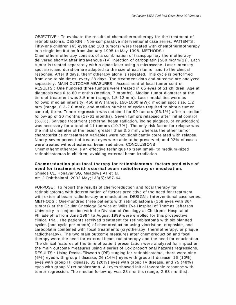
OBJECTIVE : To evaluate the results of chemothermotherapy for the treatment of retinoblastoma. DESIGN : Non-comparative interventional case series. PATIENTS : Fifty-one children (65 eyes and 103 tumors) were treated with chemothermotherapy in a single institution from January 1995 to May 1998. METHODS : Chemothermotherapy consists of a combination of transpupillary thermotherapy delivered shortly after intravenous (IV) injection of carboplatin [560 mg/m(2)]. Each tumor is treated separately with a diode laser using a microscope. Laser intensity, spot size, and duration are adapted to the size of each tumor and to the clinical response. After 8 days, thermotherapy alone is repeated. This cycle is performed from one to six times, every 28 days. The treatment data and outcome are analyzed separately. MAIN OUTCOME MEASURES : Assessment of local tumor control. RESULTS : One hundred three tumors were treated in 65 eyes of 51 children. Age at diagnosis was 0 to 60 months (median, 7 months). Median tumor diameter at the time of treatment was 3.5 mm (range, 1.5-12 mm). Laser modalities were as follows: median intensity, 450 mW (range, 150-1000 mW); median spot size, 1.2 mm (range, 0.3-2.0 mm); and median number of cycles required to obtain tumor control, three. Tumor regression was obtained for 99 tumors (96.1%) after a median follow-up of 30 months (17-61 months). Seven tumors relapsed after initial control (6.8%). Salvage treatment (external beam radiation, iodine plaques, or enucleation) was necessary for a total of 11 tumors (10.7%). The only risk factor for relapse was the initial diameter of the lesion greater than 3.5 mm, whereas the other tumor characteristics or treatment variables were not significantly correlated with relapse. Ninety-seven percent of treated eyes were able to be preserved, and 92% of cases were treated without external beam radiation. CONCLUSIONS : Chemothermotherapy is an effective technique to treat small- to medium-sized retinoblastomas in children, avoiding external beam irradiation.
Chemoreduction plus focal therapy for retinoblastoma: factors predictive of need for treatment with external beam radiotherapy or enucleation. Shields CL, Honavar SG, Meadows AT et al. Am J Ophthalmol. 2002 May;133(5):657-64. PURPOSE : To report the results of chemoreduction and focal therapy for retinoblastoma with determination of factors predictive of the need for treatment with external beam radiotherapy or enucleation. DESIGN : Interventional case series. METHODS : One-hundred three patients with retinoblastoma (158 eyes with 364 tumors) at the Ocular Oncology Service at Wills Eye Hospital of Thomas Jefferson University in conjunction with the Division of Oncology at Children’s Hospital of Philadelphia from June 1994 to August 1999 were enrolled for this prospective clinical trial. The patients received treatment for retinoblastoma with six planned cycles (one cycle per month) of chemoreduction using vincristine, etoposide, and carboplatin combined with focal treatments (cryotherapy, thermotherapy, or plaque radiotherapy). The two main outcome measures after chemoreduction and focal therapy were the need for external beam radiotherapy and the need for enucleation. The clinical features at the time of patient presentation were analyzed for impact on the main outcome measures using a series of Cox proportional hazards regressions. RESULTS : Using Reese-Ellsworth (RE) staging for retinoblastoma, there were nine (6%) eyes with group I disease, 26 (16%) eyes with group II disease, 16 (10%) eyes with group III disease, 32 (20%) eyes with group IV disease, and 75 (48%) eyes with group V retinoblastoma. All eyes showed initial favorable response with tumor regression. The median follow-up was 28 months (range, 2-63 months).

Dr Laskar IAEA Ped Rad Onco June 09 Version 1
Failure of chemoreduction and need for treatment with external beam radiotherapy occurred in 25% of eyes at 1 year, 27% at 3 years, and no further increase at 5 years. More specifically, external beam radiotherapy was necessary at 5 years in 10% of RE groups I-IV eyes and 47% of RE group V eyes. Multivariate factors predictive of treatment with external beam radiotherapy included non-Caucasian race, male sex, and RE group V disease. Failure of chemoreduction and the need for treatment with enucleation occurred in 13% eyes at 1 year, 29% at 3 years, and 34% at 5 years. More specifically, enucleation was necessary in 15% of RE groups I-IV eyes at 5 years and in 53% of RE group V at 5 years. Multivariate factors predictive of treatment with enucleation included patient age older than 12 months, single tumor in eye, and tumor proximity to foveola within 2 mm. Overall, of the 158 eyes, 50% required external beam radiotherapy or enucleation and 50% were successfully managed without these treatments. No patient developed retinoblastoma metastasis, pinealoblastoma, or second malignant neoplasms over the 5-year follow up. CONCLUSIONS : Chemoreduction offers satisfactory retinoblastoma control for RE groups I-IV eyes, with treatment failure necessitating additional external beam radiotherapy in only 10% of eyes and enucleation in 15% of eyes at 5-year follow-up. Patients with RE group V eyes require external beam radiotherapy in 47% and enucleation in 53% at 5 years.
Retnoblastoma - Radiotherapy
B) ORGAN AND VISION PRESERVATION USING RADIOTHERAPY Radiation therapy for retinoblastoma: a retrospective review of 120 patients. Pradhan DG, Sandridge AL, Mullaney P et al. Int J Radiat Oncol Biol Phys. 1997 Aug 1;39(1):3-13. PURPOSE : To characterize the patient population and treatment outcomes in patients with Retinoblastoma (RB) referred for External Beam Orbital Radiotherapy (EBORT) to King Faisal Specialist Hospital & Research Centre (KFSH&RC), Riyadh, Saudi Arabia from 1976 to 1993. METHODS AND MATERIALS : A retrospective study of 120 patients with RB affecting a total of 192 eyes. Patients were divided into three groups. Group A are 60 patients (64 eyes) treated with EBORT to the intact eye to preserve vision. Reese-Ellsworth (RE) Staging was: 1: 12%; 2: 10%; 3: 12%; 4: 23%; and 5: 43%. Twenty-eight patients (47%) also received Vincristine, Adriamycin, and Cyclophosphamide chemotherapy (C/T). Mean follow-up, per patient, was 48.5 months. Standard treatment until 1992 was 45 Gy in 12 fractions of 3.75 Gy, three times weekly over 18 days. Assuming the alpha/beta ratio for early effects and tumor control at 10, Tk = 21 days, Tpot = 5 days, then the Biological Equivalent Dose (BED) was 62 Gy10 for early effects, and 101 Gy3 for late effects. Group B are 28 patients (28 eyes) treated for curative intent with EBORT to the orbit for locally advanced disease, usually after enucleation (24 eyes). Nineteen patients (83%) also had C/T. Mean follow-up was 22.6 months. Group C are 37 patients with advanced disease treated with radiotherapy for palliation. Seventeen (46%) also received C/T. Mean follow-up was 11.7 months. RESULTS: Group A-following EBORT useful vision was retained in RE Stage 1 to 5: 7 of 7, 6 of 6, 4 of 8, 10 of 15, and 7 of 28 eyes, respectively. There was no significant difference between patients who received adjuvant chemotherapy and those who did not. Complications included cataract (27%), retinopathy (25%), vitreous hemorrhage (19%), and orbital deformities (11%). In Group B the local control rate was 71%. In Group C, 10 (27%)

Dr Laskar IAEA Ped Rad Onco June 09 Version 1
of the 37 patients were alive at last contact, and 27 (73%) were either terminal or dead of disease. None of Group A or B patients had positive CSF cytology, bone scan, or bone marrow examination. In Group C 19% had positive CSF cytology, and bone marrow, and 14% had a positive bone scan. CONCLUSIONS : 1) EBORT preserved useful vision in a significant proportion of patients even in eyes with advanced RE Stage RB, but longer follow-up is likely to reveal an even higher complication rate with this regime. 2) High dose per fraction probably contributed to the increased complications. 3) Chemotherapy did not demonstrate any effect on retaining vision in this study. 4) For disease that is confined to within the eye clinically and radiologically, invasive procedures for CSF cytology, bone marrow examination, and bone scan do not seem warranted. 5) The optimum technique, fractionation, and dosage for RB is still not well defined. Globe conserving treatment of the only eye in bilateral retinoblastoma. Lee V, Hungerford JL, Bunce C et al. Br J Ophthalmol. 2003 Nov;87(11):1374-80. AIMS : To quantify the rates of eye preservation and patient survival, local tumour relapse and recurrence, and development of new tumours in the remaining eye of children with bilateral retinoblastoma with one eye already enucleated. Also, in the same children, to describe the types of primary and secondary treatment procedures, and to define the anatomical outcome. METHODS : This is a retrospective observational case series report. The study participants consisted of 107 patients with bilateral retinoblastoma with one eye enucleated within 1 month of baseline examination and had their remaining eye treated conservatively. The main outcome measure were: primary treatment failures, new tumours, enucleation of the only eye, death, remission, and anatomical outcomes (retinal detachment, vitreous haemorrhage, and cataract). RESULTS : The median age at diagnosis was 8.4 (range 0.2-44, SD 10.1) months with a median ophthalmic follow up of 44.3 (8.1-114, SD 10.1) months. In 22 of the 107 patients (21%) the treated eye was in Reese Ellsworth groups I or II and in the remaining 85 (79%) in groups III-V at diagnosis. The primary treatment was cryotherapy in 14% (15/107) of eyes, radioactive plaque brachytherapy in 3.7% (4/107), and chemotherapy in 10% (11/107). It was lens sparing radiotherapy in 37% (40/107), whole eye radiotherapy in 29% (31/107), combined radiotherapy and chemotherapy in 2.8% (3/107), chemothermotherapy in 0.9% (1/107), and combined focal therapy in 1.8% (2/107). The primary treatment failed to achieve local tumour control during the follow up period in 37% (40/107) of eyes. In 17 eyes failure was due to inadequate control of the presenting tumour, in 16 to development of a new tumour, and in eight eyes to a combination of both. 35 (88%) of the 40 failures were managed by secondary conservative treatment and the remaining five were treated by enucleation of the only eye. There were eight (7.4%) deaths and the 3 year survival rate was 93% (100/108). Anatomical results included vitreous haemorrhage in four cases, tractional retinal detachment also in four cases, and 24 children required cataract surgery. CONCLUSIONS: Aggressive conservative treatment achieved a good rate of globe salvage without impairing survival. C) RADIOTHERAPY TARGET VOLUME : External beam irradiation for retinoblastoma: patterns of failure and dose-response analysis.

Dr Laskar IAEA Ped Rad Onco June 09 Version 1
Foote RL, Garretson BR, Schomberg PJ et al. Int J Radiat Oncol Biol Phys. 1989 Mar;16(3):823-30. Eighteen children with retinoblastoma (25 eyes) were treated with external beam radiation at the Mayo Clinic between January 1977 and January 1987; 15 eyes were in groups I to III and 10 were in groups IV and V (Reese-Ellsworth classification). The median number of tumors per eye was 3. Radiation therapy consisted of 4- or 6-MV photons. Doses varied from 39 to 51 Gy in 1.8- to 3.0-Gy fractions. Fourteen eyes were treated through lateral fields by anterior segment-sparing techniques, and 11 eyes were treated by an anterior approach with no attempt at anterior segment sparing. All patients survived (median follow-up, 31.5 months). Cataracts developed in five eyes at a median of 23 months, four in eyes treated with anterior segment-sparing techniques. Of the 15 group I to III eyes, 6 required additional treatment; 4 were salvaged with cryotherapy or photocoagulation and 2 were enucleated. Of the 10 group IV and V eyes, 8 required additional treatment; 4 were salvaged with cryotherapy or photocoagulation, 1 with persistent disease is being followed closely, and 3 were enucleated. Ten (71%) of the 14 eyes treated with anterior segment-sparing techniques required additional treatment (9 of the 10 for tumors anterior to the equator). Four (36%) of the 11 eyes treated with an anterior approach required additional treatment (3 of the 4 for tumors in the posterior pole of group IV or V eyes). Ninety percent of the tumors 10 disc diameters or smaller (1 disc diameter = 1.6 mm) were controlled independently of dose and fractionation used when they were not in the low-dose area of the anterior retina of an eye treated with an anterior segment-sparing technique. We find that use of lateral, anterior segment-sparing techniques has a high risk of anterior retinal tumor development and cataract formation and should be abandoned in favor of techniques that treat the entire retina. A dose of 45 Gy in 1.8-Gy fractions appears to be adequate for local control of tumors smaller than 10 disc diameters. Larger tumors may require additional treatment. External beam radiation therapy and retinoblastoma: long-term results in the comparison of two techniques. Blach LE, McCormick B, Abramson DH. Int J Radiat Oncol Biol Phys. 1996 Apr 1;35(1):45-51. PURPOSE : This study compares the long-term actuarial local control, eye conservation rate, survival, and ocular complications in children with retinoblastoma treated with two different external beam treatment techniques. METHODS AND MATERIALS : From 1979-1991, 182 eyes in 123 children (104 bilateral) received primary external beam radiation therapy. An anterior lens-sparing electron beam technique delivering 38 to 50 Gy in 2.5 Gy fractions was used in 67 eyes from 1979-1984 and a modified lateral beam technique, delivering 42 to 46 Gy in 2 Gy fractions, was used in 113 eyes from 1984-1991. These groups were balanced with respect to known prognostic variables. RESULTS : For Group I-III eyes, the 5- and 8-year local control was significantly improved using the modified lateral beam technique (84%) compared to (38%) using the anterior lens sparing technique (p < or = 0.0001). For Group IV-V eyes, the 5- and 8-year local control rates were not statistically different, despite a trend favoring the modified lateral beam technique. Survival endpoints including eye survival (no enucleation), cause-specific survival, and overall survival comparing the two treatment techniques were not significantly different. Overall, 22% of eyes developed cataracts. There was no difference between the two treatment groups in

Dr Laskar IAEA Ped Rad Onco June 09 Version 1
terms of cataract development. No eyes required enucleation for ocular complications. CONCLUSION : There is a significant improvement in local control using the modified lateral beam technique compared to an anterior lens-sparing approach for Group I-III eyes. However, there was no difference in survival end points between the two treatment techniques. The incidence of ocular complications using these two external beam techniques is acceptable. External beam radiotherapy for retinoblastoma: II. Lens sparing technique. Toma NM, Hungerford JL, Plowman PN et al. Br J Ophthalmol. 1995 Feb;79(2):112-7. A retrospective analysis is presented of the results of external beam radiotherapy for retinoblastoma utilising an accurate lens sparing technique. Local tumour control has been assessed in a consecutive series of 67 eyes in 53 children all of whom received external beam radiotherapy as the primary treatment of retinoblastoma. Follow up ranged from 12 to 82 months (median 35 months) with 76% of the children followed for more than 2 years. Tumour control rates have been analysed with respect to the Reese-Ellsworth classification. The role of adjuvant and salvage focal therapy is emphasised. Following lens sparing radiotherapy with prior adjuvant treatment of anterior tumours, where appropriate, the overall ocular cure rate was 72%. With salvage therapy of persistent, recurrent, or new tumours, 93% of eyes could be preserved in this series which includes mainly eyes classified in Reese-Ellsworth groups I-III. These results compare favourably with those of whole eye external beam radiotherapy for comparable tumours, and with those of lens and anterior segment sparing using other techniques. They were achieved without the ocular morbidity associated with whole eye external beam radiotherapy. D) RADIOTHERAPY DOSE : External beam radiation for retinoblastoma: results, patterns of failure, and a proposal for treatment guidelines. Hernandez JC, Brady LW, Shields JA et al. Int J Radiat Oncol Biol Phys. 1996 Apr 1;35(1):125-32. PURPOSE : To analyze treatment results and patterns of failure following external beam radiation for retinoblastoma and propose treatment guidelines according to specific clinical variables. METHODS AND MATERIALS : We analyzed 27 patients (34 eyes) with retinoblastoma who received external beam radiation as initial treatment at Hahnemann University Hospital from October 1980 to December 1991 and have been followed for at least 1 year. Of the 34 eyes, 14 were Groups I-II (Reese-Ellsworth classification), 7 were Group III, and 13 were Groups IV-V. Doses ranged from 34.5-49.5 Gy (mean 44.3 Gy, median 45 Gy) in 1.5-2.0 Gy fractions generally delivered through anterior and lateral wedged pair fields. RESULTS : At a mean follow up of 35.2 months (range 12-93 months), local tumor control was obtained in 44% (15 out of 34) of eyes with external beam radiation alone. Salvage therapy (plaque brachytherapy, cryotherapy, and/or photocoagulation) controlled an additional 10 eyes (29.5%), so that overall ocular survival has been 73.5%. Local tumor control with external beam radiotherapy alone was obtained in 78.5% (11 out of 14) of eyes in Groups I-II, but in only 20% (4 out of 20) of eyes in Groups III-V. A total of 67 existing tumors were identified prior to treatment in the 34 treated eyes

Dr Laskar IAEA Ped Rad Onco June 09 Version 1
and local control with external beam radiation alone was obtained in 87% (46 out of 53) of tumors measuring 15 mm or less and in 50% (7 out of 14) of tumors measuring more than 15 mm. When analyzing patterns of failure in the 19 eyes that relapsed, a total of 28 failure sites were identified and consisted of progression of vitreous seeds in seven instances (25% of failure sites) recurrences from previously existing tumors in 10 instances (36% of failure sites) and development of new tumors in previously uninvolved retina in 11 instances (39% of failure sites). CONCLUSIONS : 1) We find that external beam radiation to a dose of 45 Gy in fractions of 1.5 to 2.0 Gy provides adequate tumor control in retinoblastoma eyes Groups I-II (Reese-Ellsworth classification) or tumors measuring 15 mm in diameter or less. Eyes in more advanced group staging or containing tumors larger than the 15 mm seem to require higher radiation doses. We propose treatment guidelines for external beam radiation of retinoblastoma that specifically take into account the important clinical variables of tumor stage and patient age. 2) External beam radiation does not prevent the appearance of new tumors in clinically uninvolved retina. Therefore, the traditional belief that external beam radiation can treat the retina “prophylactically” should be seriously questioned. Due to this finding and their significant less morbidity, focal treatment modalities (plaque brachytherapy, photocoagulation, and/or cryotherapy), when clinically feasible, should be considered the treatment of choice for intraocular retinoblastoma. External beam radiation should be considered only when focal treatment modalities are not clinically indicated.
Use of irradiation for therapy of retinoblastoma in children more than 1 year old: the St. Jude Children’s Research Hospital experience and review of literature. Fontanesi J, Pratt CB, Hustu HO et al. Med Pediatr Oncol. 1995 May;24(5):321-6. Fifteen children (> 1 year old at diagnosis) were treated for retinoblastoma (RB) with primary irradiation at St. Jude Children’s Research Hospital between January 1963 and January 1992. Staging of the 19 treated eyes was as follows: Reese-Ellsworth (R-E) Groups I and II, n=7; Group III, n=3; Group IV, n=1; Group V, n=7; information on one globe is incomplete. Total dosage ranged from 25 to 45 Gy (median=40 Gy) in fractions of 170-225 cGy delivered 5 x/week (n=13) or 225-265 cGy delivered 4 x/week (n=4 eyes) or x 5/week (n = 2 eyes). Treatment techniques included anterior field (n=11) or lens-sparing technique (n=8). One patient has died of progressive central nervous system (CNS) disease at 13 months; one patient succumbed to second malignant neoplasm within the irradiated field at 194 months; the remaining 13 patients remain alive from 27 to 301 months (median=178 months). Local control with irradiation alone has been maintained in 12 eyes. Four eyes with local recurrence were salvaged using cryotherapy or reirradiation. Enucleation was required for three eyes for progressive disease at 4, 7, and 7 months postirradiation. Cataract formation was documented in nine eyes treated with anterior fields and in one patient treated with lens-sparing technique. At last follow-up, 7 patients had visual acuity of 20/100 or better. Radiation dose of 40 Gy appears to be adequate for local control of early stage RB (R-E Groups I-III and VB whose tumors are R-E Groups I-III) in patients > 1 year old. The results of this limited series which has lengthy follow-up is compared with the results of previously published data in an effort to define the benefits and disadvantages of the different treatment techniques which have been reported in the

Dr Laskar IAEA Ped Rad Onco June 09 Version 1
treatment of RB in children > 1 year old. Treatment outcome and dose-response relationship in infants younger than 1 year treated for retinoblastoma with primary irradiation. Fontanesi J, Pratt CB, Kun LE et al. Med Pediatr Oncol. 1996 May;26(5):297-304. Thirty-three infants ( < 1 year at diagnosis) were treated for retinoblastoma with primary irradiation at St. Jude Children’s Research Hospital (SJCRH) between 1963 and June 1992. Staging of the 44 treated eyes was as follows: Reese-Ellsworth (R-E) Groups I (n=20), Group II (n=9), Group III (n=6), Group IV (n=2), Group V (n=7). Irradiation was delivered using either a single anterior field (31 eyes) or lens-sparing techniques (13 eyes). Total doses ranged from 21-45 Gy (median=36 Gy) in fractions of 150-180 cGy (n=34) or > 180 cGy (n=10). One child died of metastatic disease at 42 months. Three patients have developed second malignant neoplasms; two have succumbed at 88 and 125 months post-RB diagnosis; the remaining patients are alive at 6-259 months postdiagnosis (median follow-up=127 months). Local control with irradiation alone and supplemented cryotherapy given within 2 months (n=2) was maintained in 29 eyes, with no statistical difference seen for total doses < or=36 Gy (21/8 eyes) vs. > 36 Gy (8/16). Of 15 eyes that required salvage therapy, tumor control has been maintained in 13. Enucleation was required for four patients, two with recurrent retinoblastoma and one with a phthisical eye. Cataract formation was documented in 23 eyes (87.5%) treated with lens-sparing techniques developed cataract. At last follow-up, 23 of 30 patients tested (77%) had visual acuity of 20/100 or better. This experience confirms early observations in that doses > or = 36 Gy do not appear to improve local control with irradiation alone in infants (< 365 days) with retinoblastoma. E) OCULAR PLAQUE THERAPY/ BRACHYTHERAPY: Plaque radiotherapy for retinoblastoma: long-term tumor control and treatment complications in 208 tumors. Shields CL, Shields JA, Cater J et al. Ophthalmology. 2001 Nov;108(11):2116-21. OBJECTIVE: To evaluate the clinical factors predictive for tumor recurrence and treatment complications in a large series of children who underwent plaque radiotherapy for retinoblastoma. DESIGN : Retrospective, noncomparative case series. PARTICIPANTS : The participants included 141 children with retinoblastoma who were managed on the Oncology Service at Wills Eye Hospital with plaque radiotherapy between July 1976 and June 1999. MAIN OUTCOME MEASURES : Tumor recurrence and treatment complications. RESULTS : There were 208 tumors managed with plaque radiotherapy. The mean patient age at plaque treatment was 19 months. Prior treatment to the retinoblastoma of concern was delivered to 148 tumors (71%) and included various combinations of treatments such as intravenous chemoreduction, external beam radiotherapy, laser photocoagulation, thermotherapy, and cryotherapy. For 72 retinoblastomas (35%), more than one therapeutic method had failed to achieve tumor control before the use of plaque radiotherapy. Of the 208 retinoblastomas managed with plaque radiotherapy, Kaplan-Meier estimates of tumor control were 83% at 1 year and 79% at 5 years. Of the 60 tumors treated only with plaque radiotherapy (primary treatment), recurrence at 1 year was 12%. Of the 148 tumors treated after failure of other methods

Dr Laskar IAEA Ped Rad Onco June 09 Version 1
(secondary treatment), specific Kaplan-Meier estimates of tumor recurrence at 1 year was detected in 8% of tumors previously treated with chemoreduction, 25% of tumors previously treated with external beam radiotherapy, 34% tumors previously treated with both chemoreduction and external beam radiotherapy, and 8% of tumors previously treated with laser photocoagulation, thermotherapy, or cryotherapy (methods other than chemoreduction and external beam radiotherapy). Using multivariable analysis, the risks for tumor recurrence included the presence of tumor seeds in the vitreous, presence of subretinal tumor seeds, and increasing patient age. Using Kaplan-Meier estimates, radiation complications at 5 years of follow-up included nonproliferative retinopathy in 27%, proliferative retinopathy in 15%, maculopathy in 25%, papillopathy in 26%, cataract in 31%, glaucoma in 11%, and scleral necrosis in 0%. CONCLUSIONS : Plaque radiotherapy for retinoblastoma provides tumor control in 79% of cases at 5 years of follow-up. It is particularly useful for those tumors that fail treatment with chemoreduction, laser photocoagulation, thermotherapy, and cryotherapy. Tumors in young patients without vitreous or subretinal seeding show the best long-term control.
Plaque radiotherapy for residual or recurrent retinoblastoma in 91 cases. Shields JA, Shields CL, De Potter P et al. J Pediatr Ophthalmol Strabismus. 1994 Jul-Aug;31(4):242-5. Plaque radiotherapy has been used successfully as primary treatment for selected solitary retinoblastomas. However, there is relatively little information on its role as a secondary treatment after other methods have failed to control the tumor. We have used solitary plaque radiotherapy to treat 91 children with residual or recurrent retinoblastoma after failure of one or more treatment modalities, which included external beam radiotherapy in 63 children, plaque radiotherapy in 9, cryotherapy in 26, and photocoagulation in 18, for a total of 116 treatments in the 91 children. Despite the fact that enucleation was considered to be the only remaining option in many of these children, tumor regression was achieved in 81 cases (89%) and recurrence developed in only 10 cases (11%) during a mean follow up lf 52 months. Treatment of the recurrence following plaque radiotherapy in these 10 cases included repeat plaque radiotherapy in 6 cases, enucleation in 3 cases, and external beam radiotherapy in 1 case. In view of the fact that enucleation was being considered as the only remaining option in many of these children, control of the tumor with plaque radiotherapy in 89% of the cases is very encouraging. Plaque radiotherapy should be considered as an important option for recurrent retinoblastoma after failure of other methods to achieve tumor control.