Diagnosis of Deep Vein Thrombosis (DVT) using Colour Duplex Imaging (CDI) versus D Dimer Test.
PVD Imaging Policy Version 1.0
Effective February 14, 2020
eviCore healthcare Clinical Decision Support Tool Diagnostic Strategies: This tool addresses common symptoms and symptom complexes. Imaging requests for individuals with atypical symptoms or clinical presentations that are not specifically addressed will require physician review. Consultation with the referring physician, specialist and/or individual’s Primary Care Physician (PCP) may provide additional insight.
CPT® (Current Procedural Terminology) is a registered trademark of the American Medical Association (AMA). CPT® five digit codes, nomenclature and other data are copyright 2018 American Medical Association. All Rights Reserved. No fee schedules, basic units, relative values or related listings are included in the CPT® book. AMA does not directly or indirectly practice medicine or dispense medical services. AMA assumes no liability for the data contained herein or not contained herein.
© 2020 eviCore healthcare. All rights reserved.
CLINICAL GUIDELINES
Peripheral Vascular Disease (PVD) Imaging Guidelines
Abbreviations and Glossary for the PVD Imaging Guidelines 3PVD-1: General Guidelines 4PVD-2: Screening for Suspected Peripheral Artery Disease/Aneurysmal Disease 10PVD-3: Cerebrovascular and Carotid Disease 13PVD-4: Upper Extremity Peripheral Vascular Disease 18PVD-5: Pulmonary Artery Hypertension 21PVD-6: Aortic Disorders, Renal Vascular Disorders and Visceral Artery Aneurysms 23PVD-7: Lower Extremity Peripheral Vascular Disease 37PVD-8: Imaging for Hemodialysis Access 44PVD-9: Arteriovenous Malformations (AVMs) 46PVD-10: Nuclear Medicine 47PVD-11: Venous Imaging General Information 48PVD-12: Acute Limb Swelling 53PVD-13 Chronic limb swelling due to chronic deep venous thrombosis/May Thurner’s syndrome 56PVD-14: Chronic limb swelling due to venous insufficiency/Venous stasis changes/Varicose veins 59PVD-15: Venous stasis ulceration 63PVD-16: IVC filters 65PVD-17: Post iliac vein stent/angioplasty 67
PVD Imaging Guidelines V1.0
______________________________________________________________________________________________________ ©2019 eviCore healthcare. All Rights Reserved. 400 Buckwalter Place Boulevard, Bluffton, SC 29910 (800) 918-8924 www.eviCore.com
Page 2 of 68
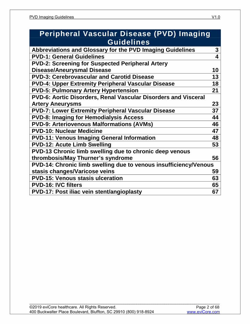
Abbreviations and Glossary for the PVD Imaging Guidelines
(See also: Cardiac Imaging Guidelines Glossary)
AAA abdominal aortic aneurysm ABI Ankle brachial index: a noninvasive, non-imaging test for arterial
insufficiency – (see toe-brachial index below). This testing can also be done after exercise if resting results are normal.
Claudication or Intermittent claudication: usually a painful cramping sensation of the legs with walking or severe leg fatigue
CTA computed tomography angiography CTV computed tomography venography
DLCO diffusion capacity: defined as the volume of carbon monoxide transferred into the blood per minute per mmHg of carbon monoxide partial pressure
DVT deep venous thrombosis ECG Electrocardiogram ENT Ears, Nose, Throat
HbA1C hemoglobin A1C: test used to determine blood sugar control for patients with diabetes
MRA magnetic resonance angiography MRV magnetic resonance venography PAD peripheral artery disease PAH pulmonary artery hypertension PFT pulmonary function tests PVD peripheral vascular disease SVC superior vena cava TIA transient ischemic attack TTE transthoracic echocardiogram
Toe-Brachial Index
useful in patients with ABI above the normal range due to non-compressible posterior tibial or dorsalis pedis arteries
V/Q Scan ventilation and perfusion scan
PVD Imaging Guidelines V1.0
______________________________________________________________________________________________________ ©2019 eviCore healthcare. All Rights Reserved. 400 Buckwalter Place Boulevard, Bluffton, SC 29910 (800) 918-8924 www.eviCore.com
Page 3 of 68
PVD-1: General Guidelines PVD-1.1: General Information 5 PVD-1.2: Procedure Coding 6 PVD-1.3: General Guidelines – Imaging 8
PVD Imaging Guidelines V1.0
______________________________________________________________________________________________________ ©2019 eviCore healthcare. All Rights Reserved. 400 Buckwalter Place Boulevard, Bluffton, SC 29910 (800) 918-8924 www.eviCore.com
Page 4 of 68
Per
iph
eral
Vas
cula
r D
isea
se (
PV
D)
Imag
ing
PVD-1.1: General Information
A current clinical evaluation (within 60 days), including medical treatments, are required prior to considering advanced imaging, which includes: Relevant history and physical examination including:
The palpation of pulses Evaluation of lower extremities for the presence of non-healing wounds or
gangrene Associated skin changes such as thickened nails, absence of hair in the feet
or calves, cool extremities Evaluation for the presence of arterial bruits Appropriate laboratory studies Non-advanced imaging modalities, such as recent ABIs (within 60 days) after
symptoms started or worsened Other meaningful contact (telephone call, electronic mail, or messaging) by an
established patient can substitute for a face-to-face clinical evaluation Risk factors for vascular disease include:
Diabetes Cigarette smoking Hypertension Hyperlipidemia Age > 50, with at least one risk factor, are considered “at risk” for vascular
disease See also: PV-17: Impotence/Erectile Dysfunction in the Pelvis Imaging
Guidelines Signs and symptoms of peripheral arterial disease
Claudication (Cramping pain in the legs, most notably back of the calves but can involve hips or thighs, after walking which is relieved with rest but recurs at a predictable distance) Symptoms that are not consistent with claudication include Generalized leg pain Nocturnal cramps Pain that is not easily relieved after a few minutes of rest Burning pain in feet
Critical limb ischemia Rest pain: Pain in the foot (not leg) at rest, particularly at night when the leg is
elevated. Pain is relieved by dangling the leg off the bed or moving to an upright position
Non healing wounds. Wounds present for >2 weeks with little to no evidence of healing
Erectile dysfunction can be associated with vascular disease Claudication and critical limb ischemia have different natural histories. Claudication
generally follows a benign indolent course. 70% of patients with claudication will have the same symptoms after five years with no progression. Critical limb ischemia, on the other hand, is associated with a high rate of limb loss (25%) and death (35%) one year after presentation
PVD Imaging Guidelines V1.0
______________________________________________________________________________________________________ ©2019 eviCore healthcare. All Rights Reserved. 400 Buckwalter Place Boulevard, Bluffton, SC 29910 (800) 918-8924 www.eviCore.com
Page 5 of 68
Per
iph
eral
Vas
cula
r D
isea
se (
PV
D)
Imag
ing
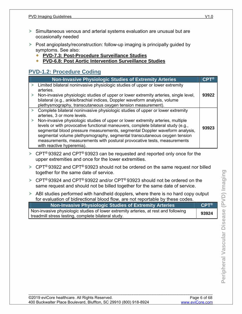
Simultaneous venous and arterial systems evaluation are unusual but are occasionally needed
Post angioplasty/reconstruction: follow-up imaging is principally guided by symptoms. See also: PVD-7.3: Post-Procedure Surveillance Studies PVD-6.8: Post Aortic Intervention Surveillance Studies
PVD-1.2: Procedure Coding Non-Invasive Physiologic Studies of Extremity Arteries CPT®
Limited bilateral noninvasive physiologic studies of upper or lower extremity arteries.
Non-invasive physiologic studies of upper or lower extremity arteries, single level, bilateral (e.g., ankle/brachial indices, Doppler waveform analysis, volume plethysmography, transcutaneous oxygen tension measurement).
93922
Complete bilateral noninvasive physiologic studies of upper or lower extremity arteries, 3 or more levels.
Non-invasive physiologic studies of upper or lower extremity arteries, multiple levels or with provocative functional maneuvers, complete bilateral study (e.g., segmental blood pressure measurements, segmental Doppler waveform analysis, segmental volume plethysmography, segmental transcutaneous oxygen tension measurements, measurements with postural provocative tests, measurements with reactive hyperemia).
93923
CPT® 93922 and CPT® 93923 can be requested and reported only once for the upper extremities and once for the lower extremities.
CPT® 93922 and CPT® 93923 should not be ordered on the same request nor billed together for the same date of service.
CPT® 93924 and CPT® 93922 and/or CPT® 93923 should not be ordered on the same request and should not be billed together for the same date of service.
ABI studies performed with handheld dopplers, where there is no hard copy output for evaluation of bidirectional blood flow, are not reportable by these codes.
Non-Invasive Physiologic Studies of Extremity Arteries CPT® Non-invasive physiologic studies of lower extremity arteries, at rest and following treadmill stress testing, complete bilateral study. 93924
PVD Imaging Guidelines V1.0
______________________________________________________________________________________________________ ©2019 eviCore healthcare. All Rights Reserved. 400 Buckwalter Place Boulevard, Bluffton, SC 29910 (800) 918-8924 www.eviCore.com
Page 6 of 68
Per
iph
eral
Vas
cula
r D
isea
se (
PV
D)
Imag
ing
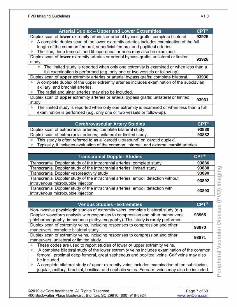
Arterial Duplex – Upper and Lower Extremities CPT® Duplex scan of lower extremity arteries or arterial bypass grafts; complete bilateral. 93925 A complete duplex scan of the lower extremity arteries includes examination of the full
length of the common femoral, superficial femoral and popliteal arteries. The iliac, deep femoral, and tibioperoneal arteries may also be examined. Duplex scan of lower extremity arteries or arterial bypass grafts; unilateral or limited study. 93926
The limited study is reported when only one extremity is examined or when less than a full examination is performed (e.g. only one or two vessels or follow-up).
Duplex scan of upper extremity arteries or arterial bypass grafts; complete bilateral. 93930 A complete duplex of the upper extremity arteries includes examination of the subclavian,
axillary, and brachial arteries. The radial and ulnar arteries may also be included. Duplex scan of upper extremity arteries or arterial bypass grafts; unilateral or limited study. 93931
The limited study is reported when only one extremity is examined or when less than a full examination is performed (e.g. only one or two vessels or follow-up).
Cerebrovascular Artery Studies CPT®
Duplex scan of extracranial arteries; complete bilateral study. 93880 Duplex scan of extracranial arteries; unilateral or limited study. 93882 This study is often referred to as a “carotid ultrasound” or “carotid duplex”. Typically, it includes evaluation of the common, internal, and external carotid arteries.
Transcranial Doppler Studies CPT®
Transcranial Doppler study of the intracranial arteries; complete study 93886 Transcranial Doppler study of the intracranial arteries; limited study 93888 Transcranial Doppler vasoreactivity study 93890 Transcranial Doppler study of the intracranial arteries; emboli detection without intravenous microbubble injection 93892
Transcranial Doppler study of the intracranial arteries; emboli detection with intravenous microbubble injection 93893
Venous Studies - Extremities CPT® Non-invasive physiologic studies of extremity veins, complete bilateral study (e.g. Doppler waveform analysis with responses to compression and other maneuvers, phleborheography, impedance plethysmography). This study is rarely performed.
93965
Duplex scan of extremity veins, including responses to compression and other maneuvers; complete bilateral study. 93970
Duplex scan of extremity veins, including responses to compression and other maneuvers; unilateral or limited study. 93971
These codes are used to report studies of lower or upper extremity veins. A complete bilateral study of the lower extremity veins includes examination of the common
femoral, proximal deep femoral, great saphenous and popliteal veins. Calf veins may also be included.
A complete bilateral study of upper extremity veins includes examination of the subclavian, jugular, axillary, brachial, basilica, and cephalic veins. Forearm veins may also be included.
PVD Imaging Guidelines V1.0
______________________________________________________________________________________________________ ©2019 eviCore healthcare. All Rights Reserved. 400 Buckwalter Place Boulevard, Bluffton, SC 29910 (800) 918-8924 www.eviCore.com
Page 7 of 68
Per
iph
eral
Vas
cula
r D
isea
se (
PV
D)
Imag
ing
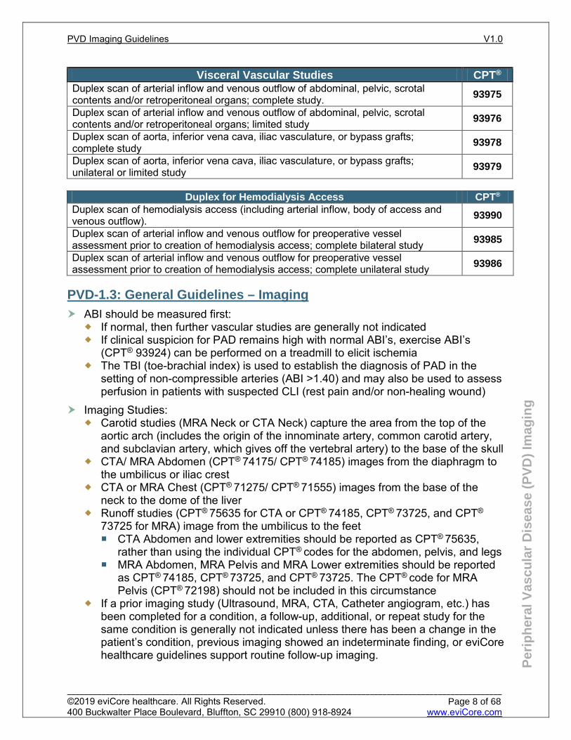
Visceral Vascular Studies CPT® Duplex scan of arterial inflow and venous outflow of abdominal, pelvic, scrotal contents and/or retroperitoneal organs; complete study. 93975
Duplex scan of arterial inflow and venous outflow of abdominal, pelvic, scrotal contents and/or retroperitoneal organs; limited study 93976
Duplex scan of aorta, inferior vena cava, iliac vasculature, or bypass grafts; complete study 93978
Duplex scan of aorta, inferior vena cava, iliac vasculature, or bypass grafts; unilateral or limited study 93979
Duplex for Hemodialysis Access CPT®
Duplex scan of hemodialysis access (including arterial inflow, body of access and venous outflow). 93990
Duplex scan of arterial inflow and venous outflow for preoperative vessel assessment prior to creation of hemodialysis access; complete bilateral study 93985
Duplex scan of arterial inflow and venous outflow for preoperative vessel assessment prior to creation of hemodialysis access; complete unilateral study 93986
PVD-1.3: General Guidelines – Imaging
ABI should be measured first: If normal, then further vascular studies are generally not indicated If clinical suspicion for PAD remains high with normal ABI’s, exercise ABI’s
(CPT® 93924) can be performed on a treadmill to elicit ischemia The TBI (toe-brachial index) is used to establish the diagnosis of PAD in the
setting of non-compressible arteries (ABI >1.40) and may also be used to assess perfusion in patients with suspected CLI (rest pain and/or non-healing wound)
Imaging Studies: Carotid studies (MRA Neck or CTA Neck) capture the area from the top of the
aortic arch (includes the origin of the innominate artery, common carotid artery, and subclavian artery, which gives off the vertebral artery) to the base of the skull
CTA/ MRA Abdomen (CPT® 74175/ CPT® 74185) images from the diaphragm to the umbilicus or iliac crest
CTA or MRA Chest (CPT® 71275/ CPT® 71555) images from the base of the neck to the dome of the liver
Runoff studies (CPT® 75635 for CTA or CPT® 74185, CPT® 73725, and CPT®
73725 for MRA) image from the umbilicus to the feet CTA Abdomen and lower extremities should be reported as CPT® 75635,
rather than using the individual CPT® codes for the abdomen, pelvis, and legs MRA Abdomen, MRA Pelvis and MRA Lower extremities should be reported
as CPT® 74185, CPT® 73725, and CPT® 73725. The CPT® code for MRA Pelvis (CPT® 72198) should not be included in this circumstance
If a prior imaging study (Ultrasound, MRA, CTA, Catheter angiogram, etc.) has been completed for a condition, a follow-up, additional, or repeat study for the same condition is generally not indicated unless there has been a change in the patient’s condition, previous imaging showed an indeterminate finding, or eviCore healthcare guidelines support routine follow-up imaging.
PVD Imaging Guidelines V1.0
______________________________________________________________________________________________________ ©2019 eviCore healthcare. All Rights Reserved. 400 Buckwalter Place Boulevard, Bluffton, SC 29910 (800) 918-8924 www.eviCore.com
Page 8 of 68
Per
iph
eral
Vas
cula
r D
isea
se (
PV
D)
Imag
ing
References Gerhard-Herman MD, Gornik HL, Barrett C, et al. 2016 AHA/ACC Guideline on the management of
patients with lower extremity peripheral artery disease. J Am Coll Cardiol. 2017 Mar 69 (11):1467-1508.
Perlstein TS and Creager MA. The ankle-brachial index as a biomarker of cardiovascular risk: it’s not just about the legs. Circulation. 2009 Nov 29; 120 (21):2033-2035. .
PVD Imaging Guidelines V1.0
______________________________________________________________________________________________________ ©2019 eviCore healthcare. All Rights Reserved. 400 Buckwalter Place Boulevard, Bluffton, SC 29910 (800) 918-8924 www.eviCore.com
Page 9 of 68
PVD-2: Screening for Suspected Peripheral Artery Disease/Aneurysmal Disease
PVD-2.1: Asymptomatic Screening 11 PVD-2.2: Screening for Vascular related genetic connective tissue Disorders (Familial Aneurysm Syndromes/Spontaneous Coronary Artery Dissection (SCAD)/Ehlers-Danlos/Marfan/Loeys-Dietz) 11 PVD-2.3: Screening for TAA in patients with bicuspid aortic valves 12
PVD Imaging Guidelines V1.0
______________________________________________________________________________________________________ ©2019 eviCore healthcare. All Rights Reserved. 400 Buckwalter Place Boulevard, Bluffton, SC 29910 (800) 918-8924 www.eviCore.com
Page 10 of 68
Per
iph
eral
Vas
cula
r D
isea
se (
PV
D)
Imag
ing
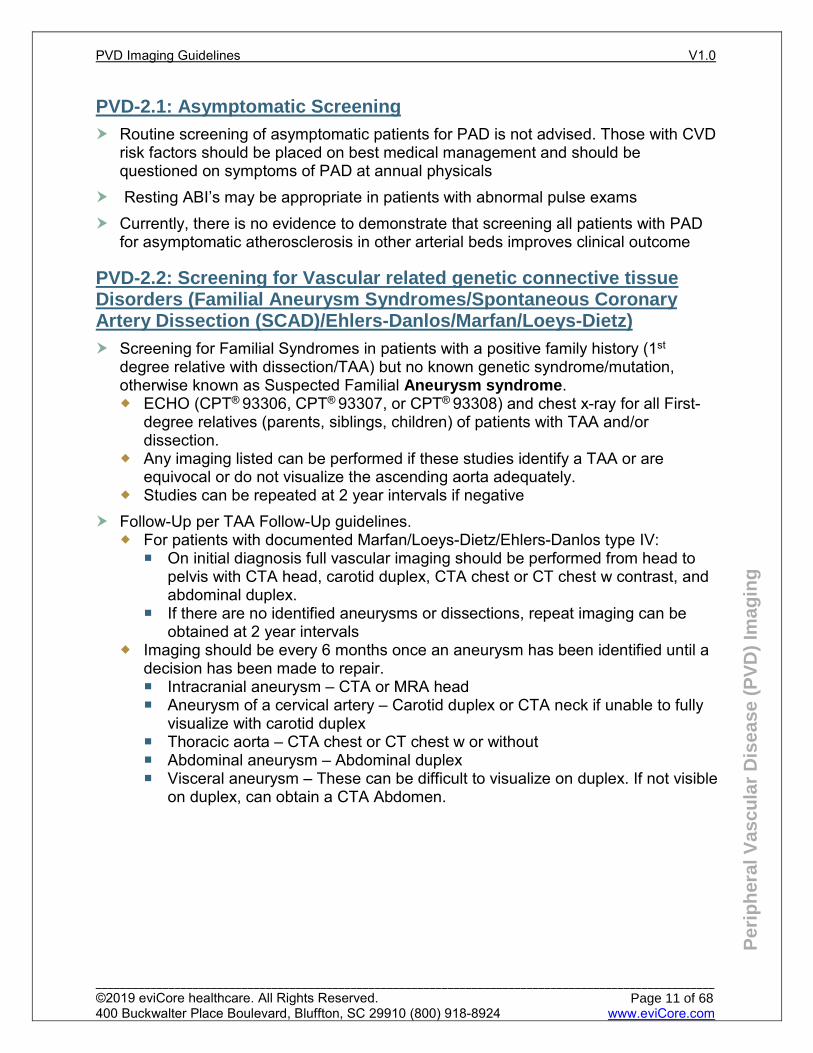
PVD-2.1: Asymptomatic Screening
Routine screening of asymptomatic patients for PAD is not advised. Those with CVD risk factors should be placed on best medical management and should be questioned on symptoms of PAD at annual physicals
Resting ABI’s may be appropriate in patients with abnormal pulse exams Currently, there is no evidence to demonstrate that screening all patients with PAD
for asymptomatic atherosclerosis in other arterial beds improves clinical outcome
PVD-2.2: Screening for Vascular related genetic connective tissue Disorders (Familial Aneurysm Syndromes/Spontaneous Coronary Artery Dissection (SCAD)/Ehlers-Danlos/Marfan/Loeys-Dietz)
Screening for Familial Syndromes in patients with a positive family history (1st degree relative with dissection/TAA) but no known genetic syndrome/mutation, otherwise known as Suspected Familial Aneurysm syndrome. ECHO (CPT® 93306, CPT® 93307, or CPT® 93308) and chest x-ray for all First-
degree relatives (parents, siblings, children) of patients with TAA and/or dissection.
Any imaging listed can be performed if these studies identify a TAA or are equivocal or do not visualize the ascending aorta adequately.
Studies can be repeated at 2 year intervals if negative Follow-Up per TAA Follow-Up guidelines.
For patients with documented Marfan/Loeys-Dietz/Ehlers-Danlos type IV: On initial diagnosis full vascular imaging should be performed from head to
pelvis with CTA head, carotid duplex, CTA chest or CT chest w contrast, and abdominal duplex.
If there are no identified aneurysms or dissections, repeat imaging can be obtained at 2 year intervals
Imaging should be every 6 months once an aneurysm has been identified until a decision has been made to repair. Intracranial aneurysm – CTA or MRA head Aneurysm of a cervical artery – Carotid duplex or CTA neck if unable to fully
visualize with carotid duplex Thoracic aorta – CTA chest or CT chest w or without Abdominal aneurysm – Abdominal duplex Visceral aneurysm – These can be difficult to visualize on duplex. If not visible
on duplex, can obtain a CTA Abdomen.
PVD Imaging Guidelines V1.0
______________________________________________________________________________________________________ ©2019 eviCore healthcare. All Rights Reserved. 400 Buckwalter Place Boulevard, Bluffton, SC 29910 (800) 918-8924 www.eviCore.com
Page 11 of 68
Per
iph
eral
Vas
cula
r D
isea
se (
PV
D)
Imag
ing
PVD-2.3: Screening for TAA in patients with bicuspid aortic valves
Screening in patients with bicuspid aortic valve: Screening, any requested imaging from the “Table of Thoracic Aorta Imaging
Options” in PVD-6.2 Thoracic Aortic Aneurysm and/or ECHO (CPT® 93306, CPT® 93307, or CPT® 93308). Additional imaging such as MRI Cardiac, CT Cardiac, or CCTA is NOT
generally indicated. There is no evidence-based data to support screening relatives of patients
with bicuspid aortic valve. Follow-up per TAA Follow-Up guidelines in PVD-6.2: Thoracic Aortic
Aneurysm (TAA) If no dilatation of the aortic root or ascending thoracic aorta is found, there is no
evidence-based data to support continued surveillance imaging.
References Gerhard-Herman MD, Gornik HL, Barrett C, et al. 2016 AHA/ACC Guideline on the Management of
Patients With Lower Extremity Peripheral Artery Disease: Executive Summary. Journal of the American College of Cardiology. 2017;69(11):1465-1508. doi:10.1016/j.jacc.2016.11.008.
Perlstein TS, Creager MA. The Ankle-Brachial Index as a Biomarker of Cardiovascular Risk. Circulation. 2009;120(21):2033-2035. doi:10.1161/circulationaha.109.907238.
Saydah SH. Poor Control of Risk Factors for Vascular Disease among Adults with Previously Diagnosed Diabetes. JAMA. 2004;291(3):335-342. doi:10.1001/jama.291.3.335.
Hennion DR, Siano KA. Diagnosis and Treatment of Peripheral Arterial Disease. Am Fam Physician. 2013 Sep 1;88(5):306-10.
Hirsch AT. Peripheral Arterial Disease Detection, Awareness, and Treatment in Primary Care. Jama. 2001;286(11):1317. doi:10.1001/jama.286.11.1317.
Mohler ER, Gornik HL, Gerhard-Herman M, Misra S, Olin JW, Zierler RE. ACCF/ACR/AIUM/ASE/ASN/ICAVL/SCAI/SCCT/SIR/SVM/SVS 2012 Appropriate Use Criteria for Peripheral Vascular Ultrasound and Physiological Testing Part I: Arterial Ultrasound and Physiological Testing. Journal of the American College of Cardiology. 2012;60(3):242-276. doi:10.1016/j.jacc.2012.02.009.
US Preventive Services Task Force, Curry SJ, Krist AH, et al. Screening for Peripheral Artery Disease and Cardiovascular Disease Risk Assessment With the Ankle-Brachial Index:US Preventive Services Task Force Recommendation Statement. JAMA. 2018;320(2):177. doi:10.1001/jama.2018.8357.
MS Conte, FB Pomposelli, DG Clair, et al. Society for Vascular Surgery practice guidelines for atherosclerotic occlusive disease of the lower extremities: Management of asymptomatic disease and claudication. Journal of Vascular Surgery 2015. Vol 6:1S-41S
MacCarrick G, Black J, Bowdin S, et al. Loeys–Dietz syndrome: a primer for diagnosis and management. Genet Med (2014).16:576–587 doi:10.1038/gim.2014.11 .
Olin JW, Gornik HL, Bacharach JM, et al. Fibromuscular Dysplasia: State of the Science and Critical Unanswered Questions. Circulation. 2014;129(9):1048-1078. doi:10.1161/01.cir.0000442577.96802.8c
Persu A, Niepen PVD, Touzé E, et al. Revisiting Fibromuscular Dysplasia. Hypertension. 2016;68(4):832-839. doi:10.1161/hypertensionaha.116.07543
Hayes SN, Kim ES, Saw J, et al. Spontaneous Coronary Artery Dissection: Current State of the Science: A Scientific Statement From the American Heart Association. Circulation. 2018;137(19). doi:10.1161/cir.0000000000000564.
Hiratzka LF, Bakris GL, Beckman JA, et al. 2010 ACCF/AHA/AATS/ACR/ASA/SCA/SCAI/SIR/STS/SVM guidelines for the diagnosis and management of patients with thoracic aortic disease. J Am Coll Cardiol 2010; 55: e27-e129.
PVD Imaging Guidelines V1.0
______________________________________________________________________________________________________ ©2019 eviCore healthcare. All Rights Reserved. 400 Buckwalter Place Boulevard, Bluffton, SC 29910 (800) 918-8924 www.eviCore.com
Page 12 of 68
PVD-3: Cerebrovascular and Carotid Disease PVD-3.1: Initial Imaging 14 PVD-3.2: Surveillance Imaging with NO History of Carotid Surgery or Intervention 15 PVD-3.3: Surveillance Imaging WITH History of Carotid Surgery or Intervention 16
PVD Imaging Guidelines V1.0
______________________________________________________________________________________________________ ©2019 eviCore healthcare. All Rights Reserved. 400 Buckwalter Place Boulevard, Bluffton, SC 29910 (800) 918-8924 www.eviCore.com
Page 13 of 68
Per
iph
eral
Vas
cula
r D
isea
se (
PV
D)
Imag
ing
PVD-3.1: Initial Imaging
Prior to considering advanced imaging, duplex ultrasound (CPT® 93880 bilateral or CPT® 93882 unilateral) should generally be used to evaluate possible carotid artery disease when any of the following apply: Hemispheric neurologic symptoms including stroke, TIA, or amaurosis fugax. Known or suspected retinal arterial emboli or Hollenhorst plaque Suspected carotid dissection Pulsatile neck masses Carotid or cervical bruit Abnormal findings on physical exam of the carotid arteries (e.g. aneurysm or
absent carotid pulses) Preoperative evaluation of patients with evidence of severe diffuse
atherosclerosis, scheduled for major cardiovascular surgical procedures Preoperative evaluation of patients prior to elective coronary artery bypass graft
(CABG) surgery in patients older than 65 years of age and in those with peripheral artery disease, history of cigarette smoking, history of stroke or TIA, or carotid bruit
Suspected Subclavian Steal Syndrome See also: CH-27: Subclavian Steal Syndrome in the Chest Imaging
Guidelines Blunt neck trauma Neurologic complaints after chiropractic neck manipulation Vasculitis potentially involving carotid arteries, i.e. Takayasu’s arteritis and
fibromuscular dysplasia (FMD) Carotid ultrasound screening in asymptomatic individuals due only to risk factors is
not indicated New signs and symptoms consistent with carotid artery disease (e.g. TIA, amaurosis
fugax, change in nature of a carotid bruit) are an indication to re-image the cervical vessels (regardless of when the previous carotid imaging was performed) using any of the following: Duplex ultrasound (CPT® 93880 bilateral study or CPT® 93882 unilateral study), MRA Neck with or without and with contrast (CPT® 70548 or 70549) CTA Neck (CPT® 70498)
For Typical Symptoms of TIA/Stroke or Carotid Dissection: See also: HD-21: Stroke/TIA
For Suspected Vertebrobasilar Pathology: Initial Imaging see also: HD-21: Stroke/TIA Surveillance Imaging
Asymptomatic or unchanged symptoms and known vertebrobasilar disease or post-stenting interval determined by Vascular Specialist
After Intracranial Hemorrhage: Initial Imaging see also: HD-13.1: Head Trauma Surveillance Imaging
Interval determined by neurosurgeon or neurologist.
PVD Imaging Guidelines V1.0
______________________________________________________________________________________________________ ©2019 eviCore healthcare. All Rights Reserved. 400 Buckwalter Place Boulevard, Bluffton, SC 29910 (800) 918-8924 www.eviCore.com
Page 14 of 68
Per
iph
eral
Vas
cula
r D
isea
se (
PV
D)
Imag
ing
For Suspected Subclavian Steal Syndrome: Initial imaging should be a carotid duplex
If initial duplex demonstrates high grade stenosis or occlusion of the subclavian artery, advanced imaging is NOT indicated unless the patient is symptomatic with arm claudication or signs of hypo-perfusion of the vertebral artery with recurrent dizziness
Surveillance of subclavian arterial disease is NOT indicated if there has not been any intervention such as a carotid-subclavian bypass or subclavian stent Advanced imaging see also: CH-27: Subclavian Steal Syndrome – General
PVD-3.2: Surveillance Imaging with NO History of Carotid Surgery or Intervention
Surveillance imaging is appropriate once a year for patients with fibromuscular dysplasia of the extracranial carotid arteries.
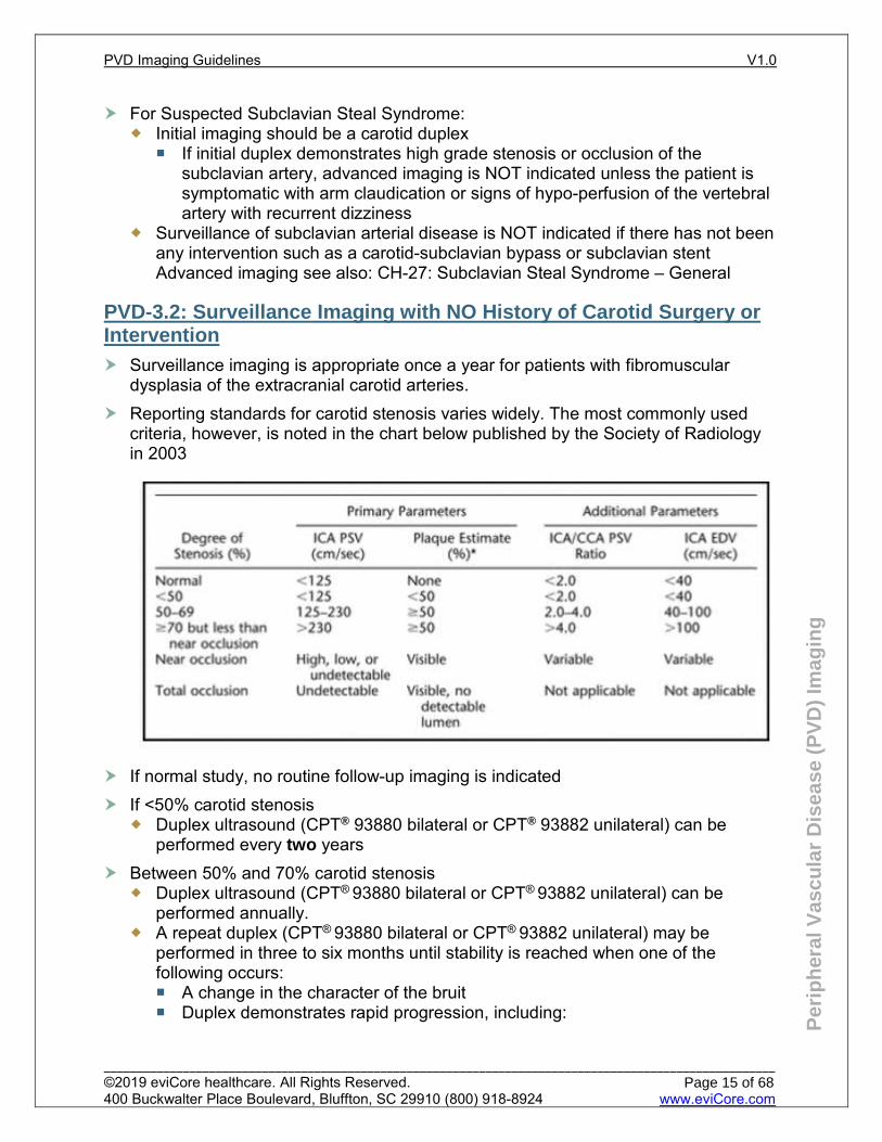
Reporting standards for carotid stenosis varies widely. The most commonly used criteria, however, is noted in the chart below published by the Society of Radiology in 2003
If normal study, no routine follow-up imaging is indicated If <50% carotid stenosis
Duplex ultrasound (CPT® 93880 bilateral or CPT® 93882 unilateral) can be performed every two years
Between 50% and 70% carotid stenosis Duplex ultrasound (CPT® 93880 bilateral or CPT® 93882 unilateral) can be
performed annually. A repeat duplex (CPT® 93880 bilateral or CPT® 93882 unilateral) may be
performed in three to six months until stability is reached when one of the following occurs: A change in the character of the bruit Duplex demonstrates rapid progression, including:
PVD Imaging Guidelines V1.0
______________________________________________________________________________________________________ ©2019 eviCore healthcare. All Rights Reserved. 400 Buckwalter Place Boulevard, Bluffton, SC 29910 (800) 918-8924 www.eviCore.com
Page 15 of 68
Per
iph
eral
Vas
cula
r D
isea
se (
PV
D)
Imag
ing
doubling of peak systolic velocities increase of the ICA/CCA ratio heavy calcification thrombus ulcerated plaque echolucent plaque
Carotid stenosis ≥70% or ICA/CCA ratio >4 Duplex ultrasound (CPT® 93880 bilateral or CPT® 93882 unilateral) or MRA Neck
with contrast (CPT® 70548) or CTA Neck (CPT® 70498) can be performed at the following intervals: Every 6 months until one of the following occurs: Intervention is performed Decision is made to not intervene
If duplex Ultrasound shows ≥ 70% occlusion/stenosis of the internal carotid artery or the ICA/CCA ratio is >4.0 even with a lower percentage of stenosis, then MRA Neck with contrast (CPT® 70548) or CTA Neck (CPT® 70498) can be performed If carotid stent is planned
MRA Head (CPT® 70544) or CTA Head (CPT® 70496) can be added
PVD-3.3: Surveillance Imaging WITH History of Carotid Surgery or Intervention
Duplex ultrasound (CPT® 93880 bilateral or CPT® 93882 unilateral) can be performed post carotid surgery or intervention at the following intervals:
1 month after procedure Every 6 months for 2 years after procedure Then annually
If ≥ 70% residual carotid stenosis is seen at 1 month after procedure Duplex ultrasound (CPT® 93880 bilateral or CPT® 93882 unilateral) can be
performed at the following intervals: Every 3-6 months for one year Then annually.
Practice Notes Carotid intima-media thickness using duplex ultrasound imaging (Category III code
0126T) is not recommended in clinical practice for risk assessment for a first ASCVD event. Although outcomes data are lacking, Texas has adopted this method in Texas Heart Attack Preventive Screening Bill (HR 1290)
Texas Heart Attack Preventive Screening Law (HR 1290) mandates that insurers in Texas cover either a calcium scoring study (CPT® 75571 or HCPCS S8092) or a carotid intima-media thickness study (ultrasound—Category III code 0126T) every five years for certain populations. To qualify, the following must apply: Must be a Texas resident. Must be a member of a fully-insured Texas health plan. Must be a man age 45 to 75 or a woman age 55 to 75.
PVD Imaging Guidelines V1.0
______________________________________________________________________________________________________ ©2019 eviCore healthcare. All Rights Reserved. 400 Buckwalter Place Boulevard, Bluffton, SC 29910 (800) 918-8924 www.eviCore.com
Page 16 of 68
Per
iph
eral
Vas
cula
r D
isea
se (
PV
D)
Imag
ing
Must have either diabetes or a Framingham cardiac risk score of intermediate or higher.
Must not have had a calcium scoring study or a carotid intima-media thickness study within the past 5 years
If ultrasound is technically difficult or confirmation of the degree of stenosis on ultrasound is needed because an interventional procedure is being considered, then MRA Neck (CPT® 70548) or CTA Neck (CPT® 70498) may be performed.
References Brott TG, Halperin JL, Abbara S, et al. 2011
ASA/ACCF/AHA/AANN/AANS/ACR/ASNR/CNS/SAIP/SCAI/SIR/SNIS/SVM/SVS Guideline on the Management of Patients With Extracranial Carotid and Vertebral Artery Disease. Journal of the American College of Cardiology. 2011;57(8). doi:10.1016/j.jacc.2010.11.006.
Gerhard-Herman M, Gardin JM, Jaff M, Mohler E, Roman M, Naqvi TZ. Guidelines for Noninvasive Vascular Laboratory Testing: A Report from the American Society of Echocardiography and the Society of Vascular Medicine and Biology. Journal of the American Society of Echocardiography. 2006;19(8):955-972. doi:10.1016/j.echo.2006.04.019.
Reutern G-MV, Goertler M-W, Bornstein NM, et al. Grading Carotid Stenosis Using Ultrasonic Methods. Stroke. 2012;43(3):916-921. doi:10.1161/strokeaha.111.636084.
ACR–AIUM–SPR–SRU. AIUM Practice Parameter for the Performance of an Ultrasound Examination of the Extracranial Cerebrovascular System. Journal of Ultrasound in Medicine. 2016;35(9):1-11. doi:10.7863/ultra.35.9.1.
Moresoli P, Habib B, Reynier P, et al. Carotid Stenting versus Endarterectomy for Asymptomatic Carotid Artery Stenosis. Stroke. 2017;48(8):2150-2157. doi:10.1161/strokeaha.117.016824.
Rogers RK, Bishu K. Optimal Treatment of Extracranial Carotid Artery Disease: Carotid Endarterectomy, Carotid Stenting, or Optimal Medical Therapy. Current Cardiology Reports. 2015;17(10). doi: 10.1007/s11886-015-0636-2.
Shakarchi JA, Lowry D, Nath J, et al. Duplex ultrasound surveillance after carotid artery endarterectomy. Journal of Vascular Surgery. 2016;63(6):1647-1650. doi:10.1016/j.jvs.2016.01.054.
Aburahma AF, Srivastava M, Aburahma Z, et al. The value and economic analysis of routine postoperative carotid duplex ultrasound surveillance after carotid endarterectomy. Journal of Vascular Surgery. 2015;62(2):378-384. doi:10.1016/j.jvs.2015.03.023.
Aboyans V, Ricco J-B, Bartelink M-LEL, et al. 2017 ESC Guidelines on the Diagnosis and Treatment of Peripheral Arterial Diseases, in collaboration with the European Society for Vascular Surgery (ESVS). European Heart Journal. 2017;39(9):763-816. doi:10.1093/eurheartj/ehx095.
Naylor A, Ricco J-B, Borst GD, et al. Editor's Choice – Management of Atherosclerotic Carotid and Vertebral Artery Disease: 2017 Clinical Practice Guidelines of the European Society for Vascular Surgery (ESVS). European Journal of Vascular and Endovascular Surgery. 2018;55(1):3-81. doi:10.1016/j.ejvs.2017.06.021.
Mohler ER, Gornik HL, Gerhard-Herman M, Misra S, Olin JW, Zierler RE. ACCF/ACR/AIUM/ASE/ASN/ICAVL/SCAI/SCCT/SIR/SVM/SVS 2012 Appropriate Use Criteria for Peripheral Vascular Ultrasound and Physiological Testing Part I: Arterial Ultrasound and Physiological Testing. Journal of the American College of Cardiology. 2012;60(3):242-276. doi:10.1016/j.jacc.2012.02.009.
Goff DC, Lloyd-Jones DM, Bennett G, et al. 2013 ACC/AHA Guideline on the Assessment of Cardiovascular Risk. Circulation. 2014;129(25 suppl 2):s49-s73. doi:10.1161/01.cir.0000437741.48606.98.
Ricotta JJ, Aburahma A, Ascher E, et al. Updated Society for Vascular Surgery guidelines for management of extracranial carotid disease. Journal of Vascular Surgery. 2011;54(3):1-31. doi:10.1016/j.jvs.2011.07.031..
Ballotta E, Giau GD, Meneghetti G, et al. Progression of atherosclerosis in asymptomatic carotid arteries after contralateral endarterectomy: A 10-year prospective study. Journal of Vascular Surgery. 2007;45(3):516-522. doi:10.1016/j.jvs.2006.11.011.
PVD Imaging Guidelines V1.0
______________________________________________________________________________________________________ ©2019 eviCore healthcare. All Rights Reserved. 400 Buckwalter Place Boulevard, Bluffton, SC 29910 (800) 918-8924 www.eviCore.com
Page 17 of 68
PVD-4: Upper Extremity Peripheral Vascular Disease
PVD-4.1: Upper Extremity PVD – Imaging 19
PVD Imaging Guidelines V1.0
______________________________________________________________________________________________________ ©2019 eviCore healthcare. All Rights Reserved. 400 Buckwalter Place Boulevard, Bluffton, SC 29910 (800) 918-8924 www.eviCore.com
Page 18 of 68
Per
iph
eral
Vas
cula
r D
isea
se (
PV
D)
Imag
ing
PVD-4.1: Upper Extremity PVD – Imaging
Signs and symptoms of arterial insufficiency include but are not limited to: Arm or hand claudication, cramping or fatigue of the unilateral extremity with use
or with raising limb overhead that is relieved with rest and is reproducible. See CH-27: Subclavian Steal Syndrome
Systolic blood pressure differential between arms of <15mmHg. See CH-27: Subclavian Steal Syndrome
Bluish discoloration of the hand or fingers Unilateral cold painful pulseless hand Non healing wound (>2 weeks with no healing or evidence of healing) or frank
gangrene For signs and symptoms of arterial insufficiency, appropriate studies include:
Arterial ultrasound of the upper extremities (CPT® 93930 or CPT® 93931) or CTA of Upper extremity (CPT® 73206) or MRA of Upper extremity (CPT® 73225)
and/or CTA Chest (CPT® 71275) or MRA Chest (CPT® 71555)
For suspected Fibromuscular Dysplasia of the brachial artery, appropriate studies include: MRA of Upper extremity (CPT® 73225) CTA of Upper extremity (CPT® 73206) Arterial Ultrasound (CPT® 93930 bilateral study or CPT® 93931 unilateral study)
Arterial Duplex (CPT® 93931) can be obtained following upper extremity arterial revascularization at: Baseline (within one month) 6 months Then annually if stable Anytime for new or worsening symptoms
For symptoms of venous insufficiency including but not limited to unilateral pain and swelling of the upper extremity Venous duplex of the upper extremities (CPT® 93970 or CPT® 93971) should be
performed initially If duplex ultrasound is nondiagnostic:
MRV Upper extremity (CPT® 73225) and/or MRV Chest (CPT® 71555), or CTV Upper extremity (CPT® 73206) and/or CTV Chest (CPT® 71275) If there is a history of exertion with the limb such as with weight lifting or in
the presence of central venous access (port, PICC line, to name a few) with a negative venous duplex, a CTV of Upper extremity (CPT® 73206) or MRV of Upper extremity (CPT® 73225), and/or CTV Chest (CPT® 71275) or MRV Chest (CPT® 71555) can be performed. See CH-31.1: Thoracic Outlet Syndrome
For Superior Vena Cava Syndrome (upper extremity and facial symptoms): CT Chest with contrast (CPT® 71260) MRV (CPT® 71555) or CTV (CPT® 71275) Chest may be considered when
stenting of the SVC is being considered
PVD Imaging Guidelines V1.0
______________________________________________________________________________________________________ ©2019 eviCore healthcare. All Rights Reserved. 400 Buckwalter Place Boulevard, Bluffton, SC 29910 (800) 918-8924 www.eviCore.com
Page 19 of 68
Per
iph
eral
Vas
cula
r D
isea
se (
PV
D)
Imag
ing
References 1. Desjardins B, Rybicki FJ, Kim HS, et al. ACR Appropriateness Criteria® Suspected Upper Extremity
Deep Vein Thrombosis. Journal of the American College of Radiology. 2012;9(9):613-619. doi:10.1016/j.jacr.2012.05.021.
2. Yoshimuta T, Akutsu K, Okajima T, et al. “String of Beads” Appearance of Bilateral Brachial Artery in Fibromuscular Dysplasia. Circulation. 2008;117(19):2542-2543. doi:10.1161/circulationaha.107.747089.
3. Birch A, Um D, Laselle B. Ultrasound Detection of Superior Vena Cava Thrombus. Western Journal of Emergency Medicine. 2014;15(6):715-718. doi:10.5811/westjem.2014.6.14006.
PVD Imaging Guidelines V1.0
______________________________________________________________________________________________________ ©2019 eviCore healthcare. All Rights Reserved. 400 Buckwalter Place Boulevard, Bluffton, SC 29910 (800) 918-8924 www.eviCore.com
Page 20 of 68
PVD-5: Pulmonary Artery Hypertension PVD-5.1: Pulmonary Artery Hypertension – Imaging 22
PVD Imaging Guidelines V1.0
______________________________________________________________________________________________________ ©2019 eviCore healthcare. All Rights Reserved. 400 Buckwalter Place Boulevard, Bluffton, SC 29910 (800) 918-8924 www.eviCore.com
Page 21 of 68

Per
iph
eral
Vas
cula
r D
isea
se (
PV
D)
Imag
ing
PVD-5.1: Pulmonary Artery Hypertension – Imaging
Pulmonary artery hypertension (PAH) comprises a spectrum of diseases which will need direct evaluation, including ECG (right ventricular hypertrophy with/without strain, right atrial dilatation); chest x-ray; arterial blood gas, PFT’s or V/Q scan. Imaging is based on suspected etiology.
Transthoracic echocardiogram (TTE) (CPT® 93306) should be performed initially and may be accompanied by: Pulmonary venous hypertension - Stress echocardiogram (CPT® 93350 or CPT®
93351) or left and/or right heart catheterization Pulmonary hypertension associated with hypoxemia - High-resolution CT Chest
(CPT® 71250) to rule out restrictive lung disorders such as idiopathic pulmonary fibrosis
Acute or chronic pulmonary embolism – CTA Chest (CPT®71275); See also in specific subsections:
CD-2.2: Transthoracic Echocardiogram (TTE)-Indications, CD-7.4: Right Heart Catheterization (RHC), CD-11.3.12: Severe Pulmonary artery hypertension (PHT) and Eisenmenger syndrome in the adult cardiac imaging guidelines
PEDCD-2.3: Congenital Heart Disease Modality Considerations, PEDCD-7: Pediatric Pulmonary Hypertension-General in the pediatric cardiac imaging guidelines
CH-25: Pulmonary Embolism (PE) in the Chest Imaging Guidelines.
References Barbosa EJM, Gupta NK, Torigian DA, et al. Current role of imaging in the diagnosis and
management of pulmonary hypertension. Am J Roeentgenol. 2012; 198 (6): 1320-1331. Galiè A, Torbicki A, Barst R, et al. Guidelines on diagnosis and treatment of pulmonary arterial
hypertension: the task force on diagnosis and treatment of pulmonary arterial hypertension of the European Society of Cardiology. Eur Heart J. 2004 Dec 1; 25 (24): 2243-2278.
Peña E, Dennie C, Veinot J, et al. Pulmonary hypertension: how the radiologist can help. RadioGraphics. 2012; 32 (1):9-32.
PVD Imaging Guidelines V1.0
______________________________________________________________________________________________________ ©2019 eviCore healthcare. All Rights Reserved. 400 Buckwalter Place Boulevard, Bluffton, SC 29910 (800) 918-8924 www.eviCore.com
Page 22 of 68
PVD-6: Aortic Disorders, Renal Vascular Disorders and Visceral Artery Aneurysms
PVD-6.1: Aortic Disorders General Information 24 PVD-6.2: Thoracic Aortic Aneurysm (TAA) 25 PVD-6.3: Abdominal Aortic Aneurysm (AAA) 27 PVD-6.4: Iliac Artery Aneurysm (IAA 28 PVD-6.5: Visceral Artery Aneurysm 28 PVD-6.6: Renovascular Hypertension/Renal Artery Stenosis 30 PVD-6.7: Aortic Dissection and Other Aortic Conditions 32 PVD-6.8: Post Aortic Endovascular/Open Surgery Surveillance Studies 34
PVD Imaging Guidelines V1.0
______________________________________________________________________________________________________ ©2019 eviCore healthcare. All Rights Reserved. 400 Buckwalter Place Boulevard, Bluffton, SC 29910 (800) 918-8924 www.eviCore.com
Page 23 of 68
Per
iph
eral
Vas
cula
r D
isea
se (
PV
D)
Imag
ing
PVD-6.1: Aortic Disorders General Information Duplex ultrasound for visceral vascular studies CPT®
Duplex scan of arterial inflow and venous outflow of abdominal, pelvic, scrotal contents and/or retroperitoneal organs; complete study. 93975
Duplex scan of arterial inflow and venous outflow of abdominal, pelvic, scrotal contents and/or retroperitoneal organs; limited study. 93976
Duplex scan of aorta, inferior vena cava, iliac vasculature, or bypass grafts; complete study. 93978
Duplex scan of aorta, inferior vena cava, iliac vasculature, or bypass grafts; unilateral or limited study. 93979
In clinical practice, CT, CTA, MRA are usually preferred to evaluate for stenosis of these vessels rather than ultrasound which can be difficult to perform (Exception: Duplex ultrasound is appropriate to rule out testicular or ovarian torsion or to evaluate an abdominal bruit or a pulsatile abdominal mass).
Mesenteric Ischemia See also: AB-6: Mesenteric/Colonic Ischemia in the Abdomen Imaging
Guidelines.
References Hirsch AT, Haskal ZJ, Hertzer NR, et al. ACC/AHA Practice Guidelines: ACC/AHA 2005 Practice
guidelines for the management of patients with peripheral arterial disease (lower extremity, renal, mesenteric, and abdominal aortic): a collaborative report from the American Association for Vascular Surgery/Society for Vascular Surgery,* Society for Cardiovascular Angiography and Interventions, Society for Vascular Medicine and Biology, Society of Interventional Radiology, and the ACC/AHA Task Force on Practice Guidelines. Circulation. 2006; 113: e463-e654.
Rooke TW, Hirsch AT, Misra S, et al. 2011 ACCF/AHA focused update of the guideline for the management of patients with peripheral artery disease (updating the 2005 guideline): a report of the American College of Cardiology Foundation/American Heart Association Task Force on Practice Guidelines. This article is co-published in the Journal of the American College of Cardiology, Catheterization and Cardiovascular Interventions, the Journal of Vascular Surgery, and Vascular Medicine. Circulation. 2011 Nov 1; 124 (18): 2020-2045.
Mohler III ER, Gornik HL, Gerhard-Herman M, et al. ACCF/ACR/AIUM/ASE/ASN/ICAVL/SCAI/SCCT/SIR/SVM/SVS 2012 Appropriate use criteria for peripheral vascular ultrasound and physiological testing part I: arterial ultrasound and physiological testing. J Am Coll Cardiol 2012;60:242–76.
PVD Imaging Guidelines V1.0
______________________________________________________________________________________________________ ©2019 eviCore healthcare. All Rights Reserved. 400 Buckwalter Place Boulevard, Bluffton, SC 29910 (800) 918-8924 www.eviCore.com
Page 24 of 68
Per
iph
eral
Vas
cula
r D
isea
se (
PV
D)
Imag
ing
PVD-6.2: Thoracic Aortic Aneurysm (TAA)
The thoracic aorta is generally divided into two segments: the ascending aorta which includes the aortic root, aortic arch and ends just distal to the left subclavian artery and the descending aorta which starts just distal to the left subclavian artery to the level of the diaphragm.
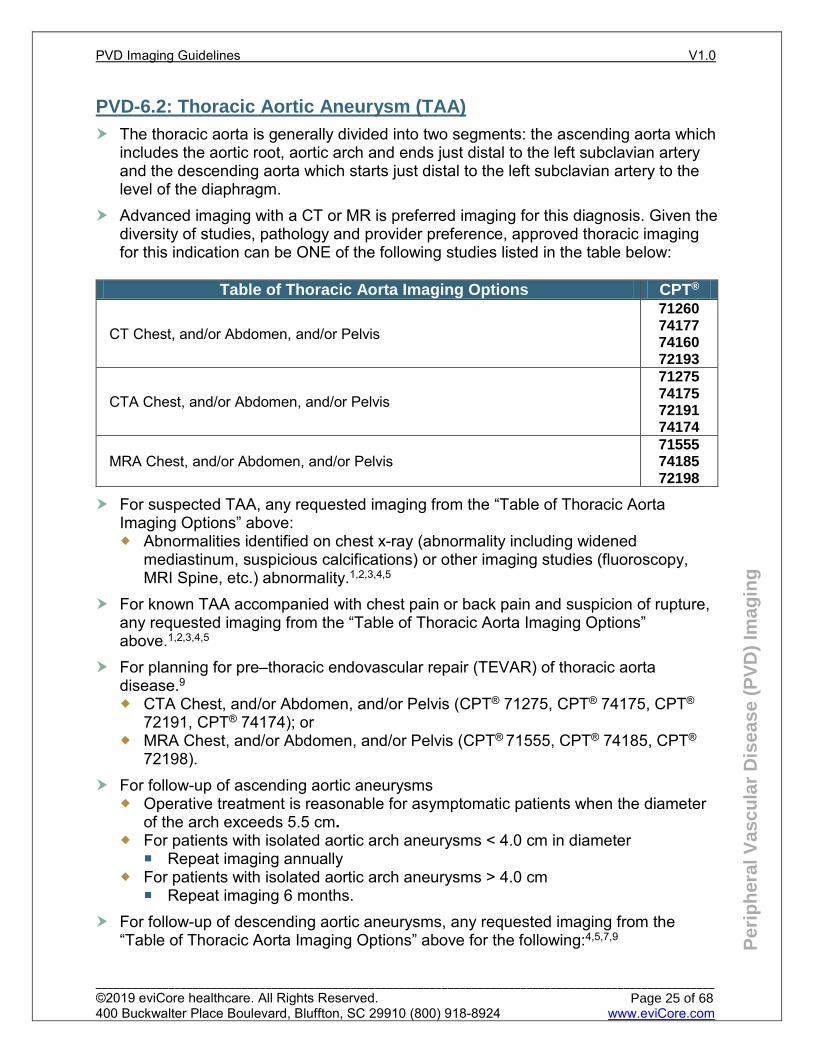
Advanced imaging with a CT or MR is preferred imaging for this diagnosis. Given the diversity of studies, pathology and provider preference, approved thoracic imaging for this indication can be ONE of the following studies listed in the table below:
Table of Thoracic Aorta Imaging Options CPT®
CT Chest, and/or Abdomen, and/or Pelvis
71260 74177 74160 72193
CTA Chest, and/or Abdomen, and/or Pelvis
71275 74175 72191 74174
MRA Chest, and/or Abdomen, and/or Pelvis 71555 74185 72198
For suspected TAA, any requested imaging from the “Table of Thoracic Aorta Imaging Options” above: Abnormalities identified on chest x-ray (abnormality including widened
mediastinum, suspicious calcifications) or other imaging studies (fluoroscopy, MRI Spine, etc.) abnormality.1,2,3,4,5
For known TAA accompanied with chest pain or back pain and suspicion of rupture, any requested imaging from the “Table of Thoracic Aorta Imaging Options” above.1,2,3,4,5
For planning for pre–thoracic endovascular repair (TEVAR) of thoracic aorta disease.9 CTA Chest, and/or Abdomen, and/or Pelvis (CPT® 71275, CPT® 74175, CPT®
72191, CPT® 74174); or MRA Chest, and/or Abdomen, and/or Pelvis (CPT® 71555, CPT® 74185, CPT®
72198). For follow-up of ascending aortic aneurysms
Operative treatment is reasonable for asymptomatic patients when the diameter of the arch exceeds 5.5 cm.
For patients with isolated aortic arch aneurysms < 4.0 cm in diameter Repeat imaging annually
For patients with isolated aortic arch aneurysms > 4.0 cm Repeat imaging 6 months.
For follow-up of descending aortic aneurysms, any requested imaging from the “Table of Thoracic Aorta Imaging Options” above for the following:4,5,7,9
PVD Imaging Guidelines V1.0
______________________________________________________________________________________________________ ©2019 eviCore healthcare. All Rights Reserved. 400 Buckwalter Place Boulevard, Bluffton, SC 29910 (800) 918-8924 www.eviCore.com
Page 25 of 68
Per
iph
eral
Vas
cula
r D
isea
se (
PV
D)
Imag
ing
“Medically” treated/observation. 3.5 to 4.4 cm TAA can be followed annually. ≥4.5 cm TAA can be followed every 6 months. ≥3.0 cm TAA when there is concern for growth can have a one-time 3 month
interval advanced imaging. Surgery or Stent treatment.
Preoperative open or endovascular (stent) repair imaging is appropriate. Suspicion of endoleaks. Open Repair imaging every 3 to 5 years.
Endovascular graft/stent. First year: 1 month, 6 months, 12 months, then annually.
Screening in the presence of other aortic aneurysms. In a patient with a known TAA, screening for AAA is appropriate with an
abdominal duplex. See PVD-6.3: Abdominal Aortic Aneurysm (AAA) in the Peripheral Vascular Disease Imaging Guidelines.
In a patient with a known AAA, screening for TAA is not supported by sufficient evidence.
Screening in patients with bicuspid aortic valve or familial TAA syndromes. See PVD-2.3: Screening for TAA in patients with bicuspid aortic valve. See PVD-2.2: Screening for Vascular related genetic connective tissue Disorders (Familial Aneurysm Syndromes/Spontaneous Coronary Artery Dissection (SCAD)/Ehlers-Danlos/Marfan/Loeys-Dietz)
References 1. Expert Panel on Cardiac Imaging. ACR Appropriateness Criteria® Acute Chest Pain -- Suspected
Aortic Dissection. American College of Radiology (ACR); 2014. 2. ACR Appropriateness Criteria® Nontraumatic Aortic Disease. American College of Radiology (ACR);
2013. 3. Hiratzka LF, Bakris GL, Beckman JA, et al. 2010
ACCF/AHA/AATS/ACR/ASA/SCA/SCAI/SIR/STS/SVM guidelines for the diagnosis and management of patients with thoracic aortic disease. J Am Coll Cardiol 2010; 55: e27-e129.
4. Loren F. Hiratzka MD, et al, 2010 ACCF/AHA/AATS/ACR/ASA/SCAI/SIR/STS/SVM Guidelines for the Diagnosis and Management of Patients With Thoracic Aortic Disease Circulation 2010; 121: e266-e369.
5. Albornoz G, Coady M, Roberts M, et al. Familial thoracic aortic aneurysms and dissections—incidence, modes of inheritance, and phenotypic patterns. Annals of Thoracic Surgery 2006 Oct; 82 (4): 1400-1405.
6. Elefteriades JA. Natural history of thoracic aortic aneurysms: indications for surgery, and surgical versus nonsurgical risks. Ann Thorac Surg 2002; 74: S1877-S1880.
7. ACR Appropriateness Criteria® thoracic aorta interventional planning and follow-up. American College of Radiology (ACR); 2017.
8. Tadros TM, Klein MD, Shapira OM. Ascending aortic dilatation associated with bicuspid aortic valve. Circulation 2009; 119: 880-890.
9. Bennett SJ, Dill KE, Hanley M, et al. ACR Appropriateness Criteria ® Suspected Thoracic Aortic Aneurysm. Journal of the American College of Radiology. 2018;15(5). doi:10.1016/j.jacr.2018.03.031.
PVD Imaging Guidelines V1.0
______________________________________________________________________________________________________ ©2019 eviCore healthcare. All Rights Reserved. 400 Buckwalter Place Boulevard, Bluffton, SC 29910 (800) 918-8924 www.eviCore.com
Page 26 of 68
Per
iph
eral
Vas
cula
r D
isea
se (
PV
D)
Imag
ing
PVD-6.3: Abdominal Aortic Aneurysm (AAA)
Ultrasound Abdominal aorta (CPT® 76706) is the preferred initial imaging study to screen and retroperitoneal ultrasound (CPT® 76775) to survey for AAA or to evaluate a pulsatile abdominal mass
Obese Individual (BMI≥ 35): CT Abdomen and Pelvis with contrast (CPT® 74177) or without contrast (CPT® 74176) can be substituted for US using the same timeline as a non-obese individual. Ultrasound of the abdominal aorta should ideally first be attempted to see if the image quality is adequate
Screening One-time screening recommendations for AAA (Ultrasound CPT® 76706)
Men and women 65 to 75 years of age with a history of tobacco use Men and women older than 75 years with a history of tobacco use and in
otherwise good health who have not previously received a screening ultrasound examination
All first-degree relatives of individuals who present with an AAA and are between 65 and 75, or in those older than 75 in good health
Medicare covers a one-time AAA screening ultrasound (CPT® 76706) if there are at least one of the following risk factors Family history of AAA The individual is a male age 65 to 75 who has smoked at least 100 cigarettes
in his lifetime If there is a documented thoracic aortic aneurysm, AAA screening is reasonable
with ultrasound (CPT® 76706); however, there is insufficient evidence to support the use of advanced imaging to screen for a thoracic aortic aneurysm in individuals with known abdominal aortic aneurysm.
Surveillance recommendations for AAA (CPT® 76775) > 2.5 cm but < 3.0 cm: 10 years 3.0 cm to 3.9 cm: 3 year intervals 4.0 cm to 4.9 cm: every 12 months 5.0 cm to 5.4 cm: every 6 months > 5.4 cm or aortic diameter has increased in size by 0.7 cm in six months, or at
least 1 cm in a year may undergo more frequent monitoring and should be evaluated by a Vascular Specialist
Additional Imaging CT of the Abdomen and Pelvis with contrast (CPT® 74177), CT of the Abdomen
and Pelvis without and with contrast (CPT® 74178), or CTA Abdomen and Pelvis (CPT® 74174), or CTA Abdomen (CPT® 74175), or CTA Pelvis (CPT® 72191). Individuals suspected to have AAA presenting with recent-onset abdominal or
back pain, particularly in the presence of a pulsatile epigastric mass or significant risk factors for AAA
Pre-operative imaging for AAA repair
PVD Imaging Guidelines V1.0
______________________________________________________________________________________________________ ©2019 eviCore healthcare. All Rights Reserved. 400 Buckwalter Place Boulevard, Bluffton, SC 29910 (800) 918-8924 www.eviCore.com
Page 27 of 68
Per
iph
eral
Vas
cula
r D
isea
se (
PV
D)
Imag
ing
PVD-6.4: Iliac Artery Aneurysm (IAA
Evaluation of a suspected IAA should begin with ultrasound (CPT® 76882 or CPT® 93925) If ultrasound is equivocal, CT Pelvis with contrast (CPT® 72193) may be
performed. Follow-up imaging studies can be performed annually with an ultrasound if an
aneurysm is > 2cm Additional Imaging
CT of the Abdomen and Pelvis with contrast (CPT® 74177), CT of the Abdomen and Pelvis without and with contrast (CPT® 74178), or CTA Abdomen and Pelvis (CPT® 74174) for preoperative imaging if endovascular or open repair is being considered
Practice Notes Isolated IAA’s are rare and are typically associated with AAA Approximately one third to one half of isolated IAA’s are bilateral at time of
presentation Abdominal Aortic aneurysm rupture usually occurs at a diameter of 5 cm or larger,
whereas common iliac aneurysms that are less than 3 cm in diameter almost never rupture.
PVD-6.5: Visceral Artery Aneurysm
Splenic artery aneurysms, the most common (60%), tend to exhibit very slow rates of growth, while the other visceral artery aneurysms are more unpredictable in their rate of growth with a greater tendency to rupture
Treatment is generally indicated for aneurysm >2cm Workup for suspected visceral artery aneurysm (spleen, kidney, liver or intestines) if
calcifications seen on plain film imaging can include: Ultrasound (CPT® 76700 or CPT® 76705), or CTA Abdomen (CPT® 74175), or CT Abdomen with contrast (CPT® 74160).
Further monitoring can be with Ultrasound (CPT® 76700 or CPT® 76705) or CTA Abdomen (CPT® 74175) or CT Abdomen with contrast (CPT® 74160) based on the intervals below or as determined by a vascular specialist: Splenic artery aneurysms:
<20mm can be imaged every three years If >25mm, they should be referred for treatment, either stent, excision or
splenectomy For all other visceral artery aneurysms:
Initial evaluation with six-month follow-up for one year Further follow-up annually if no significant enlargement is seen
PVD Imaging Guidelines V1.0
______________________________________________________________________________________________________ ©2019 eviCore healthcare. All Rights Reserved. 400 Buckwalter Place Boulevard, Bluffton, SC 29910 (800) 918-8924 www.eviCore.com
Page 28 of 68
Per
iph
eral
Vas
cula
r D
isea
se (
PV
D)
Imag
ing
CTA Abdomen (CPT® 74175), MRA Abdomen (CPT® 74185), or CT Abdomen (CPT®
74160) are indicated following stent placement at: 1 month 6 months 12 months Then every year
Practice Notes Visceral Artery Aneurysms are defined by an increase of more than 50% of the
original arterial diameter Vascular specialty consultation is beneficial in order to determine the time frame to
intervention
References Zierler RE, Jordan WD, Lal BK, et al. The Society for Vascular Surgery practice guidelines on follow-
up after vascular surgery arterial procedures. Journal of Vascular Surgery. 2018;68(1):256-284. doi:10.1016/j.jvs.2018.04.018.
Chaikof EL, Dalman RL, Eskandari MK, et al. The Society for Vascular Surgery practice guidelines on the care of patients with an abdominal aortic aneurysm. Journal of Vascular Surgery. 2018;67(1). doi:10.1016/j.jvs.2017.10.044.
Corey MR, Ergul EA, Cambria RP. The Natural History of Sphlanchnic Aneurysms and Outcome after Operative Intervention. J Vasc Surg. 2016 April 63 (4):949-57.
Erben Y, Brownstein AJ, Rajaee S. Natural History of and Management of Splanchnic Artery Aneurysms in a Single Tertiary Referral Center. J Vasc Surg 2018 Oct; 68(4): 1079-1087.
PVD Imaging Guidelines V1.0
______________________________________________________________________________________________________ ©2019 eviCore healthcare. All Rights Reserved. 400 Buckwalter Place Boulevard, Bluffton, SC 29910 (800) 918-8924 www.eviCore.com
Page 29 of 68
Per
iph
eral
Vas
cula
r D
isea
se (
PV
D)
Imag
ing
PVD-6.6: Renovascular Hypertension/Renal Artery Stenosis
Renal artery revascularization has NOT been shown to be more effective than medical therapy in most situations and should not be pursued except in extreme cases, or if there is concern for Takayasu arteritis or fibromuscular dysplasia
MRA without or with contrast (CPT® 74185) or CTA with contrast (CPT® 74175) of the Abdomen if: The individual is adherent to full doses of three blood pressure medications
(including a diuretic) yet has still not achieved goal Sudden and persistent worsening of previously controlled hypertension Onset of hypertension younger than 30 years of age Malignant hypertension with coexistent evidence of acute end-organ damage
(acute renal failure, new visual or neurological disturbance and/or advanced retinopathy) or flash pulmonary edema
Women who develop hypertension (≥ 140/90) within the first 20 weeks of pregnancy, if hypertension persists > 12 weeks post-partum
New or worsening renal function/increasing creatinine (especially after the administration of an ACE inhibitor or with angiotensin receptor blocking agent)
Unexplained atrophic kidney or discrepancy in size between kidneys of greater than 1.5 cm
Gadolinium agents may be contraindicated in patients with severe renal disease or on dialysis due to the risk of developing nephrogenic systemic sclerosis
US kidney retroperitoneal (CPT® 76775) and/or Doppler (CPT® 93975 or CPT® 93976) if expertise is available
In individuals with documented or highly suspicious renal artery stenosis due to fibromuscular dysplasia (mostly women between 15 and 50 years of age), a screening carotid duplex (CPT® 93880) is reasonable to assess for carotid involvement. Hypertensive patient with documented cervicocephalic fibromuscular dysplasia should be screened for renovascular fibromuscular dysplasia with CTA Abdomen (CPT® 74175) or MRA Abdomen (CPT® 74185). The assessment of other vascular beds should be considered if supported by suggestive symptoms or medical history.
PVD Imaging Guidelines V1.0
______________________________________________________________________________________________________ ©2019 eviCore healthcare. All Rights Reserved. 400 Buckwalter Place Boulevard, Bluffton, SC 29910 (800) 918-8924 www.eviCore.com
Page 30 of 68
Per
iph
eral
Vas
cula
r D
isea
se (
PV
D)
Imag
ing
References Harvin HJ, Verma N, Nikolaidis P, et al. ACR Appropriateness Criteria ® Renovascular Hypertension.
Journal of the American College of Radiology. 2017;14(11). doi:10.1016/j.jacr.2017.08.040. Moser M, Setaro JF. Resistant or Difficult-to-Control Hypertension. New England Journal of Medicine.
2006;355(4):385-392. doi:10.1056/nejmcp041698. Whelton PK, Carey RM, Aronow WS, et al. 2017
ACC/AHA/AAPA/ABC/ACPM/AGS/APhA/ASH/ASPC/NMA/PCNA Guideline for the Prevention, Detection, Evaluation, and Management of High Blood Pressure in Adults: A Report of the American College of Cardiology/American Heart Association Task Force on Clinical Practice Guidelines. Hypertension. 2018;71(6). doi:10.1161/hyp.0000000000000065.
Anderson JL, Halperin JL, Albert N, et al. Management of Patients With Peripheral Artery Disease (Compilation of 2005 and 2011 ACCF/AHA Guideline Recommendations). Journal of the American College of Cardiology. 2013;61(14):1555-1570. doi:10.1016/j.jacc.2013.01.004.
Persu A, Giavarini A, Touzé E, et al. European consensus on the diagnosis and management of fibromuscular dysplasia. Journal of Hypertension. 2014;32(7):1367-1378. doi:10.1097/hjh.0000000000000213.
PVD Imaging Guidelines V1.0
______________________________________________________________________________________________________ ©2019 eviCore healthcare. All Rights Reserved. 400 Buckwalter Place Boulevard, Bluffton, SC 29910 (800) 918-8924 www.eviCore.com
Page 31 of 68
Per
iph
eral
Vas
cula
r D
isea
se (
PV
D)
Imag
ing
PVD-6.7: Aortic Dissection and Other Aortic Conditions
Classic symptoms of sharp, severe acute onset of retrosternal or interscapular chest pain is seen in 96% and is best adapted to the emergent setting. Chest x-ray is imprecise; any suspicion should be considered since up to 10% of patients with aortic dissection present without classic symptoms.
CTA or MRA of the entire aorta (including arch branches) and extending through the femoral arteries for suspected aortic dissection.1,2,3,4,5
Any of the following studies can be used if acute dissection is suspected: CT Chest (CPT® 71260 or CPT® 71270) and/or CT Abdomen (CPT® 74160 or
CPT® 74170) and/or CT Pelvis (CPT® 72193 or CPT® 72194) or If CT Abdomen and Pelvis with or with and without is requested, codes:
(CPT® 74177 or CPT® 74178) are appropriate CTA Chest (CPT® 71275) and/or CTA Abdomen and/or Pelvis (CPT® 74175 or
CPT® 72191 or CPT® 74174), or MRA Chest and/or Abdomen and/or Pelvis (CPT® 71555 and/or CPT® 74185
and/or CPT® 72198) Chronic Aortic Dissections
1/3 of patients with chronic type B dissections that were not treated via open or endovascular repair will go on to develop aneurysmal disease requiring subsequent intervention. Advanced imaging of the affected segment of the aorta can be performed as follows with any of the studies noted above:
In patients with a persistent false lumen or initial aortic diameter of >4cm: Every 6 months for two years until stability has been reached Then annually
In patients with initial aortic diameter of <4cm and/or a thrombosed false lumen: Annually
Any time if the individual is symptomatic with chest pain, back pain or has any evidence of end organ ischemia: renal dysfunction, mesenteric ischemia or acute limb ischemia
In patients with Marfan syndrome/Loeys-Dietz/Ehlers-Danlos As aneurysmal expansion within a dissection can occur rapidly, post-dissection
imaging in these individuals is indicated as follows: 1 month 3 months 6 months 12 months yearly thereafter
Depending on the location of the dissection the following may be approved: CTA or MRA head Carotid duplex or CTA or MRA neck CTA or MRA chest CTA or MRA abdomen/pelvis; or CTA or MRA abdomen; or CTA or MRA
pelvis
PVD Imaging Guidelines V1.0
______________________________________________________________________________________________________ ©2019 eviCore healthcare. All Rights Reserved. 400 Buckwalter Place Boulevard, Bluffton, SC 29910 (800) 918-8924 www.eviCore.com
Page 32 of 68
Per
iph
eral
Vas
cula
r D
isea
se (
PV
D)
Imag
ing
References van Bogerijen GH, Tolenaar JL, Rampoldi V et al. Predictors of aortic growth in uncomplicated type B
aortic dissection. J Vasc Surg. 2014 Apr;59(4):1134-43. doi: 10.1016/j.jvs.2014.01.042. Masuda Y1, Yamada Z, Morooka N, Watanabe S, Inagaki Y.Prognosis of patients with medically
treated aortic dissections. Circulation. 1991 Nov;84(5 Suppl):III7-13 Shiga T, Wajima Z, Apfel CC, InoueT, Ohe Y. Diagnostic Accuracy of Transesophageal
Echocardiography, Helical Computed Tomography, and Magnetic Resonance Imaging for Suspected Thoracic Aortic Dissection: Systematic Review and Meta-analysis. Arch Intern Med 2006; 166 (13): 1350-1356.
Diercks D, Promes S, Schuur J, et al. American College of Emergency Physicians. Clinical policy: critical issues in the evaluation and management of adult patients with suspected acute nontraumatic thoracic aortic dissection. Ann Emerg Med. 2015 Jan; 65 (1) :32-42.
PVD Imaging Guidelines V1.0
______________________________________________________________________________________________________ ©2019 eviCore healthcare. All Rights Reserved. 400 Buckwalter Place Boulevard, Bluffton, SC 29910 (800) 918-8924 www.eviCore.com
Page 33 of 68
Per
iph
eral
Vas
cula
r D
isea
se (
PV
D)
Imag
ing
PVD-6.8: Post Aortic Endovascular/Open Surgery Surveillance Studies
Aortic root/ascending aortic aneurysm repair post-operative echocardiography (TEE/TTE) can be obtained
Every three months for the first year Every six months year 2 And then annually thereafter
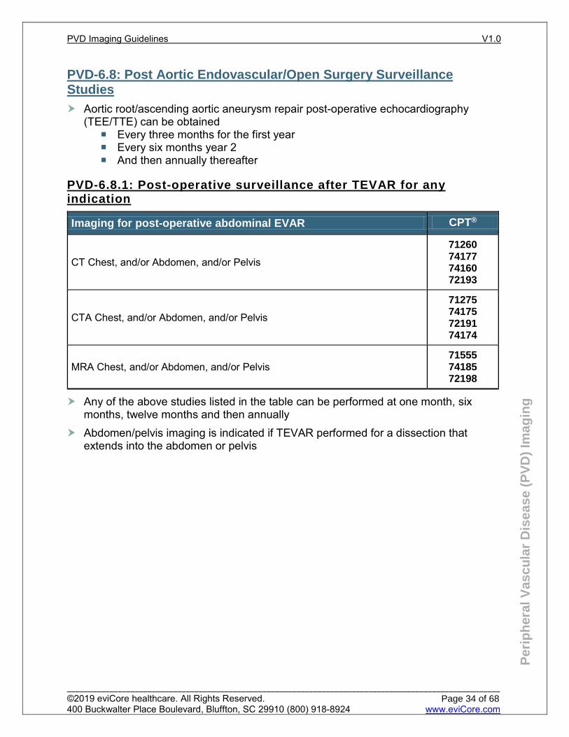
PVD-6.8.1: Post-operative surveillance after TEVAR for any indication
Imaging for post-operative abdominal EVAR CPT®
CT Chest, and/or Abdomen, and/or Pelvis
71260 74177 74160 72193
CTA Chest, and/or Abdomen, and/or Pelvis
71275 74175 72191 74174
MRA Chest, and/or Abdomen, and/or Pelvis 71555 74185 72198
Any of the above studies listed in the table can be performed at one month, six months, twelve months and then annually
Abdomen/pelvis imaging is indicated if TEVAR performed for a dissection that extends into the abdomen or pelvis
PVD Imaging Guidelines V1.0
______________________________________________________________________________________________________ ©2019 eviCore healthcare. All Rights Reserved. 400 Buckwalter Place Boulevard, Bluffton, SC 29910 (800) 918-8924 www.eviCore.com
Page 34 of 68
Per
iph
eral
Vas
cula
r D
isea
se (
PV
D)
Imag
ing
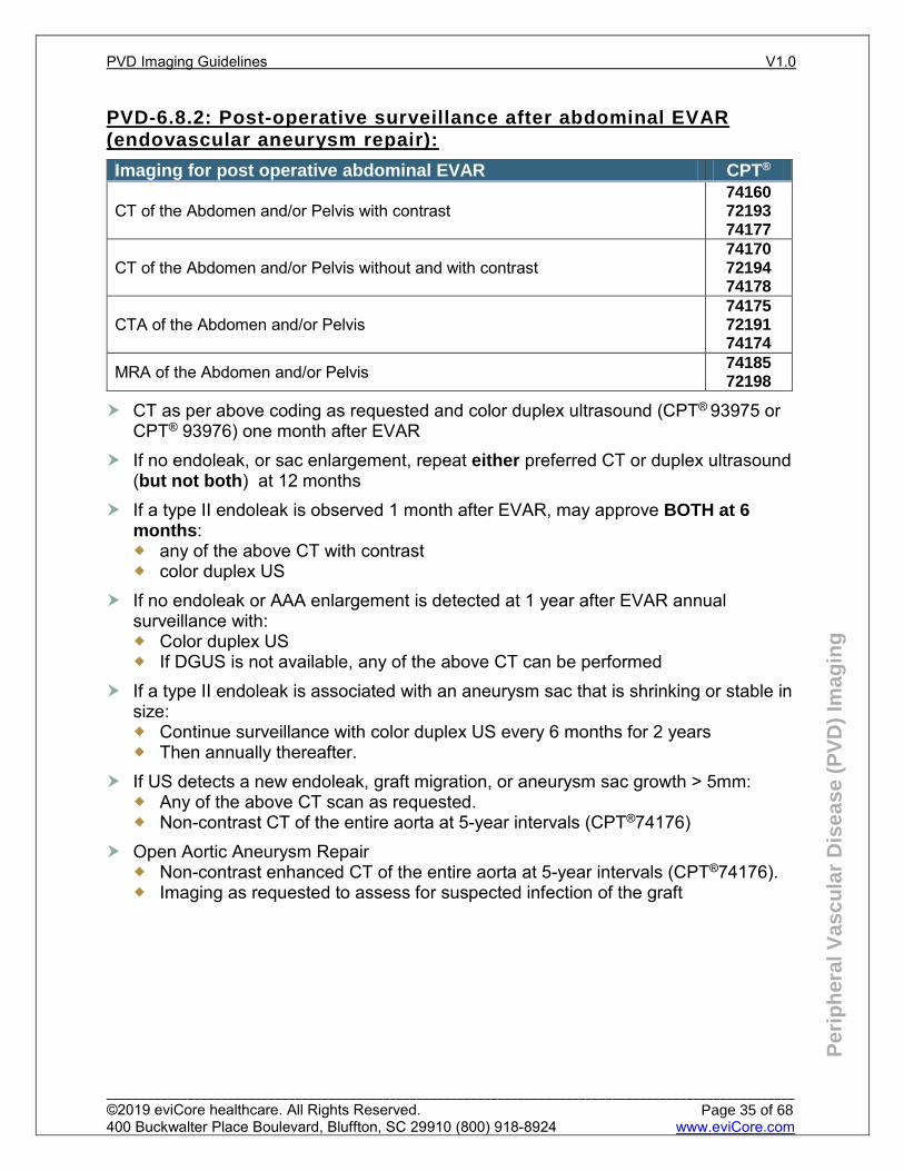
PVD-6.8.2: Post-operative surveillance after abdominal EVAR (endovascular aneurysm repair):
Imaging for post operative abdominal EVAR CPT®
CT of the Abdomen and/or Pelvis with contrast 74160 72193 74177
CT of the Abdomen and/or Pelvis without and with contrast 74170 72194 74178
CTA of the Abdomen and/or Pelvis 74175 72191 74174
MRA of the Abdomen and/or Pelvis 74185 72198
CT as per above coding as requested and color duplex ultrasound (CPT® 93975 or CPT® 93976) one month after EVAR
If no endoleak, or sac enlargement, repeat either preferred CT or duplex ultrasound (but not both) at 12 months
If a type II endoleak is observed 1 month after EVAR, may approve BOTH at 6 months: any of the above CT with contrast color duplex US
If no endoleak or AAA enlargement is detected at 1 year after EVAR annual surveillance with: Color duplex US If DGUS is not available, any of the above CT can be performed
If a type II endoleak is associated with an aneurysm sac that is shrinking or stable in size: Continue surveillance with color duplex US every 6 months for 2 years Then annually thereafter.
If US detects a new endoleak, graft migration, or aneurysm sac growth > 5mm: Any of the above CT scan as requested. Non-contrast CT of the entire aorta at 5-year intervals (CPT®74176)
Open Aortic Aneurysm Repair Non-contrast enhanced CT of the entire aorta at 5-year intervals (CPT®74176). Imaging as requested to assess for suspected infection of the graft
PVD Imaging Guidelines V1.0
______________________________________________________________________________________________________ ©2019 eviCore healthcare. All Rights Reserved. 400 Buckwalter Place Boulevard, Bluffton, SC 29910 (800) 918-8924 www.eviCore.com
Page 35 of 68
Per
iph
eral
Vas
cula
r D
isea
se (
PV
D)
Imag
ing
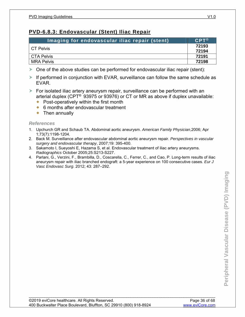
PVD-6.8.3: Endovascular (Stent) Iliac Repair
One of the above studies can be performed for endovascular iliac repair (stent): If performed in conjunction with EVAR, surveillance can follow the same schedule as
EVAR. For isolated iliac artery aneurysm repair, surveillance can be performed with an
arterial duplex (CPT® 93975 or 93976) or CT or MR as above if duplex unavailable: Post-operatively within the first month 6 months after endovascular treatment Then annually
References Upchurch GR and Schaub TA. Abdominal aortic aneurysm. American Family Physician,2006; Apr
1;73(7):1198-1204. Back M. Surveillance after endovascular abdominal aortic aneurysm repair. Perspectives in vascular
surgery and endovascular therapy, 2007;19: 395-400. Sakamoto I, Sueyoshi E, Hazama S, et al. Endovascular treatment of iliac artery aneurysms.
Radiographics October 2005;25:S213-S227. Parlani, G., Verzini, F., Brambilla, D., Coscarella, C., Ferrer, C., and Cao, P. Long-term results of iliac
aneurysm repair with iliac branched endograft: a 5-year experience on 100 consecutive cases. Eur J Vasc Endovasc Surg. 2012; 43: 287–292.
Imaging for endovascular i l iac repair (stent) CPT®
CT Pelvis 72193 72194
CTA Pelvis 72191 MRA Pelvis 72198
PVD Imaging Guidelines V1.0
______________________________________________________________________________________________________ ©2019 eviCore healthcare. All Rights Reserved. 400 Buckwalter Place Boulevard, Bluffton, SC 29910 (800) 918-8924 www.eviCore.com
Page 36 of 68
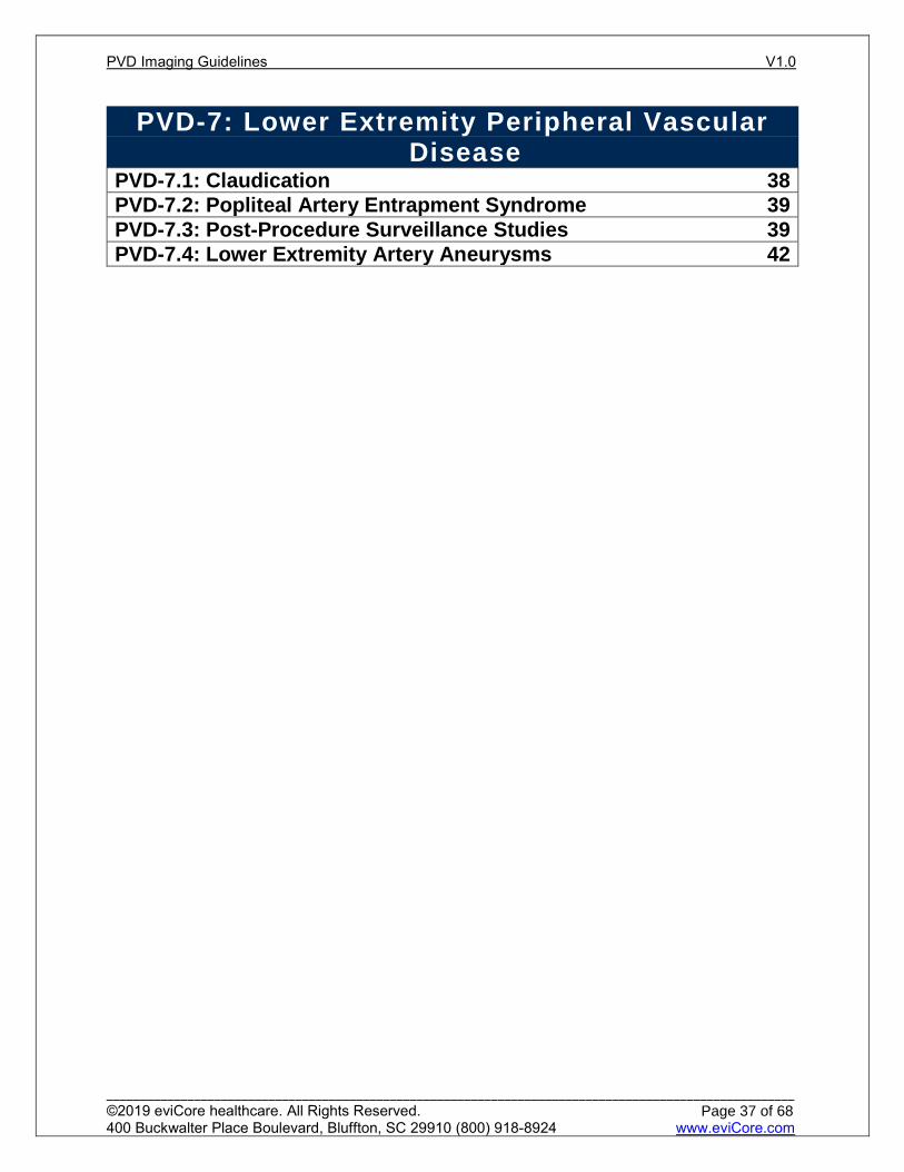
PVD-7: Lower Extremity Peripheral Vascular Disease
PVD-7.1: Claudication 38 PVD-7.2: Popliteal Artery Entrapment Syndrome 39 PVD-7.3: Post-Procedure Surveillance Studies 39 PVD-7.4: Lower Extremity Artery Aneurysms 42
PVD Imaging Guidelines V1.0
______________________________________________________________________________________________________ ©2019 eviCore healthcare. All Rights Reserved. 400 Buckwalter Place Boulevard, Bluffton, SC 29910 (800) 918-8924 www.eviCore.com
Page 37 of 68
Per
iph
eral
Vas
cula
r D
isea
se (
PV
D)
Imag
ing
PVD-7.1: Claudication
Initial evaluation for suspected PAD should be with a resting ABI. This can be accomplished at the bedside as part of the physical examination or requested as CPT® 93922 (limited Doppler ultrasound) or CPT® 93923 (multi-level complete Doppler ultrasound) CPT® 93923 may be performed once Follow-up studies may be performed with CPT® 93922 If the resting ABI is > 0.89 and PAD is still highly suspected clinically, then a
post-exercise ABI (CPT® 93924) can be performed History and physical suggestive of PAD include:
History Claudication- reproducible calf or thigh cramping with exertion that is relieved
completely with rest Critical limb ischemia Rest pain suggestive of ischemia-pain in the ball of foot when the leg is in an
elevated position particularly at night Distal non-healing wound or punched out ulcer with sharply demarcated
edges present for >2 weeks with no evidence of healing, i.e. presence of granulation tissue
Physical Examination Abnormal lower extremity pulse examination Vascular bruit Non-healing lower extremity wound Lower extremity gangrene Other suggestive lower extremity physical findings (e.g., elevation
pallor/dependent rubor) Atrophic nails, hair loss, shiny skin
If resting ABI (CPT® 93922) is normal (0.9 to 1.3) and disease is still suspected: Differentiate from “pseuodoclaudication”. See also: SP-9: Lumbar Spinal
Stenosis in the Spine Imaging Guidelines Re-measure ABI after exercise (CPT® 93924)1 A toe-brachial index may be used as further screening in patients with ABI’s
greater than 1.3 Advanced imaging is necessary only if there is consideration for invasive
therapy2,3,4,5 not to confirm diagnosis Duplex ultrasound (CPT® 93925 bilateral study or CPT® 93926 unilateral study) and
Doppler studies are adjuncts to abnormal ABI that may be used to identify location and extent of disease once there has been a decision for revascularization:6,7
MRA Aorta and Pelvic vessels, and Lower extremities (CPT® 74185, CPT® 73725 and CPT® 73725), or CTA with run-off (CPT® 75635) to further evaluate the lower extremity arteries for the purpose of preoperative planning for any of the following:2,8 Intermittent claudication (i.e. non-limb threatening ischemia) AND either:
Failed 3 months conservative medical therapy (physician supervised walking / exercise program plus medical therapy), or
PVD Imaging Guidelines V1.0
______________________________________________________________________________________________________ ©2019 eviCore healthcare. All Rights Reserved. 400 Buckwalter Place Boulevard, Bluffton, SC 29910 (800) 918-8924 www.eviCore.com
Page 38 of 68
Per
iph
eral
Vas
cula
r D
isea
se (
PV
D)
Imag
ing
Functional disability (e.g. exercise impairment sufficient to threaten the patient’s employment or to require significant alterations in the patient’s lifestyle)
Potentially limb-threatening vascular disease evidenced by: Skin breakdown Non-healing ischemic ulcers Resting leg pain Gangrene
Blue Toe Syndrome: Emboli from aortic plaque or mural thrombus Hyperviscosity syndrome Hypercoagulable states Vasculitis
Note: MRA Pelvis should not be requested/billed with CPT® 74185, CPT® 73725 and CPT® 73725
Practice Notes Claudication symptoms usually remain stable (70% to 80% of patients) and do not worsen or improve at rapid rates.9 Repeat studies to assess the efficacy of medical therapy are not indicated unless there is a negative change in clinical status for the purpose of preoperative planning for the purpose of preoperative planning such as worsening claudication or progression to critical limb ischemia
PVD-7.2: Popliteal Artery Entrapment Syndrome
Diagnosis of popliteal artery stenosis or occlusion due to compression by adjacent muscle and tendons seen in young men (ages 20 to 40).10 Ultrasound (CPT® 93926 unilateral study), CTA Lower extremity (CPT® 73706), or
MRA Lower extremity (CPT® 73725). CT or MRI of the lower extremity (contrast as requested) if requested by the
operating surgeon
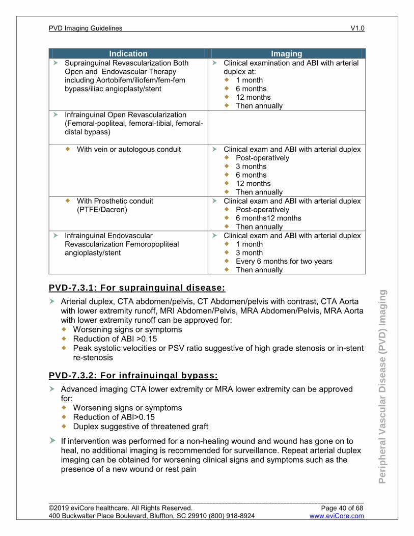
PVD-7.3: Post-Procedure Surveillance Studies
Scheduled Interval ABI (CPT® 93922) is generally appropriate following any revascularization
procedure ABI (CPT® 93922) or Duplex ultrasound (CPT® 93926 unilateral study) at each
routine follow up is appropriate generally after a history/physical has been performed
Further imaging studies such as CTA or MRA are indicated for worsening symptoms, an abnormal duplex or a significant reduction (>0.15) in the ABI
PVD Imaging Guidelines V1.0
______________________________________________________________________________________________________ ©2019 eviCore healthcare. All Rights Reserved. 400 Buckwalter Place Boulevard, Bluffton, SC 29910 (800) 918-8924 www.eviCore.com
Page 39 of 68
Per
iph
eral
Vas
cula
r D
isea
se (
PV
D)
Imag
ing
Indication Imaging Suprainguinal Revascularization Both
Open and Endovascular Therapy including Aortobifem/iliofem/fem-fem bypass/iliac angioplasty/stent
Clinical examination and ABI with arterial duplex at: 1 month 6 months 12 months Then annually
Infrainguinal Open Revascularization (Femoral-popliteal, femoral-tibial, femoral-distal bypass)
With vein or autologous conduit
Clinical exam and ABI with arterial duplex Post-operatively 3 months 6 months 12 months Then annually
With Prosthetic conduit (PTFE/Dacron)
Clinical exam and ABI with arterial duplex Post-operatively 6 months12 months Then annually
Infrainguinal Endovascular Revascularization Femoropopliteal angioplasty/stent
Clinical exam and ABI with arterial duplex 1 month 3 month Every 6 months for two years Then annually
PVD-7.3.1: For suprainguinal disease:
Arterial duplex, CTA abdomen/pelvis, CT Abdomen/pelvis with contrast, CTA Aorta with lower extremity runoff, MRI Abdomen/Pelvis, MRA Abdomen/Pelvis, MRA Aorta with lower extremity runoff can be approved for: Worsening signs or symptoms Reduction of ABI >0.15 Peak systolic velocities or PSV ratio suggestive of high grade stenosis or in-stent
re-stenosis
PVD-7.3.2: For infrainuingal bypass:
Advanced imaging CTA lower extremity or MRA lower extremity can be approved for: Worsening signs or symptoms Reduction of ABI>0.15 Duplex suggestive of threatened graft
If intervention was performed for a non-healing wound and wound has gone on to heal, no additional imaging is recommended for surveillance. Repeat arterial duplex imaging can be obtained for worsening clinical signs and symptoms such as the presence of a new wound or rest pain
PVD Imaging Guidelines V1.0
______________________________________________________________________________________________________ ©2019 eviCore healthcare. All Rights Reserved. 400 Buckwalter Place Boulevard, Bluffton, SC 29910 (800) 918-8924 www.eviCore.com
Page 40 of 68
Per
iph
eral
Vas
cula
r D
isea
se (
PV
D)
Imag
ing
References Dick F, Ricco J-B, Davies A.H, Cao P et al. Chapter VI: Follow-up after Revascularization. European
Journal of Vascular and Endovascular Surgery, Volume 42, Supplement 2, 2011 pp.S75-90. Lane TRA et al. Post-Operative Surveillance after Open Peripheral Arterial Surgery. European
Journal of Vascular and Endovascular Surgery. Volume 42, Issue 1, July 2011. Pages 59-77. Brooke BS, Beck AW, Kraiss LW, et al. Association of Quality Improvement Registry Participation
with Appropriate Follow-up after Vascular Procedures. JAMA Surgery. 2018;153(3):216. doi:10.1001/jamasurg.2017.3942.
Rooke TW, Hirsch AT, Misra S, Sidawy AN, et al. 2011 ACCF/AHA focused update of the guideline for the management of patients with peripheral artery disease (updating the 2005 guideline): a report of the American College of Cardiology Foundation/American Heart Association Task Force on Practice Guidelines: developed in collaboration with the Society for Cardiovascular Angiography and Interventions, Society of Interventional Radiology, Society for Vascular Medicine, and Society for Vascular Surgery. American College of Cardiology Foundation/American Heart Association Task Force on Practice Guidelines., Society for Cardiovascular Angiography and Interventions., Society of Interventional Radiology., Society for Vascular Medicine., Society for Vascular Surgery. J Vasc Surg. 2011 Nov; 54(5):e32-58
Conte MS, Pomposelli FB, and Clair DG. Society for Vascular Surgery practice guidelines for atherosclerotic occlusive disease of the lower extremities: Management of asymptomatic disease and claudication. Journal of Vascular Surgery. March 2015;61(3):2S-41S.e1
Bui TD, Mills JL Sr, Inhnat DM et al. The natural history of duplex-detected stenosis after femoropopliteal endovascular therapy suggests questionable clinical utility of routine duplex surveillance. J Vasc Surg. 2012 Feb 55:2:346-352
Troutman DA, Madden NJ, Dougherty MJ,et al. Duplex Ultrasound Diagnosis of Failing Stent Grafts Placed for Occlusive Disease. J Vasc Surg 2014;70:1–5.
Humphries MD, Pevec WC, Laird JR, et.al. Early Duplex Scanning after Infrainguinal Endovascular Therapy. J Vasc Surg 2011; 53:353–8.
Zierler RE, Jordan WD, Lal BK, et al. The Society for Vascular Surgery practice guidelines on follow-up after vascular surgery arterial procedures. J Vasc Surg. 2018 Jul;68(1):256-284. doi: 10.1016/j.jvs.2018.04.018.
Cooper K, Majdalany BS, Kalva SP, et al. ACR Appropriateness Criteria<sup>®</sup> Lower Extremity Arterial Revascularization—Post-Therapy Imaging. Journal of the American College of Radiology. 2018;15:S104-S115.
PVD Imaging Guidelines V1.0
______________________________________________________________________________________________________ ©2019 eviCore healthcare. All Rights Reserved. 400 Buckwalter Place Boulevard, Bluffton, SC 29910 (800) 918-8924 www.eviCore.com
Page 41 of 68
Per
iph
eral
Vas
cula
r D
isea
se (
PV
D)
Imag
ing
PVD-7.4: Lower Extremity Artery Aneurysms
For Iliac artery aneurysm see also: PVD-6.4: Iliac Artery Aneurysm (IAA) Femoral artery aneurysm
Initial imaging Ultrasound (CPT® 93925 bilateral study or CPT® 93926 unilateral study).
Surveillance imaging Symptomatic true femoral aneurysms smaller than 2.5 cm in diameter Ultrasound (CPT® 93926 unilateral study) annually
Symptomatic true femoral aneurysms larger than 2.5 cm Ultrasound (CPT® 93926 unilateral study) every 6 months
Other imaging CTA Lower extremity or MRA Lower extremity without or with contrast can be
performed when: Preoperative study for patients with no plans for invasive angiography Technically limited or abnormal ultrasound results
Popliteal artery aneurysm Initial imaging
Ultrasound (CPT® 93925 bilateral study or CPT® 93926 unilateral study) and Ultrasound to assess for a contralateral popliteal aneurysm and abdominal aortic aneurysm (CPT® 76770 or CPT® 76775)
Surveillance imaging Ultrasound (CPT® 93926 unilateral study) annually Post-interventional functional testing (ABI) (CPT® 93922) may be useful as
clinically indicated Other imaging
CTA or MRA can be performed for: Preoperative study for patients with no plans for invasive angiography Technically limited or abnormal ultrasound results
PVD Imaging Guidelines V1.0
______________________________________________________________________________________________________ ©2019 eviCore healthcare. All Rights Reserved. 400 Buckwalter Place Boulevard, Bluffton, SC 29910 (800) 918-8924 www.eviCore.com
Page 42 of 68
Per
iph
eral
Vas
cula
r D
isea
se (
PV
D)
Imag
ing
References Hirsch AT, Haskal ZJ, Hertzer NR, et al. ACC/AHA Practice Guidelines: ACC/AHA 2005 Practice
guidelines for the management of patients with peripheral arterial disease (lower extremity, renal, mesenteric, and abdominal aortic): a collaborative report from the American Association for Vascular Surgery/Society for Vascular Surgery,* Society for Cardiovascular Angiography and Interventions, Society for Vascular Medicine and Biology, Society of Interventional Radiology, and the ACC/AHA Task Force on Practice Guidelines. Circulation. 2006; 113: e463-e654.
Gerhard-Herman MD, Gornik HL, Barrett C, et al. 2016 AHA/ACC Guideline on the management of patients with lower extremity peripheral artery disease: a report of the American College of Cardiology/American Heart Association Task Force on Clinical Practice Guidelines. Circulation. 2017; 135: e726-e779.
Ahmed O, Hanley M, Bennett SJ, et al. ACR Appropriateness Criteria® Vascular claudication -Assessment for Revascularization. J Am Coll Radiol. 2017 May;14(5S):S372-S379. doi: 10.1016/j.jacr.2017.02.037.
Fowkes FG, Murray GD, Butcher I, et al. Ankle brachial index combined with Framingham Risk Score to predict cardiovascular events and mortality: a meta-analysis. JAMA. 2008 Jul 9;300 (2):197-208.
Federman DG, Kravetz JD, Bravata DM, et al. Peripheral artery disease: a marker of morbidity and mortality. Postgrad Med. 2006; 119 (2):21-27. doi:10.3810/pgm.2006.07.1746.
Willmann JK, Baumert B, Schertler T, et al. Aortoiliac and lower extremity arteries assessed with 16–detector row CT angiography: prospective comparison with digital subtraction angiography. Radiology. 2005 Sep; 236 (3):1083-1093. doi:10.1148/radiol.2362040895.
Allison MA, Hiatt WR, Hirsch AT, et al. A high ankle-brachial index is associated with increased cardiovascular disease morbidity and lower quality of life. J Am Coll Cardiol. 2008 Apr 1;51(13):1292-1298.
Macedo TA, Johnson M, Hallett Jr JW, and Breen JF. Popliteal artery entrapment syndrome: role of imaging in the diagnosis. AJR Am J Roentgen. 2003 Nov;181(5):1259-1265. doi:10.2214/ajr.181.5.1811259.
PVD Imaging Guidelines V1.0
______________________________________________________________________________________________________ ©2019 eviCore healthcare. All Rights Reserved. 400 Buckwalter Place Boulevard, Bluffton, SC 29910 (800) 918-8924 www.eviCore.com
Page 43 of 68
PVD-8: Imaging for Hemodialysis Access PVD-8.1: Preoperative Arterial Evaluation and Venous Mapping Prior to AV Fistula Creation 45
PVD Imaging Guidelines V1.0
______________________________________________________________________________________________________ ©2019 eviCore healthcare. All Rights Reserved. 400 Buckwalter Place Boulevard, Bluffton, SC 29910 (800) 918-8924 www.eviCore.com
Page 44 of 68
Per
iph
eral
Vas
cula
r D
isea
se (
PV
D)
Imag
ing
PVD-8.1: Preoperative Arterial Evaluation and Venous Mapping Prior to AV Fistula Creation
For vessel mapping prior to AV fistula creation CPT® 93985 or 93986
In some instances, MRA Upper Extremity may be needed (CPT® 73225) if duplex imaging is equivocal
Arterial evaluation to assess arterial suitability (size, degree of stenosis and calcification) prior to AV fistula creation may be appropriate CPT® 93930 or CPT® 93931 can be used to report upper extremity arterial
evaluation Venous mapping to assess venous suitability prior to AV fistula creation may be
appropriate CPT® 93970 or CPT® 93971 can be used to report venous mapping
Indications for Duplex ultrasound (CPT® 93990) of hemodialysis access include but are not limited to: Patients with decreased flow rates during hemodialysis. Development of arm swelling or discomfort after access placement surgery or a
hemodialysis session. Prolonged immaturity of a surgically created AV fistula. Suspected pseudoaneurysm. Suspected AV fistula or graft stenosis. Known or suspected fluid collection adjacent to an AV fistula or graft. Though it is, generally, not needed, one Duplex US (CPT® 93990) can be
performed after a surgically created AV fistula for assessment.
References Sidawy AN, Spergel LM, Besarab A, et al. The Society for Vascular Surgery: Clinical practice
guidelines for the surgical placement and maintenance of arteriovenous hemodialysis access. Journal of Vascular Surgery. 2008;48(5). doi:10.1016/j.jvs.2008.08.042.
PVD Imaging Guidelines V1.0
______________________________________________________________________________________________________ ©2019 eviCore healthcare. All Rights Reserved. 400 Buckwalter Place Boulevard, Bluffton, SC 29910 (800) 918-8924 www.eviCore.com
Page 45 of 68
Per
iph
eral
Vas
cula
r D
isea
se (
PV
D)
Imag
ing
PVD-9: Arteriovenous Malformations (AVMs) See: PEDPVD-2.5: Arteriovenous Malformations (AVMs) and Fistulas
PVD Imaging Guidelines V1.0
______________________________________________________________________________________________________ ©2019 eviCore healthcare. All Rights Reserved. 400 Buckwalter Place Boulevard, Bluffton, SC 29910 (800) 918-8924 www.eviCore.com
Page 46 of 68
Per
iph
eral
Vas
cula
r D
isea
se (
PV
D)
Imag
ing
PVD-10: Nuclear Medicine Nuclear medicine
Nuclear medicine studies are rarely used in the evaluation of peripheral vascular disorders, but are indicated in the following circumstances: Lymphoscintigraphy (CPT® 78195) is indicated for evaluation of lower
extremity lymphedema when a recent Doppler ultrasound is negative for valvular insufficiency.
Vascular flow imaging (CPT® 78445) is an obsolete study that has been replaced by MRA, CTA, or Duplex ultrasonography, and is not supported for any indication at this time.
Venous thrombosis imaging (CPT® 78456, CPT® 78457, and CPT® 75458) are obsolete studies that have been replaced by MRA, CTA, or Duplex ultrasonography, and are not supported for any indication at this time.
Indium 111 (111In)–labeled white blood cell (WBC) or Gallium-67 citrate studies (CPT® 78800, CPT® 78801, CPT® 78802, or CPT® 78803) can be approved for evaluation of the following: Mycotic aneurysms. Vascular graft infection. Infection of central venous catheter or other indwelling device.
PVD Imaging Guidelines V1.0
______________________________________________________________________________________________________ ©2019 eviCore healthcare. All Rights Reserved. 400 Buckwalter Place Boulevard, Bluffton, SC 29910 (800) 918-8924 www.eviCore.com
Page 47 of 68
PVD-11: Venous Imaging General Information
PVD-11.1: Abbreviations and glossary 49 PVD-11.2: Background Information 49 PVD-11.3: Procedure Coding 51 PVD-11.4: General Guidelines-Imaging 52
PVD Imaging Guidelines V1.0
______________________________________________________________________________________________________ ©2019 eviCore healthcare. All Rights Reserved. 400 Buckwalter Place Boulevard, Bluffton, SC 29910 (800) 918-8924 www.eviCore.com
Page 48 of 68
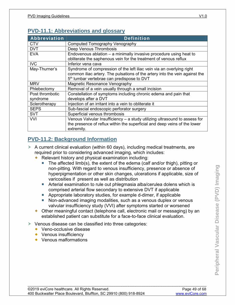
Per
iph
eral
Vas
cula
r D
isea
se (
PV
D)
Imag
ing
PVD-11.1: Abbreviations and glossary
CTV Computed Tomography Venography DVT Deep Venous Thrombosis EVA Endovenous ablation – a minimally invasive procedure using heat to
obliterate the saphenous vein for the treatment of venous reflux IVC Inferior vena cava May-Thurner’s Syndrome of compression of the left iliac vein via an overlying right
common iliac artery. The pulsations of the artery into the vein against the 5th lumbar vertebrae can predispose to DVT
MRV Magnetic Resonance Venography Phlebectomy Removal of a vein usually through a small incision Post thrombotic syndrome
Constellation of symptoms including chronic edema and pain that develops after a DVT
Sclerotherapy Injection of an irritant into a vein to obliterate it SEPS Sub-fascial endoscopic perforator surgery SVT Superficial venous thrombosis VVI Venous Valvular Insufficiency – a study utilizing ultrasound to assess for
the presence of reflux within the superficial and deep veins of the lower extremity.
PVD-11.2: Background Information
A current clinical evaluation (within 60 days), including medical treatments, are required prior to considering advanced imaging, which includes: Relevant history and physical examination including:
The affected limb(s), the extent of the edema (calf and/or thigh), pitting or non-pitting. With regard to venous insufficiency, presence or absence of hyperpigmentation or other skin changes, ulcerations if applicable, size of varicosities if present as well as distribution
Arterial examination to rule out phlegmasia alba/cerulea dolens which is comprised arterial flow secondary to extensive DVT if applicable
Appropriate laboratory studies, for example d-dimer, if applicable Non-advanced imaging modalities, such as a venous duplex or venous
valvular insufficiency study (VVI) after symptoms started or worsened Other meaningful contact (telephone call, electronic mail or messaging) by an
established patient can substitute for a face-to-face clinical evaluation. Venous disease can be classified into three categories:
Veno-occlusive disease Venous insufficiency Venous malformations
Abbreviation Definit ion
PVD Imaging Guidelines V1.0
______________________________________________________________________________________________________ ©2019 eviCore healthcare. All Rights Reserved. 400 Buckwalter Place Boulevard, Bluffton, SC 29910 (800) 918-8924 www.eviCore.com
Page 49 of 68
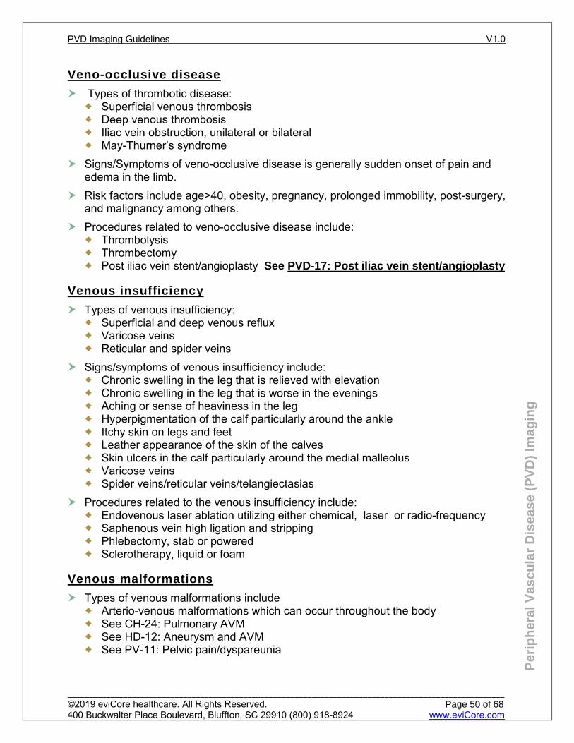
Per
iph
eral
Vas
cula
r D
isea
se (
PV
D)
Imag
ing
Veno-occlusive disease
Types of thrombotic disease: Superficial venous thrombosis Deep venous thrombosis Iliac vein obstruction, unilateral or bilateral May-Thurner’s syndrome
Signs/Symptoms of veno-occlusive disease is generally sudden onset of pain and edema in the limb.
Risk factors include age>40, obesity, pregnancy, prolonged immobility, post-surgery, and malignancy among others.
Procedures related to veno-occlusive disease include: Thrombolysis Thrombectomy Post iliac vein stent/angioplasty See PVD-17: Post iliac vein stent/angioplasty
Venous insufficiency
Types of venous insufficiency: Superficial and deep venous reflux Varicose veins Reticular and spider veins
Signs/symptoms of venous insufficiency include: Chronic swelling in the leg that is relieved with elevation Chronic swelling in the leg that is worse in the evenings Aching or sense of heaviness in the leg Hyperpigmentation of the calf particularly around the ankle Itchy skin on legs and feet Leather appearance of the skin of the calves Skin ulcers in the calf particularly around the medial malleolus Varicose veins Spider veins/reticular veins/telangiectasias
Procedures related to the venous insufficiency include: Endovenous laser ablation utilizing either chemical, laser or radio-frequency Saphenous vein high ligation and stripping Phlebectomy, stab or powered Sclerotherapy, liquid or foam
Venous malformations
Types of venous malformations include Arterio-venous malformations which can occur throughout the body See CH-24: Pulmonary AVM See HD-12: Aneurysm and AVM See PV-11: Pelvic pain/dyspareunia
PVD Imaging Guidelines V1.0
______________________________________________________________________________________________________ ©2019 eviCore healthcare. All Rights Reserved. 400 Buckwalter Place Boulevard, Bluffton, SC 29910 (800) 918-8924 www.eviCore.com
Page 50 of 68
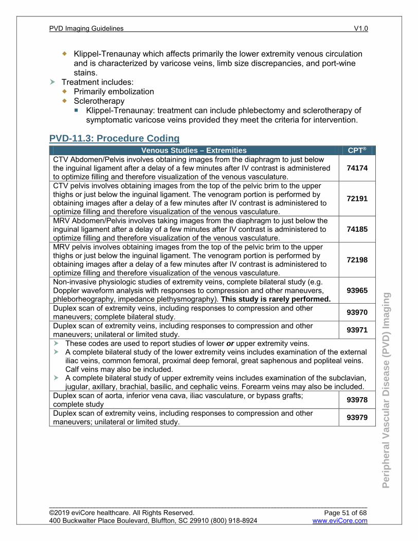
Per
iph
eral
Vas
cula
r D
isea
se (
PV
D)
Imag
ing
Klippel-Trenaunay which affects primarily the lower extremity venous circulation and is characterized by varicose veins, limb size discrepancies, and port-wine stains.
Treatment includes: Primarily embolization Sclerotherapy
Klippel-Trenaunay: treatment can include phlebectomy and sclerotherapy of symptomatic varicose veins provided they meet the criteria for intervention.
PVD-11.3: Procedure Coding Venous Studies – Extremities CPT® CTV Abdomen/Pelvis involves obtaining images from the diaphragm to just below the inguinal ligament after a delay of a few minutes after IV contrast is administered to optimize filling and therefore visualization of the venous vasculature.
74174
CTV pelvis involves obtaining images from the top of the pelvic brim to the upper thighs or just below the inguinal ligament. The venogram portion is performed by obtaining images after a delay of a few minutes after IV contrast is administered to optimize filling and therefore visualization of the venous vasculature.
72191
MRV Abdomen/Pelvis involves taking images from the diaphragm to just below the inguinal ligament after a delay of a few minutes after IV contrast is administered to optimize filling and therefore visualization of the venous vasculature.
74185
MRV pelvis involves obtaining images from the top of the pelvic brim to the upper thighs or just below the inguinal ligament. The venogram portion is performed by obtaining images after a delay of a few minutes after IV contrast is administered to optimize filling and therefore visualization of the venous vasculature.
72198
Non-invasive physiologic studies of extremity veins, complete bilateral study (e.g. Doppler waveform analysis with responses to compression and other maneuvers, phleborheography, impedance plethysmography). This study is rarely performed.
93965
Duplex scan of extremity veins, including responses to compression and other maneuvers; complete bilateral study. 93970
Duplex scan of extremity veins, including responses to compression and other maneuvers; unilateral or limited study. 93971
These codes are used to report studies of lower or upper extremity veins. A complete bilateral study of the lower extremity veins includes examination of the external
iliac veins, common femoral, proximal deep femoral, great saphenous and popliteal veins. Calf veins may also be included.
A complete bilateral study of upper extremity veins includes examination of the subclavian, jugular, axillary, brachial, basilic, and cephalic veins. Forearm veins may also be included.
Duplex scan of aorta, inferior vena cava, iliac vasculature, or bypass grafts; complete study 93978
Duplex scan of extremity veins, including responses to compression and other maneuvers; unilateral or limited study. 93979
PVD Imaging Guidelines V1.0
______________________________________________________________________________________________________ ©2019 eviCore healthcare. All Rights Reserved. 400 Buckwalter Place Boulevard, Bluffton, SC 29910 (800) 918-8924 www.eviCore.com
Page 51 of 68
Per
iph
eral
Vas
cula
r D
isea
se (
PV
D)
Imag
ing
PVD-11.4: General Guidelines-Imaging
Venous duplex (CPT® 93970, 93971) of the limb is the initial imaging of choice Follow-up duplex imaging (CPT® 93970, 93971) is not generally indicated to
document resolution and should only be obtained for new signs/symptoms or for concerns of propagation of thrombus when the treatment plan would change (Insertion of IVC filter, change of anticoagulation, etc.)
Imaging studies Venous duplex (CPT® 93970, 93971) should visualize the veins, with
demonstration of the presence or absence of compressibility and venous flow. Venous valvular insufficiency studies (CPT® 93970, 93971) visualize the veins of
the lower extremity, assess for reflux (reversal of venous antegrade flow after valve closure) and measure its duration.
CTV or MRV of the abdomen/pelvis images with contrast involves taking images from the diaphragm to just below the inguinal ligament after a delay of a few minutes after IV contrast is administered to optimize filling and therefore visualization of the venous vasculature.
References Ageno W et al. Characteristics and Management of Patients with Venous Thromboembolism: The
GARFIELD-VTE Registry. Thromg Haemost 2019. 119(2):319-327. Jones WS, Vemulapalli S, Parikh KS et al. Treatment Strategies for Patients with Lower Extremity
Chronic Venous Disease (LECVD. Agency for Healthcare Research and Quality (US); 2017 Apr. Gloviczki P, Comerota AJ, Dalsing MD, et al. The care of patients with varicose veins and associated
chronic venous diseases: Clinical practice guidelines of the Society for Vascular Surgery and the American Venous Forum. Journal of Vascular Surgery. 2011. 53(16S):1-47.
Wolpert L, Rahmani O, Stein B, et al. Magnetic Resonance Venography in the Diagnosis and Management of May-Thurner syndrome. Vasc Endovascular Surg. 2002 Jan-Feb;36(1):51-7.
PVD Imaging Guidelines V1.0
______________________________________________________________________________________________________ ©2019 eviCore healthcare. All Rights Reserved. 400 Buckwalter Place Boulevard, Bluffton, SC 29910 (800) 918-8924 www.eviCore.com
Page 52 of 68
PVD-12: Acute Limb Swelling PVD-12.1: Superficial venous thrombosis (SVT) 54 PVD-12.2: Acute deep venous thrombosis (DVT) 54 PVD-12.3: Follow-up imaging of known DVT: 55 PVD-12.4: Follow-up imaging after venous surgery: 55
PVD Imaging Guidelines V1.0
______________________________________________________________________________________________________ ©2019 eviCore healthcare. All Rights Reserved. 400 Buckwalter Place Boulevard, Bluffton, SC 29910 (800) 918-8924 www.eviCore.com
Page 53 of 68
Per
iph
eral
Vas
cula
r D
isea
se (
PV
D)
Imag
ing
PVD-12.1: Superficial venous thrombosis (SVT)
Superficial venous thrombosis (SVT) refers to acute or chronic thrombosis of thesuperficial veins in both the upper (cephalic and basilic veins) and lower extremities(greater [great] saphenous vein, lesser [small] saphenous vein, gastrocnemius andsoleal veins). Treatment: Elevation and warm compresses until pain and swellingsubsides.
The diagnosis of superficial venous thrombosis is generally made on the basis ofphysical examination. Duplex ultrasound (CPT® 93970, 93971) is the initial imaging if the diagnosis is
equivocal Follow-up duplex ultrasound (CPT® 93970, 93971) is indicated only if thrombus
in the superficial systems is encroaching onto the deep venous system (saphenofemoral or saphenopopliteal junction)
PVD-12.2: Acute deep venous thrombosis (DVT)
Deep venous thrombosis is characterized by thrombosis of a deep vein in either theupper (brachial, axillary, subclavian veins) or the lower extremity (peroneal, posteriortibial, popliteal, femoral or iliac veins).
Duplex ultrasound (CPT® 93970 bilateral study or CPT® 93971 unilateral study) is theinitial imaging study for any suspected DVT Deep venous thrombosis can present as
Symptomatic Swelling Pain Warmth Erythema Pain with dorsiflexion of the foot (Homan’s Sign) Or with progression, such as phlegmasia cerulean dolens Risk factors for DVT include age >40, obesity, malignancy, prolonged
immobilization, hypercoaguability as well as those outlined in CH-25:Pulmonary Embolism (PE) in Chest Imaging Guidelines.
CTA/CTV Abdomen and pelvis with contrast can be performed to rule out IVCthrombus secondary to the filter when there is acute bilateral lower extremityswelling in a patient with a history of an IVC filter in place.
When there is concern for proximal DVT (iliofemoral): Focused abdominal duplex can generally visualize the iliac veins and IVC to
determine the absence or presence of iliac vein thrombus in a patient. If the results are equivocal or indeterminate: CTV or MRV abdomen and pelvis with contrast (CPT® 74174 or 74185) can
be performed. For proximal DVT’s (iliac vein DVT’s or in cases of phlegmasia (extensive DVT
compromising arterial inflow), thrombectomy (rarely performed) or thrombolysis canbe performed.
PVD Imaging Guidelines V1.0
______________________________________________________________________________________________________ ©2019 eviCore healthcare. All Rights Reserved. 400 Buckwalter Place Boulevard, Bluffton, SC 29910 (800) 918-8924 www.eviCore.com
Page 54 of 68
Per
iph
eral
Vas
cula
r D
isea
se (
PV
D)
Imag
ing
If the cause of the DVT is found to be due to May-Thurner, iliac vein angioplasty followed by stenting of the left iliac vein is generally performed. See PVD-13.3 May-Thurner Syndrome
PVD-12.3: Follow-up imaging of known DVT:
Duplex ultrasound (CPT® 93970 bilateral study or CPT® 93971 unilateral study) can be repeated in order to rule out proximal extension of a calf vein DVT in those individuals who cannot be anticoagulated, most commonly after recent surgery. Time interval for follow-up study includes One week after the initial diagnosis. Serial imaging (up to 3 studies) over the first three weeks if calf DVT is not
treated. Imaging during or to terminate long-term anticoagulation therapy to determine
venous recanalization is not supported by evidence. Repeat imaging to make decisions on whether or not to continue or terminate anticoagulation is not indicated.
PVD-12.4: Follow-up imaging after venous surgery:
Venous duplex (CPT® 93971 unilateral study) can be obtained of the treated limb to rule out a DVT within seven days of endovenous ablation.
Follow-up routine imaging is not indicated after other venous procedures including: Saphenous vein ligation and stripping Phlebectomy Sclerotherapy
References Lim W, Gal GL, Bates SM, et al. American Society of Hematology 2018 guidelines for management of
venous thromboembolism: diagnosis of venous thromboembolism. Blood Advances. 2018;2(22):3226-3256. doi:10.1182/bloodadvances.2018024828.
American College of Phlebology. Treatment of Superficial Venous Disease of the Lower Leg Guidelines. American Vein & Lymphatic Society. http://www.phlebology.org/member-resources/publications/treatment-superficial-venous-disease-lower-leg-guidelines. Published 2016.
American College of Phlebology. Duplex Ultrasound Imaging of Lower Extremity Veins in Chronic Venous Disease. American Vein & Lymphatic Society. http://www.phlebology.org/member-resources/publications/duplex-ultrasound-imaging-lower-extremity-veins-chronic-venous-disease. Published 2012.
Healy DA, Kimura S, Power D, et al. A Systematic Review and Meta-analysis of Thrombotic Events Following Endovenous Thermal Ablation of the Great Saphenous Vein. European Journal of Vascular and Endovascular Surgery. 2018;56(3):410-424. doi:10.1016/j.ejvs.2018.05.008.
Jacobs CE, Pinzon MM, Orozco J, et al. Deep Venous Thrombosis after Saphenous Endovenous Radiofrequency Ablation: Is it Predictable? Annals of Vascular Surgery. 2014;28(3):679-685. doi:10.1016/j.avsg.2013.08.012.
PVD Imaging Guidelines V1.0
______________________________________________________________________________________________________ ©2019 eviCore healthcare. All Rights Reserved. 400 Buckwalter Place Boulevard, Bluffton, SC 29910 (800) 918-8924 www.eviCore.com
Page 55 of 68
PVD-13 Chronic limb swelling due to chronic deep venous thrombosis/May Thurner’s
syndrome PVD-13.1: Chronic deep venous thrombosis general information 57 PVD-13.2: Post thrombotic syndrome 57 PVD-13.3: May-Thurner’s syndrome general information 58
PVD Imaging Guidelines V1.0
______________________________________________________________________________________________________ ©2019 eviCore healthcare. All Rights Reserved. 400 Buckwalter Place Boulevard, Bluffton, SC 29910 (800) 918-8924 www.eviCore.com
Page 56 of 68
Per
iph
eral
Vas
cula
r D
isea
se (
PV
D)
Imag
ing
PVD-13.1: Chronic deep venous thrombosis general information
Chronic deep venous thrombosis is defined as an acute DVT that is greater than 14 days old.
Patient with incompletely lysed or residual DVT can develop post-thrombotic syndrome that can be characterized as chronic edema, venous stasis changes, pain and in advanced cases venous stasis ulceration. Incompletely lysed DVT can cause luminal narrowing of the vein restricting
venous outflow leading to stenosis or occlusion and /or can lead to valve dysfunction resulting in reflux of venous blood retrograde towards gravity. Both pathologies ultimately lead to chronic edema which can cause chronic pain and venous stasis disease. Imaging for post-thrombotic syndrome is not indicated unless there are signs
and symptoms suggestive of a new acute DVT or for preoperative planning for iliac vein/stenting for suspected iliac vein stenosis or occlusion.
The mainstay of treatment for chronic deep venous thrombosis is compression stockings.
In patients with a history of proximal (iliofemoral) DVT who have subsequently developed post thrombotic syndrome, imaging can be performed to evaluate for iliac venous obstruction which can result from incompletely lysed thrombus. Initial imaging should be duplex (CPT® 93970 bilateral study or CPT® 93971
unilateral study) followed by either a CT or MR venogram of the abdomen/pelvis, or CT or MR venogram of the pelvis, or venography for treatment planning purposes.
Selected patients may be a candidate for iliac vein angioplasty/stenting.
PVD-13.2: Post thrombotic syndrome
Imaging for post-thrombotic syndrome is indicated when: There are signs and symptoms suggestive of a new acute DVT and NOT for
chronic swelling that has not changed in severity or character For preoperative planning for iliac vein/stenting in the setting of known iliac
venous obstruction in those with a history of a proximal (iliofemoral) DVT. See PVD-3.1: Chronic deep venous thrombosis general information
See PVD-13.3 May-Thurner’s syndrome See PVD-15: Venous Stasis Ulceration
PVD Imaging Guidelines V1.0
______________________________________________________________________________________________________ ©2019 eviCore healthcare. All Rights Reserved. 400 Buckwalter Place Boulevard, Bluffton, SC 29910 (800) 918-8924 www.eviCore.com
Page 57 of 68
Per
iph
eral
Vas
cula
r D
isea
se (
PV
D)
Imag
ing
PVD-13.3: May-Thurner’s syndrome general information
In approximately 25% of people, the right iliac artery overlies the left iliac vein over the fifth lumbar vertebrae and its pulsations can compress the vein increasing the risk of DVT in the left extremity. Duplex (CPT® 93970 bilateral study or CPT® 93971 unilateral study) will confirm
the presence of a left common iliac vein DVT but diagnosis is made with advanced imaging such as CT or MRV abdomen/pelvis (CPT® code 74175, 74185) , venography or peri-procedural intravascular ultrasound demonstrating compression of the vein.
Treatment is with iliac vein angioplasty/stenting for both acute and chronic left-sided DVT
Prophylactic treatment of May-Thurner’s syndrome in the absence of acute or chronic DVT OR chronic left lower extremity edema and its sequelae such as varicose veins or venous stasis ulcers is NOT considered medically necessary.
References Eberhardt RT and Raffetto JD. Chronic Venous Insufficiency. Circulation. 2005; 111: 2398-2409. Partsch H, Flour M, Smith PC, et al. Indications for compression therapy in venous and lymphatic
disease. Consensus based on experimental data and scientific evidence. Int Angiol. 2008;27:193-219.
Nayak L, Vedantham S. Multifaceted Management of the Postthrombotic Syndrome. Seminars in Interventional Radiology. 2012: 29:1.
Christof T. Kaltenmeier, Erben Y, et al. Systematic review of May-Thurner syndrome with emphasis on gender differences. Journal of Vascular Surgery Venous and Lymphatic Disorders. 2018;6(3):399-407.
Rossi FH, Kambara AM, Izukawa NM, et al. Randomized double-blinded study comparing medical treatment versus iliac vein stenting in chronic venous disease. Journal of Vascular Surgery: Venous and Lymphatic Disorders. 2018;6(2):183-191.
Sermsathanasawadi N, Pruekprasert K, Pitaksantayothin W, et al. Prevalence, risk factors, and evaluation of iliocaval obstruction in advanced chronic venous insufficiency. J Vasc Surg Venous Lymphat Disord. 2019 May;7(3):441-447.
Vedantham S, Kahn SR, Goldhaber SZ, Comerota AJ, et al. Endovascular therapy for advanced post-thrombotic syndrome: Proceedings from a multidisciplinary consensus panel. Vasc Med. 2016 Aug;21(4):400-7.
PVD Imaging Guidelines V1.0
______________________________________________________________________________________________________ ©2019 eviCore healthcare. All Rights Reserved. 400 Buckwalter Place Boulevard, Bluffton, SC 29910 (800) 918-8924 www.eviCore.com
Page 58 of 68
PVD-14: Chronic limb swelling due to venous insufficiency/Venous stasis changes/Varicose
veins PVD-14.1: Venous insufficiency-General 60 PVD-14.2: Venous Reflux 60 PVD-14.3: Varicose Veins 61 PVD-14.4: Spider veins/reticular veins 61
PVD Imaging Guidelines V1.0
______________________________________________________________________________________________________ ©2019 eviCore healthcare. All Rights Reserved. 400 Buckwalter Place Boulevard, Bluffton, SC 29910 (800) 918-8924 www.eviCore.com
Page 59 of 68

Per
iph
eral
Vas
cula
r D
isea
se (
PV
D)
Imag
ing
PVD-14.1: Venous insufficiency-General
Venous insufficiency is characterized by failure of the venous blood to flow in its normal antegrade path of flow and instead reflux backwards by the force of gravity usually secondary to malfunction of the venous valves.
Risk factors include previous DVT, obesity, female sex, hereditary, and environmental factors such as prolonged standing on a hard surface.
Venous insufficiency loosely includes the diagnosis of venous reflux, varicose veins, venous stasis ulcers and spider/reticular veins.
PVD-14.2: Venous Reflux
Diagnosis is made with a venous valvular insufficiency study (CPT® 93970 bilateral study or CPT® 93971 unilateral study) which documents the presence of reflux (>500ms) in the greater saphenous vein as well as the size of the refluxing vein (3-15mm).
Symptoms of venous reflux include chronic edema, pain, and venous stasis ulcerations. Symptoms of venous reflux can be ameliorated with compression therapy with graded compression stockings, elevation, avoidance of prolonged standing and weight loss. Venous reflux can be seen in both the deep and superficial venous systems. Reflux within the deep system is not amenable to intervention. Treatment of deep venous reflux is via active compression with compression
stocks, pneumatic pumps or specialized dressings such as Unna boots. Treatment of superficial venous reflux is amenable to intervention in selected
patients who are symptomatic and have failed conservative therapy. A duplex ultrasound (CPT® 93970 bilateral study or CPT® 93971 unilateral study) demonstrating the presence of pathologic reflux within the greater and lesser saphenous veins should be undertaken within the last six months. Vein size should be documented.
Treatment of symptomatic superficial venous reflux is via endovenous laser radiofrequency ablation of the greater or lesser saphenous vein resulting in closure of the vein allowing for venous blood to be rerouted to the deep venous system.
Treatment of symptomatic superficial venous reflux can also be treated via saphenous vein ligation and stripping which has fallen out of favor but can be performed for a tortuous or enlarged (>15mm) greater or lesser saphenous vein. One complication of endovenous ablation is deep venous thrombosis.
A post ablation venous ultrasound (CPT® 93970 bilateral study or CPT® 93971 unilateral study) is indicated within seven days post procedure. If thrombus is noted within the saphenofemoral junction, repeat imaging can be performed within seven days to assess for propagation into the deep system.
Ultrasound mapping or monitoring techniques are considered medically necessary only to initially determine the extent and configuration of symptomatic varicosities or valvular insufficiency. Post procedure assessment by imaging techniques is inappropriate to confirm efficacy or outcome of the procedure.
PVD Imaging Guidelines V1.0
______________________________________________________________________________________________________ ©2019 eviCore healthcare. All Rights Reserved. 400 Buckwalter Place Boulevard, Bluffton, SC 29910 (800) 918-8924 www.eviCore.com
Page 60 of 68
Per
iph
eral
Vas
cula
r D
isea
se (
PV
D)
Imag
ing
PVD-14.3: Varicose Veins
Varicose veins are defined as enlarged, tortuous veins visible under the skin. Symptoms associated with varicose veins include achiness and heaviness of the legs and pain/discomfort over the varicosities. Varicose veins can exist both in the absence and presence of venous reflux.
Treatment involves conservative therapy such as compression stockings, avoidance of prolonged standing, intermittent elevation, weight loss (if applicable) and exercise which relieves the distention of the varicose veins ameliorating the symptoms.
If the varicosities remain symptomatic despite conservative therapy, varicose veins are treated with sclerotherapy or phlebectomy generally on the basis of size.
PVD-14.4: Spider veins/reticular veins
Spider veins are formed by the dilation of a cluster of blood vessels within the dermis – generally <3mm in diameter. Diagnosis is via physical examination. Spider veins are usually asymptomatic but can cause aching, burning and tenderness in the area overlying the abnormal veins. Spider veins can exist in the absence or presence of venous reflux. The presence of spider veins should not be an indication for treatment of venous reflux.
Treatment of spider veins is generally cosmetic except in certain cases and can be treated with sclerotherapy.
References Gloviczki P, Comerota AJ, Dalsing MC, et al. The care of patients with varicose veins and associated
chronic venous diseases: Clinical practice guidelines of the Society of Vascular Surgery and the American Venous Forum. Journal of Vascular Surgery, 2011; 53
Wittens C., Davies A.H., Bakgaard N., et al. European Society for Vascular Surgery. Editor’s Choice – Management of Chronic Venous Disease: Clinical Practice Guidelines of the European Society for Vascular Surgery (ESVS). Eur J Vasc Endovasc Surg.2015;49: 678-737.
Piazza G. Varicose Veins. Circulation. 2014; 130(7): e59-e61. Originally published August 11, 2014. Jones WS, Vemulapalli S, Parikh KS, et al. Treatment Strategies for Patients with Lower Extremity
Chronic Venous Disease (LECVD). Technology Assessment Program. Project ID: DVTT0515. (Prepared by the Duke University Evidence-based Practice Center under Contract No. 290-2015-00004-I). Rockville, MD: Agency for Healthcare Research and Quality; April 2017.
National Institute for Health and Care Excellence (NICE). Varicose veins in the legs: the diagnosis and management of varicose veins. NICE Clinical Guideline: Methods, evidence and recommendations. London: National Institute for Health and Care Excellence (UK). July, 2013.
Kahn SR, M’lan CE, Lamping DL, et al. Relationship between clinical classification of chronic venous disease and patient-reported quality of life: Results from an international cohort study. Journal of Vascular Surgery. 2003:1-6.
Khilnani NM. Duplex Ultrasound evaluation of patients with chronic venous disease of the lower extremities. AJR. 2014; 202(3): 633-642.
King T, Coulomb G, Goldman A, et al. Experience with concomitant ultrasound guided foam sclerotherapy and endovenous laser treatment in chronic venous disorder and its influence on Health Related Quality of Life: interim analysis of more than 1000 consecutive procedures. International Angiology. 2009:28 (4):289 -297.
Lurie F, Kistner RL. Trends in patient reported outcomes of conservative and surgical treatment of primary chronic venous disease contradict current practices. Annals of Surgery. 2011; 254(2):363-367.
PVD Imaging Guidelines V1.0
______________________________________________________________________________________________________ ©2019 eviCore healthcare. All Rights Reserved. 400 Buckwalter Place Boulevard, Bluffton, SC 29910 (800) 918-8924 www.eviCore.com
Page 61 of 68
Per
iph
eral
Vas
cula
r D
isea
se (
PV
D)
Imag
ing
Meissner MH, Moneta G, Burnand K, et al. The hemodynamics and diagnosis of venous disease. J Vasc Surg. 2007; 46(S):4S-24S.
Smith JJ, Garratt AM, Guest M, et al. Evaluating and improving health-related quality of life in patients with varicose veins. Journal of Vascular Surgery. 1999;30(4):710-719.
PVD Imaging Guidelines V1.0
______________________________________________________________________________________________________ ©2019 eviCore healthcare. All Rights Reserved. 400 Buckwalter Place Boulevard, Bluffton, SC 29910 (800) 918-8924 www.eviCore.com
Page 62 of 68
PVD-15: Venous stasis ulceration
PVD-15.1: Venous stasis ulcers-general 64 PVD-15.2: Venous stasis ulcers-treatment 64
PVD Imaging Guidelines V1.0
______________________________________________________________________________________________________ ©2019 eviCore healthcare. All Rights Reserved. 400 Buckwalter Place Boulevard, Bluffton, SC 29910 (800) 918-8924 www.eviCore.com
Page 63 of 68
Per
iph
eral
Vas
cula
r D
isea
se (
PV
D)
Imag
ing
PVD-15.1: Venous stasis ulcers-general
Venous stasis ulcers can arise from maceration of the skin in patients with venous insufficiency often with minimal trauma. The area over the medial malleolus is usually the most commonly affected area. The presence of chronic edema from either venous reflux, post-thrombotic syndrome or either etiology predisposes to the formation of venous stasis ulcerations.
PVD-15.2: Venous stasis ulcers-treatment
The mainstay of treatment is a sterile dressing +/- adjunctive wound care salves coupled with compression with either stockings or wraps to reduce edema.
In select patients with venous stasis ulcers felt to be due to superficial venous reflux, incompetent perforators, and/or significant varicosities, the following may be indicated: Endovenous ablation with RF or with laser for treatment of saphenous vein reflux
and incompetent perforators Saphenous vein ligation and stripping for treatment of saphenous vein reflux and
varicose veins Phlebectomy for treatment of varicose veins Sclerotherapy for treatment of incompetent perforators and varicose veins
References Abdul-Haqq R, Almaroof B, Chen BL, Panneton JM, Parent FN. Endovenous Laser Ablation of Great
Saphenous Vein and Perforator Veins Improves Venous Stasis Ulcer Healing. Annals of Vascular Surgery. 2013;27(7):932-939. doi:10.1016/j.avsg.2012.09.014.
Poblete H, Elias S. Venous Ulcers: New Options in Treatment: Minimally Invasive Vein Surgery. The Journal of the American College of Certified Wound Specialists. 2009;1(1):12-19. doi:10.1016/j.jcws.2008.10.004.
Lawrence PF, Alktaifi A, Rigberg D, Derubertis B, Gelabert H, Jimenez JC. Endovenous ablation of incompetent perforating veins is effective treatment for recalcitrant venous ulcers. Journal of Vascular Surgery. 2011;54(3):737-742. doi:10.1016/j.jvs.2011.02.068.
Harlander-Locke M, Lawrence PF, Alktaifi A, Jimenez JC, Rigberg D, Derubertis B. The impact of ablation of incompetent superficial and perforator veins on ulcer healing rates. Journal of Vascular Surgery. 2012;55(2):458-464. doi:10.1016/j.jvs.2011.08.054.
PVD Imaging Guidelines V1.0
______________________________________________________________________________________________________ ©2019 eviCore healthcare. All Rights Reserved. 400 Buckwalter Place Boulevard, Bluffton, SC 29910 (800) 918-8924 www.eviCore.com
Page 64 of 68
PVD-16: IVC filters PVD-16.1: IVC filters-General 66 PVD-16.2: IVC filters-treatment 66
PVD Imaging Guidelines V1.0
______________________________________________________________________________________________________ ©2019 eviCore healthcare. All Rights Reserved. 400 Buckwalter Place Boulevard, Bluffton, SC 29910 (800) 918-8924 www.eviCore.com
Page 65 of 68
Per
iph
eral
Vas
cula
r D
isea
se (
PV
D)
Imag
ing
PVD-16.1: IVC filters-General
IVC filters are placed in patients with known DVT that cannot be anti-coagulated, patients with poor pulmonary reserve and high risk for DVT, or prophylaxis in trauma and surgical patients.
Most IVC filters inserted are retrievable and should be removed as soon as clinically feasible. After 12 months, removal of IVC filters can become technically more difficult.
PVD-16.2: IVC filters-treatment
IVC filter insertion An initial venous duplex can be performed to assess for the presence of
thrombus in the femoral vein which would affect the approach (transjugular or transfemoral)
Advanced imaging is not indicated Advanced imaging (CT Abdomen and Pelvis CPT® 74176) can be considered for
ANY of the following: A KUB demonstrates tilting of the filter or malposition of one of the filter thongs New bilateral lower extremity swelling (venous duplex should be performed first) Filter present for >12 months, with documentation stating intent to remove
PVD Imaging Guidelines V1.0
______________________________________________________________________________________________________ ©2019 eviCore healthcare. All Rights Reserved. 400 Buckwalter Place Boulevard, Bluffton, SC 29910 (800) 918-8924 www.eviCore.com
Page 66 of 68
PVD-17: Post iliac vein stent/angioplasty
PVD-17.1: Post iliac vein stenting/angioplasty 68
PVD Imaging Guidelines V1.0
______________________________________________________________________________________________________ ©2019 eviCore healthcare. All Rights Reserved. 400 Buckwalter Place Boulevard, Bluffton, SC 29910 (800) 918-8924 www.eviCore.com
Page 67 of 68
Per
iph
eral
Vas
cula
r D
isea
se (
PV
D)
Imag
ing
PVD-17.1: Post iliac vein stenting/angioplasty
Iliac venous stents can be placed after thrombolysis for DVT associated with May-Thurner’s syndrome, DVT associated with extrinsic compression or for post thrombotic iliac obstruction. Surveillance of iliac venous stents with an arterial duplex (CPT® 93975) can be
obtained For worsening signs or symptoms including increased edema when stent
malfunction is suspected Postoperatively within the first month, at six months, twelve months and then
annually Advanced imaging CTV or MRV Abdomen and Pelvis can be obtained for an
abnormal or indeterminate duplex
References Washington State Health Care Authority. Selected Treatments for Varicose Veins, A Health
Technology Assessment. Prepared by Hayes, Inc. Final Report: April 2017. Proebstle TM, Alm BJ, Göckeritz, et al. Five-year results from the prospective European multicenter
cohort study on radiofrequency segmental thermal ablation for incompetent great saphenous veins. BJS. 2015; 102: 212-218.
Nesbitt C, Bedenis R, Bhattacharya V, et al. Endovenous ablation (radiofrequency and laser) and foam sclerotherapy versus open surgery for great saphenous vein varices (Review). Cochrane Database of Systematic Reviews. 2014; Issue 7. Art. No.: CD005624.
Weiss RA, Weiss MA, Eimpunth S, et al. Comparative Outcomes of Different Endovenous Thermal Ablation Systems on Great and Small Saphenous Vein Insufficiency: Long-Term Results. Lasers in Surgery and Medicine. 2015; 47: 156-160.
Brittenden J, Cotton SC, Elders A, et al. Clinical effectiveness and cost-effectiveness of foam sclerotherapy, endovenous laser ablation and surgery for varicose veins: results from the Comparison of LAser, Surgery and foam Sclerotherapy (CLASS) randomised controlled trial. Health Technol Assess. 2015; 19(27): 1-342.
Biemans AAM, Kockaert M, Akkersdijk GP, et al. Comparing endovenous laser ablation, foam sclerotherapy, and conventional surgery for great saphenous varicose veins. J Vasc Surg. 2013; 58: 727-734.
van der Velden SK, Biemans AA, De Maeseneer MG, et al. Five-year results of a randomized clinical trial of conventional surgery, endovenous laser ablation and ultrasound-guided foam sclerotherapy in patients with great saphenous varicose veins. Br J Surg. 2015; 102(10): 1184-1194.
Rasmussen L, Lawaetz M, Serup J, et al. Randomized clinical trial comparing endovenous laser ablation, radiofrequency ablation, foam sclerotherapy and surgical stripping for great saphenous varicose veins with 3-year follow-up. J Vasc Surg: Venous and Lym Dis. 2013; 1:349-356.
Brittenden J, Cotton SC, Elders A, et al. A randomized trial comparing treatments for varicose veins. N Engl J Med. 2014; 371(13): 1218-1227. doi:10.1056/NEJMoa1400781.
Venermo M, Saarinen J, Eskelinen E, et al. Randomized clinical trial comparing surgery, endovenous laser ablation and ultrasound-guided foam sclerotherapy for the treatment of great saphenous varicose veins. BJS. 2016; 103:1438-1444.
PVD Imaging Guidelines V1.0
______________________________________________________________________________________________________ ©2019 eviCore healthcare. All Rights Reserved. 400 Buckwalter Place Boulevard, Bluffton, SC 29910 (800) 918-8924 www.eviCore.com
Page 68 of 68